Introduction
Infertility has been recognized as a significant
public health concern in recent decades, affecting ~15% of couples
attempting to attain pregnancy (1). Approximately half of these
infertility cases are attributed to factors related to males
(2). Among all cases of male
infertility, ~10-15% of men are diagnosed with azoospermia, a
condition defined by the absence of sperm in the semen, or more
precisely, the lack of sperm in the pellet following centrifugation
(3,4).
Azoospermia is typically classified into two main
types: Obstructive azoospermia (OA) and non-obstructive azoospermia
(NOA), with the majority of cases being NOA (4). This differentiation is important in a
medical setting, as it influences treatment strategies and their
success rates (5). NOA is further
categorized into Sertoli cell-only syndrome (SCOS) and maturation
arrest (MA) (6). The prevalence of
SCOS in individuals with azoospermia ranges from 26.3 to 57.8%
(1). SCOS is characterized by the
complete absence of germ cells in seminiferous tubules, accompanied
by normal secondary sexual characteristics and reduced testicular
size (7). This disorder manifests
in two forms: Focal SCOS and complete SCOS (2). Focal SCOS shows some normal areas of
sperm production in the testis. By contrast, complete SCOS is
marked by gonocytes that fail to migrate properly to the embryonic
gonads, leading to the absence of germinal epithelium formation
(2).
The occurrence of crystals in semen is rare, with
documented instances limited to spermine phosphate crystals
(8). Due to the rarity and
pH-dependence of uric acid crystals in semen, this study aims to
report a case of uric acid crystal presence in semen of a patient
with azoospermia associated with SCOS.
Case report
Patient information
In October 2023, a 28-year-old male with a four-year
history of primary infertility visited the Smart Health Tower in
Sulaymaniyah, Iraq, and sought assistance for his fertility issues.
Gynecologists had conducted multiple examinations of the patient's
wife and determined that no noticeable female fertility issues were
present.
Clinical findings
During the physical examination, there were no signs
of gynecomastia and the patient exhibited normal secondary sexual
characteristics. While the penile size and shape were normal, both
testes were relatively small for the patient's age. However, there
was no palpable mass or significant varicocele detected.
Diagnostic approach
The patient provided a single specimen, which
underwent repeated seminal fluid analyses to ensure accuracy of the
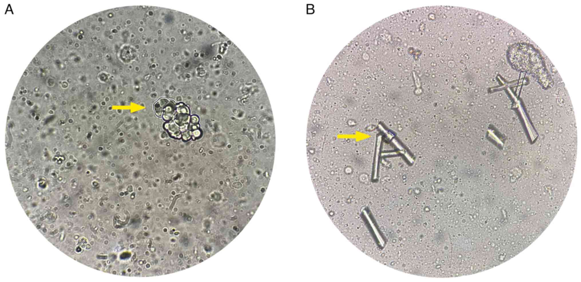
results. Both analyses revealed azoospermia with abnormal uric acid
crystals through microscopic examination. The identification of
uric acid crystals relied on microscopic examination, revealing
characteristic needle-like shapes and strong negative birefringence
under polarized light, which are specific markers for
distinguishing uric acid crystals from the crystals of other
compounds, such as phosphate crystals (9). A thorough microscopic analysis was
conducted, with polarized light microscopy being leveraged to
differentiate uric acid crystals from other potential crystalline
compounds. In the examination, phosphate crystals were specifically
ruled out, as they typically appear as prismatic or rosette-shaped
structures and exhibit different birefringence characteristics.
Positive birefringence is usually shown by phosphate crystals, in
contrast to the negative birefringence of uric acid crystals
(9). In addition, the possibility
of oxalate crystals was considered, which often present as
envelope-shaped crystals with a different pattern of birefringence
(10). By systematically comparing
the morphological features and birefringence properties under
polarized light, the presence of uric acid crystals was confirmed,
effectively excluding other crystalline forms such as phosphate and
oxalate crystals (11) (Fig. 1). The pH of the seminal fluid was
7.9 (normal value, pH ≥7.2) and the volume was 5.5 ml (normal
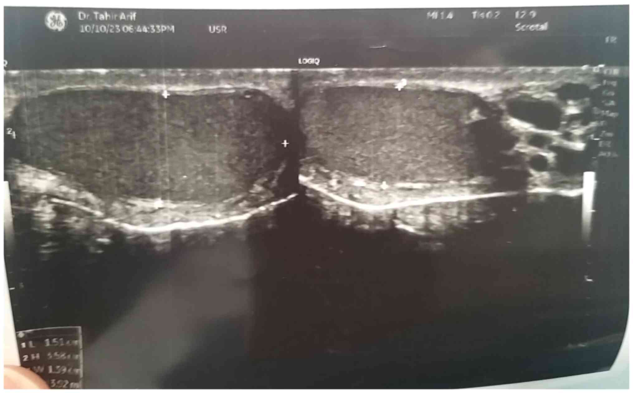
volume, >1.5 ml/ejaculation), both within normal ranges. Scrotal
Doppler ultrasound (U/S) indicated normal positioning and texture
of both testes, while they were relatively small in size (Fig. 2). The right testis measured 9 ml,
while the left testis measured ~8 ml. These measurements are below
the average testicular size range, which typically ranges from 15
to 25 ml in adult males (12).
Furthermore, the U/S revealed no focal lesion, hydrocele or
apparent inguinal hernia. Hormonal assays showed elevated levels of
follicle-stimulating hormone (FSH) (normal range, 1.5-12.4),
luteinizing hormone (LH) (normal range, 1.7-8.6), and prolactin
(normal range, 4.04-15.2) with 12.7, 16.6 mIu/ml and 35.1 ng/ml,
respectively (Table I). Genetic
analysis showed no evidence of Y-chromosome microdeletion (Table II).
 | Table ILaboratory evaluation: Semen fluid
analysis, hormonal profiling for fertility assessment. |
Table I
Laboratory evaluation: Semen fluid
analysis, hormonal profiling for fertility assessment.
| A, Semen fluid
analysis |
|---|
| Parameter | Result | WHO reference
limit |
|---|
| Volume, ml | 5.5 |
>1.5/ejaculation |
| Color | Milky white | Whitish, grey,
opalescent |
| Viscosity | Normal | Normal |
| pH | 7.9 | Basic ≥7.2 |
| Concentration,
million/ml | Azoospermia | >15 |
| Total sperm count,
million/ejaculate | Azoospermia | >39 |
| B, Hormonal profile
of testicular sperm aspiration |
| Parameter | Result | WHO reference limit
(for male) |
| FSH, mIu/ml | 12.7 | 1.5-12.4 |
| LH, mIu/ml | 16.6 | 1.7-8.6 |
| Prolactin, ng/ml | 35.1 | 4.04-15.2 |
| Testosterone,
nmol/l | 2.59 | 7.57-31.4 |
 | Table IIGenetic analysis through reverse
transcription-PCR for fertility assessment. |
Table II
Genetic analysis through reverse
transcription-PCR for fertility assessment.
| Chromosome | Target gene | Result |
|---|
| Y/STS Locus | AZFa | Normal |
| 3p24 | DAZL | Normal |
| 15q25 | POLG | Normal |
| PARs | SHOX gene | Normal |
Histological examination and
findings
Under local anesthesia, both right and left
testicular aspirations were performed for the extraction of sperm,
which retrieved a small tissue sample, ranging from 5 to 10 mg per
each testicle. The specimen was fixed for 1-3 days in 10% neutral
buffered formalin at room temperature and then processed using the
DiaPath Donatello automated processor (Diapath S.P.A). This process
involved holding the specimen in formalin (average 20 min) and
deionized water (10 min), and dehydrating it in alcohol with
increasing concentrations of 70% (1 h), 95% (1 h) and 99% (two
stages of 1 h 30 min each). Clearing was achieved through three
stages of xylene (1 h each), followed by infiltration with paraffin
wax in three stages (1 h each). The blocks were embedded in
paraffin wax using the Sakura Tissue-Tek embedding center (Sakura
Finetec), faced and sectioned using the Sakura Accu-Cut SRM
microtome. Sections were floated in a 40-50˚C water bath and placed
on glass slides, which were then oven-dried at 60-70˚C
overnight.
The following day, the slides were stained using the
DiaPath Giotto autostainer (Diapath S.P.A). The staining process
included stages of xylene (three stages of 7, 7 and 5 min), alcohol
(100% in three stages of 7, 6 and 5 min, followed by 90% for 4 min
and 70% for 3 min) and tap water (2 min). Hematoxylin Gill II
staining was applied for 8 min, followed by washing with tap water
(4 min), ammonia water (1 min) and another tap water wash (1 min).
This was followed by 70% alcohol (2 min) and Eosin Y staining (5
min), with a final wash in tap water (1 min). The slides were then
dehydrated in alcohol (70% for 15 sec, 90% for 2 min and 100% in
three stages of 3, 3, and 4 min) and cleared in xylene (three
stages of 3, 5 and 4 min). After drying for 5 min, the slides were
mounted with SurgiPath Sub-X medium (Surgipath Medical Industries,
Inc.) and covered with a coverslip. The histological examination of
both right and left testicular sperm extraction (TESA) confirmed
SCOS, with no evidence of granulomas or malignancies. Histological
findings included a reduction in the diameter of the testicular
tubules, thickened basement membranes and the presence of Sertoli
cells aligned perpendicularly to the basement membrane. These
Sertoli cells exhibited nuclear indentations with prominent
nucleoli. In addition, no germ cells were observed (data not
shown).
Follow-up and outcome
The patient's postoperative period was uneventful
and he has been undergoing routine follow-up assessments, scheduled
monthly, to monitor the patient's fertility status.
Discussion
NOA is identified by impaired spermatogenesis, which
can be caused by a variety of endogenous or exogenous
abnormalities. Histological examination of the testicular biopsy is
employed to categorize NOA into three groups: SCOS,
hypospermatogenesis and MA. Del Castillo was the first to describe
SCOS in 1947. It is characterized by the total absence of germ
cells in seminiferous tubules, accompanied by reduced testicular
size and normal secondary sexual features. The condition presents
in two forms: Focal SCOS and complete SCOS (2). Focal SCOS is defined by the presence
of residual areas of normal spermatogenesis in the testis. By
contrast, complete SCOS is identified by gonocyte failure to
migrate to the embryonic gonads, leading to the subsequent absence
of germinal epithelium formation (7). The current case was a 28-year-old
male with a primary infertility history of ~4 years and normal
secondary sexual features.
It has been observed that SCOS in infertile males
can result from diverse factors, including Y chromosome
microdeletions, cytotoxic drugs, undescended testis, radiation
exposure and viral infections (2).
The Y chromosome microdeletion is the most significant pathogenetic
defect associated with male infertility (13). According to a study by Stouffs
et al (14) on the role of
genetic investigation among azoospermic patients with SCOS, it is
evident that karyotype abnormalities, particularly Klinefelter
syndrome, are the most prevalent in azoospermic men with SCOS. A
study reported the case of a patient with azoospermia linked to
SCOS and Leydig tumors. Unlike previous studies, the patient had
normal secondary sexual features and no indication of Klinefelter
syndrome. The right testis was found to be larger than the left
one, which was a unique finding (1). In contrast to the studies mentioned,
the current study involves a patient with both testes relatively
small for their age. In addition, genetic analysis showed normal Y
chromosome microdeletion and elevated FSH, LH and
procalcitonin.
The diagnosis of azoospermia after confirmation
through the analysis of multiple semen samples can be achieved
through clinical differentiation between NOA and OA by evaluating
diagnostic factors such as physical examination, medical history
and hormonal analysis. These factors offer a prediction accuracy of
>90% in determining the type of azoospermia (5). A definitive diagnosis of NOA subtypes
is made through testicular biopsy and histopathological examination
of the specimens (2). Patients
diagnosed with NOA are often recommended to undergo genetic
testing, including cytogenetic karyotyping and molecular diagnostic
techniques, such as subtyping of Y chromosome microdeletions, in
accordance with guidelines from the American Society for
Reproductive Medicine (ASRM) (15)
and the European Association of Urology (EAU) (16). Despite the normal karyotype
observed in the majority of men with SCOS, genetic factors like
Klinefelter syndrome, Y chromosome microdeletions and other genetic
determinants can still be relevant (2). In the current case, two seminal fluid
analyses showed azoospermia with an abnormal uric acid crystal
through microscopic examination. To identify uric acid crystals in
the semen sample, thorough microscopic analysis was conducted,
leveraging polarized light microscopy to differentiate uric acid
crystals from other potential crystalline compounds. Uric acid
crystals exhibit distinctive needle-like shapes and demonstrate
strong negative birefringence under polarized light, which is a
hallmark feature. In this examination, specifically phosphate
crystals were ruled out, which typically appear as prismatic or
rosette-shaped structures and exhibit different birefringence
characteristics. Phosphate crystals usually show positive
birefringence, in contrast to the negative birefringence of uric
acid crystals. In addition, the possibility of oxalate crystals was
considered, which often present as envelope-shaped crystals with a
different pattern of birefringence. By systematically comparing the
morphological features and birefringence properties under polarized
light, the presence of uric acid crystals was confirmed. Scrotal
Doppler U/S showed that both testes had a normal position and
texture, but they were relatively small, while histopathological
examination of both right and left testicular TESA revealed Sertoli
cell-only syndrome, with no granuloma or malignancy detected.
The management of SCOS remains uncertain, with no
effective treatments for achieving biological parenthood (2). Patients with exceedingly low sperm
counts or no sperm in their semen may still be eligible for
assisted reproductive techniques. Microsurgical extraction of sperm
directly from the testes, even a minimal quantity, can be used for
intracytoplasmic sperm injection (17). The current case involved a patient
who had a sperm count of zero. Furthermore, not a single sperm
could be obtained for fertilization using intracytoplasmic sperm
injection.
Regarding the presence of crystals in semen, it is
rare and the literature mostly describes crystals composed of
spermine phosphate (8,18). The crystallization of uric acid is
typically associated with acidic environments where uric acid has
reduced solubility. Our observation of uric acid crystals in
seminal fluid with a slightly alkaline pH of 7.9 presents a
paradox, as uric acid crystals are usually expected to form in more
acidic conditions. Several factors may contribute to this atypical
finding: Localized pH variations within the fluid could have been
more acidic despite the overall alkaline measurement, specific
substances or compounds in the seminal fluid may have influenced
uric acid solubility and individual physiological or pathological
factors could affect crystallization patterns (19). Motrich et al (8) first reported uric acid crystals in
the seminal fluid of a patient with chronic prostatitis symptoms in
2005. The current case report documented uric acid crystals in the
semen of a male patient with azoospermia and SCOS. The patient's
semen had a normal volume and pH. While recent studies indicate
that factors such as dietary habits and genetic predispositions
significantly influence the occurrence of uric acid crystallization
in the urinary tract (20), their
appearance in semen is relatively rare and may suggest specific
reproductive health concerns. However, this case is significant, as
it represents the first documented instance of uric acid crystals
in the semen of an azoospermic male with SCOS in the genuine
literature (21).
The current case report faced a limitation as there
were no histopathological examination slides available for both the
right and left testicular TESA. Another limitation is that while
uric acid crystals were identified based on microscopic and
chemical analysis, the potential presence of impurities or
composite crystals cannot be definitively excluded without
additional spectroscopic techniques
In conclusion, despite uric acid crystals being
strictly dependent on pH levels, only spermine crystals composed of
phosphate have been previously reported in semen. The current study
documented a case of uric acid crystal presence in the semen of an
azoospermic male with SCOS.
Acknowledgements
Not applicable.
Funding
Funding: No funding was received.
Availability of data and materials
The datasets used and/or analyzed during the current
study are available from the corresponding author on reasonable
request.
Authors' contributions
AKHR and RQS were major contributors to the
conception of the study, as well as to the literature search for
related studies. SSF, AMM and FHK were involved in the literature
review, study design and writing the manuscript. HMM, NHAA and GMS
were involved in the literature review, the design of the study,
the critical revision of the manuscript and the processing of the
figures. FHK and SSF confirm the authenticity of all the raw data.
AAM and BOM were the radiologists who performed the assessment of
the case. All authors have read and approved the final
manuscript.
Ethics approval and consent to
participate
In our locality, ethical approval is not required
for case studies involving fewer than three cases. Written informed
consent was obtained from the patient for participation in the
present study and related investigations, including genetic
analysis.
Patient consent for publication
Written informed consent was obtained from the
patient for the publication of the present case report and any
accompanying images.
Competing interests
The authors declare that they have no competing
interests.
References
|
1
|
Bapir R, Salih RQ, Salih KM, Shabur B,
Salih AM, Kakamad FH, Abdullah HO, Fattah FH and Mohammed SH:
Simultaneous sertoli cell-only syndrome and leydig cell tumor in a
patient with azoospermia: A rare case report. Case Rep Oncol.
15:1095–1100. 2022.PubMed/NCBI View Article : Google Scholar
|
|
2
|
Ghanami Gashti N, Sadighi Gilani MA and
Abbasi M: Sertoli cell-only syndrome: Etiology and clinical
management. J Assist Reprod Genet. 38:559–572. 2021.PubMed/NCBI View Article : Google Scholar
|
|
3
|
Mazzilli R, Vaiarelli A, Dovere L,
Cimadomo D, Ubaldi N, Ferrero S, Rienzi L, Lombardo F, Lenzi A,
Tournaye H and Ubaldi FM: Severe male factor in in vitro
fertilization: Definition, prevalence, and treatment. An update.
Asian J Androl. 24:125–134. 2022.PubMed/NCBI View Article : Google Scholar
|
|
4
|
Takeshima T, Karibe J, Saito T, Kuroda S,
Komeya M, Uemura H and Yumura Y: Clinical management of
nonobstructive azoospermia: An update. Int J Urol. 31:17–24.
2024.PubMed/NCBI View Article : Google Scholar
|
|
5
|
Andrade DL, Viana MC and Esteves SC:
Differential diagnosis of azoospermia in men with infertility. J
Clin Med. 10(3144)2021.PubMed/NCBI View Article : Google Scholar
|
|
6
|
Ozturk S: Genetic variants underlying
spermatogenic arrests in men with non-obstructive azoospermia. Cell
Cycle. 22:1021–1061. 2023.PubMed/NCBI View Article : Google Scholar
|
|
7
|
Wang X, Liu X, Qu M and Li H: Sertoli
cell-only syndrome: Advances, challenges, and perspectives in
genetics and mechanisms. Cell Mol Life Sci. 80(67)2023.PubMed/NCBI View Article : Google Scholar
|
|
8
|
Motrich RD, Olmedo JJ, Molina R, Tissera
A, Minuzzi G and Rivero VE: Uric acid crystals in the semen of a
patient with symptoms of chronic prostatitis. Fertil Steril.
85:751.e1–751.e4. 2006.PubMed/NCBI View Article : Google Scholar
|
|
9
|
Pascual E, Sivera F and Andres M: Synovial
fluid analysis for crystals. Curr Opin Rheumatol. 23:161–169.
2011.PubMed/NCBI View Article : Google Scholar
|
|
10
|
Lorenz EC, Michet CJ, Milliner DS and
Lieske JC: Update on oxalate crystal disease. Curr Rheumatol Rep.
15(340)2013.PubMed/NCBI View Article : Google Scholar
|
|
11
|
Pascual E: The diagnosis of gout and CPPD
crystal arthropathy. Br J Rheumatol. 35:306–308. 1996.PubMed/NCBI View Article : Google Scholar
|
|
12
|
Arce BGP and Quanico UT: A retrospective
determination of the average testicular volume of pubertal and
post-pubertal male patients in a tertiary institution. Philippine J
Urol. 33:2023.
|
|
13
|
Krausz C, Rosta V, Swerdloff RS and Wang
C: 6-Genetics of male infertility. In: Emery and rimoin's
principles and practice of medical genetics and genomics: 121-147,
2022.
|
|
14
|
Stouffs K, Gheldof A, Tournaye H,
Vandermaelen D, Bonduelle M, Lissens W and Seneca S: Sertoli
cell-only syndrome: Behind the genetic scenes. Biomed Res Int.
2016(6191307)2016.PubMed/NCBI View Article : Google Scholar
|
|
15
|
Schlegel PN, Sigman M, Collura B, De Jonge
CJ, Eisenberg ML, Lamb DJ, Mulhall JP, Niederberger C, Sandlow JI,
Sokol RZ, et al: Diagnosis and treatment of infertility in men:
AUA/ASRM guideline part I. J Urol. 205:36–43. 2021.PubMed/NCBI View Article : Google Scholar
|
|
16
|
Jungwirth A, Giwercman A, Tournaye H,
Diemer T, Kopa Z, Dohle G and Krausz C: EAU Working Group on Male
Infertility. European Association of Urology guidelines on Male
Infertility: The 2012 update. Eur Urol. 62:324–332. 2012.PubMed/NCBI View Article : Google Scholar
|
|
17
|
Deruyver Y, Vanderschueren D and Van der
Aa F: Outcome of microdissection TESE compared with conventional
TESE in non-obstructive azoospermia: A systematic review.
Andrology. 2:20–24. 2014.PubMed/NCBI View Article : Google Scholar
|
|
18
|
Calandra RS, Rulli SB, Frungieri MB,
Suescum MO and Gonzalez-Calvar SI: Polyamines in the male
reproductive system. Acta Physiol Pharmacol Ther Latinoam.
46:209–222. 1996.PubMed/NCBI
|
|
19
|
Chhana A, Lee G and Dalbeth N: Factors
influencing the crystallization of monosodium urate: A systematic
literature review. BMC Musculoskelet Disord. 16(296)2015.PubMed/NCBI View Article : Google Scholar
|
|
20
|
Roman YM: The role of uric acid in human
health: Insights from the uricase gene. J Pers Med.
13(1409)2023.PubMed/NCBI View Article : Google Scholar
|
|
21
|
Muhialdeen AS, Ahmed JO, Baba HO, Abdullah
IY, Hassan HA, Najar KA, Mikael TM, Mustafa MQ, Mohammed DA, Omer
DA, et al: Kscien's List; A New Strategy to Discourage Predatory
Journals and Publishers (Second Version). Barw Med J. 1:2023.
|


