Open Access

Potential applications of components of aged garlic extract in mitigating pro‑inflammatory gene expression linked to human diseases (Review)

  • Authors:
    • Enzo Agostinelli
    • Giovanni Marzaro
    • Roberto Gambari
    • Alessia Finotti
  • View Affiliations

  • Published online on: May 13, 2025     https://doi.org/10.3892/etm.2025.12884
  • Article Number: 134
  • Copyright: © Agostinelli et al. This is an open access article distributed under the terms of Creative Commons Attribution License.

Metrics: Total Views: 0 (Spandidos Publications: | PMC Statistics: )
Total PDF Downloads: 0 (Spandidos Publications: | PMC Statistics: )


Abstract

In the present review, simple approaches for the screening and characterization of natural compound agents that alter pro‑inflammatory gene expression are described, with a particular focus on aged garlic extract (AGE), which has been the subject of several investigations that have supported its potential application as an anti‑inflammatory agent. Additionally, evidence regarding the possible effects and mechanisms of action of two major AGE components, S‑allyl cysteine (SAC) and S‑1‑propenyl‑l‑cysteine (S1PC), is reviewed. The proposed molecular targets of SAC and S1PC are IKKβ kinase, the Kelch‑like ECH‑associated protein 1‑nuclear factor erythroid 2‑related factor 2 complex, peroxisome proliferator‑activated receptor‑γ, histone deacetylase and toll‑like receptor 4 (TLR4). Targeting these molecules causes a marked reduction in NF‑κB activity accompanied by a notable decrease in the transcription of NF‑κB‑regulated genes. Another main objective of the present review was to discuss the possibility that AGE and its bioactive components could be employed in the treatment of several human pathologies that are characterized by a hyperinflammatory state resulting from dysregulation of the TLR4 and NF‑κB pathways. SAC is of interest in the treatment of lung pathologies, neurological diseases, osteoarthritis, muscular atrophy, cardiovascular diseases, diabetes and cancer. Additionally, the anti‑oxidative activities of AGE, SAC and S1PC are compatible with their employment in the treatment of diseases characterized by oxidative stress, such as sickle cell disease and β‑thalassemia.

1. Introduction

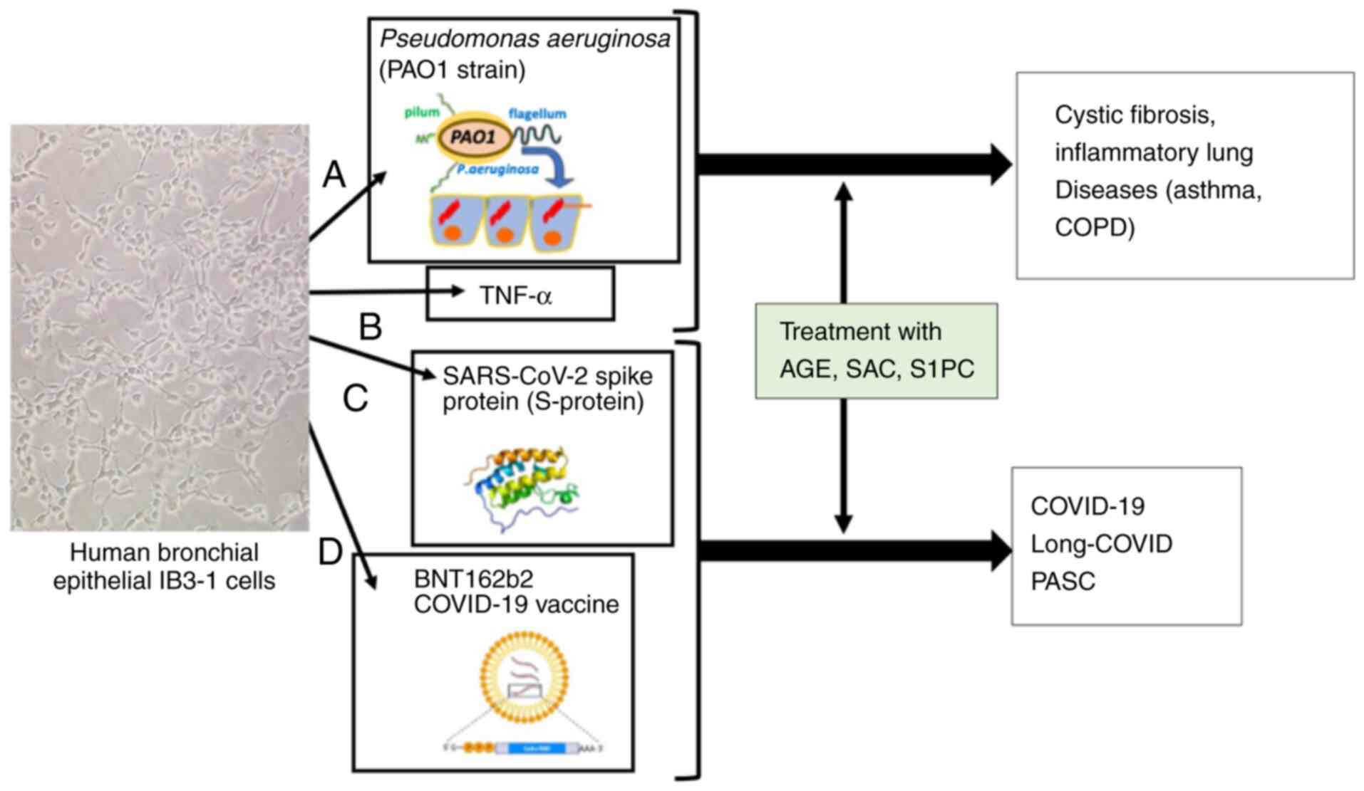
The present review is centered on the characterization and biomedical applications of natural compounds, with a particular focus on aged garlic extract (AGE). Commercially available AGE is an odorless preparation obtained by immersing fresh garlic slices in an aqueous ethanol solution for several months at room temperature (1-5). A number of studies have supported the possible employment of AGE as an anti-inflammatory agent (6-10). In this context, the availability of simple experimental approaches for the screening and characterization of molecules that interfere with pro-inflammatory gene expression is crucial for advancing the knowledge on anti-inflammatory agents present within AGE. An introductory example of experimental cellular model systems of interest in the field of respiratory diseases is shown in Fig. 1. These experimental systems may be useful in generating datasets that support the possible employment of validated agents for the treatment of human pathologies characterized by a hyperinflammatory state. The study of pro-inflammatory genes is of notable importance since the expression of these genes is critical in the development of a number of diseases, such as COVID-19, cystic fibrosis and chronic obstructive pulmonary disease (COPD). Table I reports examples of relevant pro-inflammatory genes that have been targets for anti-inflammation therapy in pre-clinical or clinical studies (11-23). For instance, interleukin (IL)-8 antagonists and monoclonal antibodies against IL-8 have been proposed for the therapy of cystic fibrosis (11,12), COPD (13), severe asthma (14) and osteoarthritis (15). Additionally, anti-IL-6 therapy has been proposed for COVID-19, long-COVID and post-acute sequelae of SARS-CoV-2 infection (16-18). Furthermore, Toll-like receptor 4 (TLR4) inhibitors may be considered for treating COVID-19 (19-21), osteoarthritis (22) and other inflammation-related diseases, such as cystic fibrosis (23).

Figure 1

Examples of experimental approaches for the screening and characterization of anti-inflammatory agents to be proposed for the experimental therapy of respiratory diseases, such as cystic fibrosis, asthma, COPD, COVID-19, PASC and long-COVID. The experimental model systems described are based on IB3-1 human bronchial epithelial cells (A) infected with PAO, or exposed to (B) TNF-α, (C) SARS-CoV-2 spike protein and (D) the COVID-19 BNT162b2 vaccine. The image of IB3-1 cells was modified from Gasparello et al (45) (copyright can be found at https://doi.org/10.3390/molecules29245938). AGE, aged garlic extract; COPD, chronic obstructive pulmonary disease; COVID-19, coronavirus disease-2019; PAO, Pseudomonas aeruginosa; PASC, post-acute sequelae of SARS-CoV-2 infection; S1PC, S-1-propenyl-l-cysteine; SAC, S-allyl cysteine; SARS-CoV-2, severe acute respiratory syndrome corona virus 2.

Table I

Examples of pro-inflammatory genes involved in human diseases characterized by hyper-inflammation.

Table I

Examples of pro-inflammatory genes involved in human diseases characterized by hyper-inflammation.

First author/s, year Clinically relevant pro-inflammatory gene target Disease Pre-clinical and clinical studies: Key results (Refs.)
McElvaney and IL-8 Cystic fibrosis IL-6 and IL-8 production is reduced by fluticasone in (11,12)
McElvaney, 2018;     cystic fibrosis bronchial epithelial cells (12)  
Escotte et al, 2003        
Rennard et al, 2015   COPD The CXCR2 antagonist MK-7123 is proposed for the treatment of COPD (NCT01006616) (13)
Planagumà et al, 2015   Severe asthma A promising anti-inflammatory treatment targeting CXC receptors 1 and 2 is described for the reduction of neutrophil migration and activation in respiratory diseases (14)
Yang et al, 2024   Osteoarthritis Osteoarthritis progression is attenuated by injecting a IL-8 neutralizing monoclonal antibody (15)
Rosas et al, 2021; Pinzon et al, 2021 IL-6 COVID-19 IL-6 inhibitors are effective therapeutic agents for COVID-19 (16,17)
Simonetti et al, 2024   Long-COVID IL-6 receptor inhibitors tocilizumab and sarilumab are proposed for long-COVID (18)
Holms, 2022   PASC Ezrin peptides are potent inhibitors of IL-1, IL-6, IL-8 and TNF-α expression, and can be used for potential therapeutic protocols for long-COVID/PASC (19)
Dinarello et al, 2012 IL-1β RA The IL-1 receptor antagonist anakinra, the soluble decoy receptor rilonacept and the neutralizing monoclonal anti-IL-1β antibody canakinumab can be employed in RA (20)
Asaba et al, 2024 TLR4 COVID-19 TLR4 inhibitors decrease inflammation and severity in COVID-19 infections (21)
Bartels et al, 2024   Osteoarthritis Inhibition of TLR4 signaling strongly inhibits joint inflammation in osteoarthritis (22)
Greene et al, 2008   Cystic fibrosis TLR4 is a therapeutic target in cystic fibrosis (23)

[i] COPD, chronic obstructive pulmonary disease; COVID-19, coronavirus disease-2019; CXCR2, C-X-C motif chemokine receptor 2; PASC, post-acute sequelae of severe acute respiratory syndrome corona virus 2 infection; RA, rheumatoid arthritis; TLR4, toll-like receptor 4.

The present review will discuss experimental model systems and their application in characterizing the activity of AGE and selected components. Then, recent evidence regarding the possible effects and mechanism of action of two major a sulfur-containing AGE components, S-allyl cysteine (SAC) (24,25) and S-1-propenyl-l-cysteine (S1PC) (26), will be reviewed. In this respect, SAC and S1PC showed stable properties under cell culture conditions, and their acute/subacute toxicity was minor in mice and rats (24,26). In particular, the pharmacokinetics of SAC was investigated on human volunteers, indicating that SAC was rapidly absorbed from the gastrointestinal tract (24).

Finally, the possible application of AGE, SAC and S1PC in the development of protocols for possible therapeutic interventions will be discussed.

2. Experimental model systems to identify and characterize inhibitors of pro-inflammatory genes

The cell models used for testing inhibitors of pro-inflammatory genes are typically mammalian cells infected with a microorganism, treated with a viral protein or treated with other agents stimulating pro-inflammatory genes. Table II provides a representative list of experimental models that have been used to characterize the effects of different classes of inhibitors of pro-inflammatory genes. One of the most used and well-characterized cell models is bronchial epithelial IB3-1 cells infected with Pseudomonas aeruginosa. Detailed descriptions of this experimental model system can be found in the publications by DiMango et al (27), who reported the origin of the IB3-1 cell line (derived from a cystic fibrosis patient with a DF508/W1282X genotype) and the protocols for infection with P. aeruginosa. The non-mucoid laboratory strain of P. aeruginosa, PAO1, is typically employed for infection. In the publication by DiMango et al (27), information can be found on PAO1 culturing and on the experimental conditions to prepare aliquots to stimulate IB3-1 cells. One of the most relevant effects of the P. aeruginosa infection of IB3-1 cells is the induction of a pro-inflammatory phenotype marked by the activation of several pro-inflammatory genes, the most relevant being the IL-8 genes. The role of transcription factors in the P. aeruginosa infection of IB3-1 cells was first reported by DiMango et al (27), who described NF-κB activation in this model. The cooperative action of the transcription factors, NF-κB, NF-IL6, activator protein 1, CHOP and cAMP responsive element binding protein, in the P. aeruginosa-dependent induction of IL-8 gene transcription in IB3-1 cells (27) was discussed in depth by Bezzerri et al (28). Notably, using this experimental model system, Finotti et al (29) demonstrated that decoy oligonucleotide molecules targeting NF-κB were able to inhibit the transcription of the IL-8 gene. Furthermore, using a chromatin immunoprecipitation assay, these authors found that the NF-κB decoy was able to strongly inhibit NF-κB recruitment to the IL-8 gene promoter (29). A microarray-based study that demonstrated the involvement of microRNAs in the P. aeruginosa infection of IB3-1 cells was reported by Fabbri et al (30), providing new information on possible employment of microRNA targeting for the development of therapeutic protocols for cystic fibrosis. Several studies have been performed using this system to demonstrate the potential anti-inflammatory properties of medicinal plant extracts or low-molecular weight drugs (31-33). For instance, Lampronti et al (31) reported the notable effects of Nigella arvensis extracts, which inhibit IL-8 expression in P. aeruginosa-infected IB3-1 cells. Furthermore, it was demonstrated that β-sitosterol is the compound responsible for this activity, while stigmasterol and campesterol are inactive in this context. These results support the conclusion that P. aeruginosa-infected IB3-1 cells serve as a suitable experimental model system to identify inhibitors of pro-inflammatory gene expression (30-33).

Table II

Examples of in vitro experimental model systems to identify inhibitors of pro-inflammatory genes.

Table II

Examples of in vitro experimental model systems to identify inhibitors of pro-inflammatory genes.

First author/s, year Experimental model system Characterized molecule (Refs.)
Di Mango et al, 1998; Fabbri et al, 2014; Lampronti et al, 2017 Cystic fibrosis IB3-1 epithelial cells infected with Pseudomonas aeruginosa Dexamethasone (27); miR-93-5p (30); β-sitosterol (31) (27,30,31)
Hawdon et al, 2010; Cerqueira et al, 2013; Aval et al, 2013 A549 alveolar epithelial cells infected with Pseudomonas aeruginosa Resveratrol (35); picetannol (36) (34-36)
Dechecchi et al, 2008; Gambari et al, 2012; Borgatti et al, 2011 Cystic fibrosis epithelial cells treated with TNF-α Miglustat (39); corilagin (40); furocoumarin derivatives (41) (39-41)
Geng et al, 1997 T-lymphoid Jurkat cells treated with TNF-α S-allyl cysteine (47) (48)
Gasparello et al, 2021; Gasparello et al, 2021b Bronchial epithelial cells treated with SARS-CoV-2 spike protein Sulforaphane (42); miR-93-5p (43) (42,43)
Cosenza et al, 2023; Gasparello et al, 2024; Papi et al, 2025 Human cell lines exposed to anti-SARS-CoV-2 BNT162b2 vaccine Aged garlic extract (45); S-allyl cysteine (45); S-1-propenyl-l-cysteine (46) (44-46)

[i] miR, microRNA; SARS-CoV-2, severe acute respiratory syndrome corona virus 2.

Similar experimental model systems have been proposed that are based on the P. aeruginosa infection of other cell lines, such as NuLi (30,31), CuFi (30,31), A549(34). Hawdon et al (34), Cerqueira et al (35) and Aval et al (36) demonstrated the potential anti-inflammatory effects of resveratrol and picetannol, using P. aeruginosa-infected A549 alveolar epithelial cells. In addition to P. aeruginosa infection, IB3-1 cells can be stimulated to upregulate pro-inflammatory genes by other stimuli, such as lipopolysaccharide (37), IL-1β (38), TNF-α (39-41) and the SARS-CoV-2 spike protein (42,43). Notably, the effects of the SARS-CoV-2 spike protein on a variety of cellular systems were reproduced by treatment with the spike-mRNA-based vaccine, BNT162b2 (44-46). Another experimental model system of particular interest in the context of the present review is the T-lymphoid Jurkat cell line, very useful to determine the effects of inhibitory agents on TLR4-dependent NFκB activation (47). Geng et al (48) used an electrophoretic mobility shift assay (EMSA) performed on nuclear extracts prepared from TNF-α stimulated Jurkat cells. This study demonstrated a powerful inhibitory effect of SAC on NF-κB activation, reinforcing the information on anti-inflammatory properties of this AGE component found using other experimental model systems (45).

In conclusion, several experimental model systems have been developed and validated for identifying agents that exhibit potent inhibitory activity on pro-inflammatory gene expression. Selected potential anti-inflammatory agents may also be tested in vivo using validated animal models of chronic P. aeruginosa lung infection that mimic cystic fibrosis, as proposed by Hoffmann et al (49). In the near future, we expect that the effects of AGE and its constituents will be analyzed using P. aeruginosa-infected primary bronchial epithelial cells. The results obtained should be considered with great attention, taking into account the anti-microbial effects of garlic-derived compounds as potential confounding factors (50,51) requiring adequate further control studies.

3. AGE: Studies supporting anti-inflammatory activity

Among the plant extracts hypothesized to retain anti-inflammatory activities, AGE is of great interest (52,53). The preparation and characterization of AGE has been described in a number of publications such as those by Ohkubo et al (1) and Kurita et al (54). Briefly, AGE can be prepared by immersing fresh garlic in 15% aqueous ethanol solution for several months at room temperature (1). AGE is a commercial odorless preparation with antioxidant properties for scavenging reactive oxygen species (ROS) (54) and retains immunomodulatory and anticancer properties (54). It should be noted that interest in the preparation of AGE and AGE-related products is evidenced by the high number of patents that have been deposited over the years (a partial list of patents and patent applications is shown in Table III). This information should be considered with great attention for the development of biomedical approaches, despite the fact that the patenting of preparations for ‘aged garlic’ is outside the major objectives of the present short review. A specialized further review would be useful to describe this interesting activity, which precedes the industrial exploitation of AGE and AGE-related compounds, considering that few reports are available on the patents on garlic and garlic-related products (55). In this context, two notable components of AGE are SAC (25) and S1PC (26). SAC and S1PC showed stable properties under cell culture conditions, and their acute/subacute toxicity was minor in mice and rats (24,26). In particular, the pharmacokinetics of SAC was investigated on human volunteers, indicating that SAC was rapidly absorbed from the gastrointestinal tract (24). Other AGE components have been described by Kodera et al (56), including S-(2-propyl) 2-propen-1-sulfinothioate (allicin), 3-(allyltrisulfanyl)-2-aminopropanoic acid, S-allylmercaptocysteine, allixin and tetrahydro-β-carboline derivatives.

Table III

Examples of in patents and patent applications to prepare ‘aged garlic’ and garlic products.

Table III

Examples of in patents and patent applications to prepare ‘aged garlic’ and garlic products.

Patent number Title and assignee Publication date Comments/description
US2554088A Extraction of garlic; assignee: Winthrop Stearns, Inc. May 22, 1951 This invention relates to an antibiotic substance and to a process for obtaining it from garlic cloves.
CN-101863807-B Preparation method of garlic extract; assignee: Sichuan Xiangzhen Enterprise Co., Ltd. June 10, 2010 A method for preparation of a garlic extract solution is described based on the following steps: Peeling, washing and slicing garlic, adding water for soaking, and filtering with a 50-100-mesh sieve.
US8187654B2 Process for preparing aged garlic; assignee: Blackgarlic, Inc. May 29, 2012 The invention concerns a method of producing aged garlic with increased antioxidation capability compared with that of raw garlic.
US20110293803 Method for producing an aged black garlic concentrate; assignee: Saenamhae Nonghyup, KR January 12, 2011 Aged black garlic concentrate is produced according to the following steps: Pretreating for cleaning; sealing the garlic in a polyethylene bag and storing in a tray; putting the tray into an aging device; and steam-treating by applying steam and heat for 30-60 min maintaining the aging device at a temperature of 80-90˚C.
JPS60262565A Preparation of garlic juice; assignee: Meiji Seika Kaisha, Ltd. December 25, 1985 A liquid containing crushed bulbs of garlic is combined with vitamin B1, saccharides and centrifuged, then the supernatant is combined with soybean powder to prepare odorless garlic juice without generation of irritating odor of garlic while they are processed.
CN110623255A Method for producing aged garlic; assignee: Pizhou Mid Autumn Food Co., Ltd. December 31, 2019 A method for producing aged garlic in which oxidation resistance is remarkably higher than that of raw materials such as increased garlic, which is used for producing garlic in which the necessary effectiveness of aged garlic as a raw material is not lowered, S-arylcysteine is not present at all in the produced garlic, and the polyphenol content is increased.
JP5968729B2 Production of black garlic extract and its use; assignee: MOMOYA KK Momoya Co Ltd. August 10, 2016 A technique for producing black garlic extract, that maintains, increases or enriches highly bioactive substances (such as S-allyl cysteine and cycloallyin) by heat-treating garlic raw material for a short time.
EP1752051A1 Method of producing aged garlic; assignee: COST PLUS KOREA Co Ltd February 14, 2007 The invention concerns a method of producing aged garlic in which its antioxidation capability is increased compared with that of raw garlic, which is used as a raw material.
WO2004077963A1 Processed plant product and method of producing the same; assignee: Wakunaga Pharmaceutical Co., Ltd. September 16, 2004 The plant product is obtained by maintaining allium in a low-oxygen or oxygen-free condition at a temperature ranging between 1˚C and 40˚C in the absence of any medium for 1 week or longer.
JP4380951B2 Garlic fermentation composition; assignee: Wakunaga Pharmaceutical Co., Ltd. December 9, 2009 An antioxidant composition obtained by fermenting enzyme-deactivated garlic with koji mold, or a composition obtained by adding beans and/or cereals to enzyme-deactivated garlic and fermenting with koji mold.
US6146638A Fermented garlic composition; assignee: Wakunaga Pharmaceutical Co., Ltd. November 14, 2000 A composition is described that is prepared by fermenting enzymatically-deactivated garlic with aspergillus and/or monascus. The composition is free from any unpleasant odor and useful as a prophylactic or therapeutic agent for several diseases, such as diabetes, hepatic diseases, cancer, immunopathy and hyperlipemia.

[i] https://patents.google.com/ (accessed November 8, 2024). http://www.freepatentsonline.com/ (accessed November 9, 2024).

4. SAC and S1PC: Proposed mechanisms of action

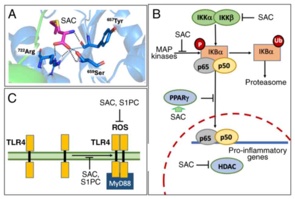
With respect to the anti-inflammatory activity of AGE, several studies are available on the effects of AGE components on the TLR4/NF-κB pathways, which play a central role in inflammatory processes (57-63). Regarding NF-κB, without external stimuli, an inactive trimer is formed in the cytoplasm that is composed of the inhibitory protein, IκB, and the p50/p65 NF-κB dimer. In these conditions, NF-κB is not translocated to the nucleus and the NF-κB regulated genes are mostly inactive, unless activated by other transcription factors (64,65). However, when external stimuli act on the corresponding receptors (for instance, when TLR4 is activated by a number of stimuli, some of which are listed in Table II), phosphorylation of IκB occurs, leading to dissociation of IκB from the trimer and its proteasome-mediated degradation, resulting in NF-κB activation. In these conditions, the p50/p65 NF-κB protein translocates to the nucleus and specifically interacts with NF-κB binding sites present in the promoters of NF-κB-regulated genes, such as the IL-6, IL-1β and IL-8 genes (66), all of which are involved in inflammatory processes that characterize several human diseases (as previously discussed in Table I). There is consensus that SAC interferes with NF-κB activation, thereby causing inhibition of the NF-κB-regulated genes (67,68). Table IV summarizes the evidence supporting the role of SAC in modulating the NF-κB pathway. Fig. 2 summarizes the biological effects of SAC (and of the analogue, S1PC) on different steps of the NF-κB pathway.

Figure 2

Proposed effects of SAC on the TLR4/NF-κB pathway. (A) Docking experiments showing the interaction between SAC and the Toll/interleukin-1 receptor domain of TLR4(45). Inhibitory effects of SAC (B) on MAP kinases, IKKβ, PPARγ and HDAC (62-64) and (C) on ROS and TLR4 dimerization are shown. HDAC, histone deacetylase; MyD88, myeloid differentiation primary response 88; P, phosphorylated; PPARγ, peroxisome proliferator-activated receptor-γ; ROS, reactive oxygen species; S1PC, S-1-propenyl-l-cysteine; SAC, S-allyl cysteine; TLR4, toll-like receptor 4; Ub, ubiquitinated.

Table IV

Effects of SAC on different molecular targets involved in the NF-κB pathway.

Table IV

Effects of SAC on different molecular targets involved in the NF-κB pathway.

First author/s, year Molecular target Employed experimental model system Methodological approach Comments (Refs.)
Park et al, 2014; Colín-Gonzáles et al, 2015; Shao et al, 2020 IKKb kinase Gastric ulcers induced in mice by intragastric administration of indomethacin (57) IKKb kinase assay (57); molecular docking (59) SAC is able to decrease IKKb activity (57) and was found to decrease phosphorylation of IkBa, leading to attenuation of NF-κB. (57-59)
Shao et al, 2020 Keap1-Nrf2 complex PPARγ HDAC In vitro: TBHP-treated murine chondrocytes; in vivo: Destabilization of the medial meniscus mice (59) Molecular docking (59) SAC is an activator of PPARγ (a strong inhibitor of NF-κB signaling). (59)
Shao et al, 2020; Gasparello et al, 2024 TRL4 BNT162b2 stimulated IB3-1 cells (45) Molecular docking (59); molecular docking and dynamics (45) The possible interactions with TLR4 are relevant, considering the TLR4 role in the regulation of NF-κB signaling. (59,45)

[i] HDAC, histone deacetylase; Keap1, Kelch-like ECH-associated protein 1; PPARγ, peroxisome proliferator-activated receptor-γ; SAC, S-allyl cysteine; TBHP, tert-butyl hydroperoxide; TLR4, toll-like receptor 4.

A notable study on the possible molecular targets of SAC has been reported by Park et al (57) and reviewed by Colín-González et al (58). In the study by Park et al (57), it was demonstrated that SAC is able to decrease IKKβ activity and the phosphorylation of IκBα, resulting in the attenuation of NF-κB. In another study, Shao et al (59) performed molecular docking analysis to determine that SAC may interact with the Kelch-like ECH-associated protein 1-nuclear factor erythroid 2-related factor 2 complex, PPARγ, histone deacetylase (HDAC)1 and TLR4. The possible interactions with TLR4 is of particular interest since this receptor is an upstream regulator of NF-κB signaling (60-63).

In our laboratory, the interaction between SAC (and the analogue S1PC) and the intracellular domain of TLR4 has been simulated using the well-known docking software, AutoDock Vina (45,46). Furthermore, to confirm the reliability of the identified molecular interactions and to predict the possible impact on TLR4 function, the computed docking models underwent molecular dynamics simulation. These studies are reported in Gasparello et al (45) and Papi et al (46) and suggest that the complexes remained stable as the hydrogen bonds formed between SAC (and S1PC) and TLR4 were retained during the entire molecular dynamics simulation, confirming a plausible interaction between the small molecules and TLR4. In addition, the results obtained suggest that binding with SAC and S1PC reduces the intermolecular interaction between the TLR4 domains of the TLR4 dimer, supporting additional effects on NF-κB, as outlined in Fig. 2. The predicted interaction between SAC and the TLR4 complex is depicted in Fig. 2A. The binding mode predicted for S1PC with TLR4 was only slightly different from SAC/TLR4, and SAC and S1PC shared interactions with both 657Tyr and 722Arg (45,46). For both small molecules, the estimated interaction energy with TLR4 was approximately -65 Kcal/mol (sum of short-range Lennard-Jones and short-range Coulomb contributions over the molecular dynamics simulation), suggesting the two compounds may have highly comparable and favorable interactions with the protein target. This finding suggests that one effect of SAC and S1PC on target cells might involve inhibition of TLR4, causing a marked reduction in NF-κB signaling and a notable inhibition of the transcription of NF-κB-regulated genes (Fig. 2B and C).

SAC reduces ROS (48), exerting clear and expected anti-inflammatory effects, as an interactive role of TLR4 and ROS has been described (69) and inhibition of ROS generation attenuates the TLR4-mediated proinflammatory phenotype, as reported by Pi et al (70). In addition to these direct and indirect effects on TLR4 (summarized in Fig. 2C), SAC might inhibit the NF-κB activity via several mechanisms of action (summarized in Fig. 2B), including via a strong inhibitory effect on IKKβ kinase, which blocks IκBα phosphorylation (64,65). This has a notable consequence for NF-κB signaling. It is well established that the activation of NF-κB requires phosphorylation of the inhibitory subunit, IκBα, the ubiquitination of IκBα (71,72) and finally the proteasome-dependent degradation of IκBα (72,73). In these conditions the p65/p50 NF-κB complex translocates to the nucleus and activates NF-κB-regulated genes. In addition to an inhibitory effect on IKKβ kinase, SAC has been demonstrated to cause a MAP kinase-mediated inhibition of IκBα phosphorylation (64), with a marked inhibition of the formation of poly-ubiquitin IκBα, of IκBα degradation and of NF-κB nuclear translocation. Finally, SAC has been reported to enhance the expression of PPARγ (74) a recognized NF-κB inhibitor (75), and to modulate HDAC activity (76).

5. TLR4, NF-κB and the expression of pro-inflammatory genes in human pathologies: Possible therapeutic applications of AGE and AGE constituents

The interplay among TLR4, NF-κB and human pathologies has been reported by a number of investigations and reviewed by several articles (77-82). For instance, Aboudounya and Heads (77) reported that SARS-CoV-2 binds and activates TLR4 to increase angiotensin converting enzyme 2 expression, facilitating entry of the virus into cells and resulting in hyperinflammation. In a recent review, Asaba et al (21) described the interplay between TLR4 and SARS-CoV-2, focusing on inflammation and the severity SARS-CoV-2 infections, suggesting TLR4 as a possible target for anti-SARS-CoV2 agents. Sahanic et al (78) demonstrated that SARS-CoV-2 activates the TLR4/MyD88 pathway in human macrophages causing a strong pro-inflammatory response in severe COVID-19. In a notable study, Alamin et al (79) identified common molecular signatures characterizing the molecular effects of SARS-CoV-2 infections and different lung diseases. This study was based on the analysis of RNA-sequencing and microarray gene expression datasets (from the Gene Expression Omnibus platform of the National Center for Biotechnology Information) and considered the following lung diseases: COPD, idiopathic pulmonary fibrosis, ILD (interstitial lung disease), asthma, tuberculosis, cystic fibrosis, pneumonia, emphysema and bronchitis. The full information about these datasets and the description of the complete workflow of the study can be found in Alamin et al (79). Notably, TLR4 was identified in most of these diseases, among the top-ranked differentially expressed genes.

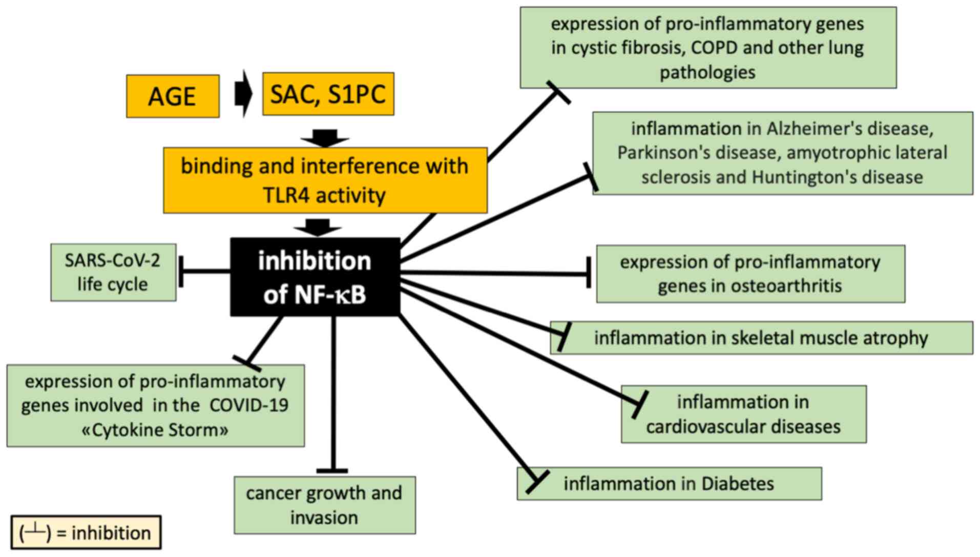
Since AGE (and its constituents, SAC and S1PC) might interfere with the TLR4 and NF-κB pathways (as outlined in Fig. 2), several pathologies caused by dysregulation of the TLR4/NF-κB axis might be affected by treatments with these natural products. Among these target pathologies, the most impactful for the worldwide health systems are COVID-19, lung infectious diseases, cystic fibrosis and COPD (https://www.healthdata.org/research-analysis/gbd; accessed February 20, 2025). Notably, and in agreement with the approach by Alamin et al (79), Hasan et al (80) reported using a differential gene expression pattern analysis, common pathophysiological processes highly similar in COVID-19 and cystic fibrosis, including the Toll-like receptor signaling pathway. Fig. 3 summarizes the pathologies with a dysregulated TLR4/NF-κB axis and/or oxidative stress, which leads to the activation of a hyperinflammatory state (81-94). In all these pathologies, novel anti-inflammatory molecules, such as AGE, SAC and S1PC, hold promise (81-94).

Figure 3

Inhibitory effects of AGE, SAC and S1PC on the NF-κB-associated expression of pro-inflammatory genes in human diseases. AGE, aged garlic extract; COPD, chronic obstructive pulmonary disease; COVID-19, coronavirus disease-2019; S1PC, S-1-propenyl-l-cysteine; SAC, S-allyl cysteine; SARS-CoV-2, severe acute respiratory syndrome corona virus 2; TLR4, toll-like receptor 4.

6. Conclusions and future perspectives

The main purpose of the present review was to discuss the possibility that AGE and its components, SAC and S1PC, might be employed in the treatment of several human pathologies that are characterized by a hyperinflammatory state. A partial list of published examples is reported in Fig. 3 (81-94). In most of these pathologies, dysregulation of the TLR4 and NF-κB pathways have been observed. In this respect, a large consensus has been reached sustaining the concept that AGE, and the most important constituents SAC and S1PC, inhibit TLR4 and NF-κB. SAC has been shown to be of interest for the treatment of lung pathologies (81,82), neurological diseases (83,84), osteoarthritis (85), muscular atrophy (86,87), cardiovascular diseases (88,89), diabetes (90,91) and cancer (92-95). In addition, in the possible use of AGE in the therapy of human pathologies, the antioxidative effects of AGE should be noted, whereby the oxidative stress of cells is altered in different pathologies. This is important for evaluating AGE and its constituents as treatments for hematological diseases characterized by oxidative stress of the erythroid cells. For instance, AGE and AGE components have been proposed for the treatment of sickle-cell diseases (95-97). In this respect, a Trade Mark for S-1-propenyl-l-cysteine (S1PCTM) has been recently obtained by Wakunaga Holdings Co., Ltd., Japan (registered on July 9, 2024; https://branddb.wipo.int/; accessed on March 20, 2025).

To maximize the impact of the studies described in the present review, further research and development are necessary. Clinical trials are expected to verify possible applications on human pathologies in the real world. Taking advantage of the increase in genomic information, one notable issue is related to the design of personalized therapeutic intervention. In this context, pharmacogenomic studies should be considered and undertaken within clinical trials, to predict the possible response (or no response) to the proposed AGE-based treatments. In this respect, combined therapies using AGE (or its constituents) alongside established therapeutic drugs or RNA- and DNA-based drugs is expected to be an attractive area of investigation. For instance, Reyes-Soto et al (94) found that SAC exhibits cytotoxicity in glioblastoma cells and improves the effect of temozolomide through the regulation of oxidative responses. Regarding the effects on TLR4, NF-κB and pro-inflammatory gene expression, we anticipate development based on the co-treatment with other currently available molecular inhibitors, including RNA-based therapies using microRNAs, used to specifically downregulate TLR4 and NF-κB, as previously suggested (98,99).

Acknowledgements

Not applicable.

Funding

Funding: The present study was supported by the MUR-FISR COVID-miRNAPNA Project (grant no. FISR2020IP_04128), the Interuniversity Consortium for Biotechnologies, Italy (CIB) (grant no. CIB-Unife-2020) and the International Polyamines Foundation ONLUS-ETS.

Availability of data and materials

The data generated in the present study may be requested from the corresponding author.

Authors' contributions

EA, GM, RG and AF conceived the study. GM and AF curated data. EA, GM, RG and AF performed formal analysis. RG, AF, EA and GM acquired funding. RG wrote the original draft. EA, GM, RG and AF reviewed and edited the manuscript. Data authentication is not applicable. All authors have read and approved the final version of the manuscript.

Ethics approval and consent to participate

Not applicable.

Patient consent for publication

Not applicable.

Competing interests

The authors declare that they have no competing interests.

References

1 

Ohkubo S, Dalla Via L, Grancara S, Kanamori Y, García-Argáez AN, Canettieri G, Arcari P, Toninello A and Agostinelli E: The anti-oxidant, aged garlic extract, exerts cytotoxic effects on wild-type and multidrug-resistant human cancer cells by altering mitochondrial permeability. Int J Oncol. 53:1257–1268. 2018.PubMed/NCBI View Article : Google Scholar

2 

Imai J, Ide N, Nagae S, Moriguchi T, Matsuura H and Itakura Y: Antioxidant and radical scavenging effects of aged garlic extract and its constituents. Planta Medica. 60:417–420. 1994.PubMed/NCBI View Article : Google Scholar

3 

Serrano JCE, Castro-Boqué E, García-Carrasco A, Morán-Valero MI, González-Hedström D, Bermúdez-López M, Valdivielso JM, Espinel AE and Portero-Otín M: Antihypertensive effects of an optimized aged garlic extract in subjects with grade I hyper-tension and antihypertensive drug therapy: A Randomized, triple-blind controlled trial. Nutrients. 15(3691)2023.PubMed/NCBI View Article : Google Scholar

4 

Yang Q, Li F, Jia G and Liu R: Aged black garlic extract inhibits the growth of estrogen receptor-positive breast cancer cells by downregulating MCL-1 expression through the ROS-JNK pathway. PLoS One. 18(e0286454)2023.PubMed/NCBI View Article : Google Scholar

5 

Das D, M K, Mitra A, Zaky MY, Pathak S and Banerjee A: A review on the efficacy of plant-derived bio-active compounds cur-cumin and aged garlic extract in modulating cancer and age-related diseases. Curr Rev Clin Exp Pharmacol. 19:146–162. 2024.PubMed/NCBI View Article : Google Scholar

6 

Borek C: Antioxidant health effects of aged garlic extract. J Nutr. 131(3s)(1010S-5S)2001.PubMed/NCBI View Article : Google Scholar

7 

Xu C, Mathews AE, Rodrigues C, Eudy BJ, Rowe CA, O'Donoughue A and Percival SS: Aged garlic extract supplementation modifies inflammation and immunity of adults with obesity: A randomized, double-blind, placebo-controlled clinical trial. Clin Nutr ESPEN. 24:148–155. 2018.PubMed/NCBI View Article : Google Scholar

8 

Libero ML, Lucarini E, Recinella L, Ciampi C, Veschi S, Piro A, Chiavaroli A, Acquaviva A, Nilofar N, Orlando G, et al: Anti-inflammatory and anti-hyperalgesic effects induced by an aqueous aged black garlic extract in rodent models of ulcerative colitis and colitis-associated visceral pain. Phytother Res. 38:4177–4188. 2024.PubMed/NCBI View Article : Google Scholar

9 

Wlosinska M, Nilsson AC, Hlebowicz J, Fakhro M, Malmsjö M and Lindstedt S: Aged garlic extract reduces IL-6: A double-blind placebo-controlled trial in females with a low risk of cardiovascular disease. Evid Based Complement Alternat Med. 2021(6636875)2021.PubMed/NCBI View Article : Google Scholar

10 

Albrakati A: Aged garlic extract rescues ethephon-induced kidney damage by modulating oxidative stress, apoptosis, inflammation, and histopathological changes in rats. Environ Sci Pollut Res Int. 28:6818–6829. 2021.PubMed/NCBI View Article : Google Scholar

11 

McElvaney OJ and McElvaney NG: Targeting IL-8 in cystic fibrosis: Enough but Not too much. Am J Respir Cell Mol Biol. 59:401–402. 2018.PubMed/NCBI View Article : Google Scholar

12 

Escotte S, Tabary O, Dusser D, Majer-Teboul C, Puchelle E and Jacquot J: Fluticasone reduces IL-6 and IL-8 production of cystic fibrosis bronchial epithelial cells via IKK-beta kinase pathway. Eur Respir J. 21:574–581. 2003.PubMed/NCBI View Article : Google Scholar

13 

Rennard SI, Dale DC, Donohue JF, Kanniess F, Magnussen H, Sutherland ER, Watz H, Lu S, Stryszak P, Rosenberg E and Staudinger H: CXCR2 antagonist MK-7123. A phase 2 proof-of-concept trial for chronic obstructive pulmonary disease. Am J Respir Crit Care Med. 191:1001–1011. 2015.PubMed/NCBI View Article : Google Scholar

14 

Planagumà A, Domènech T, Pont M, Calama E, García-González V, López R, Aulí M, López M, Fonquerna S, Ramos I, et al: Combined anti CXC receptors 1 and 2 therapy is a promising anti-inflammatory treatment for respiratory diseases by reducing neutrophil migration and activation. Pulm Pharmacol Ther. 34:37–45. 2015.PubMed/NCBI View Article : Google Scholar

15 

Yang J, Wang X, Zhang Y, He R, Fu Z, Wang R, Ma Y, Fu D, Meng S, Cai W, et al: Intra-articular injection of interleukin-8 neutralizing monoclonal antibody effectively attenuates osteoarthritis progression in rabbits. Cartilage: Mar 25, 2024 (Epub ahead of print).

16 

Rosas IO, Bräu N, Waters M, Go RC, Hunter BD, Bhagani S, Skiest D, Aziz MS, Cooper N, Douglas IS, et al: Tocilizumab in hospitalized patients with severe Covid-19 pneumonia. N Engl J Med. 384:1503–1516. 2021.PubMed/NCBI View Article : Google Scholar

17 

Pinzon RT, Wijaya VO and Buana RB: Interleukin-6 (IL-6) inhibitors as therapeutic agents for coronavirus disease 2019 (COVID-19): A systematic review and meta-analysis. J Infect Public Health. 14:1001–1009. 2021.PubMed/NCBI View Article : Google Scholar

18 

Simonetti A, Restaino A, Bernardi E, Ferrara OM, Margoni S, D'Onofrio AM, Ranieri F, Janiri D, Galluzzo V, Tosato M, et al: Effect of anti-interleukin-6 agents on psychopathology in a sample of patients with post-COVID-19 syndrome: an observational study. Brain Sci. 14(47)2024.PubMed/NCBI View Article : Google Scholar

19 

Holms R: Long COVID (PASC) is maintained by a self-sustaining pro-inflammatory TLR4/RAGE-Loop of S100A8/A9 > TLR4/RAGE signalling, inducing chronic expression of IL-1b, IL-6 and TNFa: Anti-inflammatory ezrin peptides as potential therapy. Immuno: Sep 8, 2022 (Epub ahead of print).

20 

Dinarello CA, Simon A and van der Meer JW: Treating inflammation by blocking interleukin-1 in a broad spectrum of diseases. Nat Rev Drug Discov. 11:633–652. 2012.PubMed/NCBI View Article : Google Scholar

21 

Asaba CN, Ekabe CJ, Ayuk HS, Gwanyama BN, Bitazar R and Bukong TN: Interplay of TLR4 and SARS-CoV-2: Unveiling the complex mechanisms of inflammation and severity in COVID-19 infections. J Inflamm Res. 17:5077–5091. 2024.PubMed/NCBI View Article : Google Scholar

22 

Bartels YL, van Lent PLEM, van der Kraan PM, Blom AB, Bonger KM and van den Bosch MHJ: Inhibition of TLR4 signalling to dampen joint inflammation in osteoarthritis. Rheumatology (Oxford). 63:608–618. 2024.PubMed/NCBI View Article : Google Scholar

23 

Greene CM, Branagan P and McElvaney NG: Toll-like receptors as therapeutic targets in cystic fibrosis. Expert Opin Ther Targets. 12:1481–1495. 2008.PubMed/NCBI View Article : Google Scholar

24 

Kodera Y, Suzuki A, Imada O, Kasuga S, Sumioka I, Kanezawa A, Taru N, Fujikawa M, Nagae S, Masamoto K, et al: Physical, chemical, and biological properties of s-allylcysteine, an amino acid derived from garlic. J Agric Food Chem. 50:622–632. 2002.PubMed/NCBI View Article : Google Scholar

25 

Kanamori Y, Via LD, Macone A, Canettieri G, Greco A, Toninello A and Agostinelli E: Aged garlic extract and its constituent, S-allyl-L-cysteine, induce the apoptosis of neuroblastoma cancer cells due to mitochondrial membrane depolarization. Exp Ther Med. 19:1511–1521. 2020.PubMed/NCBI View Article : Google Scholar

26 

Kodera Y, Ushijima M, Amano H, Suzuki JI and Matsutomo T: Chemical and biological properties of S-1-Propenyl-l-cysteine in aged garlic extract. Molecules. 22(570)2017.PubMed/NCBI View Article : Google Scholar

27 

DiMango E, Ratner AJ, Bryan R, Tabibi S and Prince A: Activation of NF-kappaB by adherent Pseudomonas aeruginosa in normal and cystic fibrosis respiratory epithelial cells. J Clin Invest. 101:2598–2605. 1998.PubMed/NCBI View Article : Google Scholar

28 

Bezzerri V, Borgatti M, Finotti A, Tamanini A, Gambari R and Cabrini G: Mapping the transcriptional machinery of the IL-8 gene in human bronchial epithelial cells. J Immunol. 187:6069–6081. 2011.PubMed/NCBI View Article : Google Scholar

29 

Finotti A, Borgatti M, Bezzerri V, Nicolis E, Lampronti I, Dechecchi M, Mancini I, Cabrini G, Saviano M, Avitabile C, et al: Effects of decoy molecules targeting NF-kappaB transcription factors in Cystic fibrosis IB3-1 cells: Recruitment of NF-kappaB to the IL-8 gene promoter and transcription of the IL-8 gene. Artif DNA PNA XNA. 3:97–296. 2012.PubMed/NCBI View Article : Google Scholar

30 

Fabbri E, Borgatti M, Montagner G, Bianchi N, Finotti A, Lampronti I, Bezzerri V, Dechecchi MC, Cabrini G and Gambari R: Expression of microRNA-93 and Interleukin-8 during Pseudomonas aeruginosa-mediated induction of proinflammatory responses. Am J Respir Cell Mol Biol. 50:1144–1155. 2014.PubMed/NCBI View Article : Google Scholar

31 

Lampronti I, Dechecchi MC, Rimessi A, Bezzerri V, Nicolis E, Guerrini A, Tacchini M, Tamanini A, Munari S, D'Aversa E, et al: β-sitosterol reduces the expression of chemotactic cytokine genes in cystic fibrosis bronchial epithelial cells. Front Pharmacol. 8(236)2017.PubMed/NCBI View Article : Google Scholar

32 

Tupini C, Chilin A, Rossi A, De Fino I, Bragonzi A, D'Aversa E, Cosenza LC, Vaccarin C, Sacchetti G, Borgatti M, et al: New TMA (4,6,4'-Trimethyl angelicin) analogues as anti-inflammatory agents in the treatment of cystic fibrosis lung disease. Int J Mol Sci. 23(14483)2022.PubMed/NCBI View Article : Google Scholar

33 

Montagner G, Bezzerri V, Cabrini G, Fabbri E, Borgatti M, Lampronti I, Finotti A, Nielsen PE and Gambari R: An antisense peptide nucleic acid against Pseudomonas aeruginosa inhibiting bacterial-induced inflammatory responses in the cystic fibrosis IB3-1 cellular model system. Int J Biol Macromol. 99:492–498. 2017.PubMed/NCBI View Article : Google Scholar

34 

Hawdon NA, Aval PS, Barnes RJ, Gravelle SK, Rosengren J, Khan S, Ciofu O, Johansen HK, Høiby N and Ulanova M: Cellular responses of A549 alveolar epithelial cells to serially collected Pseudomonas aeruginosa from cystic fibrosis patients at different stages of pulmonary infection. FEMS Immunol Med Microbiol. 59:207–220. 2010.PubMed/NCBI View Article : Google Scholar

35 

Cerqueira AM, Khaper N, Lees SJ and Ulanova M: The antioxidant resveratrol down-regulates inflammation in an in-vitro model of Pseudomonas aeruginosa infection of lung epithelial cells. Can J Physiol Pharmacol. 91:248–255. 2013.PubMed/NCBI View Article : Google Scholar

36 

Aval PS, Werner J, Cerqueira A, Balfour-Boehm J and Ulanova M: Piceatannol modulates lung epithelial cellular responses to Pseudomonas aeruginosa. Inflamm Allergy Drug Targets. 12:297–307. 2013.PubMed/NCBI View Article : Google Scholar

37 

De Stefano D, Ungaro F, Giovino C, Polimeno A, Quaglia F and Carnuccio R: Sustained inhibition of IL-6 and IL-8 expression by decoy ODN to NF-ĸB delivered through respirable large porous particles in LPS-stimulated cystic fibrosis bronchial cells. J Gene Med. 13:200–208. 2011.PubMed/NCBI View Article : Google Scholar

38 

Muselet-Charlier C, Roque T, Boncoeur E, Chadelat K, Clement A, Jacquot J and Tabary O: Enhanced IL-1beta-induced IL-8 production in cystic fibrosis lung epithelial cells is dependent of both mitogen-activated protein kinases and NF-kappaB signaling. Biochem Biophys Res Commun. 357:402–407. 2007.PubMed/NCBI View Article : Google Scholar

39 

Dechecchi MC, Nicolis E, Norez C, Bezzerri V, Borgatti M, Mancini I, Rizzotti P, Ribeiro CM, Gambari R, Becq F and Cabrini G: Anti-inflammatory effect of miglustat in bronchial epithelial cells. J Cyst Fibros. 7:555–565. 2008.PubMed/NCBI View Article : Google Scholar

40 

Gambari R, Borgatti M, Lampronti I, Fabbri E, Brognara E, Bianchi N, Piccagli L, Yuen MC, Kan CW, Hau DK, et al: Corilagin is a potent inhibitor of NF-kappaB activity and downregulates TNF-alpha induced expression of IL-8 gene in cystic fibrosis IB3-1 cells. Int Immunopharmacol. 13:308–315. 2012.PubMed/NCBI View Article : Google Scholar

41 

Borgatti M, Chilin A, Piccagli L, Lampronti I, Bianchi N, Mancini I, Marzaro G, dall'Acqua F, Guiotto A and Gambari R: Development of a novel furocoumarin derivative inhibiting NF-κB dependent biological functions: Design, synthesis and biological effects. Eur J Med Chem. 46:4870–4877. 2011.PubMed/NCBI View Article : Google Scholar

42 

Gasparello J, D'Aversa E, Papi C, Gambari L, Grigolo B, Borgatti M, Finotti A and Gambari R: Sulforaphane inhibits the expression of interleukin-6 and interleukin-8 induced in bronchial epithelial IB3-1 cells by exposure to the SARS-CoV-2 Spike protein. Phytomedicine. 87(153583)2021.PubMed/NCBI View Article : Google Scholar

43 

Gasparello J, d'Aversa E, Breveglieri G, Borgatti M, Finotti A and Gambari R: In vitro induction of interleukin-8 by SARS-CoV-2 Spike protein is inhibited in bronchial epithelial IB3-1 cells by a miR-93-5p agomiR. Int Immunopharmacol. 101(108201)2021.PubMed/NCBI View Article : Google Scholar

44 

Cosenza LC, Marzaro G, Zurlo M, Gasparello J, Zuccato C, Finotti A and Gambari R: Inhibitory effects of SARS-CoV-2 spike protein and BNT162b2 vaccine on erythropoietin-induced globin gene expression in erythroid precursor cells from patients with β-thalassemia. Exp Hematol. 129(104128)2024.PubMed/NCBI View Article : Google Scholar

45 

Gasparello J, Papi C, Marzaro G, Macone A, Zurlo M, Finotti A, Agostinelli E and Gambari R: Aged Garlic Extract (AGE) and its constituents S-allyl-cysteine (SAC) inhibit the expression of pro-inflammatory genes induced in bronchial epithelial IB3-1 cells by exposure to the SARS-CoV-2 Spike protein and the BNT162b2 vaccine. Molecules. 29(5938)2024.PubMed/NCBI View Article : Google Scholar

46 

Papi C, Gasparello J, Marzaro G, Macone A, Zurlo M, Di Padua F, Fino P, Agostinelli E, Gambari R and Finotti A: S-1-propenyl-l-cysteine (S1PC), a major constituent of Aged Garlic Extract (AGE), exhibits inhibitory effects on pro-inflammatory gene expression in bronchial epithelial IB3-1 cells exposed to the BNT162b2 vaccine. Exp Ther Med (In press).

47 

Puck A, Künig S, Modak M, May L, Fritz P, Battin C, Radakovics K, Steinberger P, Reipert BM, Crowe BA and Stöckl J: The soluble cytoplasmic tail of CD45 regulates T-cell activation via TLR4 signaling. Eur J Immunol. 51:3176–3185. 2021.PubMed/NCBI View Article : Google Scholar

48 

Geng Z, Rong Y and Lau BH: S-allyl cysteine inhibits activation of nuclear factor kappa B in human T cells. Free Radic Biol Med. 23:345–350. 1997.PubMed/NCBI View Article : Google Scholar

49 

Hoffmann N, Rasmussen TB, Jensen PØ, Stub C, Hentzer M, Molin S, Ciofu O, Givskov M, Johansen HK and Høiby N: Novel mouse model of chronic Pseudomonas aeruginosa lung infection mimicking cystic fibrosis. Infect Immun. 73:2504–2514. 2005.PubMed/NCBI View Article : Google Scholar

50 

Bjarnsholt T, Jensen PØ, Rasmussen TB, Christophersen L, Calum H, Hentzer M, Hougen HP, Rygaard J, Moser C, Eberl L, et al: Garlic blocks quorum sensing and promotes rapid clearing of pulmonary Pseudomonas aeruginosa infections. Microbiology (Reading). 151:3873–3880. 2005.PubMed/NCBI View Article : Google Scholar

51 

Smyth AR, Cifelli PM, Ortori CA, Righetti K, Lewis S, Erskine P, Holland ED, Givskov M, Williams P, Cámara M, et al: Garlic as an inhibitor of Pseudomonas aeruginosa quorum sensing in cystic fibrosis-a pilot randomized controlled trial. Pediatr Pulmonol. 45:356–362. 2010.PubMed/NCBI View Article : Google Scholar

52 

Libero ML, Montero-Hidalgo AJ, Recinella L, Luque RM, Generali D, Acquaviva A, Orlando G, Ferrante C, Menghini L, Di Simone SC, et al: The protective effects of an aged black garlic water extract on the prostate. Nutrients. 16(3025)2024.PubMed/NCBI View Article : Google Scholar

53 

Liu J: Aged garlic therapeutic intervention targeting inflammatory pathways in pathogenesis of bowel disorders. Heliyon. 10(e33986)2024.PubMed/NCBI View Article : Google Scholar

54 

Kurita M, Matsutomo T and Kodera Y: 3-Allyltrisulfanyl-alanine formation during the preparation of aged garlic extract. J Agric Food Chem. 68:14577–14583. 2020.PubMed/NCBI View Article : Google Scholar

55 

Padiya R and Banerjee SK: Garlic as an anti-diabetic agent: Recent progress and patent reviews. Recent Pat Food Nutr Agric. 5:105–127. 2013.PubMed/NCBI View Article : Google Scholar

56 

Kodera Y, Kurita M, Nakamoto M and Matsutomo T: Chemistry of aged garlic: Diversity of constituents in aged garlic extract and their production mechanisms via the combination of chemical and enzymatic reactions. Exp Ther Med. 19:1574–1584. 2020.PubMed/NCBI View Article : Google Scholar

57 

Park JM, Han YM, Kangwan N, Lee SY, Jung MK, Kim EH and Hahm KB: S-allyl cysteine alleviates nonsteroidal anti-inflammatory drug-induced gastric mucosal damages by increasing cyclooxygenase-2 inhibition, heme oxygenase-1 induction, and histone deacetylation inhibition. J Gastroenterol Hepatol 29 Suppl. 4:S80–S92. 2014.PubMed/NCBI View Article : Google Scholar

58 

Colín-González AL, Ali SF, Túnez I and Santamaría A: On the antioxidant, neuroprotective and anti-inflammatory properties of S-allyl cysteine: An update. Neurochem Int. 89:83–91. 2015.PubMed/NCBI View Article : Google Scholar

59 

Shao Z, Pan Z, Lin J, Zhao Q, Wang Y, Ni L, Feng S, Tian N, Wu Y, Sun L, et al: S-allyl cysteine reduces osteoarthritis pathology in the tert-butyl hydroperoxide-treated chondrocytes and the destabilization of the medial meniscus model mice via the Nrf2 signaling pathway. Aging (Albany NY). 12:19254–19272. 2020.PubMed/NCBI View Article : Google Scholar

60 

Zhang G and Ghosh S: Toll-like receptor-mediated NF-kappaB activation: A phylogenetically conserved paradigm in innate immunity. J Clin Invest. 107:13–19. 2001.PubMed/NCBI View Article : Google Scholar

61 

Liu T, Zhang L, Joo D and Sun SC: NF-κB signaling in inflammation. Sig Transduct Target Ther. 2(17023)2017.PubMed/NCBI View Article : Google Scholar

62 

Li C, Wang J, Zhao M, Zhang S and Zhang Y: Toll-like receptor 4 antagonist FP7 alleviates lipopolysaccharide-induced septic shock via NF-kB signaling pathway. Chem Biol Drug Des. 97:1151–1157. 2021.PubMed/NCBI View Article : Google Scholar

63 

Doyle SL and O'Neill LA: Toll-like receptors: From the discovery of NFkappaB to new insights into transcriptional regulations in innate immunity. Biochem Pharmacol. 72:1102–1113. 2006.PubMed/NCBI View Article : Google Scholar

64 

Vallabhapurapu S and Karin M: Regulation and function of NF-kappaB transcription factors in the immune system. Annu Rev Immunol. 27:693–733. 2009.PubMed/NCBI View Article : Google Scholar

65 

Israel A: The IKK complex, a central regulator of NF-kappaB activation. Cold Spring Harb Perspect Biol. 2(a000158)2010.PubMed/NCBI View Article : Google Scholar

66 

Kawai T and Akira S: Signaling to NF-kappaB by Toll-like receptors. Trends Mol Med. 13:460–469. 2007.PubMed/NCBI View Article : Google Scholar

67 

Orozco-Morales M, Hernández-Pedro NY, Barrios-Bernal P, Arrieta O, Ruiz-Godoy LM, Aschner M, Santamaría A and Colín-González AL: S-allylcysteine induces cytotoxic effects in two human lung cancer cell lines via induction of oxidative damage, downregulation of Nrf2 and NF-ĸB, and apoptosis. Anticancer Drugs. 32:117–126. 2021.PubMed/NCBI View Article : Google Scholar

68 

Basu C, Chatterjee A, Bhattacharya S, Dutta N and Sur R: S-allyl cysteine inhibits TNF-α-induced inflammation in HaCaT keratinocytes by inhibition of NF-κB-dependent gene expression via sustained ERK activation. Exp Dermatol. 28:1328–1335. 2019.PubMed/NCBI View Article : Google Scholar

69 

Qin L, Li G, Qian X, Liu Y, Wu X, Liu B, Hong JS and Block ML: Interactive role of the toll-like receptor 4 and reactive oxygen species in LPS-induced microglia activation. Glia. 52:78–84. 2005.PubMed/NCBI View Article : Google Scholar

70 

Pi Y, Zhang LL, Li BH, Guo L, Cao XJ, Gao CY and Li JC: Inhibition of reactive oxygen species generation attenuates TLR4-mediated proinflammatory and proliferative phenotype of vascular smooth muscle cells. Lab Invest. 93:880–887. 2013.PubMed/NCBI View Article : Google Scholar

71 

Karin M and Ben-Neriah Y: Phosphorylation meets ubiquitination: The control of NF-[kappa]B activity. Annu Rev Immunol. 18:621–663. 2000.PubMed/NCBI View Article : Google Scholar

72 

Tanaka K, Kawakami T, Tateishi K, Yashiroda H and Chiba T: Control of IkappaBalpha proteolysis by the ubiquitin-proteasome pathway. Biochimie. 83:351–356. 2001.PubMed/NCBI View Article : Google Scholar

73 

Kanarek N and Ben-Neriah Y: Regulation of NF-κB by ubiquitination and degradation of the IκBs. Immunol Rev. 246:77–94. 2012.PubMed/NCBI View Article : Google Scholar

74 

Ortuño Sahagún D, Márquez-Aguirre AL, Quintero-Fabián S, López-Roa RI and Rojas-Mayorquín AE: Modulation of PPAR-γ by nutraceutics as complementary treatment for obesity-related disorders and inflammatory diseases. PPAR Res. 2012(318613)2012.PubMed/NCBI View Article : Google Scholar

75 

Remels AH, Langen RC, Gosker HR, Russell AP, Spaapen F, Voncken JW, Schrauwen P and Schols AM: PPARgamma inhibits NF-kappaB-dependent transcriptional activation in skeletal muscle. Am J Physiol Endocrinol Metab. 297:E174–E183. 2009.PubMed/NCBI View Article : Google Scholar

76 

Nian H, Delage B, Ho E and Dashwood RH: Modulation of histone deacetylase activity by dietary isothiocyanates and allyl sulfides: Studies with sulforaphane and garlic organosulfur compounds. Environ Mol Mutagen. 50:213–221. 2009.PubMed/NCBI View Article : Google Scholar

77 

Aboudounya MM and Heads RJ: COVID-19 and toll-like receptor 4 (TLR4): SARS-CoV-2 may bind and activate TLR4 to increase ACE2 expression, facilitating entry and causing hyperinflammation. Mediators Inflamm. 2021(8874339)2021.PubMed/NCBI View Article : Google Scholar

78 

Sahanic S, Hilbe R, Dünser C, Tymoszuk P, Löffler-Ragg J, Rieder D, Trajanoski Z, Krogsdam A, Demetz E, Yurchenko M, et al: SARS-CoV-2 activates the TLR4/MyD88 pathway in human macrophages: A possible correlation with strong pro-inflammatory responses in severe COVID-19. Heliyon. 9(e21893)2023.PubMed/NCBI View Article : Google Scholar

79 

Alamin MH, Rahaman MM, Ferdousi F, Sarker A, Ali MA, Hossen MB, Sarker B, Kumar N and Mollah MNH: In-silico discovery of common molecular signatures for which SARS-CoV-2 infections and lung diseases stimulate each other, and drug repurposing. PLoS One. 19(e0304425)2024.PubMed/NCBI View Article : Google Scholar

80 

Hasan MT, Abdulrazak LF, Alam MK, Islam MR, Sathi YH, Al-Zahrani FA, Ahmed K, Bui FM and Moni MA: Discovering common pathophysiological processes between COVID-19 and cystic fibrosis by differential gene expression pattern analysis. Biomed Res Int. 2022(8078259)2022.PubMed/NCBI View Article : Google Scholar

81 

Rais N, Ved A, Ahmad R, Kumar M, Barbhai MD, Chandran D, Dey A, Dhumal S, Senapathy M, Deshmukh VP, et al: S-Allyl-L-Cysteine-A garlic Bioactive: Physicochemical nature, mechanism, pharmacokinetics, and health promoting activities. J Funct Foods. 107(105657)2023.

82 

Nie Y, Yu K, Li B, Hu Y, Zhang H, Xin R, Xiong Y, Zhao P and Chai G: S-allyl-l-cysteine attenuates bleomycin-induced pulmonary fibrosis and inflammation via AKT/NF-κB signaling pathway in mice. J Pharmacol Sci. 139:377–384. 2019.PubMed/NCBI View Article : Google Scholar

83 

Ray B, Chauhan NB and Lahiri DK: The ‘aged garlic extract:’ (AGE) and one of its active ingredients S-allyl-L-cysteine (SAC) as potential preventive and therapeutic agents for Alzheimer's disease (AD). Curr Med Chem. 18:3306–3313. 2011.PubMed/NCBI View Article : Google Scholar

84 

Kosuge Y: Neuroprotective mechanisms of S-allyl-L-cysteine in neurological disease. Exp Ther Med. 19:1565–1569. 2020.PubMed/NCBI View Article : Google Scholar

85 

Elmazoglu Z, Aydın Bek Z, Sarıbaş SG, Özoğul C, Goker B, Bitik B, Aktekin CN and Karasu Ç: S-allylcysteine inhibits chondrocyte inflammation to reduce human osteoarthritis via targeting RAGE, TLR4, JNK, and Nrf2 signaling: comparison with colchicine. Biochem Cell Biol. 99:645–654. 2021.PubMed/NCBI View Article : Google Scholar

86 

Gupta P, Dutt V, Kaur N, Kalra P, Gupta S, Dua A, Dabur R, Saini V and Mittal A: S-allyl cysteine: A potential compound against skeletal muscle atrophy. Biochim Biophys Acta Gen Subj. 1864(129676)2020.PubMed/NCBI View Article : Google Scholar

87 

Dutt V, Saini V, Gupta P, Kaur N, Bala M, Gujar R, Grewal A, Gupta S, Dua A and Mittal A: S-allyl cysteine inhibits TNFα-induced skeletal muscle wasting through suppressing proteolysis and expression of inflammatory molecules. Biochim Biophys Acta Gen Subj. 1862:895–906. 2018.PubMed/NCBI View Article : Google Scholar

88 

Chuah SC, Moore PK and Zhu YZ: S-allylcysteine mediates cardioprotection in an acute myocardial infarction rat model via a hydrogen sulfide-mediated pathway. Am J Physiol Heart Circ Physiol. 293:H2693–H2701. 2007.PubMed/NCBI View Article : Google Scholar

89 

Yue LJ, Zhu XY, Li RS, Chang HJ, Gong B, Tian CC, Liu C, Xue YX, Zhou Q, Xu TS and Wang DJ: S-allyl-cysteine sulfoxide (alliin) alleviates myocardial infarction by modulating cardiomyocyte necroptosis and autophagy. Int J Mol Med. 44:1943–1951. 2019.PubMed/NCBI View Article : Google Scholar

90 

Sheela CG and Augusti KT: Antidiabetic effects of S-allyl cysteine sulphoxide isolated from garlic Allium sativum Linn. Indian J Exp Biol. 30:523–526. 1992.PubMed/NCBI

91 

Zhai B, Zhang C, Sheng Y, Zhao C, He X, Xu W, Huang K and Luo Y: Hypoglycemic and hypolipidemic effect of S-allyl-cysteine sulfoxide (alliin) in DIO mice. Sci Rep. 8(3527)2018.PubMed/NCBI View Article : Google Scholar

92 

Pandey P, Khan F, Alshammari N, Saeed A, Aqil F and Saeed M: Updates on the anticancer potential of garlic organosulfur compounds and their nanoformulations: Plant therapeutics in cancer management. Front Pharmacol. 14(1154034)2023.PubMed/NCBI View Article : Google Scholar

93 

Miraghajani M, Rafie N, Hajianfar H, Larijani B and Azadbakht L: Aged garlic and cancer: A systematic review. Int J Prev Med. 9(84)2018.PubMed/NCBI View Article : Google Scholar

94 

Reyes-Soto CY, Ramírez-Carreto RJ, Ortíz-Alegría LB, Silva-Palacios A, Zazueta C, Galván-Arzate S, Karasu Ç, Túnez I, Tinkov AA, Aschner M, et al: S-allyl-cysteine triggers cytotoxic events in rat glioblastoma RG2 and C6 cells and improves the effect of temozolomide through the regulation of oxidative responses. Discov Oncol. 15(272)2024.PubMed/NCBI View Article : Google Scholar

95 

Ohnishi ST and Ohnishi T: In vitro effects of aged garlic extract and other nutritional supplements on sickle erythrocytes. J Nutr. 131(3s):1085S–1092S. 2001.PubMed/NCBI View Article : Google Scholar

96 

Takasu J, Uykimpang R, Sunga MA, Amagase H and Niihara Y: Aged garlic extract is a potential therapy for sickle-cell anemia. J Nutr. 136 (Suppl 3):803S–805S. 2006.PubMed/NCBI View Article : Google Scholar

97 

Takasu J, Uykimpang R, Sunga M, Amagase H and Niihara Y: Aged garlic extract therapy for sickle cell anemia patients. BMC Blood Disord. 2(3)2002.PubMed/NCBI View Article : Google Scholar

98 

Gambari R and Finotti A: Interplay of TLR4 and SARS-CoV-2: Possible involvement of microRNAs [Letter]. J Inflamm Res. 17:7963–7964. 2024.PubMed/NCBI View Article : Google Scholar

99 

Gambari R, Papi C, Gasparello J, Agostinelli E and Finotti A: Preliminary results and a theoretical perspective of co-treatment using a miR-93-5p mimic and aged garlic extract to inhibit the expression of the pro-inflammatory interleukin-8 gene. Exp Ther Med. 29(85)2025.PubMed/NCBI View Article : Google Scholar

Related Articles

Journal Cover

July-2025
Volume 30 Issue 1

Print ISSN: 1792-0981
Online ISSN:1792-1015

Sign up for eToc alerts

Recommend to Library

Copy and paste a formatted citation
x
Spandidos Publications style
Agostinelli E, Marzaro G, Gambari R and Finotti A: Potential applications of components of aged garlic extract in mitigating pro‑inflammatory gene expression linked to human diseases (Review). Exp Ther Med 30: 134, 2025.
APA
Agostinelli, E., Marzaro, G., Gambari, R., & Finotti, A. (2025). Potential applications of components of aged garlic extract in mitigating pro‑inflammatory gene expression linked to human diseases (Review). Experimental and Therapeutic Medicine, 30, 134. https://doi.org/10.3892/etm.2025.12884
MLA
Agostinelli, E., Marzaro, G., Gambari, R., Finotti, A."Potential applications of components of aged garlic extract in mitigating pro‑inflammatory gene expression linked to human diseases (Review)". Experimental and Therapeutic Medicine 30.1 (2025): 134.
Chicago
Agostinelli, E., Marzaro, G., Gambari, R., Finotti, A."Potential applications of components of aged garlic extract in mitigating pro‑inflammatory gene expression linked to human diseases (Review)". Experimental and Therapeutic Medicine 30, no. 1 (2025): 134. https://doi.org/10.3892/etm.2025.12884