Spandidos Publications Logo
  • About
    • About Spandidos
    • Aims and Scopes
    • Abstracting and Indexing
    • Editorial Policies
    • Reprints and Permissions
    • Job Opportunities
    • Terms and Conditions
    • Contact
  • Journals
    • All Journals
    • Oncology Letters
      • Oncology Letters
      • Information for Authors
      • Editorial Policies
      • Editorial Board
      • Aims and Scope
      • Abstracting and Indexing
      • Bibliographic Information
      • Archive
    • International Journal of Oncology
      • International Journal of Oncology
      • Information for Authors
      • Editorial Policies
      • Editorial Board
      • Aims and Scope
      • Abstracting and Indexing
      • Bibliographic Information
      • Archive
    • Molecular and Clinical Oncology
      • Molecular and Clinical Oncology
      • Information for Authors
      • Editorial Policies
      • Editorial Board
      • Aims and Scope
      • Abstracting and Indexing
      • Bibliographic Information
      • Archive
    • Experimental and Therapeutic Medicine
      • Experimental and Therapeutic Medicine
      • Information for Authors
      • Editorial Policies
      • Editorial Board
      • Aims and Scope
      • Abstracting and Indexing
      • Bibliographic Information
      • Archive
    • International Journal of Molecular Medicine
      • International Journal of Molecular Medicine
      • Information for Authors
      • Editorial Policies
      • Editorial Board
      • Aims and Scope
      • Abstracting and Indexing
      • Bibliographic Information
      • Archive
    • Biomedical Reports
      • Biomedical Reports
      • Information for Authors
      • Editorial Policies
      • Editorial Board
      • Aims and Scope
      • Abstracting and Indexing
      • Bibliographic Information
      • Archive
    • Oncology Reports
      • Oncology Reports
      • Information for Authors
      • Editorial Policies
      • Editorial Board
      • Aims and Scope
      • Abstracting and Indexing
      • Bibliographic Information
      • Archive
    • Molecular Medicine Reports
      • Molecular Medicine Reports
      • Information for Authors
      • Editorial Policies
      • Editorial Board
      • Aims and Scope
      • Abstracting and Indexing
      • Bibliographic Information
      • Archive
    • World Academy of Sciences Journal
      • World Academy of Sciences Journal
      • Information for Authors
      • Editorial Policies
      • Editorial Board
      • Aims and Scope
      • Abstracting and Indexing
      • Bibliographic Information
      • Archive
    • International Journal of Functional Nutrition
      • International Journal of Functional Nutrition
      • Information for Authors
      • Editorial Policies
      • Editorial Board
      • Aims and Scope
      • Abstracting and Indexing
      • Bibliographic Information
      • Archive
    • International Journal of Epigenetics
      • International Journal of Epigenetics
      • Information for Authors
      • Editorial Policies
      • Editorial Board
      • Aims and Scope
      • Abstracting and Indexing
      • Bibliographic Information
      • Archive
    • Medicine International
      • Medicine International
      • Information for Authors
      • Editorial Policies
      • Editorial Board
      • Aims and Scope
      • Abstracting and Indexing
      • Bibliographic Information
      • Archive
  • Articles
  • Information
    • Information for Authors
    • Information for Reviewers
    • Information for Librarians
    • Information for Advertisers
    • Conferences
  • Language Editing
Spandidos Publications Logo
  • About
    • About Spandidos
    • Aims and Scopes
    • Abstracting and Indexing
    • Editorial Policies
    • Reprints and Permissions
    • Job Opportunities
    • Terms and Conditions
    • Contact
  • Journals
    • All Journals
    • Biomedical Reports
      • Information for Authors
      • Editorial Policies
      • Editorial Board
      • Aims and Scope
      • Abstracting and Indexing
      • Bibliographic Information
      • Archive
    • Experimental and Therapeutic Medicine
      • Information for Authors
      • Editorial Policies
      • Editorial Board
      • Aims and Scope
      • Abstracting and Indexing
      • Bibliographic Information
      • Archive
    • International Journal of Epigenetics
      • Information for Authors
      • Editorial Policies
      • Editorial Board
      • Aims and Scope
      • Abstracting and Indexing
      • Bibliographic Information
      • Archive
    • International Journal of Functional Nutrition
      • Information for Authors
      • Editorial Policies
      • Editorial Board
      • Aims and Scope
      • Abstracting and Indexing
      • Bibliographic Information
      • Archive
    • International Journal of Molecular Medicine
      • Information for Authors
      • Editorial Policies
      • Editorial Board
      • Aims and Scope
      • Abstracting and Indexing
      • Bibliographic Information
      • Archive
    • International Journal of Oncology
      • Information for Authors
      • Editorial Policies
      • Editorial Board
      • Aims and Scope
      • Abstracting and Indexing
      • Bibliographic Information
      • Archive
    • Medicine International
      • Information for Authors
      • Editorial Policies
      • Editorial Board
      • Aims and Scope
      • Abstracting and Indexing
      • Bibliographic Information
      • Archive
    • Molecular and Clinical Oncology
      • Information for Authors
      • Editorial Policies
      • Editorial Board
      • Aims and Scope
      • Abstracting and Indexing
      • Bibliographic Information
      • Archive
    • Molecular Medicine Reports
      • Information for Authors
      • Editorial Policies
      • Editorial Board
      • Aims and Scope
      • Abstracting and Indexing
      • Bibliographic Information
      • Archive
    • Oncology Letters
      • Information for Authors
      • Editorial Policies
      • Editorial Board
      • Aims and Scope
      • Abstracting and Indexing
      • Bibliographic Information
      • Archive
    • Oncology Reports
      • Information for Authors
      • Editorial Policies
      • Editorial Board
      • Aims and Scope
      • Abstracting and Indexing
      • Bibliographic Information
      • Archive
    • World Academy of Sciences Journal
      • Information for Authors
      • Editorial Policies
      • Editorial Board
      • Aims and Scope
      • Abstracting and Indexing
      • Bibliographic Information
      • Archive
  • Articles
  • Information
    • For Authors
    • For Reviewers
    • For Librarians
    • For Advertisers
    • Conferences
  • Language Editing
Login Register Submit
  • This site uses cookies
  • You can change your cookie settings at any time by following the instructions in our Cookie Policy. To find out more, you may read our Privacy Policy.

    I agree
Search articles by DOI, keyword, author or affiliation
Search
Advanced Search
presentation
Experimental and Therapeutic Medicine
Join Editorial Board Propose a Special Issue
Print ISSN: 1792-0981 Online ISSN: 1792-1015
Journal Cover
January-2026 Volume 31 Issue 1

Full Size Image

Sign up for eToc alerts
Recommend to Library

Journals

International Journal of Molecular Medicine

International Journal of Molecular Medicine

International Journal of Molecular Medicine is an international journal devoted to molecular mechanisms of human disease.

International Journal of Oncology

International Journal of Oncology

International Journal of Oncology is an international journal devoted to oncology research and cancer treatment.

Molecular Medicine Reports

Molecular Medicine Reports

Covers molecular medicine topics such as pharmacology, pathology, genetics, neuroscience, infectious diseases, molecular cardiology, and molecular surgery.

Oncology Reports

Oncology Reports

Oncology Reports is an international journal devoted to fundamental and applied research in Oncology.

Experimental and Therapeutic Medicine

Experimental and Therapeutic Medicine

Experimental and Therapeutic Medicine is an international journal devoted to laboratory and clinical medicine.

Oncology Letters

Oncology Letters

Oncology Letters is an international journal devoted to Experimental and Clinical Oncology.

Biomedical Reports

Biomedical Reports

Explores a wide range of biological and medical fields, including pharmacology, genetics, microbiology, neuroscience, and molecular cardiology.

Molecular and Clinical Oncology

Molecular and Clinical Oncology

International journal addressing all aspects of oncology research, from tumorigenesis and oncogenes to chemotherapy and metastasis.

World Academy of Sciences Journal

World Academy of Sciences Journal

Multidisciplinary open-access journal spanning biochemistry, genetics, neuroscience, environmental health, and synthetic biology.

International Journal of Functional Nutrition

International Journal of Functional Nutrition

Open-access journal combining biochemistry, pharmacology, immunology, and genetics to advance health through functional nutrition.

International Journal of Epigenetics

International Journal of Epigenetics

Publishes open-access research on using epigenetics to advance understanding and treatment of human disease.

Medicine International

Medicine International

An International Open Access Journal Devoted to General Medicine.

Journal Cover
January-2026 Volume 31 Issue 1

Full Size Image

Sign up for eToc alerts
Recommend to Library

  • Article
  • Citations
    • Cite This Article
    • Download Citation
    • Create Citation Alert
    • Remove Citation Alert
    • Cited By
  • Similar Articles
    • Related Articles (in Spandidos Publications)
    • Similar Articles (Google Scholar)
    • Similar Articles (PubMed)
  • Download PDF
  • Download XML
  • View XML
Case Report Open Access

Hepatic perivascular epithelioid cell tumor: A rare mesenchymal tumor with epithelioid features: A case report

  • Authors:
    • Mahmoud Abbas
    • Benjamin Strücker
    • Mehmet Haluk Morgül
    • Andreas Pascher
    • Wolfgang Hartmann
    • Eva Wardelmann
  • View Affiliations / Copyright

    Affiliations: Gerhard‑Domagk‑Institute of Pathology and Cytology, University Hospital Muenster, D‑48149 Muenster, Germany, Department of General, Visceral and Transplant Surgery, University Hospital Muenster, D‑48149 Muenster, Germany
    Copyright: © Abbas et al. This is an open access article distributed under the terms of Creative Commons Attribution License.
  • Article Number: 12
    |
    Published online on: October 29, 2025
       https://doi.org/10.3892/etm.2025.13007
  • Expand metrics +
Metrics: Total Views: 0 (Spandidos Publications: | PMC Statistics: )
Metrics: Total PDF Downloads: 0 (Spandidos Publications: | PMC Statistics: )
Cited By (CrossRef): 0 citations Loading Articles...

This article is mentioned in:


Abstract

Perivascular epithelioid cell tumors (PEComas) are rare mesenchymal neoplasms characterized by distinctive histological and immunohistochemical features. The tumor family originally included renal angiomyolipoma and pulmonary clear cell ‘sugar’ tumor. The present report describes a case of a hepatic PEComa in a 43‑year‑old man with no comorbidities and otherwise good health, who presented with abdominal discomfort and right upper quadrant tenderness. Laboratory findings were unremarkable. The patient underwent robotic‑assisted left hemihepatectomy, and the results indicated negative margins. Histologically, the tumor consisted of epithelioid, polyhedral cells with central to eccentric nuclei and abundant eosinophilic cytoplasm, arranged in nests and separated by a delicate capillary network. Central necrosis was present, closely resembling hepatocellular carcinoma. Immunohistochemistry demonstrated strong expression of Melan‑A and HMB‑45, focal positivity for smooth muscle actin, and negativity for other lineage markers. The Ki‑67 proliferation index was <5%. CD34 immunostaining demonstrated strong positivity in the endothelial cells, thereby highlighting the rich capillary network associated with the tumor. Molecular analysis supported the diagnosis. Next‑generation sequencing identified pathogenic variants in TSC2 (exons 10 and 27), while fluorescence in situ hybridization excluded a TFE3 gene rearrangement at Xp11.23, ruling out TFE3‑associated PEComa. The current case illustrated the diagnostic challenges of hepatic PEComas, given their rarity and morphologic overlap with primary liver tumors. It emphasizes the necessity of integrating histopathology, immunohistochemistry and molecular testing for accurate diagnosis, thereby guiding appropriate clinical management and avoiding misclassification.

Introduction

Perivascular epithelioid cell tumors (PEComas) are a rare group of mesenchymal neoplasms characterized by distinctive histological and immunohistochemical features. This tumor family was first described by Bonetti et al (1) in 1992, initially encompassing renal angiomyolipoma and the clear cell ‘sugar’ tumor of the lung.

Subsequently, additional neoplasms were incorporated into the PEComa family based on their characteristic co-expression of melanocytic markers (HMB-45, Melan-A) and smooth muscle markers (SMA), including lymphangioleiomyomatosis (2). The same research group later expanded the category to include clear cell myomelanocytic tumors of the falciform ligament (ligamentum teres) and other clear cell tumors across multiple organs (3), such as the urinary bladder, prostate, uterus, ovary, vulva, vagina, lung, pancreas and liver (hepatic angiomyolipoma). Molecular studies have established a strong link between PEComas and the tuberous sclerosis complex (TSC), an autosomal dominant disorder caused by mutations or deletions of the TSC1 (9q34) or TSC2 (16p13.3) genes (4,5). Clinically, TSC is associated with intellectual disability, seizures, and various neoplasms, including angiomyolipomas, subependymal giant cell astrocytomas, cutaneous angiofibromas, cardiac rhabdomyomas, lymphangioleiomyomatosis, and multifocal micronodular pneumocyte hyperplasia. Further research has demonstrated that TSC1/TSC2 loss results in activation of Rheb/mTOR/p70S6K signaling pathway, a key driver of tumorigenesis in PEComas and the biological rationale for the use of mTOR inhibitors in selected cases (6-8). It is noteworthy, however, that PEComas do not occur more frequently in patients with clinical TSC. Instead, most PEComas harbor somatic mutations in TSC1 or, more commonly, TSC2 without affected individuals meeting the diagnostic criteria for systemic TSC. This distinction highlights the role of TSC gene alterations as driver mutation in PEComa tumorigenesis, rather than as manifestations of germline disease.

In 2005, Folpe et al (9) reported on 26 cases of soft tissue and gynecologic PEComas, have proposed histological criteria for risk classification into benign, uncertain malignant potential, and malignant categories. Parameters associated with aggressive behavior include tumor size >5 cm, infiltrative growth, high nuclear grade, necrosis, and mitotic activity >1/50 high-power fields. Surgical excision remains the standard treatment, particularly for tumors with high-risk features.

Yamasaki et al (10) have reported the first description of hepatic PEComa. in 2000. Since then, additional reports, including malignant variants, have been described, with a total of 224 primary hepatic PEComas described across 75 publications (11,12).

Here, we present an unusual case of hepatic PEComa diagnosed in the hepatobiliary surgery center of the University Hospital Münster, Germany. With a particular focus on histopathological evaluation, this case highlights the importance of distinguishing PEComas from morphologically overlapping hepatic neoplasms, particularly hepatocellular carcinoma, in order to ensure accurate diagnosis and appropriate treatment.

Case report

A 43-year-old man with no known comorbidities and in good health (height: 188 cm, weight: 75 kg, BMI: 21.2 kg/m²) presented with abdominal discomfort and right upper quadrant tenderness. Initial laboratory tests were unremarkable. The alpha-fetoprotein (AFP) was within the reference range (2.4 ng/ml; reference <7 ng/ml). During admission, fluctuating elevations of GPT and GOT were noted. Dynamic contrast-enhanced CT multi-slice 
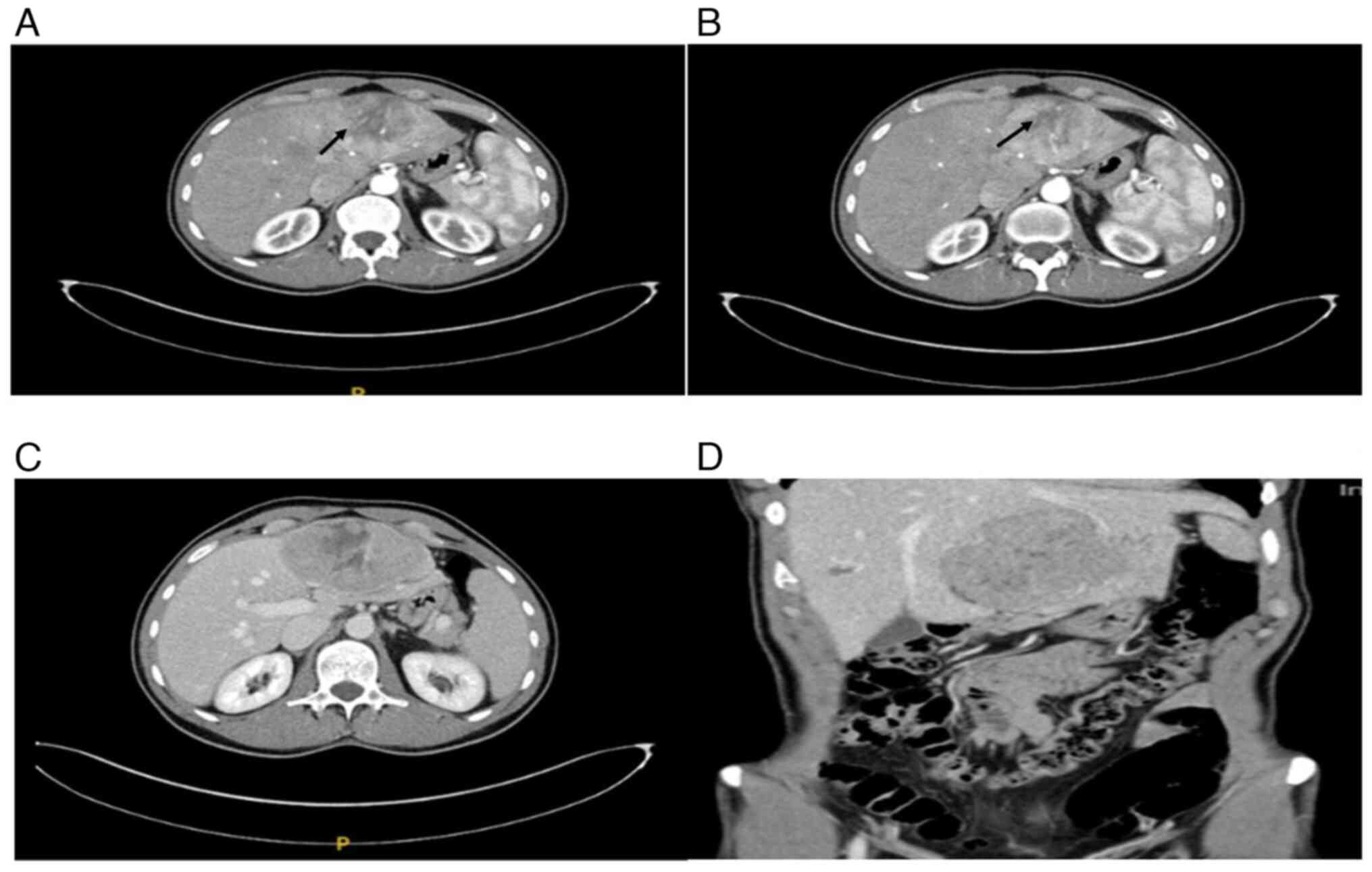
(MSCT) revealed a large tumor in the hepatic left lobe measuring 11.6x10.3x6.8 cm (Fig. 1A-D). The lesion was inhomogeneous, peripherally hypervascular, and showed partial washout in the portal venous phase. Based on imaging, a liver adenoma was suspected, and the interdisciplinary tumor board recommended resection.

(A) Dynamic multi-slice
contrast-enhanced CT imaging demonstrated a well-circumscribed
(arrow), hypervascular mass in the left lobe of the liver. (B)
Dynamic multi-slice contrast-enhanced CT imaging revealed that the
lesion showed heterogeneous enhancement in the arterial phase, with
progressive washout (arrow) or persistent enhancement in the portal
venous and delayed phases. (C) Dynamic multi-slice
contrast-enhanced CT imaging: The lesion was inhomogeneous,
peripherally hypervascular and showed partial washout in the portal
venous phase. (D) Dynamic multi-slice contrast-enhanced CT imaging
delineated the tumor within the left lobe of the liver and its
spatial relationship to adjacent organs.

Figure 1

(A) Dynamic multi-slice contrast-enhanced CT imaging demonstrated a well-circumscribed (arrow), hypervascular mass in the left lobe of the liver. (B) Dynamic multi-slice contrast-enhanced CT imaging revealed that the lesion showed heterogeneous enhancement in the arterial phase, with progressive washout (arrow) or persistent enhancement in the portal venous and delayed phases. (C) Dynamic multi-slice contrast-enhanced CT imaging: The lesion was inhomogeneous, peripherally hypervascular and showed partial washout in the portal venous phase. (D) Dynamic multi-slice contrast-enhanced CT imaging delineated the tumor within the left lobe of the liver and its spatial relationship to adjacent organs.

The patient underwent robotic-assisted left hemihepatectomy. Postoperative recovery was uneventful, and he was discharged on day 5 with negative resection margins.

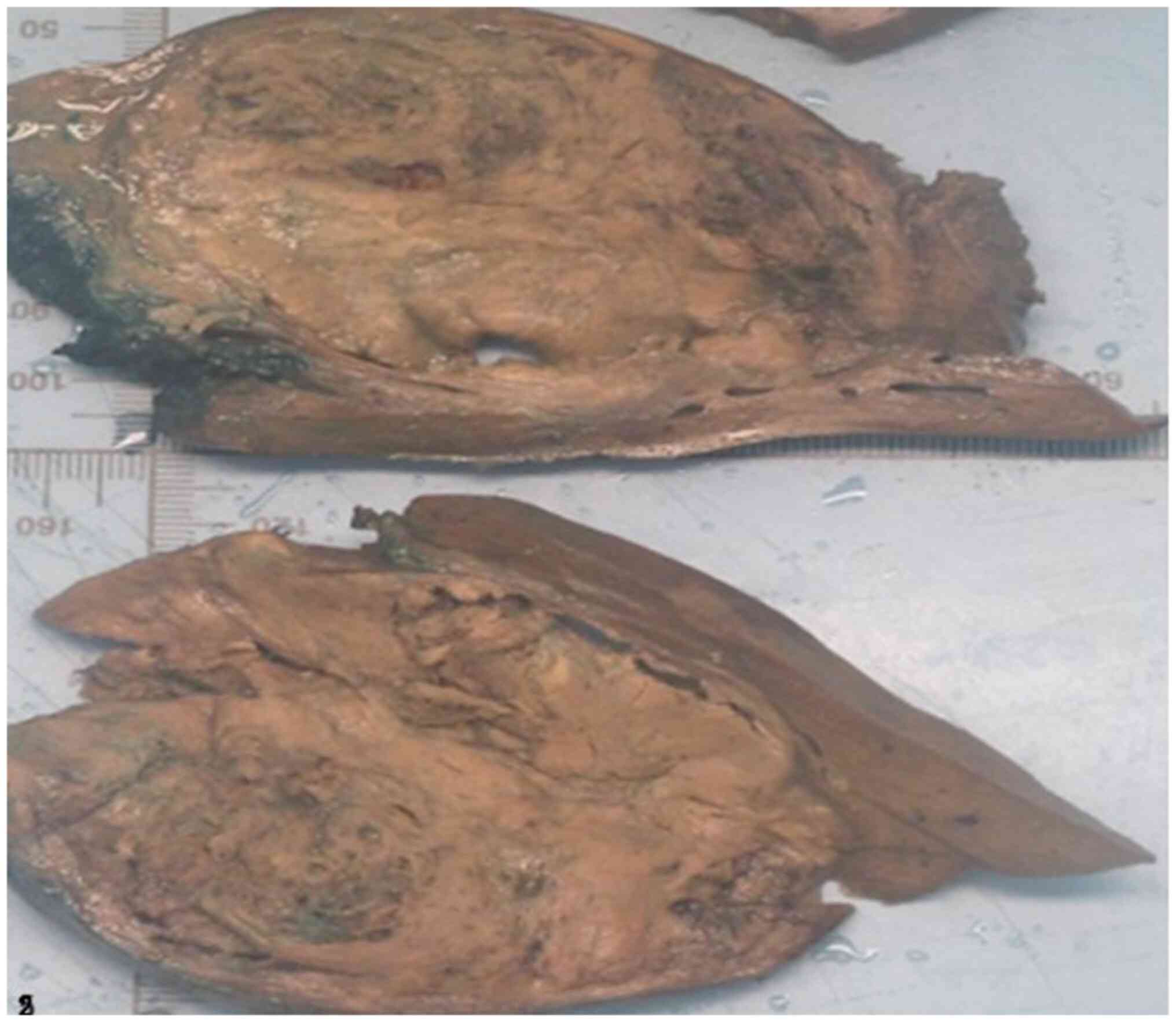
Gross pathology revealed a liver specimen weighing 604 g containing a lobulated, heterogeneous, pseudocapsulated tumor measuring 12x8x5.5 cm, located 2 mm from the resection margin (Fig. 2).

Macroscopic view of a liver segment
weighing 604 g, containing a heterogeneous, lobulated,
pseudocapsulated tumor measuring 12x8x5.5 cm. The tumor was located
2 mm from the resection margin.

Figure 2

Macroscopic view of a liver segment weighing 604 g, containing a heterogeneous, lobulated, pseudocapsulated tumor measuring 12x8x5.5 cm. The tumor was located 2 mm from the resection margin.

Microscopy (Fig. 3A-E) showed epithelioid, polyhedral cells with central to eccentric nuclei and abundant eosinophilic cytoplasm, arranged in nests and separated by a delicate capillary network. Central necrosis (Fig. 3B; arrow) was present, and, at the periphery, the tumor infiltrated its pseudocapsule with vascular invasion (Fig. 3D and E; arrow). Nuclei displayed irregular contours with focal nucleolar prominence, and the mitotic rate was up to 6/10 HPF. Based on these features and the epithelioid morphology, the initial histopathological differential diagnosis was moderately differentiated hepatocellular carcinoma (HCC).
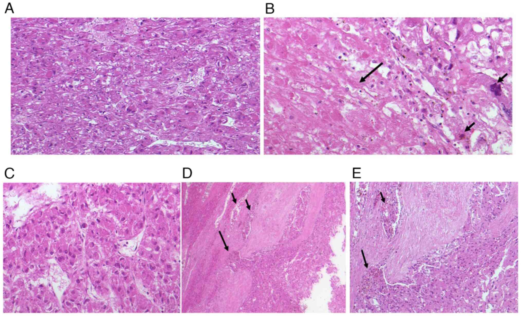
(A) Microscopic examination showed
epithelioid, polyhedral tumor cells with central to eccentric
nuclei and abundant eosinophilic cytoplasm, arranged in nests and
separated by a delicate capillary network (H&E staining;
magnification, x20). (B) Central necrosis (long arrow) was present.
Nuclei displayed irregular contours with focal nucleolar prominence
and abnormal mitotic figures (short arrow) (H&E staining;
magnification, x20). (C) Microscopic examination revealed
polyhedral epithelioid tumor cells with centrally to eccentrically
located nuclei and abundant eosinophilic cytoplasm. The cells were
arranged in trabeculae, separated by a fine capillary network
(H&E staining; magnification, x20). (D) Microscopic examination
demonstrated tumor cell infiltration into blood vessels and
capillaries (short arrow) as well as penetration of the
pseudocapsule (long arrow) (H&E staining; magnification, x10).
(E) At higher magnification, peripheral tumor infiltration was
evident, with the long arrow indicating pseudocapsule penetration
and the short arrow indicating vascular invasion (H&E staining;
magnification, x20).

Figure 3

(A) Microscopic examination showed epithelioid, polyhedral tumor cells with central to eccentric nuclei and abundant eosinophilic cytoplasm, arranged in nests and separated by a delicate capillary network (H&E staining; magnification, x20). (B) Central necrosis (long arrow) was present. Nuclei displayed irregular contours with focal nucleolar prominence and abnormal mitotic figures (short arrow) (H&E staining; magnification, x20). (C) Microscopic examination revealed polyhedral epithelioid tumor cells with centrally to eccentrically located nuclei and abundant eosinophilic cytoplasm. The cells were arranged in trabeculae, separated by a fine capillary network (H&E staining; magnification, x20). (D) Microscopic examination demonstrated tumor cell infiltration into blood vessels and capillaries (short arrow) as well as penetration of the pseudocapsule (long arrow) (H&E staining; magnification, x10). (E) At higher magnification, peripheral tumor infiltration was evident, with the long arrow indicating pseudocapsule penetration and the short arrow indicating vascular invasion (H&E staining; magnification, x20).

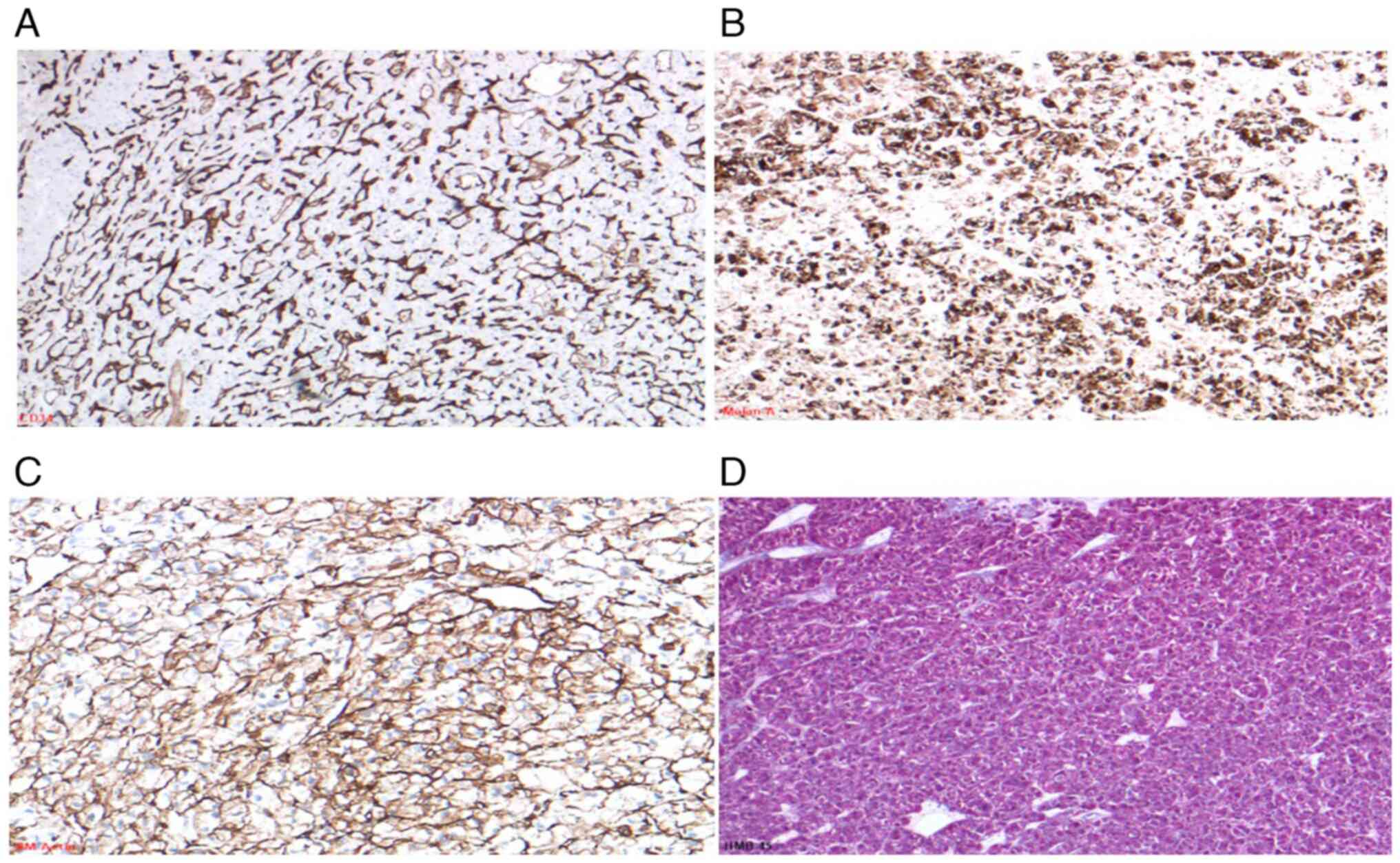
Immunohistochemistry (IHC) demonstrated complete negative for HepPar-1, Arginase-1, Glypican-3, and Glutamine Synthetase. HSP-70 showed weak, non-specific positivity. The Ki-67 proliferation index was <5% and CD34 staining highlighted the peritumoral capillary network.

To refine these unexpected findings, an extended IHC panel was applied. The tumor was negative for β-catenin, Serum Amyloid A (SAA), L-FABP, CD10, CK7, CK20, and pancytokeratin, effectively excluding HCC and hepatocellular adenoma. Further testing revealed no expression of SOX10, S100, PAX8, Chromogranin A, Vimentin, CD117 (c-Kit), Desmin, H-Caldesmon, Myogenin, MyoD1, OCT-4, and SALL4. BRG1 and INI-1 expression were preserved. Notably, tumor cells showed strong, diffuse expression of Melan-A and HMB-45, with focal weak positivity for SMA (Fig. 4A-D), consistent with a perivascular epithelioid cell tumor (PEComa).

(A) CD34 immunostaining highlighted
the endothelial cells of the intercellular capillary network within
the tumor (magnification, x20). (B) Melan-A immunohistochemistry
showed diffuse, strong cytoplasmic positivity in tumor cells
(magnification, x20). (C) SM-Actin staining revealed focal
cytoplasmic positivity in a subset of tumor cells (magnification,
x20). (D) HMB-45 immunostaining demonstrated positive cytoplasmic
expression in tumor cells, confirming melanocytic differentiation
(magnification, x20). SM-Actin, smooth muscle actin.

Figure 4

(A) CD34 immunostaining highlighted the endothelial cells of the intercellular capillary network within the tumor (magnification, x20). (B) Melan-A immunohistochemistry showed diffuse, strong cytoplasmic positivity in tumor cells (magnification, x20). (C) SM-Actin staining revealed focal cytoplasmic positivity in a subset of tumor cells (magnification, x20). (D) HMB-45 immunostaining demonstrated positive cytoplasmic expression in tumor cells, confirming melanocytic differentiation (magnification, x20). SM-Actin, smooth muscle actin.

Molecular analysis supported this diagnosis. NGS revealed truncating mutations in exons 10 and 27 of the TSC2 gene (Table I). FISH analysis excluded a TFE3 rearrangement at Xp11.23, ruling out TFE3-associated PEComa.

Table I

Detected mutations.

Table I

Detected mutations.

GeneReference sequencesExonAlteration (interpretation)AF, %Database ID
TSC1 (HGNC:12362) NM_000368/NP_000359-/--/--/--/-
TSC2 (HGNC:12363) NM_000548/NP_00053910c.913G>T; p. Gly305* (likely loss of function, likely pathogenic)11,3ClinVar: n.a.
TSC2 (HGNC:12363) NM_000548/NP_00053927c.3093dup; p. Arg1032Serfs*13 (likely loss of function, likely pathogenic)13,3ClinVar: 2910389

[i] Molecular pathological analysis using next-generation sequencing revealed mutations in exons 10 and 27 of the TSC2 gene. AF is given. The gene nomenclature follows the guidelines of the HGNC. n.a. is stated when no data were available. AF, allele frequency; HGNC, Human Genome Organisation Gene Nomenclature Committee; n.a., not applicable.

The final pathology report summarized the histological and molecular features, highlighting adverse prognostic indicators: large tumor size, infiltrative growth, necrosis, and mitotic activity (7). The case was reviewed at the multidisciplinary tumor board, and close clinical follow-up was recommended. The patient has been treated surgically to date, and follow-up was recommended. The most recent whole-body imaging, performed five months after the left hemihepatectomy, showed no metastases or residual lesions.

The Ethics Committee of University Hospital Münster (Approval No. 2019-636-f-S) approves this case.

Discussion

Most hepatic PEComas reported to date are benign. However, histological and clinical features predictive of aggressive behavior have been defined. Yoo et al (12) have identified ‘worrisome features’, including tumor size ≥7 cm, infiltrative borders, mitotic activity >1/10 mm², necrosis, vascular invasion, and classification as PEComa not otherwise specified (NOS). Risk stratification is based on these features: high-risk if ≥3 are present, intermediate-risk if 1-2 are present, and low-risk if none are identified. In the present case, multiple adverse features indicated high-risk disease.

Molecular findings further supported this assessment. The identified truncating mutations in TSC2 are typically loss-of-function variants that drive mTOR pathway activation and are more frequently associated with aggressive clinical behavior, whereas missense variants may have variable consequences depending on their functional domains. Pan et al (7) have demonstrated that TSC2 alterations correlate with poor prognosis, highlighting the biological relevance of mutation type.

Despite their malignant potential, PEComas generally have a low recurrence (3.1%) and metastasis rate (2.7%), as reported by Kvietkauskas et al (13). Complete surgical resection with negative margins remains the standard treatment. In selected cases, neoadjuvant mTOR inhibitors have been shown to reduce tumor size and resection without complications (14).

Histological subtypes may also guide diagnostic interpretation. Bennett et al (15) have emphasized that epithelioid PEComas, such as in our patient, often lack strong smooth muscle marker expression, in contrast to spindle cell-dominant PEComas, which generally stain strongly positive. Recognition of this pattern is essential to avoid misdiagnosis.

In summary, we report a rare case of hepatic PEComa with multiple high-risk features and pathogenic TSC2 mutations. This underscores the importance of a thorough immunohistochemical and molecular workup in epithelioid liver tumors to differentiate PEComa from morphologically similar entities such as hepatocellular carcinoma Awareness of this diagnostic pitfall is crucial for pathologists and clinicians to ensure accurate diagnosis, appropriate patient management, and optimal therapeutic decision-making.

Acknowledgements

The authors would like to thank Dr Kim Falkenberg (Gerhard-Domagk-Institute of Pathology and Cytology, University Hospital Muenster, D-48149 Muenster, Germany) for processing the next-generation sequencing analysis, interpreting the results and providing the data link, and Mrs. Petra Abbas (Muenster, Germany) for proofreading.

Funding

Funding: No funding was received.

Availability of data and materials

The next-generation sequencing data generated in the present study may be found in the Sequence Read Archive database under accession number PRJNA1332571 or at the following URL: https://www.ncbi.nlm.nih.gov/sra/PRJNA1332571. The other data generated in the present study are not publicly available due to data privacy and protection regulations but may be requested from the corresponding author.

Authors' contributions

MA was involved in detection and diagnosis of the rare case, performance and analysis of immunohistochemistry and molecular pathology, development of the study concept, and manuscript writing and final proofreading. BS participated in the surgical procedure, was involved in case discussion, and critically reviewed and proofread the manuscript, including validation of the overall concept. MHM was involved in case discussion, patient follow-up and proofreading of the manuscript. AP was involved in case discussion, and critical review and reading of the manuscript, with validation of the study concept. WH was involved in detection and diagnosis of the rare case, contributed to the study concept, analyzed immunohistochemistry and molecular pathology data, and proofread the manuscript. EW was involved in detection and diagnosis of the rare case, contributed to the study concept, analyzed immunohistochemistry and molecular pathology data, and proofread the manuscript. MA and WH confirm the authenticity of all the raw data. All authors have read and approved the final version of the manuscript.

Ethics approval and consent to participate

The Ethics Committee of University Hospital Münster (approval no. 2019-636-f-S; Muenster, Germany) approved this case.

Patient consent for publication

The patient provided written consent for publication.

Competing interests

The authors declare that they have no competing interests.

References

1 

Bonetti F, Pea M, Martignoni G and Zamboni G: PEC and sugar. Am J Surg Pathol. 16:307–308. 1992.PubMed/NCBI View Article : Google Scholar

2 

Pea M, Martignoni G, Zamboni G and Bonetti F: Perivascular epithelioid cell. Am J Surg Pathol. 20:1149–1153. 1996.PubMed/NCBI View Article : Google Scholar

3 

Martignoni G, Pea M, Reghellin D, Zamboni G and Bonetti F: PEComas: The past, the present and the future. Virchows Arch. 452:119–132. 2008.PubMed/NCBI View Article : Google Scholar

4 

European Chromosome 16 Tuberous Sclerosis Consortium. Identification and characterization of the tuberous sclerosis gene on chromosome 16. Cell. 75:1305–1315. 1993.PubMed/NCBI View Article : Google Scholar

5 

van Slegtenhorst M, de Hoogt R, Hermans C, Nellist M, Janssen B, Verhoef S, Lindhout D, van den Ouweland A, Halley D, Young J, et al: Identification of the tuberous sclerosis gene TSC1 on chromosome 9q34. Science. 277:805–808. 1997.PubMed/NCBI View Article : Google Scholar

6 

Kwiatkowski DJ: Tuberous sclerosis: From tubers to mTOR. Ann Hum Genet. 67:87–96. 2003.PubMed/NCBI View Article : Google Scholar

7 

Pan CC, Chung MY, Ng KF, Liu CY and Wang JS: (2012). Constant allelic alteration of the TSC2 gene in PEComas: A genetic hallmark? Modern Pathology, 25(3), 393–398.

8 

Kenerson H, Folpe AL, Takayama TK and Yeung RS: Activation of the mTOR pathway in sporadic PEComas. Journal of Pathology. 211:445–453. 2007.PubMed/NCBI View Article : Google Scholar

9 

Folpe AL, Mentzel T, Lehr HA, Fisher C, Balzer BL and Weiss SW: Perivascular epitheliod cell neoplasms of soft tissue and gynecologic origin: A clinicopathologic study of 26 cases and review of the literature. Am J Surg Pathol. 29:1558–1575. 2005.PubMed/NCBI View Article : Google Scholar

10 

Yamasaki S, Tanaka S, Fujii H, Matsumoto T, Okuda C, Watanabe G and Suda K: Monotypic epithelioid angiomyolipoma of the liver. Histopathology. 36:451–456. 2000.PubMed/NCBI View Article : Google Scholar

11 

Fang SH, Zhou LN, Jin M and Hu JB: Perivascular epithelioid cell tumor of the liver: A report of two cases and review of the literature. World J Gastroenterol. 13:5537–5539. 2007.PubMed/NCBI View Article : Google Scholar

12 

Yoo Y, Kim J and Song IH: Risk prediction criteria for the primary hepatic perivascular epithelioid cell tumour family, including angiomyolipoma: Analysis of 132 cases with a literature review. Histopathology. 86:979–992. 2025.PubMed/NCBI View Article : Google Scholar

13 

Kvietkauskas M, Samuolyte A, Rackauskas R, Luksaite-Lukste R, Karaliute G, Maskoliunaite V, Valkiuniene RB, Sokolovas V and Strupas K: Primary liver perivascular epithelioid cell tumor (PEComa): Case report and literature review. Medicina (Kaunas). 60(409)2024.PubMed/NCBI View Article : Google Scholar

14 

Bergamo F, Maruzzo M, Basso U, Montesco MC, Zagonel V, Gringeri E and Cillo U: Neoadjuvant sirolimus for a large hepatic perivascular epithelioid cell tumor (PEComa). World J Surg Oncol. 12(46)2014.PubMed/NCBI View Article : Google Scholar

15 

Bennett JA, Braga AC, Pinto A, Van de Vijver K, Cornejo K, Pesci A, Zhang L, Morales-Oyarvide V, Kiyokawa T, Zannoni GF, et al: Uterine PEComas: A morphologic, immunohistochemical, and molecular analysis of 32 tumors. Am J Surg Pathol. 42:1370–1383. 2018.PubMed/NCBI View Article : Google Scholar

Related Articles

  • Abstract
  • View
  • Download
  • Twitter
Copy and paste a formatted citation
Spandidos Publications style
Abbas M, Strücker B, Morgül MH, Pascher A, Hartmann W and Wardelmann E: Hepatic perivascular epithelioid cell tumor: A rare mesenchymal tumor with epithelioid features: A case report. Exp Ther Med 31: 12, 2026.
APA
Abbas, M., Strücker, B., Morgül, M.H., Pascher, A., Hartmann, W., & Wardelmann, E. (2026). Hepatic perivascular epithelioid cell tumor: A rare mesenchymal tumor with epithelioid features: A case report. Experimental and Therapeutic Medicine, 31, 12. https://doi.org/10.3892/etm.2025.13007
MLA
Abbas, M., Strücker, B., Morgül, M. H., Pascher, A., Hartmann, W., Wardelmann, E."Hepatic perivascular epithelioid cell tumor: A rare mesenchymal tumor with epithelioid features: A case report". Experimental and Therapeutic Medicine 31.1 (2026): 12.
Chicago
Abbas, M., Strücker, B., Morgül, M. H., Pascher, A., Hartmann, W., Wardelmann, E."Hepatic perivascular epithelioid cell tumor: A rare mesenchymal tumor with epithelioid features: A case report". Experimental and Therapeutic Medicine 31, no. 1 (2026): 12. https://doi.org/10.3892/etm.2025.13007
Copy and paste a formatted citation
x
Spandidos Publications style
Abbas M, Strücker B, Morgül MH, Pascher A, Hartmann W and Wardelmann E: Hepatic perivascular epithelioid cell tumor: A rare mesenchymal tumor with epithelioid features: A case report. Exp Ther Med 31: 12, 2026.
APA
Abbas, M., Strücker, B., Morgül, M.H., Pascher, A., Hartmann, W., & Wardelmann, E. (2026). Hepatic perivascular epithelioid cell tumor: A rare mesenchymal tumor with epithelioid features: A case report. Experimental and Therapeutic Medicine, 31, 12. https://doi.org/10.3892/etm.2025.13007
MLA
Abbas, M., Strücker, B., Morgül, M. H., Pascher, A., Hartmann, W., Wardelmann, E."Hepatic perivascular epithelioid cell tumor: A rare mesenchymal tumor with epithelioid features: A case report". Experimental and Therapeutic Medicine 31.1 (2026): 12.
Chicago
Abbas, M., Strücker, B., Morgül, M. H., Pascher, A., Hartmann, W., Wardelmann, E."Hepatic perivascular epithelioid cell tumor: A rare mesenchymal tumor with epithelioid features: A case report". Experimental and Therapeutic Medicine 31, no. 1 (2026): 12. https://doi.org/10.3892/etm.2025.13007
Follow us
  • Twitter
  • LinkedIn
  • Facebook
About
  • Spandidos Publications
  • Careers
  • Cookie Policy
  • Privacy Policy
How can we help?
  • Help
  • Live Chat
  • Contact
  • Email to our Support Team