Spandidos Publications Logo
  • About
    • About Spandidos
    • Aims and Scopes
    • Abstracting and Indexing
    • Editorial Policies
    • Reprints and Permissions
    • Job Opportunities
    • Terms and Conditions
    • Contact
  • Journals
    • All Journals
    • Oncology Letters
      • Oncology Letters
      • Information for Authors
      • Editorial Policies
      • Editorial Board
      • Aims and Scope
      • Abstracting and Indexing
      • Bibliographic Information
      • Archive
    • International Journal of Oncology
      • International Journal of Oncology
      • Information for Authors
      • Editorial Policies
      • Editorial Board
      • Aims and Scope
      • Abstracting and Indexing
      • Bibliographic Information
      • Archive
    • Molecular and Clinical Oncology
      • Molecular and Clinical Oncology
      • Information for Authors
      • Editorial Policies
      • Editorial Board
      • Aims and Scope
      • Abstracting and Indexing
      • Bibliographic Information
      • Archive
    • Experimental and Therapeutic Medicine
      • Experimental and Therapeutic Medicine
      • Information for Authors
      • Editorial Policies
      • Editorial Board
      • Aims and Scope
      • Abstracting and Indexing
      • Bibliographic Information
      • Archive
    • International Journal of Molecular Medicine
      • International Journal of Molecular Medicine
      • Information for Authors
      • Editorial Policies
      • Editorial Board
      • Aims and Scope
      • Abstracting and Indexing
      • Bibliographic Information
      • Archive
    • Biomedical Reports
      • Biomedical Reports
      • Information for Authors
      • Editorial Policies
      • Editorial Board
      • Aims and Scope
      • Abstracting and Indexing
      • Bibliographic Information
      • Archive
    • Oncology Reports
      • Oncology Reports
      • Information for Authors
      • Editorial Policies
      • Editorial Board
      • Aims and Scope
      • Abstracting and Indexing
      • Bibliographic Information
      • Archive
    • Molecular Medicine Reports
      • Molecular Medicine Reports
      • Information for Authors
      • Editorial Policies
      • Editorial Board
      • Aims and Scope
      • Abstracting and Indexing
      • Bibliographic Information
      • Archive
    • World Academy of Sciences Journal
      • World Academy of Sciences Journal
      • Information for Authors
      • Editorial Policies
      • Editorial Board
      • Aims and Scope
      • Abstracting and Indexing
      • Bibliographic Information
      • Archive
    • International Journal of Functional Nutrition
      • International Journal of Functional Nutrition
      • Information for Authors
      • Editorial Policies
      • Editorial Board
      • Aims and Scope
      • Abstracting and Indexing
      • Bibliographic Information
      • Archive
    • International Journal of Epigenetics
      • International Journal of Epigenetics
      • Information for Authors
      • Editorial Policies
      • Editorial Board
      • Aims and Scope
      • Abstracting and Indexing
      • Bibliographic Information
      • Archive
    • Medicine International
      • Medicine International
      • Information for Authors
      • Editorial Policies
      • Editorial Board
      • Aims and Scope
      • Abstracting and Indexing
      • Bibliographic Information
      • Archive
  • Articles
  • Information
    • Information for Authors
    • Information for Reviewers
    • Information for Librarians
    • Information for Advertisers
    • Conferences
  • Language Editing
Spandidos Publications Logo
  • About
    • About Spandidos
    • Aims and Scopes
    • Abstracting and Indexing
    • Editorial Policies
    • Reprints and Permissions
    • Job Opportunities
    • Terms and Conditions
    • Contact
  • Journals
    • All Journals
    • Biomedical Reports
      • Information for Authors
      • Editorial Policies
      • Editorial Board
      • Aims and Scope
      • Abstracting and Indexing
      • Bibliographic Information
      • Archive
    • Experimental and Therapeutic Medicine
      • Information for Authors
      • Editorial Policies
      • Editorial Board
      • Aims and Scope
      • Abstracting and Indexing
      • Bibliographic Information
      • Archive
    • International Journal of Epigenetics
      • Information for Authors
      • Editorial Policies
      • Editorial Board
      • Aims and Scope
      • Abstracting and Indexing
      • Bibliographic Information
      • Archive
    • International Journal of Functional Nutrition
      • Information for Authors
      • Editorial Policies
      • Editorial Board
      • Aims and Scope
      • Abstracting and Indexing
      • Bibliographic Information
      • Archive
    • International Journal of Molecular Medicine
      • Information for Authors
      • Editorial Policies
      • Editorial Board
      • Aims and Scope
      • Abstracting and Indexing
      • Bibliographic Information
      • Archive
    • International Journal of Oncology
      • Information for Authors
      • Editorial Policies
      • Editorial Board
      • Aims and Scope
      • Abstracting and Indexing
      • Bibliographic Information
      • Archive
    • Medicine International
      • Information for Authors
      • Editorial Policies
      • Editorial Board
      • Aims and Scope
      • Abstracting and Indexing
      • Bibliographic Information
      • Archive
    • Molecular and Clinical Oncology
      • Information for Authors
      • Editorial Policies
      • Editorial Board
      • Aims and Scope
      • Abstracting and Indexing
      • Bibliographic Information
      • Archive
    • Molecular Medicine Reports
      • Information for Authors
      • Editorial Policies
      • Editorial Board
      • Aims and Scope
      • Abstracting and Indexing
      • Bibliographic Information
      • Archive
    • Oncology Letters
      • Information for Authors
      • Editorial Policies
      • Editorial Board
      • Aims and Scope
      • Abstracting and Indexing
      • Bibliographic Information
      • Archive
    • Oncology Reports
      • Information for Authors
      • Editorial Policies
      • Editorial Board
      • Aims and Scope
      • Abstracting and Indexing
      • Bibliographic Information
      • Archive
    • World Academy of Sciences Journal
      • Information for Authors
      • Editorial Policies
      • Editorial Board
      • Aims and Scope
      • Abstracting and Indexing
      • Bibliographic Information
      • Archive
  • Articles
  • Information
    • For Authors
    • For Reviewers
    • For Librarians
    • For Advertisers
    • Conferences
  • Language Editing
Login Register Submit
  • This site uses cookies
  • You can change your cookie settings at any time by following the instructions in our Cookie Policy. To find out more, you may read our Privacy Policy.

    I agree
Search articles by DOI, keyword, author or affiliation
Search
Advanced Search
presentation
Experimental and Therapeutic Medicine
Join Editorial Board Propose a Special Issue
Print ISSN: 1792-0981 Online ISSN: 1792-1015
Journal Cover
May-2026 Volume 31 Issue 5

Full Size Image

Sign up for eToc alerts
Recommend to Library

Journals

International Journal of Molecular Medicine

International Journal of Molecular Medicine

International Journal of Molecular Medicine is an international journal devoted to molecular mechanisms of human disease.

International Journal of Oncology

International Journal of Oncology

International Journal of Oncology is an international journal devoted to oncology research and cancer treatment.

Molecular Medicine Reports

Molecular Medicine Reports

Covers molecular medicine topics such as pharmacology, pathology, genetics, neuroscience, infectious diseases, molecular cardiology, and molecular surgery.

Oncology Reports

Oncology Reports

Oncology Reports is an international journal devoted to fundamental and applied research in Oncology.

Experimental and Therapeutic Medicine

Experimental and Therapeutic Medicine

Experimental and Therapeutic Medicine is an international journal devoted to laboratory and clinical medicine.

Oncology Letters

Oncology Letters

Oncology Letters is an international journal devoted to Experimental and Clinical Oncology.

Biomedical Reports

Biomedical Reports

Explores a wide range of biological and medical fields, including pharmacology, genetics, microbiology, neuroscience, and molecular cardiology.

Molecular and Clinical Oncology

Molecular and Clinical Oncology

International journal addressing all aspects of oncology research, from tumorigenesis and oncogenes to chemotherapy and metastasis.

World Academy of Sciences Journal

World Academy of Sciences Journal

Multidisciplinary open-access journal spanning biochemistry, genetics, neuroscience, environmental health, and synthetic biology.

International Journal of Functional Nutrition

International Journal of Functional Nutrition

Open-access journal combining biochemistry, pharmacology, immunology, and genetics to advance health through functional nutrition.

International Journal of Epigenetics

International Journal of Epigenetics

Publishes open-access research on using epigenetics to advance understanding and treatment of human disease.

Medicine International

Medicine International

An International Open Access Journal Devoted to General Medicine.

Journal Cover
May-2026 Volume 31 Issue 5

Full Size Image

Sign up for eToc alerts
Recommend to Library

  • Article
  • Citations
    • Cite This Article
    • Download Citation
    • Create Citation Alert
    • Remove Citation Alert
    • Cited By
  • Similar Articles
    • Related Articles (in Spandidos Publications)
    • Similar Articles (Google Scholar)
    • Similar Articles (PubMed)
  • Download PDF
  • Download XML
  • View XML

  • Supplementary Files
    • Supplementary_Data1.mp4
    • Supplementary_Data2.pdf
Case Report Open Access

Direct venous‑to‑arterial communication as a novel mechanism for ectopic embolism following esophageal variceal embolization: A case report

  • Authors:
    • Youchun Lei
    • You Ke
    • Tianwen Yang
    • Zhengguo Xu
  • View Affiliations / Copyright

    Affiliations: Department of Gastroenterology, The First Affiliated Hospital of Chongqing Medical and Pharmaceutical College, Chongqing 400000, P.R. China, Department of Hepatobiliary Surgery, The Second Affiliated Hospital, Chongqing Medical University, Chongqing 400000, P.R. China, Department of Gastroenterology, The Second Affiliated Hospital, The Third Military Medical University, Chongqing 400000, P.R. China
    Copyright: © Lei et al. This is an open access article distributed under the terms of Creative Commons Attribution License.
  • Article Number: 130
    |
    Published online on: March 6, 2026
       https://doi.org/10.3892/etm.2026.13125
  • Expand metrics +
Metrics: Total Views: 0 (Spandidos Publications: | PMC Statistics: )
Metrics: Total PDF Downloads: 0 (Spandidos Publications: | PMC Statistics: )
Cited By (CrossRef): 0 citations Loading Articles...

This article is mentioned in:


Abstract

Ectopic embolism is an infrequent but potentially life‑threatening complication following esophageal variceal embolization. Although it has been hypothesized to result from right‑to‑left shunting via pulmonary arteriovenous malformations or patent foramen ovale, definitive anatomical routes often remain undetected. The present case study reports a 57‑year‑old male patient with decompensated cirrhosis who developed acute bilateral visual loss and lower limb weakness within 24 h after undergoing transjugular intrahepatic portosystemic shunt placement combined with endovascular embolization of esophageal varices. Brain imaging confirmed multiple acute cerebral infarctions; notably, angiographic review revealed an abnormal direct venous connection between the esophageal variceal plexus and the left heart/ascending aorta, bypassing the pulmonary circuit. No cardiac or pulmonary shunts were identified. This direct communication likely permitted embolic material to enter the systemic circulation, causing cerebral artery occlusion. In conclusion, to the best of our knowledge, the present case provides the first angiographic evidence of a direct vascular route from esophageal varices to the systemic arterial system. The present finding underscores the importance of comprehensive imaging before embolization and suggests that solid embolic agents such as coils may be preferable in select patients to prevent ectopic embolism.

Introduction

Esophageal varices are a common and life-threatening complication of portal hypertension in patients with liver cirrhosis (1). Endovascular embolization, often in combination with transjugular intrahepatic portosystemic shunt (TIPS), is a standard therapeutic approach to reduce variceal bleeding (2,3). Although generally effective, complications such as ectopic embolism may occur, occasionally resulting in catastrophic outcomes including cerebral, renal or mesenteric infarction (4,5); the underlying anatomical basis for such events has not been fully elucidated. In patients with cirrhosis, treatment for ectopic embolism caused by esophageal varices is relatively rare. Within the literature, to the best of our knowledge, there is only 1 case of cerebral embolism (6) and 3 cases of spinal cord embolism (7-9), but the specific cause of embolism is unclear.

To the best of our knowledge, the present case is the first reported case of angiographically confirmed direct venous-to-arterial communication causing ectopic embolism. Traditionally, right-to-left shunting mechanisms-such as pulmonary arteriovenous malformations or intracardiac defects-have been implicated (7,10); however, in several cases, including the present case, such mechanisms are absent (11-14).

Case report

Case presentation

A 57-year-old man with a long-standing history of hepatitis B virus-related cirrhosis (Child-Pugh class B; model for end-stage liver disease score 15) (15) was admitted to The Second Affiliated Hospital of The Third Military Medical University (Chongqing, China) in June 2023, for recurrent upper gastrointestinal bleeding and progressive ascites, which was unresponsive to diuretics. Previous endoscopic therapy with ligation had been performed twice over the past year, with partial but temporary hemostasis.

On admission, the patient was hemodynamically stable. Laboratory investigations revealed anemia (hemoglobin, 82 g/l; reference range, 110-160 g/l), thrombocytopenia (platelets, 55x109/l; reference range, 125-350x109/l), hypoalbuminemia (albumin, 28 g/l; reference range, 35-50 g/l) and elevated total bilirubin (34 µmol/l; reference range, 3-22 µmol/l). Imaging using contrast-enhanced abdominal CT showed marked splenomegaly, large esophageal varices and ascites. After multidisciplinary discussion, the patient underwent elective TIPS combined with embolization of esophageal varices.

Imaging and interventions

The TIPS procedure was performed in June 2023, under local anesthesia via a right internal jugular approach. Puncture of the left portal vein branch was achieved via the middle hepatic vein. Splenic venography revealed large, tortuous esophageal varices. After balloon dilatation of the puncture channel (Cook 7x8 mm), an 8x80 mm GORE VIATORR stent graft was placed.

Embolization of the varices was performed through a 4F Cobra catheter by slow injection of a 1:1 mixture of 2 ml tissue adhesive (GLUBRAN2) and 2 ml iodized oil under fluoroscopic control. Pre- and post-TIPS portal pressure gradients were 32 and 17 mmHg, respectively, indicating successful decompression. At ~24 
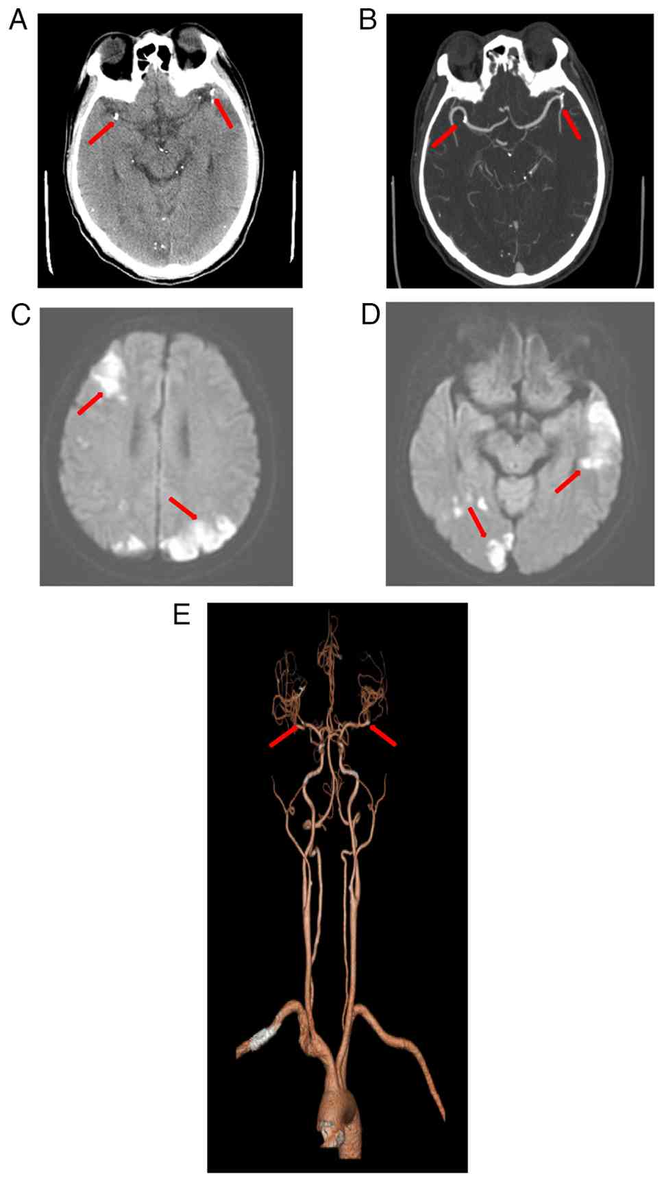
h post-surgery, the patient developed sudden bilateral vision loss and lower limb weakness (grade III muscle strength bilaterally). Magnetic resonance angiography showed embolic occlusion of branches of the middle cerebral artery. Emergent neuroimaging including non-contrast CT and diffusion-weighted MRI revealed multiple acute infarctions in the bilateral frontal lobe, parietal lobe, occipital lobe, temporal lobe, left thalamus and cerebellar hemisphere (Fig. 1A-E).

Multimodal imaging evaluation
revealing acute cerebral infarction. (A) Axial head CTA
demonstrates spotty and nodular high-density filling of the
sclerosing agent within the distal M1 segments of the bilateral
MCAs. (B) Head CTA maximum intensity projection reveals cast-like
high-density filling in the distal M1 segments of the bilateral
MCAs. (C) Brain MRI (DWI, b=800) shows multiple patchy hyperintense
lesions in the right frontal lobe and bilateral parietal lobes. (D)
Brain MRI DWI (b=800) displays multiple patchy hyperintense lesions
in the right temporo-occipital region and the left temporal lobe.
(E) Head CTA volume rendering depicts the abrupt cutoff of the
distal M1 segments of the bilateral MCAs, accompanied by sparse and
reduced distal branching. CTA, CT angiography; MCA, middle cerebral
artery; DWI, diffusion-weighted imaging. (A, B and E) Arrows refer
to the positions filled with sclerosing agents in the cerebral
arteries. (C and D) Arrows refer to the part of the cerebral
infarction caused by arterial blockage.

Figure 1

Multimodal imaging evaluation revealing acute cerebral infarction. (A) Axial head CTA demonstrates spotty and nodular high-density filling of the sclerosing agent within the distal M1 segments of the bilateral MCAs. (B) Head CTA maximum intensity projection reveals cast-like high-density filling in the distal M1 segments of the bilateral MCAs. (C) Brain MRI (DWI, b=800) shows multiple patchy hyperintense lesions in the right frontal lobe and bilateral parietal lobes. (D) Brain MRI DWI (b=800) displays multiple patchy hyperintense lesions in the right temporo-occipital region and the left temporal lobe. (E) Head CTA volume rendering depicts the abrupt cutoff of the distal M1 segments of the bilateral MCAs, accompanied by sparse and reduced distal branching. CTA, CT angiography; MCA, middle cerebral artery; DWI, diffusion-weighted imaging. (A, B and E) Arrows refer to the positions filled with sclerosing agents in the cerebral arteries. (C and D) Arrows refer to the part of the cerebral infarction caused by arterial blockage.

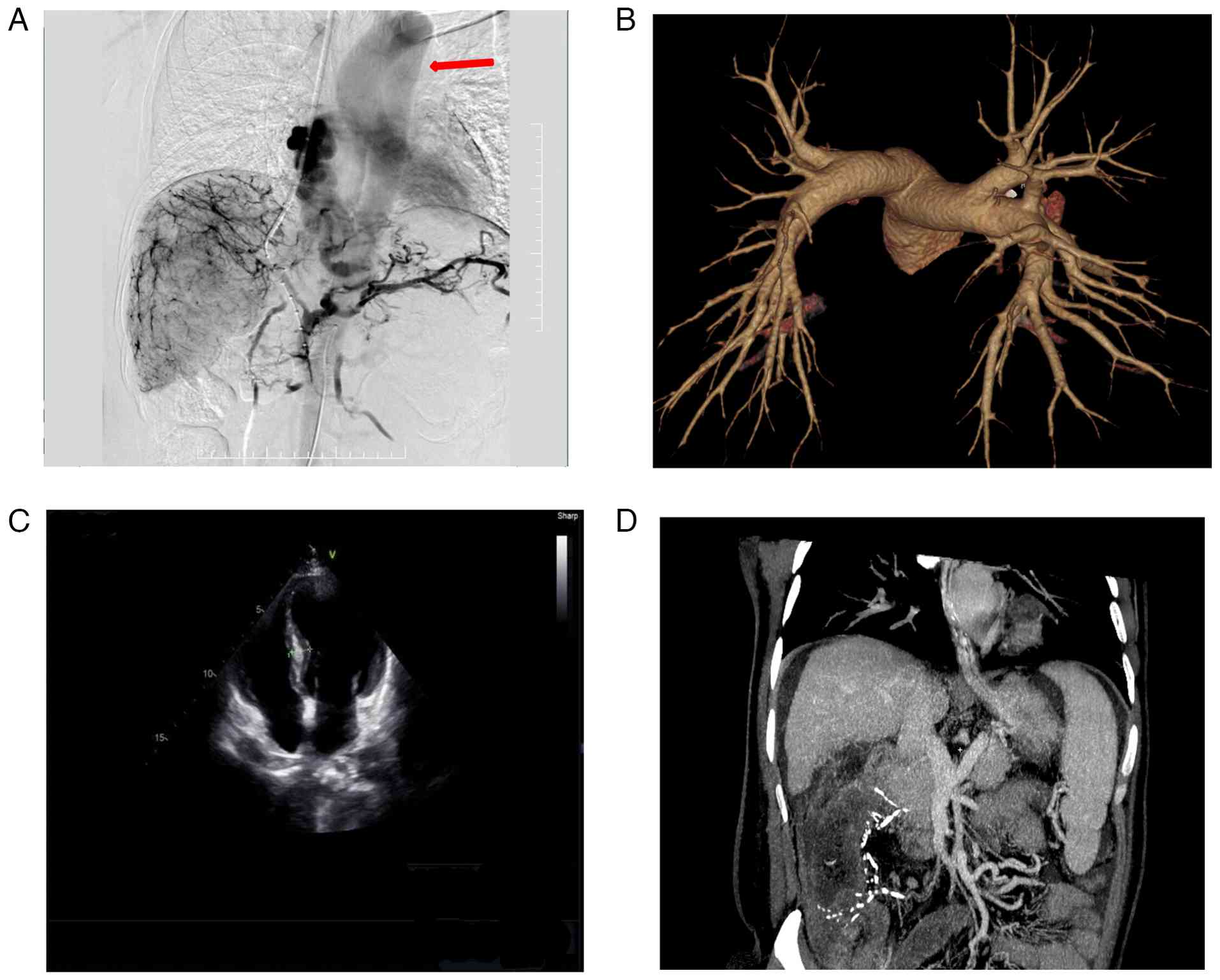
To identify the embolic source, further evaluation was conducted. Echocardiography excluded patent foramen ovale or intracardiac thrombus (Fig. 2C). Additionally, no arteriovenous malformations or pulmonary embolism was found on pulmonary CT angiography (Fig. 2B).

Intraoperative angiography
demonstrated direct drainage from the esophageal variceal plexus to
the left heart and ascending aorta, exclusion of conventional
shunts and underlying abdominal pathology. (A) Sequential
fluoroscopic images obtained during the procedure reveal the
abnormal vascular pathway in this single patient. Contrast medium
injected into the esophageal variceal plexus drains directly into
the left heart and subsequently fills the ascending aorta
(indicated by arrows), completely bypassing the pulmonary
circulation. This rare anatomical variant provided the direct
conduit for the embolic material to enter the systemic arterial
circulation. (B) Pulmonary CT angiography shows no evidence of
pulmonary arteriovenous malformations or pulmonary embolism, ruling
out intrapulmonary shunting. (C) Echocardiography reveals normal
cardiac chambers with no signs of patent foramen ovale or
intracardiac thrombus, excluding intracardiac shunting sources. (D)
Contrast-enhanced abdominal CT scan demonstrates marked
splenomegaly, extensive esophageal varices and ascites, confirming
the background of decompensated cirrhosis and portal
hypertension.

Figure 2

Intraoperative angiography demonstrated direct drainage from the esophageal variceal plexus to the left heart and ascending aorta, exclusion of conventional shunts and underlying abdominal pathology. (A) Sequential fluoroscopic images obtained during the procedure reveal the abnormal vascular pathway in this single patient. Contrast medium injected into the esophageal variceal plexus drains directly into the left heart and subsequently fills the ascending aorta (indicated by arrows), completely bypassing the pulmonary circulation. This rare anatomical variant provided the direct conduit for the embolic material to enter the systemic arterial circulation. (B) Pulmonary CT angiography shows no evidence of pulmonary arteriovenous malformations or pulmonary embolism, ruling out intrapulmonary shunting. (C) Echocardiography reveals normal cardiac chambers with no signs of patent foramen ovale or intracardiac thrombus, excluding intracardiac shunting sources. (D) Contrast-enhanced abdominal CT scan demonstrates marked splenomegaly, extensive esophageal varices and ascites, confirming the background of decompensated cirrhosis and portal hypertension.

Notably, retrospective analysis of fluoroscopic angiography demonstrated direct drainage from the esophageal variceal plexus to the left atrium and ascending aorta, bypassing the pulmonary circulation (Fig. 2A and D and Video S1). This anatomical variant likely accounted for the cerebral embolism.

Due to the recent hemorrhagic history, anticoagulation was withheld, and the patient was managed supportively with neuroprotective agents (0.25 g citicoline once daily for 5 days). Over 2 weeks, bilateral visual acuity markedly improved, and motor function partially recovered. At the most recent follow-up, the patient remained clinically stable with normal mental status and appetite. He reported no recurrence of gastrointestinal bleeding (hematemesis or melena), nausea, vomiting, abdominal pain or ascites.

Discussion

The present case represents a novel mechanism of ectopic embolism in the setting of esophageal variceal embolization. To the best of our knowledge, the present case is the first reported case of angiographically confirmed direct venous-to-arterial communication causing ectopic embolism. Traditionally, right-to-left shunting via patent foramen ovale or pulmonary arteriovenous fistulas has been considered necessary for systemic embolization following venous embolization procedures (16-18). However, in the present case, such conventional shunts were definitively ruled out.

Instead, angiography revealed a rare and direct venous drainage route from the esophageal variceal plexus to the systemic circulation via the left heart, presumably through pathological dilation of small bronchial or mediastinal collaterals (19,20). To the best of our knowledge, this anatomic variant has not been previously described in the literature, in the context of post-embolization cerebral infarction (16).

A similar case of spinal cord infarction after sclerotherapy was reported by Seidman et al (7) in 1984, but no vascular mechanism was identified. Other studies postulated that the patent foramen ovale may allow embolic material into the cerebral circulation during TIPS, but in the absence of identifiable shunts, the mechanism remained speculative (21-25). The present case confirms the hypothesis of an alternative, direct venous-to-arterial conduit.

Due to the difficulty of detecting this vascular pathway with preoperative routine vascular CT, it is recommended to extend the display time and fully understand its flow direction in gastric coronary angiography during TIPS surgery, especially paying attention to whether the heart and aortic circulatory system are visible. From the perspective of surgical safety, if direct esophageal venous to the systemic arterial communication is found during surgery, it is recommended to avoid using liquid embolic agents due to their inherent migration risk (26,27). Solid embolic agents such as coils or embolization are unlikely to penetrate small caliber blood vessels and may be a safer option, particularly when imaging suspicion or abnormal communication is observed prior to embolization (28,29).

In conclusion, the present case highlights a previously unreported cause of systemic embolization following esophageal variceal embolization; direct venous communication between the esophageal variceal plexus and the left heart/ascending aorta. If the cardiac and aortic circulatory systems are found to be visualized during gastric coronary angiography during TIPS surgery, it is recommended to use large-diameter or solid embolic materials such as coils, which may prevent potentially life-threatening ectopic embolic events.

Supplementary Material

Dynamic angiography of the present case. This video demonstrates the direct vascular communication between the esophageal variceal plexus and the left heart/ascending aorta during the transjugular intrahepatic portosystemic shunt procedure, which corresponds to the sequential images shown in Fig. 2A.
Supplementary data.

Acknowledgements

Not applicable.

Funding

Funding: The authors declare that financial support was received for the research, authorship and publication of this article. This study was funded by the General Project of Chongqing Science and Health Joint Medical Research Project (grant no. 2024MSXM081), the Science and Technology Research Project of Chongqing Education Commission (grant no. KJQN202302828), the 2023 Chongqing Nan'an District Science and Health Union Public Medical Research Project (grant no. 2023-05), the Open Research Project of the Chongqing Key Laboratory for Occupational Disease Prevention and Poisoning Treatment in 2021 (grant no. 2021ZYBKF07), the Chongqing Pharmaceutical Vocational Education Group General Project (grant no. CQZJ202352), the General Project of the Incubation Fund within The First Affiliated Hospital of Chongqing Medical and pharmaceutical College (grant no. 2022-2023MS02) and the 2023 Chongqing Nan'an District Public Health Key Specialty (Disease Prevention and Control) Construction Project.

Availability of data and materials

The data generated in the present study may be requested from the corresponding author.

Authors' contributions

YL and ZX contributed to designing the study and performed the surgery. YK obtained the medical images. TY advised on patient treatment and analyzed patient data. ZX and YK confirm the authenticity of all the raw data. All authors have read and approved the final manuscript.

Ethics approval and consent to participate

The Ethics Committee of the Second Affiliated Hospital of the Third Military Medical University (Chongqing, China) exempted the present study from ethical approval. The participant provided written informed consent to participate in this study.

Patient consent for publication

Written informed consent was obtained from the patient for the publication of clinical details and any accompanying images.

Competing interests

The authors declare that they have no competing interests.

References

1 

Zhang CM and Wang X: Suspected cerebrovascular air embolism during endoscopic esophageal varices ligation under sedation with fatal outcome: A case report. World J Clin Cases. 10:371–380. 2022.PubMed/NCBI View Article : Google Scholar

2 

Abuelazm MT, Cheema HA, Jafar U, Awad AK, Atef M, Abdalshafy H, Alashwah M, Shahid A, Awan RU, Afifi AM, et al: Transjugular intrahepatic portosystemic shunt with or without variceal embolization to prevent variceal rebleeding: An updated meta-analysis. Expert Rev Gastroenterol Hepatol. 17:741–751. 2023.PubMed/NCBI View Article : Google Scholar

3 

Betancourt-Torres M, Perez-Torres A, Figueroa-Diaz L and Labat Alvarez EJ: Iatrogenic cerebral air embolism during esophago-gastroduodenoscopy. Am J Case Rep. 21(e925046)2020.PubMed/NCBI View Article : Google Scholar

4 

Chevallier O, Comby PO, Guillen K, Pellegrinelli J, Mouillot T, Falvo N, Bardou M, Midulla M, Aho-Glélé S and Loffroy R: Efficacy, safety and outcomes of transcatheter arterial embolization with N-butyl cyanoacrylate glue for non-variceal gastrointestinal bleeding: A systematic review and meta-analysis. Diagn Interv Imaging. 102:479–487. 2021.PubMed/NCBI View Article : Google Scholar

5 

Davis JPE, Lim JK, Francis FF and Ahn J: AGA clinical practice update on management of portal vein thrombosis in patients with cirrhosis: Expert review. Gastroenterology. 168:396–404.e1. 2025.PubMed/NCBI View Article : Google Scholar

6 

Wang G, Zhang C and Shi Y: Cerebral embolism due to portopulmonary venous anastomosis during endoscopic therapy for gastric varices. Am J Gastroenterol. 114(1833)2019.PubMed/NCBI View Article : Google Scholar

7 

Seidman E, Weber AM, Morin CL, Ethier R, Lamarche JB, Guerguérian AJ, Geoffroy G and Roy CC: Spinal cord paralysis following sclerotherapy for esophageal varices. Hepatology. 4:950–954. 1984.PubMed/NCBI View Article : Google Scholar

8 

Mueller D and Gilden DH: Brown-Sequard syndrome after esophageal sclerotherapy and crack cocaine abuse. Neurology. 58:1129–1130. 2002.PubMed/NCBI View Article : Google Scholar

9 

Heller SL, Meyer JR and Russell EJ: Spinal cord venous infarction following endoscopic sclerotherapy for esophageal varices. Neurology. 47:1081–1085. 1996.PubMed/NCBI View Article : Google Scholar

10 

Xu SH, Wu F, Guo LH, Zhang WB and Xu HX: Liver fibrosis index-based nomograms for identifying esophageal varices in patients with chronic hepatitis B related cirrhosis. World J Gastroenterol. 26:7204–7221. 2020.PubMed/NCBI View Article : Google Scholar

11 

de Sousa CP, Carvalho C, Sousa C and Amaral R: Treatment of gastric varices with cyanoacrylate complicated by systemic embolization. Treatment of gastric varices with cyanoacrylate complicated by systemic embolization. Rev Gastroenterol Peru. 44:67–70. 2024.PubMed/NCBI

12 

Gong T, Tsauo J, Ding M, Jin L, Duan F, Yu Y and Li X: Transcatheter arterial embolization for cancer-related non-variceal upper gastrointestinal bleeding: A multicenter retrospective study of 107 patients. Diagn Interv Imaging. 104:60–66. 2023.PubMed/NCBI View Article : Google Scholar

13 

Lee EW, Eghtesad B, Garcia-Tsao G, Haskal ZJ, Hernandez-Gea V, Jalaeian H, Kalva SP, Mohanty A, Thabut D and Abraldes JG: AASLD practice guidance on the use of TIPS, variceal embolization, and retrograde transvenous obliteration in the management of variceal hemorrhage. Hepatology. 79:224–250. 2024.PubMed/NCBI View Article : Google Scholar

14 

Jiang N, Wang WS, Zhu XL and Shen J: Liver failure after percutaneous transhepatic variceal embolization: A case report. Asian J Surg. 46:2857–2858. 2023.PubMed/NCBI View Article : Google Scholar

15 

D'Amico G, Garcia-Tsao G and Pagliaro L: Natural history and prognostic indicators of survival in cirrhosis: A systematic review of 118 studies. J Hepatol. 44:217–231. 2006.PubMed/NCBI View Article : Google Scholar

16 

Kim JS, Kim BW, Kim DH, Park CH, Lee H, Joo MK, Jung DH, Chung JW, Choi HS, Baik GH, et al: Guidelines for non-variceal upper gastrointestinal bleeding. Korean J Gastroenterol. 75:322–332. 2020.PubMed/NCBI View Article : Google Scholar : (In Korean).

17 

Kolb JM and Samarasena JB: EUS-guided splenic artery embolization for variceal hemorrhage: Balancing creativity and innovation in Endo-hepatology with caution. Gastrointest Endosc. 95:184–186. 2022.PubMed/NCBI View Article : Google Scholar

18 

Lai HY, Wu KT, Liu Y, Zeng ZF and Zhang B: Angiography and transcatheter arterial embolization for non-variceal gastrointestinal bleeding. Scand J Gastroenterol. 55:931–940. 2020.PubMed/NCBI View Article : Google Scholar

19 

Liu B and Li G: Progress in endoscopic and interventional treatment of esophagogastric variceal bleeding. Dis Markers. 2022(2940578)2022.PubMed/NCBI View Article : Google Scholar

20 

Ohs Z, Jones M, Sharma N and Loveridge K: Percutaneous transhepatic embolization of ectopic varices in a patient with portal hypertension presenting with hemorrhagic shock. Cureus. 13(e18209)2021.PubMed/NCBI View Article : Google Scholar

21 

Onishi Y, Shimizu H, Tsunoda S, Obama K and Nakamoto Y: Direct percutaneous access to a mesenteric vein for antegrade embolization of esophageal varices: A case report. Radiol Case Rep. 16:2491–2495. 2021.PubMed/NCBI View Article : Google Scholar

22 

Pal A, Blanzy J, Gómez KJR, Preul MC and Vernon BL: Liquid embolic agents for endovascular embolization: A review. Gels. 9(378)2023.PubMed/NCBI View Article : Google Scholar

23 

Patidar Y, Chatterjee N, Mukund A and Sarin SK: Evaluation of clinical outcome and predictors of mortality in patients undergoing antegrade transvenous variceal embolization in adjunct to salvage transjugular intrahepatic portosystemic shunt for active uncontrolled gastric variceal bleeding. Br J Radiol. 97:1791–1798. 2024.PubMed/NCBI View Article : Google Scholar

24 

Sioutas GS, Vivanco-Suarez J, Shekhtman O, Matache IM, Salem MM, Burkhardt JK, Srinivasan VM and Jankowitz BT: Liquid embolic agents for middle meningeal artery embolization in chronic subdural hematoma: Institutional experience with systematic review and meta-analysis. Interv Neuroradiol. 15(15910199231183132)2023.PubMed/NCBI View Article : Google Scholar

25 

Swaminathan N and Chaudhary S: Oesophageal variceal-pulmonary venous fistula A rare cause of a right-to-left shunt. Eur J Case Rep Intern Med. 7(001482)2020.PubMed/NCBI View Article : Google Scholar

26 

Vollherbst DF, Chapot R, Bendszus M and Möhlenbruch MA: Glue, onyx, squid or PHIL? Liquid embolic agents for the embolization of cerebral arteriovenous malformations and dural arteriovenous fistulas. Clin Neuroradiol. 32:25–38. 2022.PubMed/NCBI View Article : Google Scholar

27 

Wang DX, Wu XJ, Yu JZ, Zhan JY, Xing FF, Liu W, Chen JM, Liu P, Liu CH and Mu YP: Visualizing global progress and challenges in esophagogastric variceal bleeding. World J Gastrointest Surg. 17(102020)2025.PubMed/NCBI View Article : Google Scholar

28 

Wang Z, Zeng Z, Chen L, Shi C, Jin J, Zhang F, Zhang Q, Mei X and Kong D: Endoscopic ultrasonography-guided injection of cyanoacrylate in the treatment of gastroesophageal varices type 1: A single-center randomized study. Surg Endosc. 37:8277–8284. 2023.PubMed/NCBI View Article : Google Scholar

29 

Wong F: Management of refractory ascites. Clin Mol Hepatol. 29:16–32. 2023.PubMed/NCBI View Article : Google Scholar

Related Articles

  • Abstract
  • View
  • Download
  • Twitter
Copy and paste a formatted citation
Spandidos Publications style
Lei Y, Ke Y, Yang T and Xu Z: Direct venous‑to‑arterial communication as a novel mechanism for ectopic embolism following esophageal variceal embolization: A case report. Exp Ther Med 31: 130, 2026.
APA
Lei, Y., Ke, Y., Yang, T., & Xu, Z. (2026). Direct venous‑to‑arterial communication as a novel mechanism for ectopic embolism following esophageal variceal embolization: A case report. Experimental and Therapeutic Medicine, 31, 130. https://doi.org/10.3892/etm.2026.13125
MLA
Lei, Y., Ke, Y., Yang, T., Xu, Z."Direct venous‑to‑arterial communication as a novel mechanism for ectopic embolism following esophageal variceal embolization: A case report". Experimental and Therapeutic Medicine 31.5 (2026): 130.
Chicago
Lei, Y., Ke, Y., Yang, T., Xu, Z."Direct venous‑to‑arterial communication as a novel mechanism for ectopic embolism following esophageal variceal embolization: A case report". Experimental and Therapeutic Medicine 31, no. 5 (2026): 130. https://doi.org/10.3892/etm.2026.13125
Copy and paste a formatted citation
x
Spandidos Publications style
Lei Y, Ke Y, Yang T and Xu Z: Direct venous‑to‑arterial communication as a novel mechanism for ectopic embolism following esophageal variceal embolization: A case report. Exp Ther Med 31: 130, 2026.
APA
Lei, Y., Ke, Y., Yang, T., & Xu, Z. (2026). Direct venous‑to‑arterial communication as a novel mechanism for ectopic embolism following esophageal variceal embolization: A case report. Experimental and Therapeutic Medicine, 31, 130. https://doi.org/10.3892/etm.2026.13125
MLA
Lei, Y., Ke, Y., Yang, T., Xu, Z."Direct venous‑to‑arterial communication as a novel mechanism for ectopic embolism following esophageal variceal embolization: A case report". Experimental and Therapeutic Medicine 31.5 (2026): 130.
Chicago
Lei, Y., Ke, Y., Yang, T., Xu, Z."Direct venous‑to‑arterial communication as a novel mechanism for ectopic embolism following esophageal variceal embolization: A case report". Experimental and Therapeutic Medicine 31, no. 5 (2026): 130. https://doi.org/10.3892/etm.2026.13125
Follow us
  • Twitter
  • LinkedIn
  • Facebook
About
  • Spandidos Publications
  • Careers
  • Cookie Policy
  • Privacy Policy
How can we help?
  • Help
  • Live Chat
  • Contact
  • Email to our Support Team