Introduction
Epithelioid fibrous histiocytoma (EFH), also
referred to as epithelioid cell histiocytoma, is an uncommon benign
mesenchymal tumor of the skin that was first described by Jones
et al in 1989(1). As a
distinct entity within the spectrum of cutaneous fibrous
histiocytic lesions, EFH has garnered increasing attention due to
its unique clinicopathological features and distinctive molecular
signature. Clinically, it typically presents as a solitary,
slow-growing red to reddish-brown nodule or plaque, usually
measuring <2 cm in diameter (1,2). The
tumor occurs predominantly on the lower extremities of
young-to-middle-aged adults, with a mean age of 42 years and a
slight female predominance (1,2).
Despite its generally characteristic presentation, EFH remains
underrecognized in clinical practice, particularly when it occurs
in atypical locations or in unusual age groups.
Histopathologically, EFH is characterized by a
dermal-based proliferation of large, rounded epithelioid cells,
often arranged in nested or sheet-like patterns. These cells
exhibit abundant eosinophilic cytoplasm, vesicular nuclei and
distinct cell borders. Overlying epidermal hyperplasia is
frequently observed, which can mimic melanocytic or epithelial
neoplasms and contribute to diagnostic confusion. For numerous
years, EFH was considered merely a variant of conventional benign
fibrous histiocytoma (BFH); however, advances in molecular
pathology have fundamentally revised this understanding. A landmark
discovery was the identification of recurrent anaplastic lymphoma
kinase (ALK) gene rearrangements in EFH, most commonly involving
fusion partners such as sequestosome 1 and vinculin (VCL) (2-4).
These rearrangements lead to constitutive activation of the ALK
receptor tyrosine kinase and subsequent ALK protein overexpression,
which can be reliably detected by immunohistochemistry (2-4).
Notably, conventional BFH consistently lacks ALK expression,
rendering ALK immunohistochemistry a highly sensitive and specific
diagnostic marker for distinguishing EFH from its histologic
mimics.
The accurate diagnosis of EFH relies heavily on a
panel of immunohistochemical biomarkers, each serving to exclude
specific entities in the differential diagnosis. CD68 and CD163 are
histiocytic markers that indicate a histiocytic lineage, commonly
positive in EFH but also expressed in juvenile xanthogranuloma and
other histiocytic disorders. S-100 is a marker of melanocytic and
neural differentiation; its expression in EFH can lead to potential
confusion with Spitz nevus or melanoma (5). CD1a is a classic marker for
Langerhans cell histiocytosis, and its occasional expression in EFH
necessitates careful differentiation from dendritic cell neoplasms.
CD10, a mesenchymal and germinal center marker, may be positive in
a subset of EFH and helps distinguish it from other spindle cell
lesions. Most importantly, ALK immunohistochemistry serves as a key
diagnostic tool, as ALK overexpression due to underlying gene
rearrangements is a highly specific feature of EFH that reliably
distinguishes it from its histologic mimics, all of which are
consistently ALK-negative (2). The
diagnostic utility of these markers lies not in any single positive
result, but in the comprehensive staining profile when interpreted
in conjunction with histomorphology.
Although EFH is classically considered a tumor
occurring in young to middle-aged adults, rare cases have been
documented in atypical anatomical locations, including the eyelids,
nasal region, oral cavity and other areas of the head and neck
(3). Furthermore, sporadic reports
have extended the age range of affected individuals to include
children and the elderly (5). Such
atypical presentations pose significant diagnostic challenges, as
the differential diagnosis expands to encompass a variety of benign
and malignant entities more commonly encountered in these
locations, including Spitz nevi, juvenile xanthogranuloma,
pilomatrixoma, basal cell carcinoma and various adnexal tumors. In
such cases, histopathological evaluation alone may be insufficient,
and ancillary studies-particularly ALK immunohistochemistry-are
essential for establishing an accurate diagnosis (6,7).
In the present study, a rare case of ALK-positive
EFH occurring in the eyelid of a 7-year-old male patient was
documented. To the best of our knowledge, the occurrence of EFH in
this demographic-a prepubertal child- and at this specific
periocular location has been seldom described in the literature
(8). This case not only expands
the known clinical spectrum of EFH but also underscores the
importance of considering this entity in the differential diagnosis
of cutaneous nodules in children and at atypical anatomical sites.
Furthermore, it highlights the critical role of molecular ancillary
testing in achieving a correct diagnosis, thereby preventing
unnecessary overtreatment or misdiagnosis. Given the benign nature
of EFH and its excellent prognosis following complete local
excision, accurate recognition of this entity carries important
clinical implications.
Case report
A 7-year-old male patient was referred to Jinan
Mingshui Eye Hospital outpatient department (Jinan, China) in June
2025, for the evaluation of a slowly growing mass on the right
upper eyelid, which had been present for 3 months. The lesion was
asymptomatic, with no history of trauma, inflammation or visual
disturbance. Systemic review and family history revealed
unremarkable results. Ophthalmic examination revealed a yellow-red,
round, well-circumscribed mass at the medial canthus of the right
upper eyelid, near the upper lacrimal punctum, measuring 6x5x5 mm
(Fig. 1A). The surface was smooth
and intact, with no tenderness or involvement of the lacrimal
punctum. The bulbar conjunctiva, cornea and fundus were normal.
Surgical excision was performed under general anesthesia 4 days
later in June 2025. The operation was performed under general
anesthesia and surgical microscope. A tear point probe was placed
in the upper tear point to identify and protect the tear point
(Fig. 1B). Subsequently, fine
micro-scissors were used to perform sharp separation along the
outside of the tumor capsule, where the tumor was completely
removed without damaging the lacrimal point during the operation
(Fig. 1C). Due to the small size
of the tumor and clinical consideration of it being benign, to
reduce the operation time, no intraoperative frozen section
examination was performed. The final status of the surgical margin
was confirmed by postoperative paraffin pathological evaluation
(data not shown). Microscopic examination revealed squamous
epithelium overlying a dense proliferation of small spindle cells,
with occasional mitotic figures and scattered Touton-like giant
cells (Fig. 1D and E). Immunohistochemical staining showed
partial positivity for CD68, CD163, S-100, CD1a and CD10, along
with cytoplasmic ALK expression (Fig.
1F-K). The Ki-67 labeling index was ~10% (Fig. 1L). Based on these findings, a
diagnosis of ALK-positive EFH was confirmed. The patient was
followed up 9 days after surgery in June 2025, at which time the
surgical site was well-healed, with no signs of inflammation or
recurrence (Fig. 1M). The patient
was followed up at 1, 3 and 6 months postoperatively. At each
visit, clinical examination of the surgical site revealed no
evidence of local recurrence, and the patient remained
asymptomatic. No recurrence was observed during the 6-month
follow-up period.
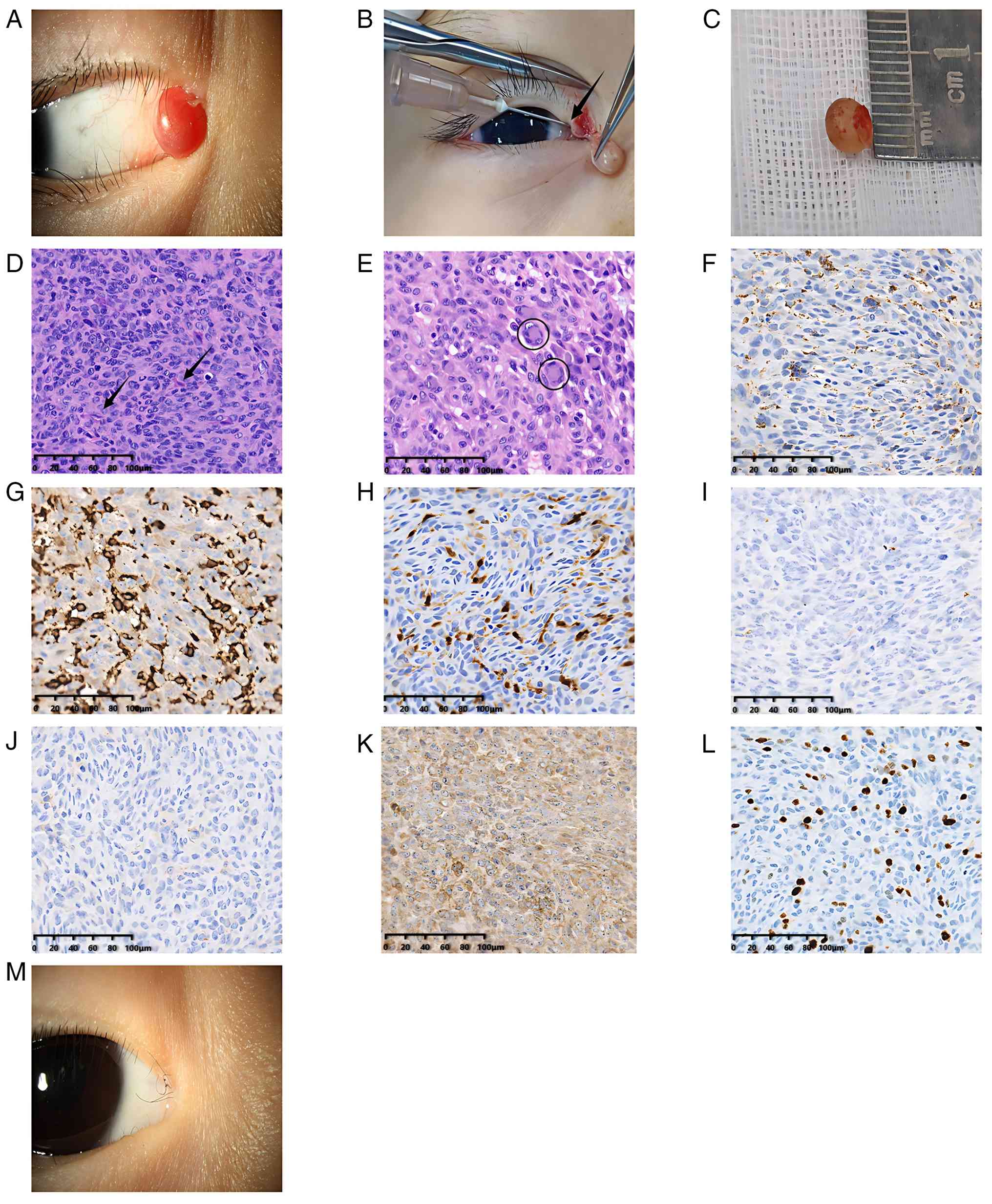 | Figure 1Clinical and pathological features of
adjoining masses in the right eye. (A) Preoperative appearance
reveals a clear boundary, yellow-red round mass with a size of
~6x5x5 mm in the adjacent part of the right upper eyelid and
adjacent to the upper lacrimal point. (B) Intraoperative image:
After the tumor was completely removed, the upper lacrimal point
(shown by the arrow) was well preserved. (C) Gross specimen:
Resected tumor specimens, ~5x5x5 mm in size. (D) Under squamous
epithelium, dense proliferation of small spindle cells is seen;
mitotic figures are clearly visible (arrows). (E) Scattered
Dutton-like giant cells can be observed among the proliferating
spindle cells (in circles). Immunohistochemical staining: The tumor
cells were partially positive for (F) CD68, (G) CD163, (H) S-100,
(I) CD1a and (J) CD10 and showed (K) diffuse strong positivity for
cytoplasmic ALK. (L) Ki-67 proliferation index was ~10%
(magnification, x200; scale bars, 100 µm). (M) Postoperative
appearance: The surgical incision healed well without redness,
swelling and recurrence. ALK, anaplastic lymphoma kinase. |
Materials and methods
Tissue processing and
histopathological examination
The excised tumor specimen was immediately fixed in
10% neutral-buffered formalin for 24 h at room temperature.
Following fixation, the tissue was routinely dehydrated, cleared,
embedded in paraffin wax and sectioned at a thickness of 4 µm.
Sections were stained with hematoxylin and eosin for routine
histopathological evaluation. All histopathological examinations
were performed under a light microscope (Olympus BX53; Olympus
Corporation) by two experienced pathologists independently.
Immunohistochemistry. Immunohistochemical
staining was performed on 4-µm-thick paraffin sections using the
EnVision FLEX polymer detection system (Dako; Agilent Technologies,
Inc.). In brief, sections were deparaffinized in xylene and
rehydrated through a graded ethanol series. Antigen retrieval was
performed by heating the sections in EDTA buffer (pH 8.0) using a
pressure cooker for 2.5 min at 121˚C, followed by cooling to room
temperature. Endogenous peroxidase activity was blocked with 3%
hydrogen peroxide for 10 min. The sections were then incubated with
primary antibodies at 4˚C overnight. The following primary
antibodies were used: Anti-CD68 (dilution, 1:200; cat. no. M0814;
Dako; Agilent Technologies, Inc.), anti-CD163 (dilution, 1:200;
cat. no. M0892; Dako), anti-S-100 (polyclonal; dilution, 1:500;
cat. no. Z0311; Dako), anti-CD1a (dilution, 1:50; cat. no. M3571;
Dako), anti-CD10 (dilution, 1:100; cat. no. M7302; Dako), anti-ALK
(dilution, 1:100; cat. no. 3633; Cell Signaling Technology, Inc.)
and anti-Ki-67 (dilution, 1:100; cat. no. M7240; Dako). After
washing with PBS, sections were incubated with reagents from the
EnVision FLEX polymer detection system (Dako) for 30 min at room
temperature. The reaction was visualized with 3,3'-diaminobenzidine
chromogen and sections were counterstained with hematoxylin.
Positive controls included known ALK-positive
anaplastic large cell lymphoma tissue provided by an external
collaborating institution, Jinan KingMed Diagnostics Co., Ltd.
(Jinan, China). Appropriate tissue controls for other markers were
similarly sourced from the same institution. Negative controls were
prepared by substituting the primary antibody with PBS.
Immunohistochemical staining was evaluated independently by two
pathologists. ALK positivity was defined as distinct cytoplasmic
granular staining in >10% of tumor cells.
Discussion
ALK-positive EFH is a rare cutaneous neoplasm that
typically presents on the extremities of adults; its occurrence in
the periocular region of children is exceptionally uncommon. To the
best of our knowledge, only one case of ALK-positive EFH occurring
in the eyelid of a child has been reported in the English-language
literature prior to the present case described a 12-year-old female
patient who presented with a nodule on the lower eyelid (9). Histopathological examination revealed
an intradermal proliferation of epithelioid cells with abundant
eosinophilic cytoplasm, and immunohistochemistry demonstrated
positivity for CD68 and vimentin, with no mention of ALK testing,
as ALK rearrangements in EFH had not yet been described at the time
of that report. The tumor was treated by complete local excision
with no recurrence documented during follow-up. In comparison, the
present case occurred in a younger patient (7-year-old male) and
involved the upper eyelid adjacent to the lacrimal punctum, a
location that posed specific surgical considerations to preserve
lacrimal function. Furthermore, the present case benefited from
comprehensive immunohistochemical evaluation, including ALK
testing, which confirmed the diagnosis with greater precision and
highlighted the value of incorporating ALK immunohistochemistry in
the workup of pediatric histiocytic-appearing eyelid tumors.
Histologically, EFH is characterized by epithelioid
cells with abundant eosinophilic cytoplasm, vesicular nuclei and
small nucleoli. The presence of Touton-like giant cells and low
mitotic activity are also common features (10). Immunohistochemically, EFH typically
shows variable expression of histiocytic markers, such as CD68 and
CD163, which it is consistently ALK-positive due to underlying gene
rearrangements (11). The
characteristic molecular change of EFH is ALK gene rearrangement,
which leads to the overexpression of ALK protein. ALK fusion
partners reported in the current literature include VCL, dynactin
1, SP100 and cAMP-dependent protein kinase type II-α regulatory
subunit (6). Different fusion
partners can affect the subcellular localization and signal
activation intensity of ALK protein. VCL-ALK fusion is mostly
located under the plasma membrane, whilst SP100-ALK fusion is
typically distributed in the nucleus (11,12).
Identifying the type of ALK fusion has auxiliary value for the
pathological diagnosis and differential diagnosis of EFH, where it
can also provide the molecular basis for potential targeted
intervention. In the present case, CD1a and CD10 showed partial
immunoreactivity. CD1a is classically considered to be a marker of
Langerhans cell histiocytosis. However, its expression can
occasionally be detected in other histiocytic lesions, including
indeterminate dendritic cell histiocytosis, certain subtypes of
histiocytic sarcoma and reactive dendritic cell proliferative
lesions (13), where its
significance is not fully understood but may be associated with the
presence of activated or immature dendritic cell populations. By
contrast, CD10 is a membrane-associated peptidase expressed in a
variety of mesenchymal cells and neoplasms, where its expression
has also been reported in a subset of EFH. Positivity for these two
aforementioned markers is not specific for EFH. However, when
included in an immunohistochemical panel, they contribute to
establishing a comprehensive staining profile and assist in
excluding other entities in the differential diagnosis. Langerhans
cell histiocytosis characteristically shows strong and diffuse
expression of both CD1a and Langerin, which was not identified in
the present case. In view of the notion that EFH is a benign tumor,
local treatment is typically the main clinical treatment, where
systemic targeted therapy is generally not required. Differential
diagnosis should therefore be combined with morphology and
immunophenotype. Juvenile xanthogranuloma (JXG) typically presents
as Touton giant cells appearing morphologically, but it also
expresses FXIIIa and CD14 whilst not expressing ALK. In addition,
its Ki-67 proliferation index is low (<5%) (14,15).
In the present case, Ki-67 was ~10%, which was higher compared with
that of typical JXG, supporting EFH diagnosis. Spitz nevus is
another similar condition that is common in children, but its
cellular structure is typically distributed in nests, expressing
S-100 and SOX10, whilst not expressing ALK and CD68(16). By contrast, cutaneous
myoepithelioma can express S-100 and cytokeratin, but rarely
express CD68 and ALK (17).
Epithelioid sarcoma is a malignancy that frequently expresses CK
and epithelial membrane antigen and is frequently accompanied by a
lack of integrase interactor 1 expression (18). In granulosa cell tumor, S-100 and
CD68 expression are typically positive, but the cytoplasm is
characteristically granular and ALK expression is negative
(19,20). The main histomorphological and
immunophenotypic features distinguishing EFH from its key
differential diagnoses are summarized in Table I.
 | Table IMain differential diagnoses of
EFH. |
Table I
Main differential diagnoses of
EFH.
| Disease | Histomorphological
features | Key
immunophenotype | (Refs.) |
|---|
| EFH | Proliferation of
epithelioid or spindle-like cells, occasionally with Touton-like
giant cells | ALK(+), CD68(+/-),
CD163(+/-), S-100(-/+) | (2) |
| Juvenile
xanthogranuloma | Touton giant cells,
foamy histiocytes | FXIIIa(+), CD14(+),
ALK(-), CD1a(-) | (15) |
| Spitz nevus | Junctional or
compound nevus, nests of epithelioid/spindle cells | S-100(+), SOX10(+),
ALK(-), CD68(-) | (16) |
| Cutaneous
myoepithelioma | Plasmacytoid or
spindle cells, may show trabecular pattern | S-100(+), CK(+),
p63(+), ALK(-), CD68(-) | (17) |
| Epithelioid
sarcoma | Nodules of
epithelioid cells, often with central necrosis | CK(+), EMA(+), loss
of INI1 expression, ALK(-) | (18) |
| Granular cell
tumor | Cells with abundant
eosinophilic granular cytoplasm | S-100(+), CD68(+),
ALK(-), inhibin(+) | (19) |
To conclude, the present case illustrates an unusual
presentation of ALK-positive EFH in the eyelid of a young male
patient aged 7 years. Accurate diagnosis relied on a combination of
clinical, histopathological and immunohistochemical findings.
Complete surgical excision remained the treatment of choice, with
favorable outcomes reported. Therefore, awareness of this entity is
important for ophthalmologists and pathologists to ensure
appropriate management and follow-up.
Acknowledgements
Not applicable.
Funding
Funding: The present report was supported by the scientific
research project of Shandong School Health Association in 2025
(grant no. SDW2025067).
Availability of data and materials
The data generated in the present study may be
requested from the corresponding author.
Authors' contributions
ZS was the attending physician in charge of the
case, responsible for diagnostic and therapeutic decisions and
final review of the manuscript. ZZ and YuL contributed equally to
collecting clinical data, literature review and writing the
original draft. PS, WC and YaL were responsible for the analysis
and interpretation of laboratory and pathological data and patient
follow-up, respectively. ZS and YaL confirm the authenticity of all
the raw data. JG provided essential clinical nursing information.
All authors reviewed the manuscript and have read and approved the
final version of the manuscript.
Ethics approval and consent to
participate
The present case report was approved by the Ethics
Committee of Jinan Mingshui Eye Hospital (approval no. 2025-016-01;
Jinan, China).
Patient consent for publication
Written informed consent was obtained from the
patient's legal guardian for the publication of the anonymized
clinical information and pathological images.
Competing interests
The authors declare that they have no competing
interests.
References
|
1
|
Jones EW, Cerio R and Smith NP:
Epithelioid cell histiocytoma: A new entity. Br J Dermatol.
120:185–195. 1989.PubMed/NCBI View Article : Google Scholar
|
|
2
|
Doyle LA, Mariño-Enríquez A, Fletcher CDM
and Hornick JL: ALK rearrangement and overexpression in epithelioid
fibrous histiocytoma. Mod Pathol. 28:904–912. 2015.PubMed/NCBI View Article : Google Scholar
|
|
3
|
Dickson BC, Swanson D, Charames GS,
Fletcher CD and Hornick JL: Epithelioid fibrous histiocytoma:
Molecular characterization of ALK fusion partners in 23 cases. Mod
Pathol. 31:753–762. 2018.PubMed/NCBI View Article : Google Scholar
|
|
4
|
Agaimy A and Michal M and Michal M:
Epithelioid fibrous histiocytoma: ALK fusion partners and beyond.
Genes Chromosomes Cancer. 61:471–480. 2022.
|
|
5
|
DeSimone MS, Odintsov I, Tsai HK, Dickson
BC, Alomari AK, Hornick JL, Fletcher CDM and Papke DJ Jr:
Epithelioid fibrous histiocytoma is on a continuum with superficial
ALK-rearranged myxoid spindle cell neoplasm: A clinicopathologic
series of 35 cases including alternate RET and NTRK3 fusions. Am J
Surg Pathol. 48:1568–1579. 2024.PubMed/NCBI View Article : Google Scholar
|
|
6
|
Felty CC and Linos K: Epithelioid fibrous
histiocytoma: A concise review. Am J Dermatopathol. 41:879–883.
2019.PubMed/NCBI View Article : Google Scholar
|
|
7
|
Schafer GL, Thompson K and Topping K:
Eyelid pilomatrixomas: A case report and comprehensive literature
review. Cureus. 17(e84639)2025.PubMed/NCBI View Article : Google Scholar
|
|
8
|
Wright GR, Archibald CW, Fontaine D,
Dakin-Hache K and Walsh NM: Epithelioid fibrous histiocytoma with
chondroblastoma-like features: A report of a rare entity and
discussion of related diagnostic challenges. Am J Dermatopathol.
44:e11–e15. 2022.PubMed/NCBI View Article : Google Scholar
|
|
9
|
Singh Gomez C, Calonje E and Fletcher CD:
Epithelioid benign fibrous histiocytoma of skin:
Clinico-pathological analysis of 20 cases of a poorly known
variant. Histopathology. 24:123–129. 1994.PubMed/NCBI View Article : Google Scholar
|
|
10
|
Kazakov DV, Kyrpychova L, Martinek P,
Grossmann P, Steiner P, Vanecek T, Pavlovsky M, Bencik V and Michal
M and Michal M: ALK gene fusions in epithelioid fibrous
histiocytoma: A study of 14 cases, with new histopathological
findings. Am J Dermatopathol. 40:805–814. 2018.PubMed/NCBI View Article : Google Scholar
|
|
11
|
Russell-Goldman E, Dong F, Laga A and
Hanna J: A novel fusion partner, SP100, drives nuclear dot
localization of ALK in epithelioid fibrous histiocytoma. Am J
Dermatopathol. 45:539–543. 2023.PubMed/NCBI View Article : Google Scholar
|
|
12
|
Zhao M, Song J, Yin X, Xu J, Teng X and
Wang J: ALK-rearranged mesenchymal neoplasms: A clinicopathological
and molecular study of eight additional cases of an emerging group
of tyrosine kinase fusion mesenchymal tumours. J Clin Pathol: June
4, 2024 (Epub ahead of print).
|
|
13
|
Frater JL, Kling CW, Obadiah JM, Gardner
LJ, Grosso LE, Resh B and Hurley MY: Histiocytic sarcoma with
secondary involvement of the skin and expression of CD1a: Evidence
of indeterminate cell differentiation? J Cutan Pathol. 33:437–442.
2006.PubMed/NCBI View Article : Google Scholar
|
|
14
|
Cypel TKS and Zuker RM: Juvenile
xanthogranuloma: Case report and review of the literature. Can J
Plast Surg. 16:175–177. 2008.PubMed/NCBI View Article : Google Scholar
|
|
15
|
Chen R, Liu S, Tang L, Yu X, Meng Z, Hu Y,
Li J and Liang X: On the knowledge of solitary juvenile
xanthogranuloma of the eyelid: A case series and literature review.
Graefes Arch Clin Exp Ophthalmol. 260:2339–2345. 2022.PubMed/NCBI View Article : Google Scholar
|
|
16
|
Yang X, Chen Q, Yang X and Qian H:
Analysis of clinical and pathological characteristics of 51 cases
of Spitz nevus in children. J Dermatol Venereol. 46:271–273.
2024.
|
|
17
|
Plaza JA, Brenn T, Chung C, Salim S, Linos
KD, Jour G, Duran Rincon J, Wick M, Sangueza M and Gru AA:
Histomorphological and immunophenotypical spectrum of cutaneous
myoepitheliomas: A series of 35 cases. J Cutan Pathol. 48:847–855.
2021.PubMed/NCBI View Article : Google Scholar
|
|
18
|
Song L, Stashek KM, Benyounes A, Davis DL,
Mulligan ME, Ng VY and Kallen ME: Epithelioid sarcoma with retained
INI1 (SMARCB1) expression. Histopathology. 78:464–466.
2021.PubMed/NCBI View Article : Google Scholar
|
|
19
|
Bai L and Zhai Z: Clinical and
histopathological features of 10 cases of cutaneous granulosa cell
tumor. Chin J Dermatol Venereol. 38:881–886. 2024.
|
|
20
|
Liu J, Wu Y, Yao X and Du X: Progress in
the diagnosis and treatment of granular cell tumors of the
extremities. Chin J Bone Joint. 14:182–186. 2025.
|


