Spandidos Publications Logo
  • About
    • About Spandidos
    • Aims and Scopes
    • Abstracting and Indexing
    • Editorial Policies
    • Reprints and Permissions
    • Job Opportunities
    • Terms and Conditions
    • Contact
  • Journals
    • All Journals
    • Oncology Letters
      • Oncology Letters
      • Information for Authors
      • Editorial Policies
      • Editorial Board
      • Aims and Scope
      • Abstracting and Indexing
      • Bibliographic Information
      • Archive
    • International Journal of Oncology
      • International Journal of Oncology
      • Information for Authors
      • Editorial Policies
      • Editorial Board
      • Aims and Scope
      • Abstracting and Indexing
      • Bibliographic Information
      • Archive
    • Molecular and Clinical Oncology
      • Molecular and Clinical Oncology
      • Information for Authors
      • Editorial Policies
      • Editorial Board
      • Aims and Scope
      • Abstracting and Indexing
      • Bibliographic Information
      • Archive
    • Experimental and Therapeutic Medicine
      • Experimental and Therapeutic Medicine
      • Information for Authors
      • Editorial Policies
      • Editorial Board
      • Aims and Scope
      • Abstracting and Indexing
      • Bibliographic Information
      • Archive
    • International Journal of Molecular Medicine
      • International Journal of Molecular Medicine
      • Information for Authors
      • Editorial Policies
      • Editorial Board
      • Aims and Scope
      • Abstracting and Indexing
      • Bibliographic Information
      • Archive
    • Biomedical Reports
      • Biomedical Reports
      • Information for Authors
      • Editorial Policies
      • Editorial Board
      • Aims and Scope
      • Abstracting and Indexing
      • Bibliographic Information
      • Archive
    • Oncology Reports
      • Oncology Reports
      • Information for Authors
      • Editorial Policies
      • Editorial Board
      • Aims and Scope
      • Abstracting and Indexing
      • Bibliographic Information
      • Archive
    • Molecular Medicine Reports
      • Molecular Medicine Reports
      • Information for Authors
      • Editorial Policies
      • Editorial Board
      • Aims and Scope
      • Abstracting and Indexing
      • Bibliographic Information
      • Archive
    • World Academy of Sciences Journal
      • World Academy of Sciences Journal
      • Information for Authors
      • Editorial Policies
      • Editorial Board
      • Aims and Scope
      • Abstracting and Indexing
      • Bibliographic Information
      • Archive
    • International Journal of Functional Nutrition
      • International Journal of Functional Nutrition
      • Information for Authors
      • Editorial Policies
      • Editorial Board
      • Aims and Scope
      • Abstracting and Indexing
      • Bibliographic Information
      • Archive
    • International Journal of Epigenetics
      • International Journal of Epigenetics
      • Information for Authors
      • Editorial Policies
      • Editorial Board
      • Aims and Scope
      • Abstracting and Indexing
      • Bibliographic Information
      • Archive
    • Medicine International
      • Medicine International
      • Information for Authors
      • Editorial Policies
      • Editorial Board
      • Aims and Scope
      • Abstracting and Indexing
      • Bibliographic Information
      • Archive
  • Articles
  • Information
    • Information for Authors
    • Information for Reviewers
    • Information for Librarians
    • Information for Advertisers
    • Conferences
  • Language Editing
Spandidos Publications Logo
  • About
    • About Spandidos
    • Aims and Scopes
    • Abstracting and Indexing
    • Editorial Policies
    • Reprints and Permissions
    • Job Opportunities
    • Terms and Conditions
    • Contact
  • Journals
    • All Journals
    • Biomedical Reports
      • Information for Authors
      • Editorial Policies
      • Editorial Board
      • Aims and Scope
      • Abstracting and Indexing
      • Bibliographic Information
      • Archive
    • Experimental and Therapeutic Medicine
      • Information for Authors
      • Editorial Policies
      • Editorial Board
      • Aims and Scope
      • Abstracting and Indexing
      • Bibliographic Information
      • Archive
    • International Journal of Epigenetics
      • Information for Authors
      • Editorial Policies
      • Editorial Board
      • Aims and Scope
      • Abstracting and Indexing
      • Bibliographic Information
      • Archive
    • International Journal of Functional Nutrition
      • Information for Authors
      • Editorial Policies
      • Editorial Board
      • Aims and Scope
      • Abstracting and Indexing
      • Bibliographic Information
      • Archive
    • International Journal of Molecular Medicine
      • Information for Authors
      • Editorial Policies
      • Editorial Board
      • Aims and Scope
      • Abstracting and Indexing
      • Bibliographic Information
      • Archive
    • International Journal of Oncology
      • Information for Authors
      • Editorial Policies
      • Editorial Board
      • Aims and Scope
      • Abstracting and Indexing
      • Bibliographic Information
      • Archive
    • Medicine International
      • Information for Authors
      • Editorial Policies
      • Editorial Board
      • Aims and Scope
      • Abstracting and Indexing
      • Bibliographic Information
      • Archive
    • Molecular and Clinical Oncology
      • Information for Authors
      • Editorial Policies
      • Editorial Board
      • Aims and Scope
      • Abstracting and Indexing
      • Bibliographic Information
      • Archive
    • Molecular Medicine Reports
      • Information for Authors
      • Editorial Policies
      • Editorial Board
      • Aims and Scope
      • Abstracting and Indexing
      • Bibliographic Information
      • Archive
    • Oncology Letters
      • Information for Authors
      • Editorial Policies
      • Editorial Board
      • Aims and Scope
      • Abstracting and Indexing
      • Bibliographic Information
      • Archive
    • Oncology Reports
      • Information for Authors
      • Editorial Policies
      • Editorial Board
      • Aims and Scope
      • Abstracting and Indexing
      • Bibliographic Information
      • Archive
    • World Academy of Sciences Journal
      • Information for Authors
      • Editorial Policies
      • Editorial Board
      • Aims and Scope
      • Abstracting and Indexing
      • Bibliographic Information
      • Archive
  • Articles
  • Information
    • For Authors
    • For Reviewers
    • For Librarians
    • For Advertisers
    • Conferences
  • Language Editing
Login Register Submit
  • This site uses cookies
  • You can change your cookie settings at any time by following the instructions in our Cookie Policy. To find out more, you may read our Privacy Policy.

    I agree
Search articles by DOI, keyword, author or affiliation
Search
Advanced Search
presentation
Experimental and Therapeutic Medicine
Join Editorial Board Propose a Special Issue
Print ISSN: 1792-0981 Online ISSN: 1792-1015
Journal Cover
August-2026 Volume 32 Issue 2

Full Size Image

Sign up for eToc alerts
Recommend to Library

Journals

International Journal of Molecular Medicine

International Journal of Molecular Medicine

International Journal of Molecular Medicine is an international journal devoted to molecular mechanisms of human disease.

International Journal of Oncology

International Journal of Oncology

International Journal of Oncology is an international journal devoted to oncology research and cancer treatment.

Molecular Medicine Reports

Molecular Medicine Reports

Covers molecular medicine topics such as pharmacology, pathology, genetics, neuroscience, infectious diseases, molecular cardiology, and molecular surgery.

Oncology Reports

Oncology Reports

Oncology Reports is an international journal devoted to fundamental and applied research in Oncology.

Experimental and Therapeutic Medicine

Experimental and Therapeutic Medicine

Experimental and Therapeutic Medicine is an international journal devoted to laboratory and clinical medicine.

Oncology Letters

Oncology Letters

Oncology Letters is an international journal devoted to Experimental and Clinical Oncology.

Biomedical Reports

Biomedical Reports

Explores a wide range of biological and medical fields, including pharmacology, genetics, microbiology, neuroscience, and molecular cardiology.

Molecular and Clinical Oncology

Molecular and Clinical Oncology

International journal addressing all aspects of oncology research, from tumorigenesis and oncogenes to chemotherapy and metastasis.

World Academy of Sciences Journal

World Academy of Sciences Journal

Multidisciplinary open-access journal spanning biochemistry, genetics, neuroscience, environmental health, and synthetic biology.

International Journal of Functional Nutrition

International Journal of Functional Nutrition

Open-access journal combining biochemistry, pharmacology, immunology, and genetics to advance health through functional nutrition.

International Journal of Epigenetics

International Journal of Epigenetics

Publishes open-access research on using epigenetics to advance understanding and treatment of human disease.

Medicine International

Medicine International

An International Open Access Journal Devoted to General Medicine.

Journal Cover
August-2026 Volume 32 Issue 2

Full Size Image

Sign up for eToc alerts
Recommend to Library

  • Article
  • Citations
    • Cite This Article
    • Download Citation
    • Create Citation Alert
    • Remove Citation Alert
    • Cited By
  • Similar Articles
    • Related Articles (in Spandidos Publications)
    • Similar Articles (Google Scholar)
    • Similar Articles (PubMed)
  • Download PDF
  • Download XML
  • View XML
Case Report Open Access

Spontaneous cavernous sinus dural arteriovenous fistula with remote cerebellar hemorrhage: A case report

  • Authors:
    • Tianjun Bai
    • Jie Liu
    • Yuezhen Shen
    • Shulai Zhu
    • Yonghong Wang
  • View Affiliations / Copyright

    Affiliations: Department of Magnetic Resonance Imaging, The Second People's Hospital of Liao Cheng, Linqing, Shandong 252600, P.R. China, Department of Neurosurgery, The Second People's Hospital of Liao Cheng, Linqing, Shandong 252600, P.R. China, Department of Internal Neurology, The Second People's Hospital of Liao Cheng, Linqing, Shandong 252600, P.R. China
    Copyright: © Bai et al. This is an open access article distributed under the terms of Creative Commons Attribution License.
  • Article Number: 225
    |
    Published online on: June 24, 2026
       https://doi.org/10.3892/etm.2026.13220
  • Expand metrics +
Metrics: Total Views: 0 (Spandidos Publications: | PMC Statistics: )
Metrics: Total PDF Downloads: 0 (Spandidos Publications: | PMC Statistics: )
Cited By (CrossRef): 0 citations Loading Articles...

This article is mentioned in:


Abstract

Cavernous sinus dural arteriovenous fistula (CS‑DAVF) is a rare vascular malformation that can lead to severe clinical symptoms, including ocular manifestations and intracranial hemorrhage. The current study reports a case of a spontaneous CS‑DAVF complicated by remote cerebellar hemorrhage. A 52‑year‑old man presented with nausea, vomiting and decreased appetite, with a history of hypertension but no trauma. Initially misdiagnosed with conjunctivitis, the patient showed no clinical improvement after standard treatment. Physical examination and auxiliary tests showed conjunctival congestion of the left eye with impaired ocular abduction. A subacute cerebral hemorrhage in the left cerebellar hemisphere and a left CS fistula were identified via head magnetic resonance imaging and magnetic resonance angiography. This diagnosis was confirmed by angiography, demonstrating a dural arteriovenous fistula in the CS region. Subsequently, the patient underwent DAVF interventional embolization on day 6 post‑presentation. Postoperatively, the symptoms showed notable clinical improvement, with resolution of the left conjunctival edema and restoration of normal limb movements. This case highlights the potential complications and diagnostic challenges associated with DAVFs, providing valuable clinical insights and underscoring the critical role of imaging studies in the diagnostic process. Despite the inherent limitations in generalizability associated with a single‑case study, this report provides critical clinical implications and meaningful reference for clinical practice.

Introduction

Spontaneous dural arteriovenous fistula (DAVF) is a relatively rare pathological condition, representing 10-15% of all intracranial arteriovenous malformations, including ~6% of supratentorial and 35% of infratentorial vascular malformations (1). The reported annual incidence is ~0.15 per 100,000 individuals, and DAVFs are most frequently diagnosed in patients aged 40-60 years (2). DAVF is primarily characterized by arterial blood flow that drains directly into the cavernous sinus (CS) via an abnormal arteriovenous fistula. This abnormal drainage may lead to increased intracranial pressure and various cerebral complications (3). This condition exhibits a predilection for middle-aged men and is often associated with hypertension, trauma or other vascular pathologies. Epidemiological data on DAVF indicate a low incidence rate, and its clinical symptoms are complex and varied, which increases the difficulty of diagnosis (2). Typical clinical manifestations include ocular symptoms, such as exophthalmos and conjunctival congestion, as well as neurological symptoms. In the early stages, these symptoms may be atypical and are easily misdiagnosed as common ocular diseases, such as conjunctivitis (4). The current study reports a case of spontaneous CS-DAVF complicated by remote cerebellar hemorrhage. The study highlights the potential complications and diagnostic challenges associated with this condition, offering valuable insights for similar cases.

Case report

A 52-year-old man presented to The Second People's Hospital of Liao Cheng (Linqing, China) in December 2023 with nausea, vomiting and decreased appetite. A total of 10 days prior to this, the patient had developed redness of the left conjunctiva accompanied by a foreign body sensation in the left eye and was initially diagnosed with conjunctivitis. Oral antibiotic therapy with amoxicillin was administered and yielded poor results, with persisting symptoms; the exact dose and duration are not available. The patient had a history of hypertension and denied any trauma. On admission, a physical examination revealed the following: Temperature, 36.5˚C (normal range, 36.0-37.3˚C); pulse, 64 beats/min (normal range, 60-100 beats/min); respiratory rate, 16 breaths/minute (normal range, 12-20 breaths/min); and blood pressure, 154/104 mmHg (normal range, <140/90 mmHg for an adult male). No facial weakness, sensory disturbance, dysarthria, ataxia or gait instability was observed. Bilateral pupillary light reflexes were intact, and no visual acuity deterioration, proptosis or tinnitus was noted. The patient was conscious and oriented, with left conjunctival congestion and mild limitation of the abduction of the left eye. An abdominal examination revealed a soft abdomen without tenderness or rebound pain. No hepatic or renal percussion tenderness was elicited. Bowel sounds were normal, and the lower extremities were free of edema. Routine laboratory investigations, including coagulation function, were unremarkable. Further workup 
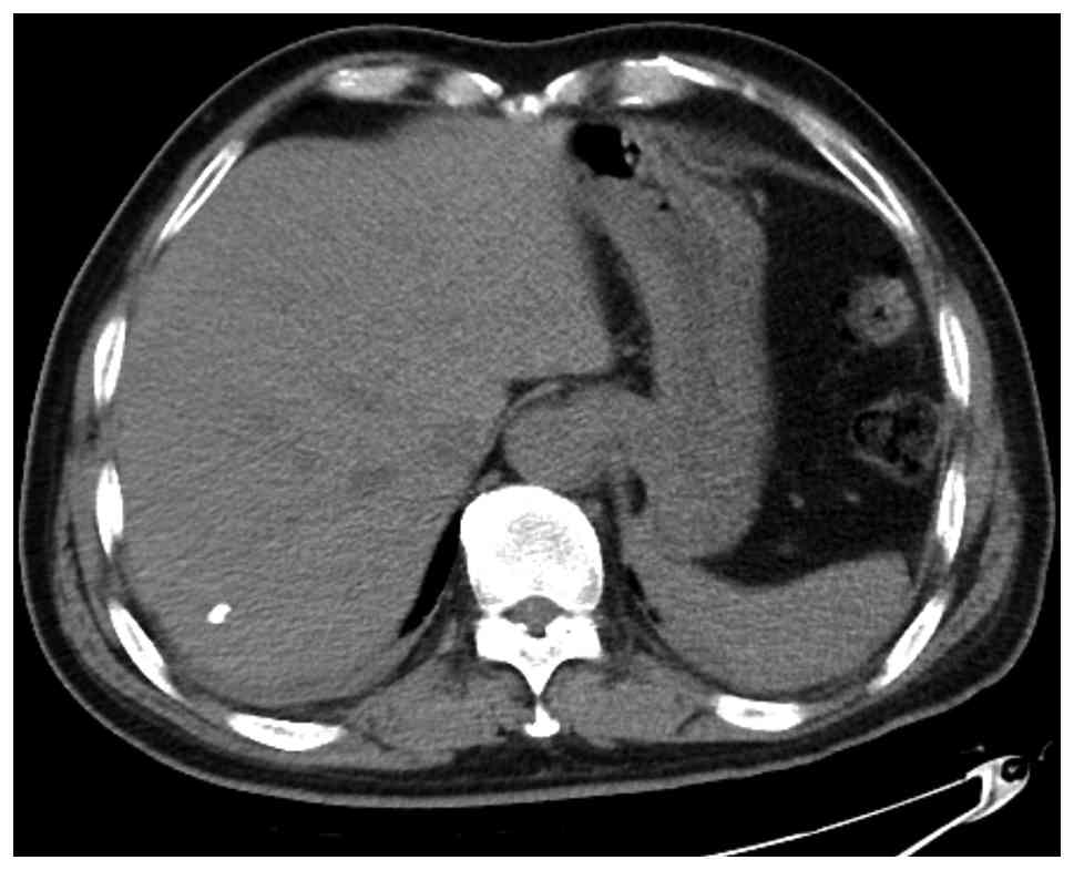
with a non-contrast computed tomography scan of the whole abdomen demonstrated hepatic calcifications (Fig. 1). Despite symptomatic supportive treatment, including antiemetics and hydration, which yielded mild improvement in the vomiting, the left conjunctival congestion 
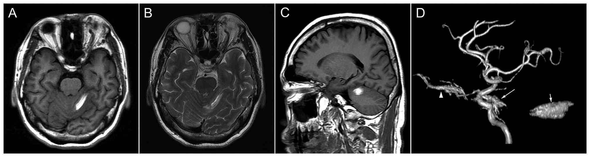
persisted severely. Further head magnetic resonance imaging demonstrated a subacute intracerebral hemorrhage in the left cerebellar hemisphere (Fig. 2A-C), and magnetic resonance angiography revealed a left CS fistula (Fig. 2D). Therefore, the patient was transferred to the Department of Neurosurgery for further management.

Axial non-contrast computed tomography
image of the abdomen demonstrating hepatic calcifications.

Figure 1

Axial non-contrast computed tomography image of the abdomen demonstrating hepatic calcifications.

Head magnetic resonance imaging
showing a subacute intracerebral haemorrhage in the left cerebellar
hemisphere. (A) Axial T1WI and (B) T2WI revealed a strip-like
hyperintense signal in the left cerebellar hemisphere with a
characteristic ‘zebra sign’. (C) Sagittal T1WI further demonstrated
a similar hyperintense signal, suggestive of subacute haemorrhage
in the same region. WI, weighted imaging. (D) Three-dimensional
magnetic resonance angiography demonstrating a left CS dural
arteriovenous fistula with dilation of the ophthalmic vein and
abnormal venous drainage adjacent to the CS. CS, cavernous sinus.
Long arrow, CS-DAVF; short arrow, cerebellar hematoma; arrowhead,
ophthalmic vein.

Figure 2

Head magnetic resonance imaging showing a subacute intracerebral haemorrhage in the left cerebellar hemisphere. (A) Axial T1WI and (B) T2WI revealed a strip-like hyperintense signal in the left cerebellar hemisphere with a characteristic ‘zebra sign’. (C) Sagittal T1WI further demonstrated a similar hyperintense signal, suggestive of subacute haemorrhage in the same region. WI, weighted imaging. (D) Three-dimensional magnetic resonance angiography demonstrating a left CS dural arteriovenous fistula with dilation of the ophthalmic vein and abnormal venous drainage adjacent to the CS. CS, cavernous sinus. Long arrow, CS-DAVF; short arrow, cerebellar hematoma; arrowhead, ophthalmic vein.

After the exclusion of surgical contraindications, 
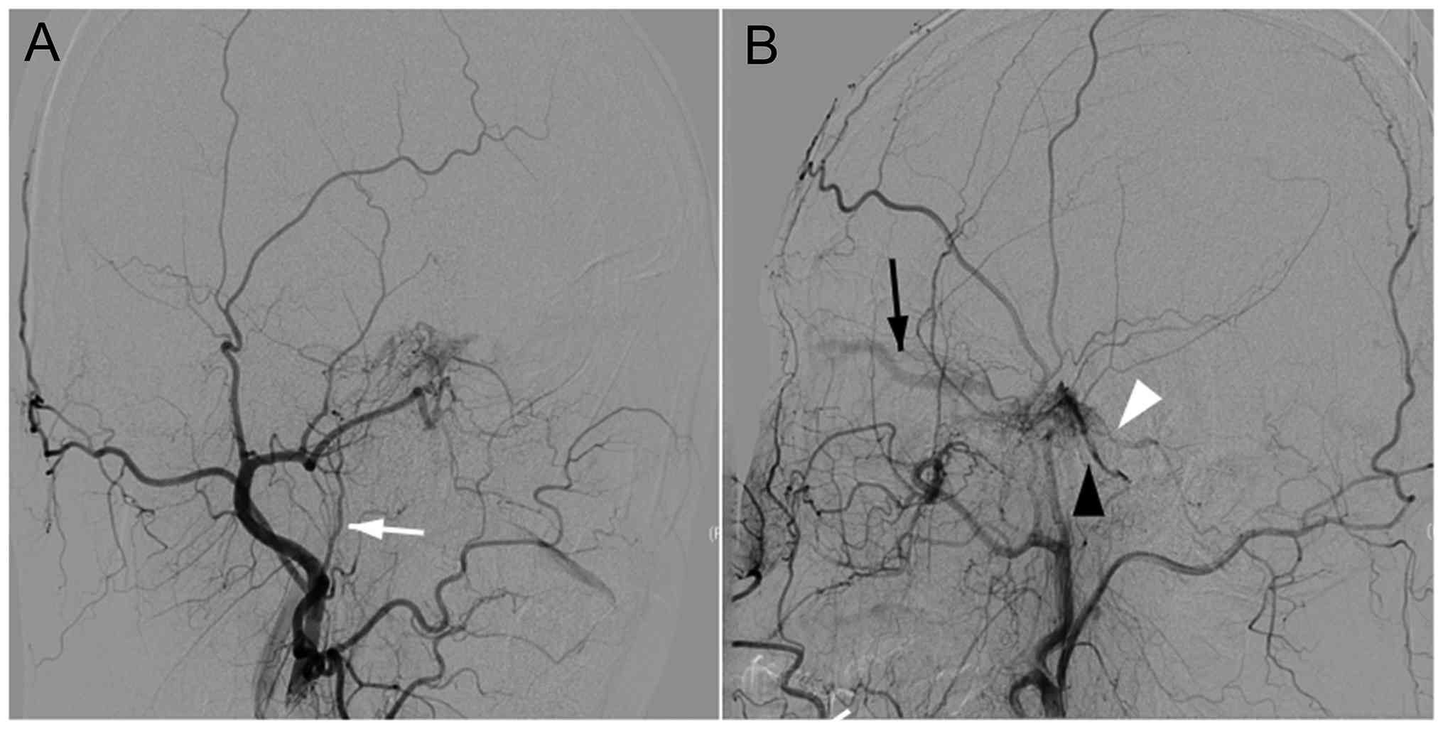
the patient underwent a diagnostic angiography in January 2024, for a comprehensive assessment of the vascular pathology. Selective angiography of the left external carotid artery revealed a vascular network arising from the distal branches of the ascending pharyngeal artery and supplying the CS. Drainage was observed into the ophthalmic vein, inferior petrosal sinus and superior petrosal sinus, confirming the presence of a CS-DAVF (Fig. 3).

(A) Angiography of the left external
carotid artery depicting the vascular supply of the CS-dural
arteriovenous fistula (the white arrow indicates the ascending
pharyngeal artery). (B) Angiography of the left external carotid
artery depicting that the distal branches of the ascending
pharyngeal artery form a vascular network supplying the CS, with
drainage into the ophthalmic vein, inferior petrosal sinus and
superior petrosal sinus (the white arrowhead indicates the superior
petrosal sinus; black arrowhead indicates the inferior petrosal
sinus; and the black arrow indicates the ophthalmic vein). CS,
cavernous sinus.

Figure 3

(A) Angiography of the left external carotid artery depicting the vascular supply of the CS-dural arteriovenous fistula (the white arrow indicates the ascending pharyngeal artery). (B) Angiography of the left external carotid artery depicting that the distal branches of the ascending pharyngeal artery form a vascular network supplying the CS, with drainage into the ophthalmic vein, inferior petrosal sinus and superior petrosal sinus (the white arrowhead indicates the superior petrosal sinus; black arrowhead indicates the inferior petrosal sinus; and the black arrow indicates the ophthalmic vein). CS, cavernous sinus.

The patient underwent an interventional embolization for the DAVF on day 6 post-presentation. During the procedure, a 5F single-curve angiographic catheter was selectively advanced into the left internal jugular vein over a Glidewire® (Terumo Medical Corporation), with the tip positioned at the junction of the inferior petrosal sinus and jugular vein. The patency of the inferior petrosal sinus was confirmed by Glidewire exploration, which successfully accessed the CS. A pre-shaped Echelon-10 (Medtronic plc) microcatheter was then navigated over a microwire into the anterior CS, adjacent to the origin of the ophthalmic vein. Microcatheter angiography confirmed the intracavernous position. Multiple coils were deployed in a basket configuration, embolizing the ophthalmic vein origin and anterior CS. After preparing the microcatheter with a dimethyl sulfoxide flush, Onyx-18 (Medtronic plc) was slowly injected through the microcatheter, permeating the ophthalmic vein origin, CS compartments and fistula sites.

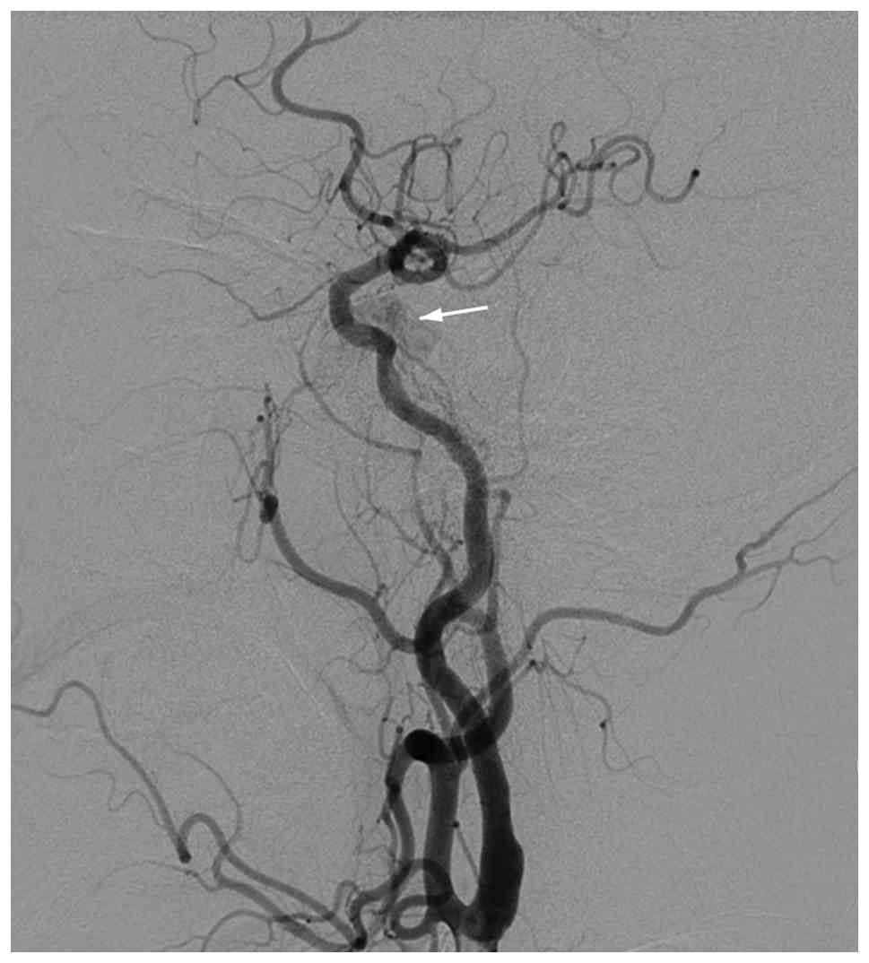
Post-embolization bilateral common carotid angiography revealed complete occlusion of the left CS, with visible coil mesh and Onyx cast. No opacification was identified in the ophthalmic vein and superior petrosal sinus, while the delayed venous phase showed minimal faint opacification in the posterosuperior CS draining into the inferior petrosal sinus (Fig. 4). The procedure was successfully completed with adequate anesthesia. Postoperatively, the patient received nutritional support, dexamethasone (5 mg, intravenous, stat), glycerol fructose (250 ml, intravenous drip, every 12 h) to reduce intracranial pressure, and vitamin B6 (0.2 g, intravenous drip, once daily) for neurotrophic support. At the 1-week follow-up, the left conjunctival edema had improved compared with the preprocedural status, with mild restriction of left ocular movement and preserved limb mobility, muscle strength and muscle tone. No postoperative hemorrhagic, ischemic or infectious complications were observed during hospitalization. At the 1-month follow-up, the patient showed marked improvement in conjunctival congestion and ocular motility, without evidence of recurrent hemorrhage or neurological deficits. The patient is scheduled for regular clinical and imaging follow-up every 3 months to monitor for recurrence. The prognosis of the patient is considered favorable based on the complete occlusion of the fistula and the progressive resolution of symptoms.

Postoperative angiography improvement
following embolization of the cavernous sinus-dural arteriovenous
fistula. Postoperative angiography of the left common carotid
artery showing that most of the abnormal vascular shadow has
disappeared, with only a minimal amount of residual contrast
enhancement remaining locally (white arrow indicates a minimal
amount of residual contrast enhancement remaining locally).

Figure 4

Postoperative angiography improvement following embolization of the cavernous sinus-dural arteriovenous fistula. Postoperative angiography of the left common carotid artery showing that most of the abnormal vascular shadow has disappeared, with only a minimal amount of residual contrast enhancement remaining locally (white arrow indicates a minimal amount of residual contrast enhancement remaining locally).

Discussion

Spontaneous DAVF in the CS region is an uncommon vascular malformation, typically characterized by ophthalmic and neurological symptoms, such as ocular motility disorders, elevated intraocular pressure and headaches (4). Current literature indicates that patients are frequently misdiagnosed with mild ocular conditions, such as conjunctivitis, at the initial presentation, leading to a delay in appropriate treatment (3,5). In the current case, the patient presented with remote cerebellar hemorrhage, an atypical manifestation that may be attributed to hemodynamic changes in the CS, especially in cases of obstructed venous drainage of the orbit (6,7). This unusual manifestation underscores the complexity of DAVF and the severity of its associated symptoms.

Clinically, the diagnosis of DAVF often faces challenges, as early symptoms can easily be confused with other diseases. Patients may seek medical attention due to ocular symptoms such as conjunctival congestion, and the preliminary diagnosis is often misidentified as conjunctivitis (4). Therefore, in radiological evaluation, it is crucial to pay close attention to the abnormal signals on cranial MR and the manifestations of cerebral hemorrhage, particularly in patients presenting with concomitant ocular and neurological signs. Therapeutically, interventional embolization has demonstrated favorable outcomes in managing DAVF, effectively alleviating symptoms and improving quality of life (8,9). The treatment plan for the present case was determined through a multidisciplinary consultation, considering the patient's high-risk factors, particularly hypertension, emphasizing the necessity of early recognition and intervention (10). Future research should further explore the optimal management strategies and epidemiological characteristics of different DAVF subtypes, thereby optimizing clinical outcomes and mitigating complications (11,12).

From the pathological perspective, the development of CS arteriovenous fistula is closely associated with hypertension, particularly in non-traumatic cases. The etiology of DAVF is likely multifactorial, involving hypertension, chronic venous obstruction and other vascular lesions. Arteriovenous fistulas in non-traumatic patients usually occur in the setting of elevated venous pressure. Long-term venous hypertension may lead to vascular wall fragility and pathological remodeling, thereby promoting the formation of fistulas, resulting in the direct shunting of arterial blood into the venous system (13,14). This abnormal blood flow pattern may lead to venous hypertension, especially in cases where venous drainage from the eye is obstructed. When the pressure within the CS increases, it may induce hemodynamic alterations at distant sites. Common causes of distant cerebellar hemorrhage include arterial embolism and ischemia, lesions of the venous system, traumatic factors, tumors, cerebral infections or inflammation (15). At presentation, the current patient lacked common precipitating factors, such as arterial embolism, ischemia or trauma. Therefore, the condition was attributed to the DAVF-induced abnormal hemodynamics.

The clinical significance of the present case lies in the accompanying remote cerebellar hemorrhage, underscoring the intricate pathological mechanisms associated with DAVF. Through detailed analysis, the case reminds clinicians to maintain a high level of vigilance for similar symptoms to prevent a misdiagnosis and delays in treatment. Imaging studies play a key role in the diagnosis of DAVF, especially when the patient presents with ocular and neurological symptoms (4).

There are certain limitations to the present study. Firstly, this is a single-case report describing a rare presentation of CS-DAVF associated with remote cerebellar hemorrhage. Therefore, the findings may not be generalizable to all patients with DAVF. Secondly, the retrospective nature of this report may have introduced incomplete clinical information and potential publication bias related to the rarity of the condition. Thirdly, the follow-up period was relatively short, limiting comprehensive evaluation of the long-term prognosis and recurrence risk after endovascular treatment. In addition, the diagnostic evaluation and treatment strategy were based on single-center clinical experience, which may differ across institutions. Further multicenter studies with larger sample sizes are needed to better clarify the clinical characteristics, pathophysiological mechanisms and optimal management strategies of DAVF. Despite the inherent limitations of this study, it offers valuable insights for clinical practice and emphasizes the importance of early recognition and intervention for DAVF. This report paves the way for further investigations, aiming to improve the understanding of DAVF and subsequently enhance the overall prognosis for patients.

Acknowledgements

Not applicable.

Funding

Funding: No funding was received.

Availability of data and materials

The data generated in the present study may be requested from the corresponding author.

Authors' contributions

TJB and JL contributed to the conception and design of the case report, analysis and interpretation of the clinical data, and critical revision of the manuscript for important intellectual content. YZS and SLZ acquired and organized the clinical data, including laboratory results, medical images and follow-up information, and participated in the analysis and interpretation of the patient's clinical course. YHW contributed to the study design, data acquisition and interpretation, performed the literature review, and drafted the manuscript. All authors have read and approved the final manuscript. TJB, JL, YZS, SLZ and YHW confirm the authenticity of all the raw data.

Ethics approval and consent to participate

Not applicable.

Patient consent for publication

The patient in this study received standard clinical practice and provided written informed consent for the publication of any medical data and images.

Competing interests

The authors declare that they have no competing interests.

References

1 

Rhim JK, Cho YD, Yoo DH, Kang HS, Cho WS, Kim JE, Cho MJ, Hwang G, Kwon OK and Han MH: Endovascular treatment of bilateral cavernous sinus dural arteriovenous fistula: Therapeutic strategy and follow-up outcomes. Korean J Radiol. 19:334–341. 2018.PubMed/NCBI View Article : Google Scholar

2 

Maciejewski K, Pinkiewicz M, Mruk B, Knap D, Zaczyński A, Walecki J and Zawadzki M: A practical approach to intracranial dural arteriovenous fistulas: Pathogenesis, classification and management. J Clin Med. 14(6895)2025.PubMed/NCBI View Article : Google Scholar

3 

Roy T, van den Berg R, Brandsen RP and Saeed PO: Likelihood of spontaneous closure of cavernous sinus dural arteriovenous fistulas based on clinical and radiological findings. Interv Neuroradiol. 31:344–351. 2023.PubMed/NCBI View Article : Google Scholar

4 

Thiyagarajam K, Chong MF and Mohd Khialdin S: The diagnostic challenges in carotid cavernous fistula: A case series. Cureus. 13(e19696)2021.PubMed/NCBI View Article : Google Scholar

5 

Kato M, Maki Y, Nakada Y, Shirai S and Ono Y: Three cases of spontaneous bilateral external carotid-cavernous sinus fistula (dural arteriovenous shunts in the region of the cavernous sinus). No Shinkei Geka (Japanese). 3:607–613. 1975.PubMed/NCBI

6 

Moster ML, Sergott RC and Grossman RI: Dural carotid-cavernous sinus vascular malformation with facial nerve paresis. Can J Ophthalmol. 23:27–29. 1988.PubMed/NCBI

7 

Dixit N, Sharma A and Hosur B: Remote cerebellar hemorrhage: A bleed not too far! Neurol. India. 71:1088–1089. 2023.PubMed/NCBI View Article : Google Scholar

8 

He HW, Jiang CH, Wu ZX, Li YX, Lü XL and Wang ZC: Transvenous embolization with a combination of detachable coils and onyx for a complicated cavernous dural arteriovenous fistula. Chin Med J (Engl). 121:1651–1655. 2008.PubMed/NCBI

9 

Kato S, Ishihara H, Nakayama H, Fujii M, Fujisawa H, Kajiwara K, Nomura S, Sadanaga H and Suzuki M: Transvenous embolization for dural arteriovenous shunt of the cavernous sinus. Comparison of multi-staged transvenous embolization and transvenous embolization with sinus packing. Interv Neuroradiol. 13:353–358. 2007.PubMed/NCBI View Article : Google Scholar

10 

Nakagawa H, Kubo S, Nakajima Y, Izumoto S and Fujita T: Shifting of dural arteriovenous malformation from the cavernous sinus to the sigmoid sinus to the transverse sinus after transvenous embolization. A case of left spontaneous carotid-cavernous sinus fistula. Surg Neurol. 37:30–38. 1992.PubMed/NCBI View Article : Google Scholar

11 

Komiyama M, Nishikawa M and Yasui T: Transient monocular blindness during manual carotid compression for carotid-cavernous sinus fistulas-Two case reports. Neurol Med Chir (Tokyo). 36:805–807. 1996.PubMed/NCBI View Article : Google Scholar

12 

Li W, Liu Y, Chu F and Wang YL: Interventional embolization of unilateral cavernous sinus with onxy glue combined with coils for the treatment of bilateral dural arteriovenous fistula. J Craniofac Surg. 35:e451–e454. 2024.PubMed/NCBI View Article : Google Scholar

13 

Li H, Wang Y, Li L, Niu X and Zhang J: Clinical characteristics, imaging, treatment, and prognosis of dural arteriovenous fistula presented with parkinsonism-A systematic review. Neurosurg Rev. 48(625)2025.PubMed/NCBI View Article : Google Scholar

14 

Shapiro M, Raz E, Litao M, Becske T, Riina H and Nelson PK: Investigating the pathophysiology of intracranial dural arteriovenous fistulas with associated cerebellar hemorrhage. Stroke. 52:1381–1387. 2021.

15 

Arismendi Morillo GJ, Fernández Abreu MC, Cardozo Sosa DP and Cardozo JJ: Lesiones ocupantes de espacio no neoplasicas que simulan tumores del sistema nervioso central. Rev Neurol. 38:427–430. 2004.PubMed/NCBI

Related Articles

  • Abstract
  • View
  • Download
  • Twitter
Copy and paste a formatted citation
Spandidos Publications style
Bai T, Liu J, Shen Y, Zhu S and Wang Y: Spontaneous cavernous sinus dural arteriovenous fistula with remote cerebellar hemorrhage: A case report. Exp Ther Med 32: 225, 2026.
APA
Bai, T., Liu, J., Shen, Y., Zhu, S., & Wang, Y. (2026). Spontaneous cavernous sinus dural arteriovenous fistula with remote cerebellar hemorrhage: A case report. Experimental and Therapeutic Medicine, 32, 225. https://doi.org/10.3892/etm.2026.13220
MLA
Bai, T., Liu, J., Shen, Y., Zhu, S., Wang, Y."Spontaneous cavernous sinus dural arteriovenous fistula with remote cerebellar hemorrhage: A case report". Experimental and Therapeutic Medicine 32.2 (2026): 225.
Chicago
Bai, T., Liu, J., Shen, Y., Zhu, S., Wang, Y."Spontaneous cavernous sinus dural arteriovenous fistula with remote cerebellar hemorrhage: A case report". Experimental and Therapeutic Medicine 32, no. 2 (2026): 225. https://doi.org/10.3892/etm.2026.13220
Copy and paste a formatted citation
x
Spandidos Publications style
Bai T, Liu J, Shen Y, Zhu S and Wang Y: Spontaneous cavernous sinus dural arteriovenous fistula with remote cerebellar hemorrhage: A case report. Exp Ther Med 32: 225, 2026.
APA
Bai, T., Liu, J., Shen, Y., Zhu, S., & Wang, Y. (2026). Spontaneous cavernous sinus dural arteriovenous fistula with remote cerebellar hemorrhage: A case report. Experimental and Therapeutic Medicine, 32, 225. https://doi.org/10.3892/etm.2026.13220
MLA
Bai, T., Liu, J., Shen, Y., Zhu, S., Wang, Y."Spontaneous cavernous sinus dural arteriovenous fistula with remote cerebellar hemorrhage: A case report". Experimental and Therapeutic Medicine 32.2 (2026): 225.
Chicago
Bai, T., Liu, J., Shen, Y., Zhu, S., Wang, Y."Spontaneous cavernous sinus dural arteriovenous fistula with remote cerebellar hemorrhage: A case report". Experimental and Therapeutic Medicine 32, no. 2 (2026): 225. https://doi.org/10.3892/etm.2026.13220
Follow us
  • Twitter
  • LinkedIn
  • Facebook
About
  • Spandidos Publications
  • Careers
  • Cookie Policy
  • Privacy Policy
How can we help?
  • Help
  • Live Chat
  • Contact
  • Email to our Support Team