Introduction
Congenital patellar dislocation (CPD) is a rare
condition with an unclear etiology, affecting approximately 1 in
100,000 individuals (1). Numerous
patients only seek medical attention in the late stages of the
disease when they develop severe knee osteoarthritis (2). Lateral fixed patellar dislocation
during knee flexion and extension is the most typical clinical
manifestation (3). As the
condition progresses, certain patients may develop knee
deformities, such as valgus knee, tibial external rotation and
femoral internal rotation. Recent case reports have suggested that
total knee arthroplasty (TKA) is the most successful treatment for
such patients (4,5). However, there is currently no
consensus regarding the optimal surgical approach for the extensor
mechanism, among tibial tubercle medialization, patellar
replacement and medial patellofemoral ligament (MPFL)
reconstruction, or on the choice of knee prosthesis (6). Reconstructing patellar tracking and
stability, restoring the alignment of the extensor mechanism and
ensuring soft tissue balance are crucial; compared with standard
primary TKA, surgery for patients with patellar dislocation and
valgus knee deformity presents certain technical challenges. The
pie-crusting release technique is a reliable method for lateral
retinacular release. By gradually releasing and immediately
reassessing, it can accurately restore soft-tissue tension balance,
safely reduce the patella and ensure stable tracking. The tight
overlapping suturing of the medial patellar retinaculum is a
relatively simple and effective technique for medial patellar
stabilization. The present case report explored in detail the
treatment of bilateral CPD leading to severe knee osteoarthritis
using the adopted surgical method of TKA combined with the
pie-crusting release and overlap suture techniques. No additional
reconstructive procedures were performed. This case highlights the
potential of these two techniques in managing atypical and complex
joint disorders, particularly in settings with limited medical
resources and technical capabilities. Furthermore, it underscores
the importance of comprehensive evaluation when addressing such
rare and challenging cases.
Case report
Patient information
A 60-year-old female farmer with a 30-year history
of bilateral knee pain, presented to Zhangye People's Hospital
Affiliated to Hexi University (Zhangye, China) in March 2024
complaining of an exacerbated pain and restricted mobility in the
right knee over the past year. Pertinent to the patient's medical
history was the delayed onset of walking; no other syndromic
features, hereditary conditions or intellectual impairments were
noted. Since childhood, the patient had experienced persistent
bilateral knee weakness and instability. As a young adult, the
patient could not participate in heavy physical labor and struggled
with basic movements, such as standing up from a squatting
position. At the age of 30 years, the patient visited a local
county hospital where an X-ray was taken (no relevant imaging data
was available) and a diagnosis of CPD was formed, which required
surgical treatment. However, the surgery was deferred due to
financial constraints. The patient was unable to climb stairs or
use a toilet, which severely impacted daily life. At the present
admission, the patient entered the clinic with a noticeable limp
while using a walking aid. Physical examination revealed that the
patient could stand independently with a 15˚ valgus deformity.
Range of motion (ROM) of the knee joint was restricted: There was a
30˚ extension contracture deformity in both knees, and flexion was
limited to 80˚ due to pain. Both knee joints exhibited mild
swelling. The patella was positioned laterally to the lateral
femoral condyle and could not be reduced medially, even with
flexion and extension attempts. Both lateral stabilities testing
and anterior-posterior drawer testing demonstrated normal
ligamentous integrity. The ROM in the right knee was 25-80˚, while
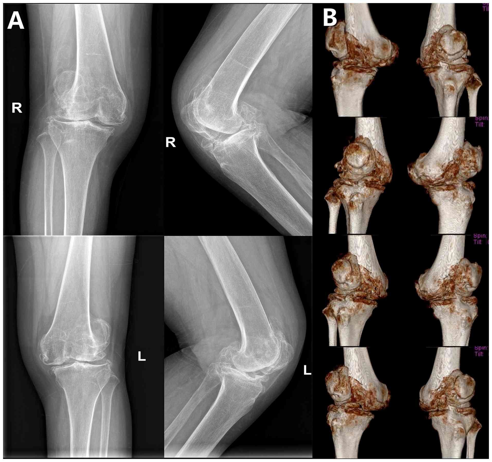
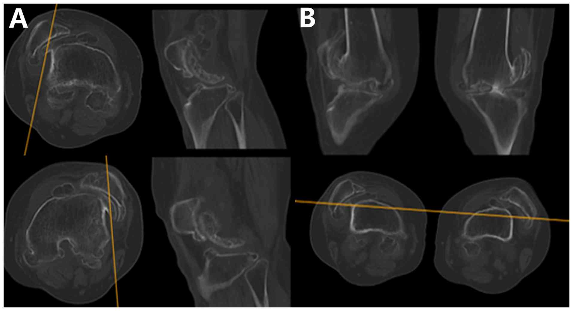
that in the left knee was 35-80˚. Preoperative X-ray and computed
tomography imaging revealed bilateral patellar dislocation,
abnormal development of the femoral trochlea and irregular patellar
morphology, along with extensive osteophyte proliferation around
the knee joint (Figs. 1A and
B, and 2A and B). Preoperative functional assessment
revealed severe impairment, with Knee Society Scores (KSS) of 42
(clinical) and 50 (functional), Oxford Knee Scores (OKS) of 36
(7,8), and a Visual Analog Scale (VAS) score
of 8, consistent with advanced osteoarthritis.
Case analysis and surgical
reconstruction strategy
TKA was selected as a treatment option to address
three main problem areas: Knee osteoarthritis, patellar dislocation
and knee flexion-extension dysfunction. The purpose of TKA combined
with the pie-crusting and overlapping techniques is to treat
osteoarthritis and reconstruct patellar tracking and stability
(9). The preoperative lateral
stress test under general anesthesia revealed good stability of the
medial and lateral collateral ligaments. If intraoperative findings
had shown irreparable patellar tracking and alignment,
reconstruction of the MPFL would have been necessary to achieve
balance. A pneumatic tourniquet was applied at the upper thigh, and
the knee was maintained in extreme flexion to avoid interference
from the extensor mechanism. A modified Insall approach
(parapatellar approach) was used (10). First, a midline skin incision was
made, then a superficial fascial incision, and finally, the joint
incision 8 cm above the patella. A longitudinal incision was made
along the medial third of the quadriceps tendon, extending medially
and distally through the patellar ligament to the tibial tubercle.
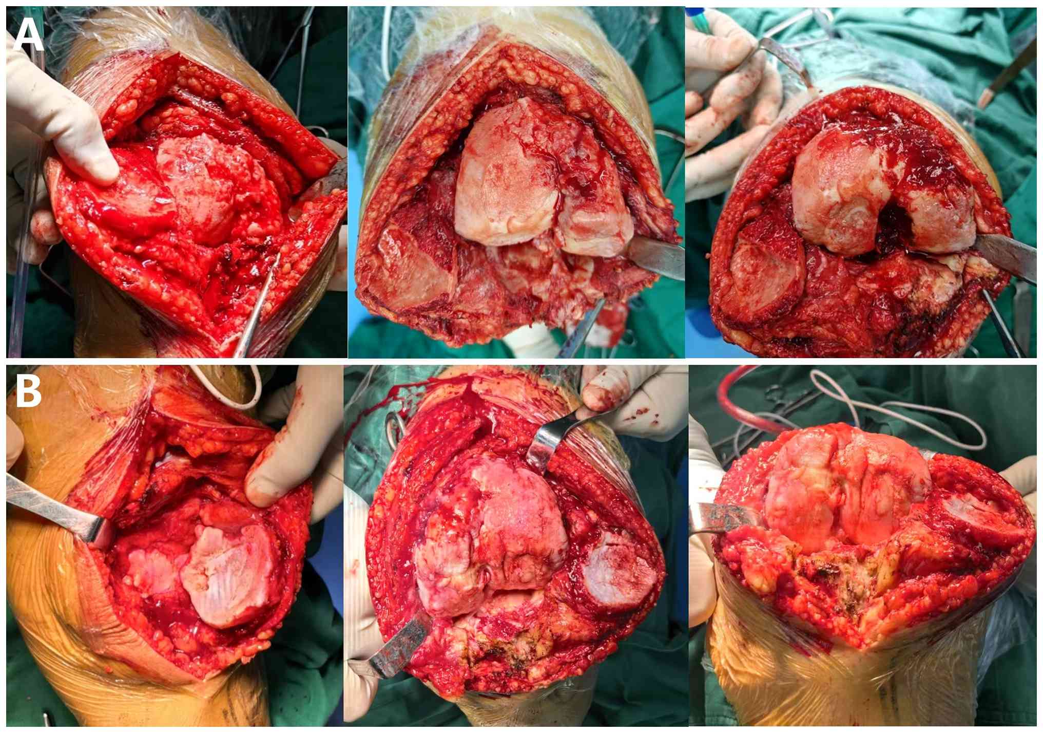
Intraoperative findings revealed fixed patellar dislocation with
dysplastic morphology, and extensive osteophyte formation had
created a concave articular surface, along with a ball-socket-like
pseudojoint with the lateral femoral condyle. Weight-bearing
occurred on the anterior-lateral femoral condyle and tibial
intercondylar eminence, and osteophytes covered the femoral
intercondylar notch. The distal femur exhibited equinus deformity,
with articular cartilage wear, joint space narrowing, subchondral
bone sclerosis and severe posterior femoral condyle destruction
(Fig. 3A and B). The lateral retinaculum and iliotibial
band were markedly contracted. The standard neutral mechanical
alignment principle was adopted for the osteotomy, and the trial
test showed good stability in both flexion and extension. Next, a
reciprocating saw was used to perform patellar surface
reconstruction, reshaping the articular surface and central ridge
for proper alignment with the femoral trochlear groove. The
significant lateral patellar maltracking and subluxation risk were
addressed. A deep capsular release was first performed.
Subsequently, a pie-crusting release of the lateral retinaculum and
the iliotibial band was conducted using an #11 blade. With the knee
held at 90˚ flexion and a laminar spreader gently distracting the
lateral compartment, multiple horizontal stab incisions (8-10
punctures spaced 0.5-1 cm apart) were made parallel to the joint
line over the palpably tight lateral structures. This stepwise
release was performed until the lateral structures demonstrated
adequate lengthening. Patellar tracking was assessed following each
series of punctures using the ‘no-thumb’ test. The release was
considered sufficient when the patella remained centered within the
trochlear groove throughout a full range of passive knee flexion
and extension without manual pressure, and a rectangular flexion
gap was achieved. To reinforce medial stability, a medial
retinacular overlap suturing technique was employed. The redundant
medial retinacular tissue was advanced laterally and superiorly.
Using #2 Ethibond sutures, an interrupted vertical mattress suture
pattern was utilized to imbricate the medial retinaculum. The
sutures were placed to achieve an overlap of 1.0-1.5 cm, starting
proximally at the vastus medialis obliquus (VMO) expansion and
extending distally to the medial border of the patellar tendon. The
suturing was performed with the knee positioned at 30-45˚ flexion,
and the patella was reduced in the trochlear groove, ensuring
appropriate tissue tension that was neither too tight to cause
medial over-constraint nor too loose to allow residual lateral
shift. This created a double-layered reinforcement: The deep layer
involved imbricating the medial capsule and residual MPFL tissue to
the patellar border, while the superficial layer involved advancing
the VMO fascia anterolaterally and suturing it to the anterior
surface of the patella to provide dynamic medial stabilization.
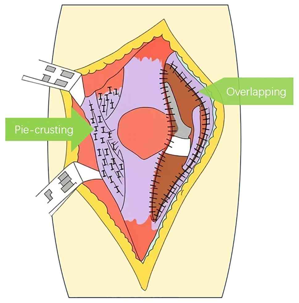
Fig. 4 illustrates the extent of
the release of the lateral patellar retinaculum (marked as zone
‘I’). The medial patellar retinaculum and its continuity with the
vastus medialis muscle were reinforced by an overlapping suture
technique using Ethibond sutures, ensuring stability and providing
adequate soft tissue coverage.
Postoperative management
Following the surgery, the patient wore a hinged
knee brace in an extended position. On the day of the surgery, the
patient performed ankle pump exercises, and on post-operative day
2, isometric and isotonic exercises of the quadriceps were
initiated. At 1-week post-surgery, the patient actively performed
knee flexion and extension exercises within a pain-free ROM. At 2
weeks post-surgery, the patient walked with partial weight-bearing
on the ground, assisted by a walker. All surgical incisions
achieved primary healing without wound dehiscence or infection, and
the sutures were removed 2 weeks after surgery. At 4-weeks
post-surgery, the knee brace was removed, and the patient was able
to walk with full weight-bearing and continued to perform the
straight leg raising exercise.
Follow-up results
The patient underwent left knee surgery 12 months
after right knee surgery using the same approach. The final
follow-up was conducted 27 months after right knee surgery and 15
months after left knee surgery. The patient demonstrated a
pain-free gait (Video S1), with
active leg extension capability. The knee joint extension was
comparable and symmetrical between both sides. The 27-month
postoperative X-rays showed satisfactory results, with good
prosthesis stability and no signs of osteolysis or patellar
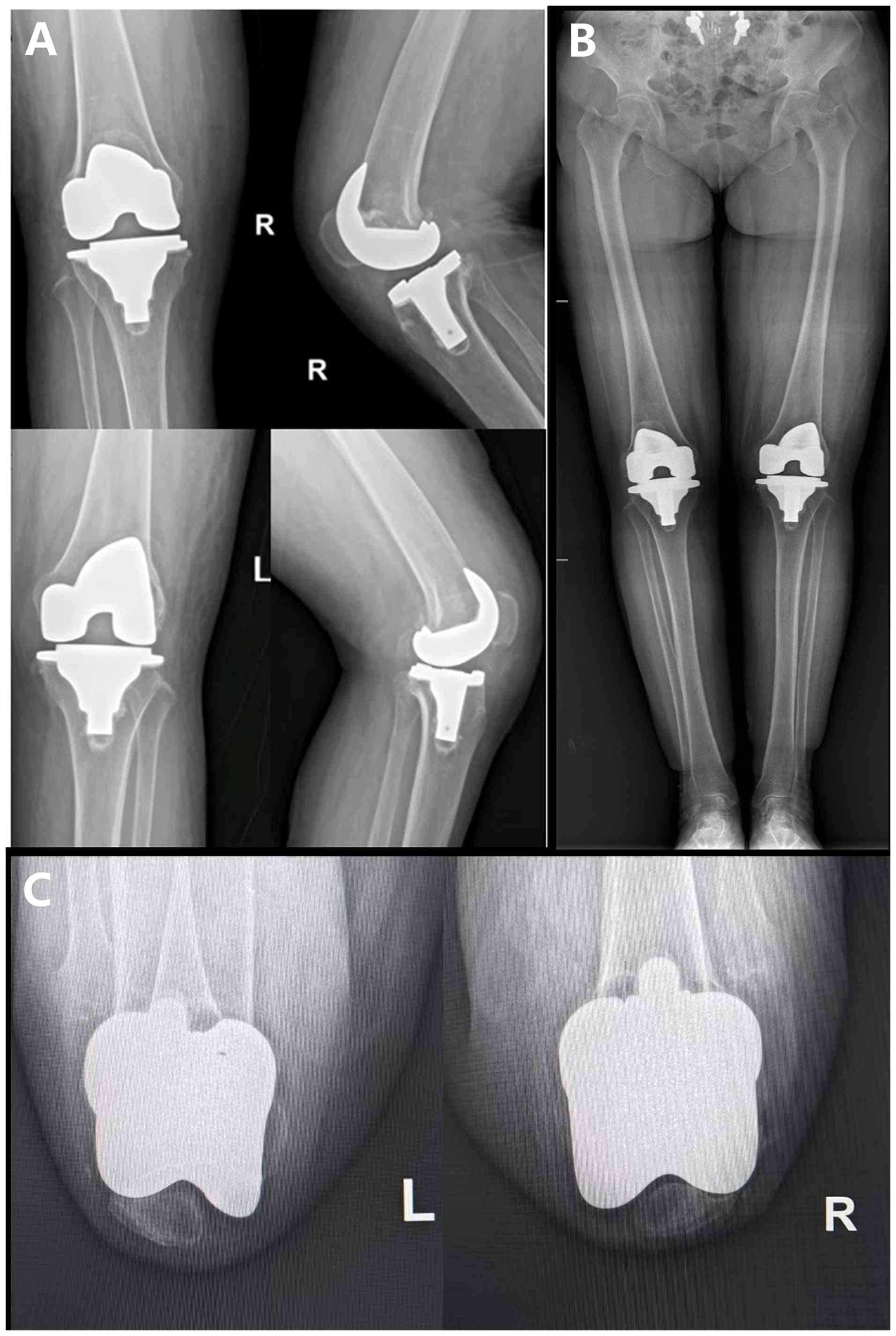
subluxation (Fig. 5A and B). Sunrise view X-ray of the patella
revealed good patellofemoral alignment with stable patellar
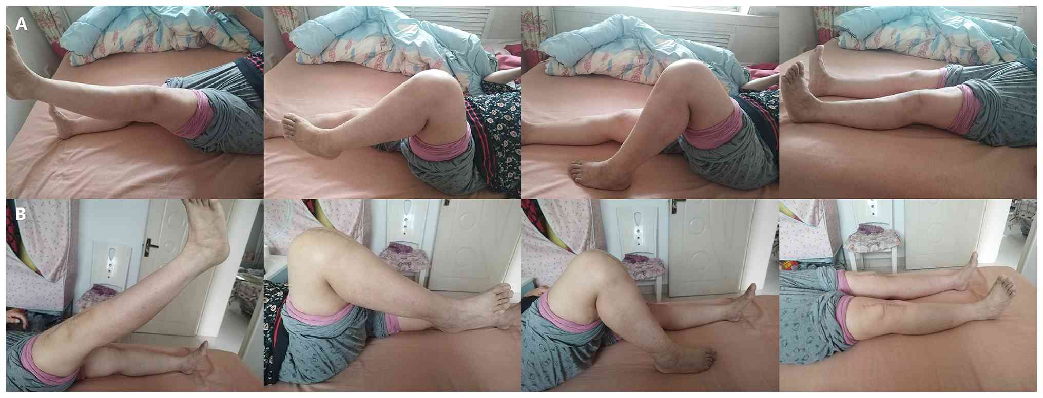
tracking (Fig. 5C). The ROM was
0-115˚ for the right knee and 0-115˚ for the left knee (Fig. 6A and B). Patellar tracking and stability were
excellent (Video S2). The KSS
results of the right knee were 94 points (clinical) and 92 points
(functional), and the OKS was 13 points (7,8).
Meanwhile, the KSS clinical score of the left knee joint was 93
points, the functional score was 90 points and the OKS score was 14
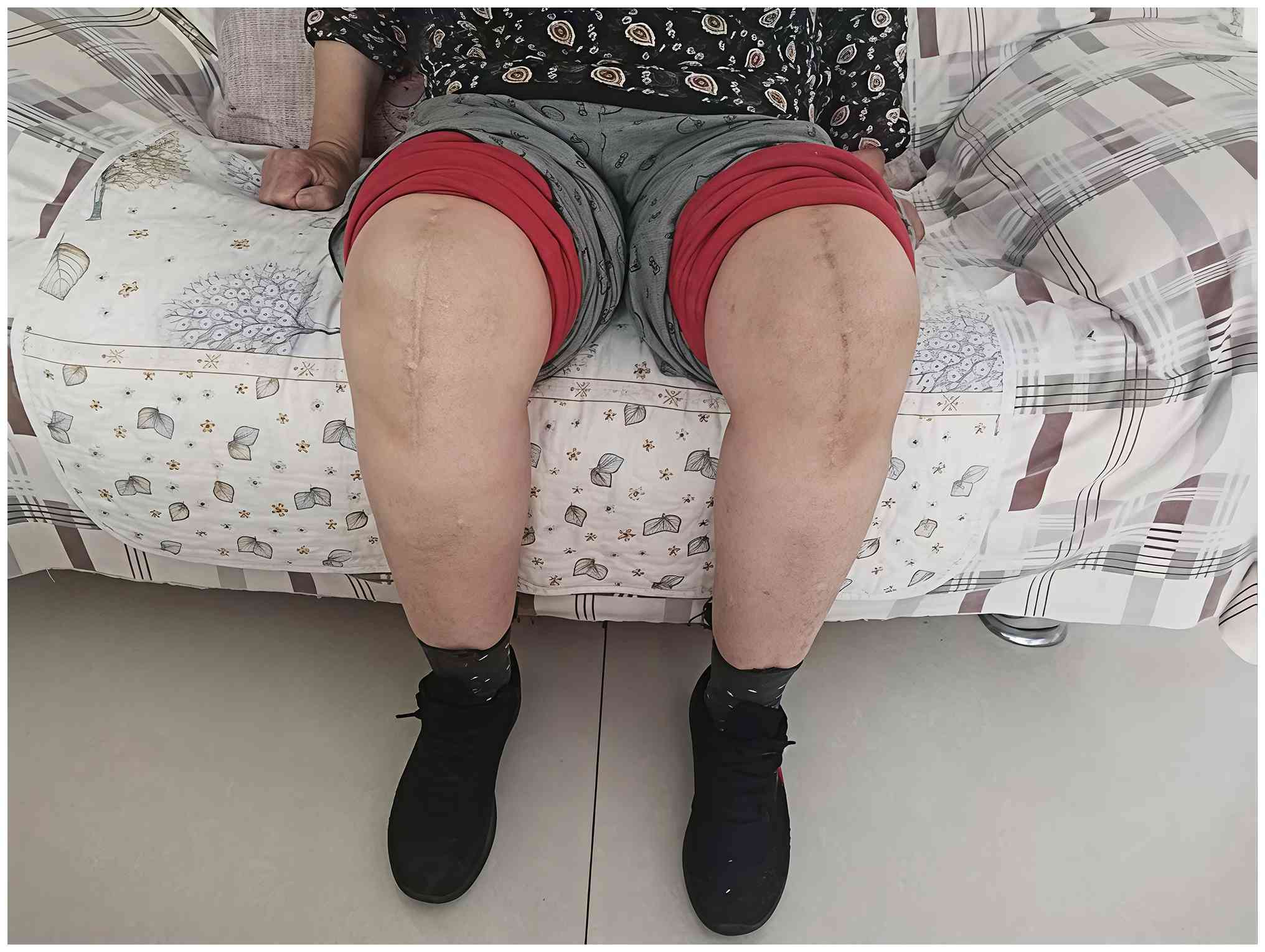
points. The rotational alignment of both knee joints was
successfully corrected and restored post-operatively (Fig. 7). A detailed comparison of the
clinical outcomes before and after the surgery is presented in
Table I. At the final follow-up,
no early complications, such as wound infection, periprosthetic
infection, thrombosis, prosthesis loosening, patellar instability
or recurrent dislocation, were observed. The patient will be
followed up regularly for at least 2 years post-operatively, with
clinical evaluations and radiographic assessments scheduled every 3
months during the first year, every 6 months during the second
year, and annually thereafter. Follow-up assessments will include
the KSS, OKS, VAS for pain, and active ROM measurements.
Additionally, long-term monitoring for potential complications such
as implant loosening, periprosthetic infection or recurrent
patellar dislocation will be conducted through physical
examinations and periodic radiographs.
 | Table IComparison of knee joint function
scores before and after total knee arthroplasty. |
Table I
Comparison of knee joint function
scores before and after total knee arthroplasty.
| | | KSS | |
|---|
| Timeframe | Knee | Clinical score | Functional score | OKS | VAS | ROM, ˚ |
|---|
| Preoperative | Right | 42 | 50 | 36 | 8 | 10-85 |
| | Left | 52 | 55 | 32 | 7 | 5-90 |
| Postoperative | Right | 94 | 92 | 13 | 0 | 0-115 |
| | Left | 93 | 90 | 14 | 0 | 0-115 |
Discussion
Irreducible patellar dislocation is a rare disease.
According to recent studies, its causes are mainly divided into
congenital and acquired types. Most patients with acquired patellar
dislocation have an obvious history of knee joint trauma. In
adults, chronic dislocations often go undiagnosed as symptoms of
secondary osteoarthritis appear late and patients adapt to their
limited function (2,11). To further understand patellar
dislocation, relevant literature was retrieved, including 10 case
reports and 1 retrospective case series study. The specific
descriptions are shown in Table
II (2-6,11-16).
Among them, 5 case reports explicitly mentioned the description of
acute patellar dislocation following a sprain or fall in the
medical history (3,5,11-13).
In addition to acquired cases, some patients with CPD have a higher
probability of knee joint injury during exercise or labor than the
normal population. Among patients with a history of CPD who present
with permanent patellar dislocation, some cases may be attributable
to trauma-related factors. In addition, CPD-related or pathological
anatomical factors may contribute, including poor hypoplastic
patellar and femoral condylar development, a flattened femoral
trochlea and inherent patellar instability. From the pathological
anatomy in the literature review (Table II), it can be concluded that in
most cases, dysplasia of the femoral trochlea can be observed in
the patients undergoing TKA (2,3,5,6,12,14).
Therefore, some cases may have multiple etiologies. Furthermore,
most patients lack previous imaging data, have a long history of
dislocation and deformity, and also present with severe knee
osteoarthritis (hyperplastic osteophytes, disorder of anatomical
structures or ligament contracture). All these factors affect the
clinical diagnosis. Regardless of the difficulty in determining the
diagnosis and etiology, for patients with patellar dislocation
combined with end-stage osteoarthritis, TKA has been widely adopted
and achieved good clinical efficacy. However, the most important
aspect of the surgical technique is how to reconstruct the
patellofemoral trajectory and restore patellar stability during the
surgery.
| Table IISummary of published literature on TKA
for knee osteoarthritis secondary to patellar dislocation. |
Table II
Summary of published literature on TKA
for knee osteoarthritis secondary to patellar dislocation.
| First author,
year | Country | Article type | Pathological
characteristics | Surgical methods | Surgical
approach | Prosthesis type | Follow-up time,
months | Clinical
efficacy | (Refs.) |
|---|
| Ishigaki et
al, 2018 | Japan | Case report | Bilateral lateral
patellar dislocation, valgus deformity (7-15˚), trochlear
hypoplasia | Simultaneous
bilateral TKA+lateral release + vastus medialis plication + fat
pad/joint capsule coverage | Lateral
parapatellar | Cruciate-retaining
(NAKASHIMA FINE) | 24 | ROM: 0-120˚; JOA
score: 40→90. Stable patellar tracking, No recurrence. | (6) |
| Figueroa et
al, 2018 | Chile | Case report | Chronic lateral
dislocation, valgus/varus deformity, tricompartmental OA | TKA+tibial tubercle
osteotomy (medialization) + vastus medialis advancement | Medial
parapatellar | Posterior-stabilized
(PFC Sigma®) | 24 | Pain-free ambulation;
ROM: 0-100˚; KSS improved. No instability. | (4) |
| Yamanaka et
al, 2015 | Japan | Case report | Bilateral congenital
dislocation, valgus (10˚), trochlear dysplasia, hypoplastic
patella | Bilateral staged TKA
(right: no realignment; left: lateral release + tibial tubercle
transfer + quadriceps lengthening) | Medial parapatellar
(both) | Posterior-stabilized
(NexGen LPS-Flex) | 24 | Right: ROM 0-125˚
(extension lag 45˚); Left: ROM 0-90˚. KSS improved. Left tibial
fracture post-op (conservative management). | (15) |
| Albuquerque et
al, 2018 | Brazil | Case report | Permanent lateral
dislocation, valgus/varus OA, trochlear dysplasia | TKA+lateral
retinacular release | Medial
parapatellar | Posterior-stabilized
(PFC Sigma) | 24 | KSS: 42→91 (Case 1),
25→82 (Case 2). Stable patellar tracking, No recurrence. | (5) |
| Pradhan et al,
2001 | Japan | Case report | Bilateral congenital
dislocation, severe valgus (FTA 160˚), OA | Staged
TKA+ITB/PLC/LCL release + gastrocnemius release + biceps
Z-lengthenin (right failed → constrained revision) | Not specified | MG-II prosthesis
(primary); Constrained (Biomet Maxim) (revision) | 14 | Right knee
dislocation at 14 months → revision. Left knee pain-free at 6
years. | (16) |
| Junqueira et
al, 2016 | Brazil | Case report | Chronic
post-traumatic dislocation, valgus, hypoplastic trochlea | TKA+lateral
release | Subvastus | Medial-pivot
(unspecified) | 24 | KSS: 49→93; ROM:
0-105˚. Bone scan confirmed patellar viability. No recurrence. | (13) |
| Chouhan et
al, 2024 | India | Case report | Habitual
dislocation (55 years), valgus (FTA 166˚), trochlear dysplasia,
tibial slope reversal | TKA+lateral release
+ ITB Z-lengthening + medial plication + quadriceps snip | Medial
parapatellar |
Posterior-stabilized (Scorpio-TS) | 23 | ROM: 5-95˚; Kujala
score: 39→83. Stable patella, no recurrence. | (2) |
| Kamada et
al, 2017 | Japan | Case report | Permanent lateral
dislocation, valgus (12˚), trochlear dysplasia, OA | Navigation-assisted
TKA+lateral release | Medial
parapatellar | Cruciate-retaining
mobile-bearing (ATTUNE™) | 12 | ROM: 5-110˚; KSS:
73→93. Stable patellar tracking. | (12) |
| Kumagai et
al, 2016 | Japan and UK | Case report | Prolonged
dislocation (55 years), valgus, trochlear dysplasia | TKA+lateral release
+ medial plication | Medial
parapatellar | Constrained
condylar (Scorpio-TS) | 12 | ROM: 0-105˚; HSS
patellar score: 15→70. No recurrence. | (14) |
| Kim et al,
2024 | South Korea | Case series | Neglected lateral
dislocation, valgus (mean 8.2˚), trochlear dysplasia, patellar
hypoplasia | TKA+lateral release
± medial plication ± quadriceps lengthening (snip/V-Y)±constrained
implant | Medial
parapatellar | Varied
(Triathlon/Persona/Attune) | 68 | KSS knee:
36.7→88.3; WOMAC: 52.8→14.3. ROM improved. 1 case asymptomatic
subluxation; no revisions. | (11) |
| Garcia-Mansilla
et al, 2020 | Argentina | Case report | Chronic
dislocation, OA, trochlear irregularity | TKA + MPFL
reconstruction (tibialis anterior allograft) + lateral release | Medial
parapatellar | Cruciate-retaining
(Legion Oxinium) | 6 | ROM: 0-115˚; stable
patellar position (lateral tilt but no dislocation). | (3) |
There are differences in patellar treatment
strategies and opposing views on reconstructing the extensor
mechanism of the knee. Marmor (17) and Pradhan et al (16) reported that patients with CPD often
adapt to abnormal anatomy, and that satisfactory functional
outcomes can still be achieved with TKA even without altering the
patellar position. However, in the case reported by Pradhan et
al (16), tibiofemoral joint
dislocation occurred 14 months after the surgery, suggesting that a
high-restraint prosthesis is required following extensive
soft-tissue release. Ishigaki et al (6) and Kim et al (11) advocated for combined lateral
support band release, medial overlapping sutures and patellar
replacement to reconstruct the trajectory (6,11).
In addition, the 68-month follow-up in the case series reported by
Kim et al (11) showed that
there was only 1 case of asymptomatic subluxation (from 8
patients), supporting the reliability of this protocol. Regarding
key techniques for soft-tissue balance, all studies have emphasized
the necessity of releasing the lateral support band, but further
surgeries need to be individualized. Kim et al (11) indicated that quadricep lengthening
is suitable for those with a severe contracture. No severe
contracture was observed in the bilateral quadriceps of the patient
in the present case. Yamanaka et al (15) reported the risk of postoperative
tibial fractures following tibial tubercle osteotomy (TTO) after a
patient sustained a left tibial fracture 5 weeks postoperatively,
just below the osteotomy/transfer site after a fall. There is
controversy over prosthesis selection and the specific types of
constraints in the prevention and control of complications. For the
present patient, a PS prosthesis was used during primary TKA.
Overlap and pie-crusting techniques were employed to achieve
balanced soft tissues, resulting in good patellar tracking and
stability as supported by the technical principles previously
outlined (18). Pradhan et
al (16) switched to a hinged
knee due to dislocation failure; however, we consider that a
posterior-stabilized (PS) prosthesis combined with soft-tissue
surgery was sufficient to maintain stability in the present case.
When considering patellar replacement, the advantages include
reduced anterior knee pain and an optimized trajectory. However,
there is an ongoing risk of an increased rate of dislocation, as
seen in the subsequent failed cases in study by Marmor (17). In the present study, a patellar
replacement was not performed, as the patella 12-mm thick following
the removal of the osteophytes. A reciprocating saw was used for
patelloplasty, with the aim of restoring the anatomical structure
of the patella as much as possible.
The pie-crusting release technique is a reliable
method for lateral supporting band release. By gradually releasing
and evaluating immediately, it can accurately restore soft-tissue
tension balance, safely reduce the patella and ensure stable
tracking. In addition, the tight overlapping suturing of the medial
patellar retinaculum is a relatively simple and effective technique
for medial patellar stabilization.
When performing TKA for CPD combined with severe
patellofemoral arthritis, the choice of surgical strategy should be
based on specific anatomical and pathological features, after
carefully evaluating the trade-offs. No severe lateralization of
the tibial tubercle was found in the preoperative evaluation of the
present case; therefore, performing TTO would unnecessarily
increase surgical complexity, operative time, and the risks of
non-union and implant-related complications. While MPFL
reconstruction is considered the ‘gold standard’ for restoring
medial static stability of the patella, it requires a graft and a
high level of technical expertise, with potential risks of
excessive constraint leading to abnormal patellofemoral joint
pressure or limited knee flexion. In the present case, following
intraoperative release of the lateral contracted structures using
the precise pie-crusting technique, the patellar trajectory was
already balanced. When combined with a medial overlapping suture,
this approach provided reliable static stability, thereby obviating
the additional costs and complication risks associated with MPFL
reconstruction. Regarding patellar replacement, although it can
improve patellofemoral joint congruity and potentially reduce
anterior knee pain, it also introduces long-term risks, such as
prosthesis loosening, fracture and patellar clunk. In the present
study, although the patient's patellar cartilage was severely worn,
the bony structure was intact and of sufficient thickness;
therefore, patelloplasty was selected over replacement. We
acknowledge that postoperative imaging (for example, axial views)
suggested a slight lateralization of the patella, a condition that
might have been corrected to achieve better congruity through
patellar replacement. However, given the excellent clinical scores
and stability achieved, preserving the native patella avoided
prosthesis-related risks, which we believe was a more reasonable
choice. In conclusion, under the specific conditions of the present
case, including limited resources, adequate patellar bone stock and
stable collateral ligaments, the combined soft-tissue balancing
technique using a standard prosthesis, lateral pie-crusting release
and medial overlapping suture directly targeted the core pathology
of ‘tight lateral, loose medial’. This approach achieved stable
reconstruction of the patellar trajectory with minimal complexity
and cost, suggesting that it can be a safe, effective and
goal-oriented strategy.
In addition, when confronted with limited medical
resources or specific medical insurance payment policies, when
there is a strict pre-operative evaluation and proper planning,
combining standard prostheses with soft-tissue balance techniques
can achieve satisfactory clinical efficacy. Given these challenges,
surgeons must conduct thorough preoperative assessments.
The findings of the present study should be
interpreted taking several limitations into account, including the
relatively short follow-up duration (2 years for the right knee and
1 year for the left), potentially limiting the assessment of the
long-term survivorship of the prostheses. The single-case design
constrains the generalizability of these findings, and the success
of this technique requires validation in larger cohorts. The
absence of objective functional assessments, such as isokinetic
muscle strength testing and instrumented gait analysis, precludes
the acquisition of more granular quantitative biomechanical
data.
In conclusion, TKA for end-stage osteoarthritis due
to patellar dislocation is surgically challenging, requiring
thorough preoperative assessment. In patients with stable
collateral ligaments, primary TKA with pie-crusting lateral release
can effectively restore patellar tracking and kinematics. The
present case confirms that pie-crusting and medial overlap suturing
can successfully correct patellar tracking and restore stability.
Successful outcomes rely on systematic planning, meticulous
technique and patient compliance. This combined approach represents
a promising treatment strategy.
Supplementary Material
At the final follow-up, the patient
was able to walk unassisted and without pain.
The bilateral knee range of motion at
the final follow-up.
Supplementary Data.
Acknowledgements
Not applicable.
Funding
Funding: The present study was supported by the President's Fund
of Hexi University (grant no. 2025YB043).
Availability of data and materials
The data generated in the present study may be
requested from the corresponding author.
Authors' contributions
Conceptualization was performed by XD. Data curation
was the responsibility of HL, DY and XY. YW contributed to the
collection of clinical data and imaging analysis. HL and DY wrote
the original draft. HL, DY and XY reviewed and edited the
manuscript. All authors have read and approved the manuscript. HL
and XY confirm the authenticity of all the raw data.
Ethics approval and consent to
participate
The present study was performed in accordance with
the ethical standards of the Declaration of Helsinki 1964. The
patient provided written informed consent for participation in the
present case report.
Patient consent for publication
The patient provided written informed consent for
the publication of the present case report and all accompanying
images, including those that contain potentially identifiable
features.
Competing interests
The authors declare that they have no competing
interests.
References
|
1
|
Curtis BH and Fisher RL: Congenital
hyperextension with anterior subluxation of the knee. Surgical
treatment and long-term observations. J Bone Joint Surg Am.
51:255–269. 1969.PubMed/NCBI
|
|
2
|
Chouhan DK, Thami T and Dadra A: Total
knee arthroplasty for neglected habitual dislocation of patella
with knee osteoarthritis: A case report. JBJS Case Connect.
14:2024.PubMed/NCBI View Article : Google Scholar
|
|
3
|
Garcia-Mansilla I, Jones KJ and Sassoon
AA: Combined total knee arthroplasty and medial patellofemoral
ligament reconstruction for chronic patellar dislocation and severe
osteoarthritis. Arthroplast Today. 7:47–53. 2020.PubMed/NCBI View Article : Google Scholar
|
|
4
|
Figueroa D, Garín A and Figueroa F: Total
knee replacement in patients with osteoarthritis and concomitant
inveterate patellar dislocation. Arthroplast Today. 5:68–72.
2018.PubMed/NCBI View Article : Google Scholar
|
|
5
|
Albuquerque RPE, Sousa Filho PGT, Loures
FB, Cobra H, Barretto JM and Cavanellas N: Total knee arthroplasty
in patients with permanent patella dislocation. Report of two cases
and literature review. Rev Bras Ortop. 53:503–509. 2018.PubMed/NCBI View Article : Google Scholar
|
|
6
|
Ishigaki K, Aoki H, Saito M, Takamatsu R,
Takahashi H, Tsuchiya K and Nakamura T: A case of simultaneous
total knee arthroplasty in congenital dislocation of the Patella.
Open J Orthop. 08:267–272. 2018.
|
|
7
|
Miralles-Muñoz FA, Gonzalez-Parreño S,
Martinez-Mendez D, Gonzalez-Navarro B, Ruiz-Lozano M,
Lizaur-Utrilla A and Alonso-Montero C: A validated outcome
categorization of the knee society score for total knee
arthroplasty. Knee Surg Sports Traumatol Arthrosc. 30:1266–1272.
2022.PubMed/NCBI View Article : Google Scholar
|
|
8
|
Ingelsrud LH, Terluin B, Gromov K, Price
A, Beard D and Troelsen A: Which Oxford knee score level represents
a satisfactory symptom state after undergoing a total knee
replacement? Acta Orthop. 92:85–90. 2021.PubMed/NCBI View Article : Google Scholar
|
|
9
|
Motififard M, Ravanbod H, Rahmati R and
Saleki M: Pie-crusting technique of medial collateral ligament for
total knee arthroplasty in varus deformity: A systematic review.
Adv Biomed Res. 12(138)2023.PubMed/NCBI View Article : Google Scholar
|
|
10
|
Hosseini-Monfared P, Mirahmadi A, Amiri S,
Minaie R, Ghafouri MH and Kazemi SM: Comparable long-term
functional outcomes of subvastus and medial parapatellar approach
in total knee arthroplasty: A 10-year follow-up study. J Exp
Orthop. 11(e70035)2024.PubMed/NCBI View Article : Google Scholar
|
|
11
|
Kim SE, Kim SH, Lee JI, Han HS, Lee MC and
Ro DH: Primary total knee arthroplasty for treating osteoarthritic
knees with neglected patellar dislocation. Medicina (Kaunas).
60(1492)2024.PubMed/NCBI View Article : Google Scholar
|
|
12
|
Kamada K, Matsumoto T, Takayama K, Araki
D, Hashimoto S, Hayashi S, Matsushita T and Kuroda R: Mobile
bearing total knee arthroplasty for valgus knee osteoarthritis with
permanent patellar dislocation: A case report and review of the
literature. Case Rep Orthop. 2017(1230412)2017.PubMed/NCBI View Article : Google Scholar
|
|
13
|
Junqueira JJ, Helito CP, Bonadio MB,
Pécora JR and Demange MK: Total knee arthroplasty with subvastus
approach in patient with chronic post-traumatic patellar
dislocation. Rev Bras Ortop. 51:614–618. 2016.PubMed/NCBI View Article : Google Scholar
|
|
14
|
Kumagai K, Kubo M, Maeda T, Araki S,
Shioji S, Okumura N, Fujikawa H, Kawasaki T, Imai S and Matsusue Y:
Total knee arthroplasty for treatment of osteoarthritis with
prolonged patellar dislocation. Arthroplast Today. 3:25–28.
2016.PubMed/NCBI View Article : Google Scholar
|
|
15
|
Yamanaka H, Kawamoto T, Tamai H, Suzuki M,
Kobayashi T, Eguchi Y and Nakajima H: Total knee arthroplasty in a
patient with bilateral congenital dislocation of the patella
treated with a different method in each knee. Case Rep Orthop.
2015(890315)2015.PubMed/NCBI View Article : Google Scholar
|
|
16
|
Pradhan RL, Watanabe W, Itoi E, Yamada S,
Shimada Y and Sato K: Total knee arthroplasty in bilateral
congenital dislocation of the patella-a case report. Acta Orthop
Scand. 72:422–424. 2001.PubMed/NCBI View Article : Google Scholar
|
|
17
|
Marmor L: Total knee arthroplasty in a
patient with congenital dislocation of the patella. Case report.
Clin Orthop Relat Res. 226:129–133. 1988.PubMed/NCBI
|
|
18
|
Zhang Y, Ye LY, Liu HX and Wen H:
Quadriceps tendon pie-crusting release of stiff knees in total knee
arthroplasty. J Orthop Sci. 20:669–674. 2015.PubMed/NCBI View Article : Google Scholar
|