1. Introduction
Lactate, the end product of glycolysis, has long
been considered a metabolic waste product of glucose metabolism
under hypoxic conditions, and was therefore not considered a
notable research topic (1). In the
Warburg effect, tumor cells tend to undergo glycolysis and produce
large amounts of lactate even in the presence of sufficient oxygen.
Lactate accumulates in the cells and is exported to the
extracellular environment through the activation of transporters on
the cell membrane, ultimately leading to the formation of an acidic
tumor microenvironment (TME) (2).
In 2019, Zhang et al (3)
identified the lysine lactylation (Kla) of histones for the first
time, discovering a new metabolite-associated posttranslational
modification (PTM). In addition to histones, there is growing
evidence that non-histone proteins can also be modified by
lactylation (4). Both L-lactate
and lactyl-coenzyme A are the direct substrates for protein
lactylation (5,6). The discovery of protein lactylation
generated a new era for more in-depth analysis of lactate
metabolism and provided a novel mechanism for determining the
pathophysiological mechanisms of lactate in tumors and inflammatory
diseases (7,8).
Over the years, protein lactylation, as a key
regulatory mechanism in cellular metabolism, immune response and
tumor biology, has gradually become a cutting-edge focus of
scientific research. Lactylation not only plays a marked role in
the regulation of energy metabolism, but also notably affects the
regulation of gene expression, the activation of signaling pathways
and cell proliferation and migration (1,2)
(Fig. 1). Specifically,
lactylation provides tumor cells with a notable energetic and
survival advantage by altering the chemistry of specific histone
sites [such as histone H3 lysine 18 (H3K18)], increasing the
activity of glycolysis-related enzymes and facilitating the
activation of a variety of signaling pathways that promote tumor
development (9,10). At the metabolic level, lactylation
promotes glycolysis and other metabolic pathways by upregulating
the expression or activity of key enzymes, creating a malignant
positive feedback loop that further exacerbates the process of
tumor development (11,12). In addition, lactylation promotes
the expression of specific genes, such as NF-kB (p65), YY1, CCND1
and NRP2, which play key roles in tumor cell proliferation,
migration and invasion, by regulating their transcription (13,14).
In terms of the immune response, lactylation results in the
formation of an immunosuppressive microenvironment that is
conducive to tumor growth by increasing the generation and
stability of regulatory T cells (Tregs) while suppressing the
function of effector T cells. For example, in pancreatic ductal
adenocarcinoma (PDAC), the increased lactylation level at the K72
of the moesin protein promotes its interaction with transforming
growth factor β (TGF-β) receptor I and its downstream SMAD family
member 3 (SMAD3) signaling, which promotes the generation and
stabilization of Tregs and consequently enhances the
immunosuppressive state (10).
Similarly, in non-small cell lung cancer (NSCLC), increased
lactylation at the K70 of the apolipoprotein C2 (APOC2) protein
promotes extracellular lipolysis and induces Treg accumulation,
which further exacerbates the phenomenon of immunoresistance
(9). Notably, lactylation also
plays a role in tumor metastasis. Studies have shown that
lactylation promotes the migration and invasion ability of tumor
cells by activating specific signaling pathways [such as Hippo and
platelet derived growth factor receptor beta (PDGFRβ)]. For
example, in glioblastoma (GBM), histone H3 lysine 9 lactylation
(H3K9la) activates LUC7-like 2 (LUC7L2) transcription to promote
its expression, and LUC7L2 mediates the retention of intron 7 of
MutL homolog 1 (MLH1) and reduces MLH1 expression, leading
to temozolomide resistance (15).
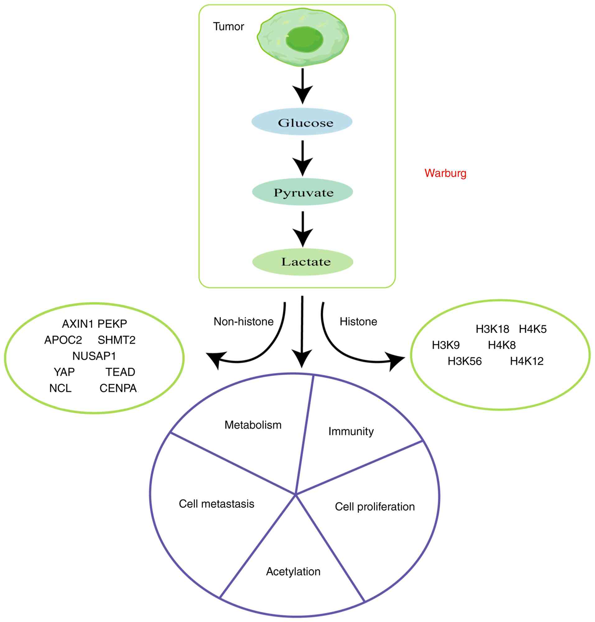 | Figure 1Tumor cells produce lactic acid
through the Warburg effect. The produced lactic acid can serve as a
substrate for lactylation modification, promoting the lactylation
of histones and non-histones during cell proliferation, metastasis,
immunity and metabolism. AXIN1, axis inhibition protein 1; PEKP,
phosphofructokinase, platelet; APOC2, Apolipoprotein C2; NUSAP1,
nucleolar and spindle-associated protein 1; YAP, yes-associated
protein; TEAD, TEA domain; NCL, nucleocapsid; CENPA, centromeric
protein A; SHMT2, serine hydroxymethyltransferase-2; H3K18, histone
H3 lysine 18; H4K5, histone H4 lysine 5; H3K9, histone H3 lysine 9;
H4K8, histone H4 lysine 8; H3K56, histone H3 lysine 56; H4K12, H4
lysine 12. |
In the present review the associations between
lactylation and tumorigenesis are discussed in depth, laying the
foundation for further understanding of its role in disease
progression and aimed to provide some insights for new therapeutic
strategies (16).
2. Lactylation in tumor immunity
Lactate is notable metabolite in the TME that serves
as fuel for mitochondrial metabolism and plays an integral role in
shaping the function of immune cells, which should not be
overlooked in cancer immunotherapy because it can regulate immune
cell metabolism and inhibit the activation and proliferation of
immune cells (17-19)
(Fig. 2). Lactate is a signaling
molecule that plays a notable role in regulating the immune
response of tumor cells and influencing immune surveillance and
escape related behaviors (17-19)
(Fig. 2). The present section
specifically discusses lactate-mediated anti-tumor immunity and
focuses on the research investigating NSCLC, gastric cancer (GC)
and glioma.
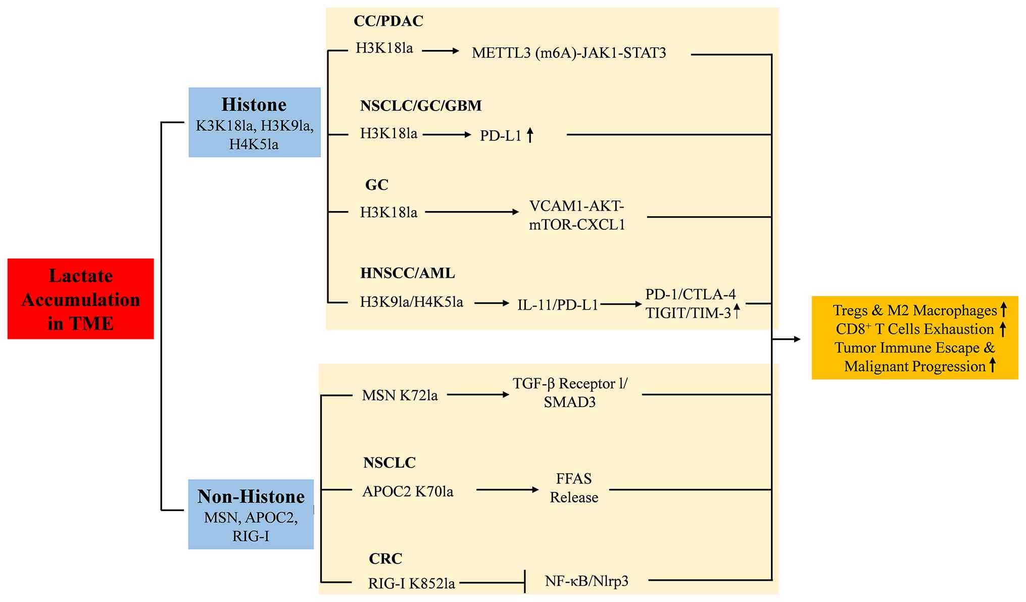 | Figure 2Histone and non-histone lactylation
in tumor immunity. Lactate accumulation in TME reshapes the
immunomicroenvironment. IL-11, interlukin-11; H3K9, histone H3
lysine 9; JAK1, janus kinase 1; STAT3, signal transducer and
activator of transcription 3; H3K18, histone H3 lysine 18; PD-L1,
Programmed death-ligand 1; STAT5, signal transducer and activator
of transcription 5; H4K5, histone H4 lysine 5; VCAM1, vascular cell
adhesion molecule-1; CXCL1, C-X-C motif ligand 1; SMAD3, small
mother against decapentaplegic family member 3; Rig-1, retinoic
acid-inducible gene I; Nlrp3, nod-like receptor protein 3; APOC2,
apolipoprotein C2; METTL3, methyltransferase-like 3; TME, tumor
microenvironment; Tregs, regulatory T cells; FFAS, free fatty
acids; MSN, moesin; CC, cervical cancer; PDAC, pancreatic ductal
adenocarcinoma; NSCLC, non-small cell lung cancer; GC, gastric
cancer; GBM, glioblastoma; HNSCC, head and neck squamous cell
carcinoma; AML, acute myeloid leukemia; CRC, colorectal cancer. |
The accumulation of lactate in the TME induces
histone H3K18 lactylation (H3K18la), upregulating the expression of
the RNA methyltransferase methyltransferase-like 3 (METTL3) in
tumor-infiltrating myeloid cells. METTL3 enhances the translation
of Janus kinase 1 (JAK1) mRNA through m6A modification,
activating the JAK1-STAT3 signaling pathway. This activity
strengthens the immunosuppressive functions of myeloid cells (such
as promoting T-cell exhaustion and Treg-cell infiltration), thereby
driving tumor immune evasion and malignant progression. Notably, a
preclinical study confirmed that genetic ablation of METTL3 in
myeloid cells, treatment with the METTL3 inhibitor STM2457 or
blockade of lactylation (for example using the p300 inhibitor C646)
can notably inhibit tumor growth and remodel the immune
microenvironment (20). Recent
findings suggest that targeting the lactylation-immunosuppression
axis represents a potential antitumor immunotherapy strategy for
patients with NSCLC (21), GC
(22), glioma (23), ovarian cancer (OC) (24), cervical cancer (CC) (25), colorectal cancer (CRC) (26), GBM (27), head and neck squamous cell
carcinoma (HNSCC) (28), acute
myeloid leukemia (AML) (29) and
PDAC (30). These studies have
revealed that histone lactylation not only participates in the
malignant transformation process of tumor cells themselves but also
promotes immune escape and immunosuppressive states in the TME
through complex molecular mechanisms.
Specifically, a study in NSCLC has shown that
elevated H3K18la levels directly activate the POM121 transmembrane
nucleoporin/MYC/programmed death ligand 1 (PD-L1) pathway, increase
PD-L1 expression and enhance the immune escape ability of tumor
cells (21).
In GC, H3K18la upregulates vascular cell adhesion
molecule-1 (VCAM1) transcription, activates the AKT-mTOR-C-X-C
motif ligand (CXCL)1 signaling pathway, and promotes the
recruitment of human GC-derived mesenchymal stem cells and M2
macrophages. Both in vivo and in vitro experiments
confirmed that MK2206 (an AKT inhibitor) notably inhibits the
proliferation, migration and tumor growth of GC cells induced by
VCAM1 overexpression. Targeting the AKT pathway effectively
reverses its procancer effects (22). In addition, lysyl oxidase (LOX)
secreted by cancer-associated fibroblasts upregulates insulin-like
growth factor 1 (IGF1) expression through activation of the TGF-β
signaling pathway, which increases the rate of glycolysis and
lactate accumulation, leading in turn to an increase in the level
of H3K18la, which further facilitates the transcription of PD-L1
and enhances immune evasion in GC cells. Targeting LOX, IGF1 or
glycolysis [such as with dasatinib or lactate dehydrogenase A
(LDHA) inhibitors] can effectively block lactylation, thereby
inhibiting PD-L1 expression and tumor progression (31).
Similarly, in gliomas, lactate-induced H3K18la
promotes the expression of tumor necrosis factor superfamily member
9 in a histone-lactylation-dependent manner, leading to M2
polarization and enhanced immune escape and promoting the
migration, invasion, colony formation and in vivo tumor
growth of glioma cells (23). In
other types of cancers such as OC (24), CC (25), CRC (26) and GBM (27), H3K18la promotes tumor progression
and constructs an immunosuppressive microenvironment by regulating
specific gene promoter activities (such as PD-L1 and
glycerol-3-phosphate dehydrogenase 2) and transcriptional
repression of the retinoic acid receptor γ gene.
In HNSCC, elevated levels of H3K9la promote
interlukin-11 (IL-11) transcription and CD8+ T cell
depletion and upregulate immune checkpoints such as programmed
death-1 (PD-1), TIGIT, cytotoxic T-lymphocyte antigen-4 (CTLA-4)
and TIM-3 on CD8+ T cells, resulting in T-cell
exhaustion and immune escape. Targeting IL-11 (for example with
chol-siIL11) effectively blocks the lactate/H3K9la/IL-11/JAK2/STAT3
axis, reversing CD8+ T-cell exhaustion and restoring
their cytotoxic function. Combining this approach with anti-PD-1
therapy can result in synergistic effects, offering a novel
combination strategy for HNSCC immunotherapy (28). In AML, high expression of STAT5
promotes glycolysis and lactate accumulation, increases E3-binding
protein nuclear translocation, raises the level of histone H4
lysine 5 lactylation and promotes PD-L1 transcription, thereby
inhibiting CD8+ T cell activation through PD-1/PD-L1
interaction, leading to immune escape. Patients with high STAT5
expression may benefit from PD-1/PD-L1 blockade therapy (29). The epidermal growth factor
receptor/extracellular signal-regulated kinase (ERK)/acetyl
coenzyme A synthetase 2 (ACSS2)/lysine acetyltransferase 2A (KAT2A)
axis directly upregulates PD-L1 expression via H3K18la, leading to
CD8+ T-cell dysfunction and immune escape. Targeting the
interaction between ACSS2 and KAT2A with a blocking peptide
effectively suppresses PD-L1 expression, restores T-cell function,
and synergizes with anti-PD-1 therapy, offering a novel
metabolic-immune combination strategy for GBM immunotherapy
(6). In PDAC, CCCTC-binding factor
interacts with heterogeneous nuclear ribonucleoprotein U through a
FLG-AS1-dependent mechanism, promotes E1A-associated protein
recruitment and m6A reader IGF2BP2 activation, enhances
colony-stimulating factor 1 (CSF1) and MYC mRNA
stability, regulates CSF1-selective splicing and promotes M2
polarization of tumor associated macrophages and enhances immune
escape (30).
In summary, elevated levels of lactylation not only
promote the transcription of specific genes (such as IL-11 and
PD-L1) but also affect the function of immune cells through
specific signaling pathways (such as JAK2/STAT3), especially by
weakening CD8+ T-cell activity or promoting the
polarization of immunosuppressive cells such as M2 macrophages.
Together, these changes construct an immunosuppressive
microenvironment that is conducive to tumor growth and development.
Thus, the marked role of histone lactylation modifications in a
variety of cancer types provides a theoretical basis and new
perspectives for the development of novel anticancer therapies
based on this mechanism. A deeper understanding of these complex
regulatory networks can aid in more precisely understanding the
cancer process and improve therapeutic outcomes.
Protein lactylation plays notable roles in the
progression and immune escape of numerous cancers. First,
lactylation promotes the generation and stabilization of Tregs in
the TME by enhancing the interaction of key proteins with signaling
pathways, as revealed by Chen et al (9) and Gu et al (10) and. An increase in the lactylation
level of the K72 locus of the moesin protein enhanced its
interaction with TGF-β receptor I and its downstream SMAD3
signaling, which promoted the generation and stabilization of Tregs
in the TME, thereby contributing to the formation of an
immunosuppressive TME. Targeting lactate metabolism [for example
with LDHi (GSK2837808A)] or directly blocking moesin lactylation
inhibited Treg function and enhanced the efficacy of anti-PD-1
therapy. Clinical samples also show that moesin lactylation levels
are negatively associated with the response to PD-1 therapy
(10). In NSCLC, tumor-derived
lactate, via P300-mediated lactylation of APOC2 at K70, stabilizes
the APOC2 protein, promoting extracellular lipolysis and the
release of free fatty acids (FFAs). These FFAs drive the
accumulation of Tregs and the upregulation of immune checkpoints
such as CTLA-4, thereby suppressing the function of
PD-1+ CD8+ T-cells and leading to resistance
to anti-PD-1 therapy. Targeting APOC2 lactylation (using an
anti-APOC2 K70-lactylation antibody) or lactate metabolism (with
FX11) reverses this resistance and synergizes with anti-PD-1
treatment. In clinical samples, APOC2 K70 lactylation levels are
positively associated with immunotherapy resistance, suggesting a
novel combination strategy and a potential predictive biomarker for
NSCLC immunotherapy (9). Second,
lactylation inhibits the transcription of immune-related genes. In
CRC, the tumor-resident microbiota (for example E. coli)
enhances glycolysis and lactate production in tumor cells, driving
RIG-I lactylation at K852 in macrophages. This activity inhibits
the NF-κB-Nlrp3 axis, promoting M2 polarization and Treg
accumulation (along with the upregulation of the expression of
checkpoints such as CTLA-4), thereby suppressing CD8+
T-cell function and facilitating CRC liver metastasis. Targeting
RIG-I lactylation [for example with
7-(carboxymethyl)-10-methyl-10H-phenothiazin-2-yl acetic acid]
reverses this immunosuppression and increases the efficacy of
chemotherapy (32). In summary,
protein lactylation promotes tumor development and immune escape
through diverse mechanisms in different types of cancers,
suggesting that this modification may be a potential therapeutic
target.
3. Lactylation in tumor metabolism
A major hallmark of cancer is metabolic
reprogramming (33). Metabolism
regulates glycolysis, oxidative phosphorylation and other metabolic
pathways by modulating gene expression and signaling pathways
(34) (Fig. 3). The most well-known example is
the Warburg effect, where tumor cells preferentially use glycolysis
for energy production, even in the presence of oxygen, instead of
oxidative phosphorylation. This metabolic switch leads to the
accumulation of large amounts of lactate, which in tumors not only
exists as a metabolic byproduct but also functions as a metabolic
intermediate and signaling molecule (35,36).
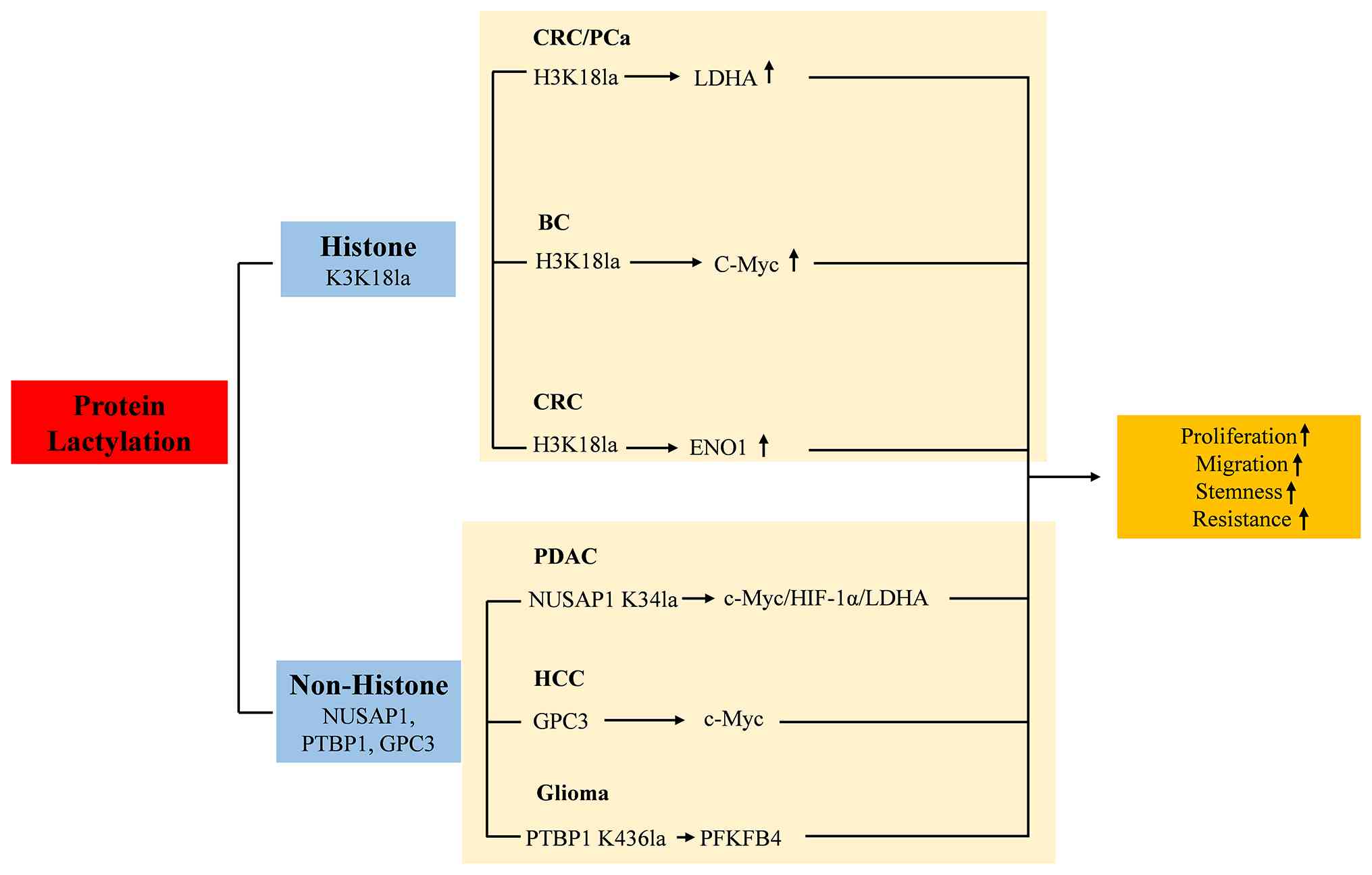 | Figure 3Histone and non-histone lactylation
in tumor metabolism. The tumor metabolism was regulated by H3K18la
and the lactylation of NUSAP1, YAP and GPC3. NUSAP1, nucleolar and
spindle-associated protein 1; LDHA, lactate dehydrogenase A; PFKP,
phosphofructokinase, platelet; YAP, yes-associated protein; PFKFB4,
6-phosphofructo-2-kinase/fructose-2,6-biphosphatase 4; H3K18,
histone H3 lysine 18; LDHA, lactate dehydrogenase A; ENO1, enolase
1; HIF-1α, hypoxia-inducible factor-1α; GPC3, glypican-3; CRC,
colorectal cancer; PCa, prostate cancer; BC, bladder cancer; PDAC,
pancreatic ductal adenocarcinoma; HCC, hepatocellular
carcinoma. |
Histone lactylation contributes to tumorigenesis and
progression through complex metabolic and signaling networks in
multiple cancer types. In PDAC, P300 acts as a potential writer
leading to elevated levels of H3K18la, which activates the
transcription of TTK protein kinase (TTK) and BUB1 mitotic
checkpoint serine/threonine kinase B (BUB1B) to form a positive
feedback loop of glycolysis/H3K18la/TTK/BUB1B. It was observed that
knockdown of TTK inhibited the activation of LDHA via Y239
phosphorylation, which established a positive feedback loop of
glycolysis and histone lactylation and positive feedback loops of
cell cycle genes, suggesting a new mechanism of PDAC and providing
new insights for its treatment (37). In CRC, G protein-coupled receptor
37 promotes LDHA expression through the hippo pathway, increases
H3K18la levels and upregulates the expression of the chemokines
CXCL1 and CXCL5(38). Chen et
al (39) reported that enolase
1 (ENO1) upregulation enhances glycolysis and lactate accumulation.
On the one hand, lactate activates NSUN2 transcription through
H3K18 lactylation; on the other hand, it directly induces K356
lactylation of the NSUN2 protein, increasing its RNA capture
ability. This establishes a self-reinforcing
‘NSUN2-ENO1-lactate-NSUN2’ positive feedback loop. Targeting this
circuit with the small-molecule inhibitor Nsu2-i4 effectively
blocks this feedback mechanism. In anaplastic thyroid carcinoma,
the BRAFV600E mutation enhances aerobic glycolytic flux, leading to
elevated levels of histone H4 lysine 12 lactylation (40). In endometrial carcinoma, H3K18la is
enriched in the ubiquitin-specific protease 39 (USP39) promoter
region. USP39 deubiquitinates and stabilizes phosphoglycerate
kinase 1 (PGK1) and activates glycolysis through the
phosphatidylinositol 3-kinase (PI3K)/AKT/hypoxia-inducible
factor-1alpha (HIF-1α) signaling pathway, promoting cell
proliferation and migration. The lactic acid produced by glycolysis
further stimulates histone lactylation. Targeting lactylation or
USP39 can effectively block this loop (41). In breast cancer, glycolysis-derived
lactate directly activates c-Myc transcription through H3K18
lactylation. c-Myc in turn upregulates SRSF10, driving the protumor
effects of MDM4 and Bcl-x and forming a positive feedback loop of
‘lactate-H3K18la/c-Myc/SRSF10/glycolysis’, and targeting glycolysis
or P300 could disrupt this axis (42). In gliomas, NF-κB activation drives
glycolysis and lactate accumulation, promoting H3K18la deposition
and subsequently activating LINC01127 transcription. LINC01127,
which acts as an RNA scaffold, recruits RNA polymerase II subunit A
to the promoter of mitogen-activated protein kinase kinase kinase
kinase 4 (MAP4K4) in a cis manner, thereby activating the
MAP4K4-c-Jun N-terminal kinase (JNK) pathway and subsequently
activating NF-κB. This positive feedback loop sustains glioma stem
cell stemness and drives tumor progression. The targeting of
LINC01127 or JNK effectively disrupts this axis (43). In prostate cancer, DNA
topoisomerase II α (TOP2A) results in the production of lactate
through the upregulation of LDHA activity and glycolysis, forming a
positive feedback loop of TOP2A/LDHA/lactylation and thus promoting
the proliferation, migration, invasion and epithelial-mesenchymal
transition (EMT) of prostate cancer cells (44). Together, these mechanisms increase
glycolysis and lactate accumulation through the upregulation of key
enzymes (such as LDHA and PGK1) and the formation of a positive
feedback loop that further exacerbates tumor progression. In
addition, lactate modification regulates the transcription of
specific genes (such as TTK, BUB1B, CXCL1, CXCL5, USP39 and c-Myc)
and affects metabolic pathways and signaling pathways, thereby
promoting the proliferation and survival of tumor cells.
Protein lactylation in multiple cancer types
collectively contributes to tumor development by promoting
metabolic reprogramming, stabilizing key proteins, modulating
signaling pathways and influencing gene expression; specifically,
lactylation enhances glycolysis and other metabolic pathways by
upregulating key enzymes [such as LDHA, alanyl-tRNA synthetase 1
(AARS1), phosphofructokinase, platelet (PFKP)], which provide an
energetic advantage to tumor cells. In addition, lactylation
stabilizes key proteins [such as nucleolar and spindle-associated
protein 1 (NUSAP1) and X-ray cross complementing 1 (XRCC1)], which
prolongs their half-life and enhances their functions. Furthermore,
lactylation affects multiple aspects of the TME by activating or
inhibiting specific signaling pathways (such as TGF-β/SMAD3, Hippo
and MAPK/ERK). Lactylation is also enriched in promoter regions
that regulate the transcription of specific genes, further
contributing to tumorigenesis and progression. In PDAC, the binding
of NUSAP1 to c-Myc and HIF-1α promotes LDHA expression, after which
the lactylation of NUSAP1 stabilizes its expression, resulting in
the formation of a positive feedback loop that promotes metastasis;
targeting NUSAP1 or blocking its lactylation can effectively
inhibit this loop (45).
In GC, AARS1 is translocated to the nucleus as a
lactylation writer, and lactylation of yes-associated protein (YAP)
and TEA domain transcription factor (TEAD) promotes AARS1
transcription through the Hippo signaling pathway, driving the
malignant progression of GC. AARS1 is notably expressed in GC
tissues and is positively associated with YAP/TEAD1 expression and
poor patient prognosis. Moreover, the GC-associated R77Q mutation
increases the lactyltransferase activity of AARS1(46). In GBM, interaction of aldehyde
dehydrogenase 1 family member A3 (ALDH1A3) and pyruvate kinase M2
(PKM2) increases glucose metabolism, and promotes lactylation of
XRCC1 at lysine 247 and enhances DNA repair, leading to resistance
to chemoradiotherapy; the small molecule D34-919, which targets the
ALDH1A3-PKM2 interaction, effectively reverses this mechanism and
synergizes with chemoradiotherapy to inhibit tumor growth both
in vivo and in vitro (11).
In intrahepatic cholangiocarcinoma, nucleolin (NCL)
is lactylated by P300 at lysine 477, and lactylated NCL binds to
MAP kinase-activating death domain protein (MADD) pre-mRNA
to prevent aberrant splicing, thereby ensuring efficient
translation of MADD and activating ERK signaling through the MAPK
pathway, thus driving tumorigenesis (47). In pancreatic adenocarcinoma, P300
catalyzes the lactylation of lysine 128 of nicotinamide nucleotide
adenylyltransferase 1 (NMNAT1), inhibits DNA damage-inducible
transcript 3 transcription and promotes the survival of tumor cells
under glucose-deprived conditions. Targeting the lactylation of
NMNAT1 or its upstream metabolic pathways could inhibit tumor
growth (48). In lung
adenocarcinoma, basic leucine zipper and W2 domains 2 (BZW2)
promotes glycolysis to increase lactate accumulation, which in turn
upregulates isocitrate dehydrogenase subunit expression via H3K18
lactylation, thereby driving tumor cell proliferation, migration,
invasion and apoptosis resistance. Targeting glycolysis (for
example with 2-DG or oxamate) or BZW2 effectively inhibits this
axis (12).
In gliomas, polypyrimidine tract-binding protein 1
(PTBP1) promotes glycolysis by promoting the lactylation at lysine
436 of PTBP1, increasing the RNA binding capacity and stabilizing
the mRNA of the metabolic enzyme
6-phosphofructo-2-kinase/fructose-2,6-biphosphatase 4(49). In the hypoxic TME of hepatocellular
carcinoma (HCC), the upregulation of glypican-3 (GPC3) stabilizes
the c-Myc protein by increasing its lactylation, thereby promoting
the proliferation, migration, invasion, stemness and glycolysis of
HCC cells. Targeting GPC3 or inhibiting glycolysis reduces c-Myc
lactylation and suppresses the malignant phenotype of tumors
(50). In OC, lactylation of PFKP
upregulates phosphatase and tensin homolog expression and inhibits
glycolytic processes (51). In
esophageal cancer, hypoxia induces an increase in the level of
Axin1 protein lactylation at lysine 147, promotes Axin1
ubiquitination degradation and relieves the inhibition of
glycolysis, driving metabolic reprogramming and stemness
maintenance in esophageal cancer cells (52).
4. Lactylation in tumor cell
proliferation
The lactic acid produced by glycolysis affects not
only the transcription of the downstream molecule laminin γ 2
(LAMC2), AP001885.4, through histone lactylation, but also affects
the transcription of cyclin D1 (CCND1)/neuropilin-2 (NRP2) through
the non-histone lactylation of centromeric protein A (CENPA). In
esophageal squamous cell carcinoma (ESCC), hypoxia specifically
activates LAMC2 transcription by inducing H3K9la, thereby driving
the proliferation, migration, invasion and metastasis of ESCC cells
through the PI3K/AKT/VEGFA axis (53). Fu et al (13) reported that elevated H3K18la
promotes the transcription of AP001885.4, thereby promoting the
proliferation of ESCC cells. In HCC, CENPA is lactylated at lysine
124, and then promotes the transcription of CCND1/NRP2, thereby
enhancing the proliferation of hepatoma cells (14). In CRC, lysine acetyltransferase 8
(KAT8) was identified as a lactate transferase, lactylating
eukaryotic translation elongation factor-1, α-2 at lysine 408,
increasing translation elongation and protein synthesis and driving
malignant tumor progression; targeting KAT8 can effectively
suppress tumor growth (54).
Taken together, these findings suggest that protein
lactylation collectively promotes cell proliferation in different
types of cancers by increasing the transcription of key genes,
activating specific signaling pathways, and promoting metabolic
reprogramming (Fig. 4).
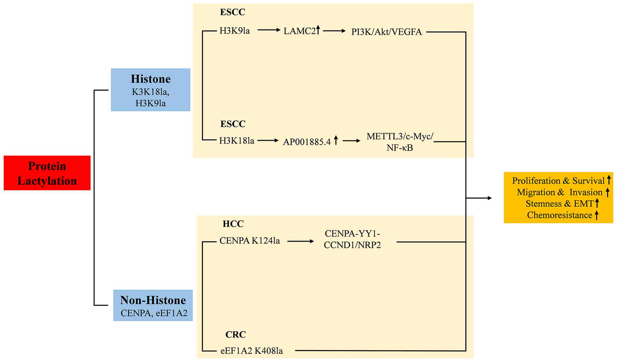 | Figure 4Histone and non-histone lactylation
in tumor cell proliferation. Protein lactylation promotes the
proliferation of tumor cells by regulating PI3K/Akt/VEGFA,
METTL3/c-Myc/NF-kB, and CENPA-YY1-CCND1/NRP2 signaling pathways.
H3K9, histone H3 lysine 9; LAMC2, laminin γ 2; CENPA, centromeric
protein A; YY1, yin yang 1; CCND1, cyclin D1; NRP2, neuropilin-2;
METTL3, methyltransferase-like 3; NF-κb, nuclear factor κB; H3K18,
histone H3 lysine 18; VEGFA, vascular endothelial growth factor A;
ESCC, esophageal squamous cell carcinoma; HCC, hepatocellular
carcinoma; CRC, colorectal cancer; EMT, epithelial-mesenchymal
transition. |
5. Lactate in cell metastasis
Protein lactylation promotes migration and invasion
by regulating the transcription and signaling pathways of key genes
in multiple cancer types. Specifically, in bladder cancer, the
expression of circXRN2 is aberrantly downregulated in bladder
cancer tissues and cell lines. CircXRN2 prevents large tumor
suppressor 1 from speckle-type POZ protein (SPOP)-mediated
degradation by binding to SPOP degron, which then activates the
Hippo signaling pathway, reducing the levels of H3K18la and
lipocalin 2 (LCN2) expression, thus inhibiting migration of cancer
cells (55). In addition, Wang
et al (56) reported that
phosphofructokinase-1 (PFK-1) inhibited histone lactylation and
transcription activity of zinc-finger E-box-binding homeobox 1,
thereby inhibiting the migration and invasion of bladder cancer
cells (56).
In breast cancer, potassium two pore domain channel
subfamily K member 1 increases glycolysis and lactate production by
binding to and activating LDHA, which results in elevated levels of
H3K18la and thus creates malignant positive feedback that reduces
tumor cell stiffness and adhesion, ultimately leading to the
proliferation, invasion and metastasis of breast cancer cells
(57). In clear cell renal cell
carcinoma (ccRCC), the inactivation of von Hippel-Lindau activates
PDGFRβ transcription via the HIF-glycolysis-lactate-H3K18la axis,
while PDGFRβ signaling in turn promotes glycolysis and lactylation,
resulting in the formation of a positive feedback loop that drives
malignant progression of ccRCC. Targeting glycolysis (for example
with oxamate) or PDGFRβ (for example with axitinib) can effectively
disrupt this loop, and the combination therapy has synergistic
effects (58).
In CRC, intestinal bacteria-derived
lipopolysaccharide promotes the transcription and upregulates the
expression of LINC00152 by increasing the level of lactylation of
histone H4 lysine 8 on the promoter, and the overexpression of
LINC00152 promotes the migration and invasion of cells (59). In GC, elevated glucose transporter
protein 3 promotes elevated levels of LDHA, L-lactyl, H3K9la,
H3K18la and histone H3 lysine 56 lactylation (H3K56la), and
promotes the metastasis and invasive ability of GC cells (60). In esophageal cancer, hypoxia
induces elevated levels of serine hydroxymethyltransferase-2
(SHMT2) protein lactylation and upregulates SHMT2 expression, which
in turn increases methylenetetrahydrofolate dehydrogenase 1 like
expression and promotes esophageal cancer cell migration and
invasion (61). In pancreatic
cancer (PC), overexpression of the small Rho GTPase Rif promotes
the upregulation of c-Myc to promote PKM2 transcription, which in
turn promotes the production of lactic acid from glycolysis and
subsequently induces the lactylation of Snail1, which facilitates
EMT and accelerates PC cell migration and invasion (62). In HCC, elevated levels of H3K9la
and H3K56la promote endothelial cell-specific molecule 1 (ESM1)
transcription, upregulate ESM1 expression and promote the
metastasis of HCC cells (63).
In summary, protein lactylation promotes migration
and invasion in different types of cancers through the following
common mechanisms. First, lactylation promotes glycolysis and other
metabolic pathways by upregulating key enzymes (such as LDHA and
PFK-1), providing an energy advantage to tumor cells and forming a
malignant positive feedback loop. Second, lactylation is enriched
in the promoter region, regulating the transcription of specific
genes (such as LCN2, LINC00152 and ESM1) and promoting tumor cell
migration and invasion. Furthermore, lactylation affects multiple
aspects of the TME by activating or inhibiting specific signaling
pathways (such as Hippo, PDGFRβ and EMT). Finally, lactylation
stabilizes key proteins (such as SHMT2 and Snail1), prolonging
their half-life and enhancing their function, thereby further
promoting tumor development (Fig.
5). These findings suggest that further attention to molecules
related to protein lactylation is important for the treatment of
different tumors.
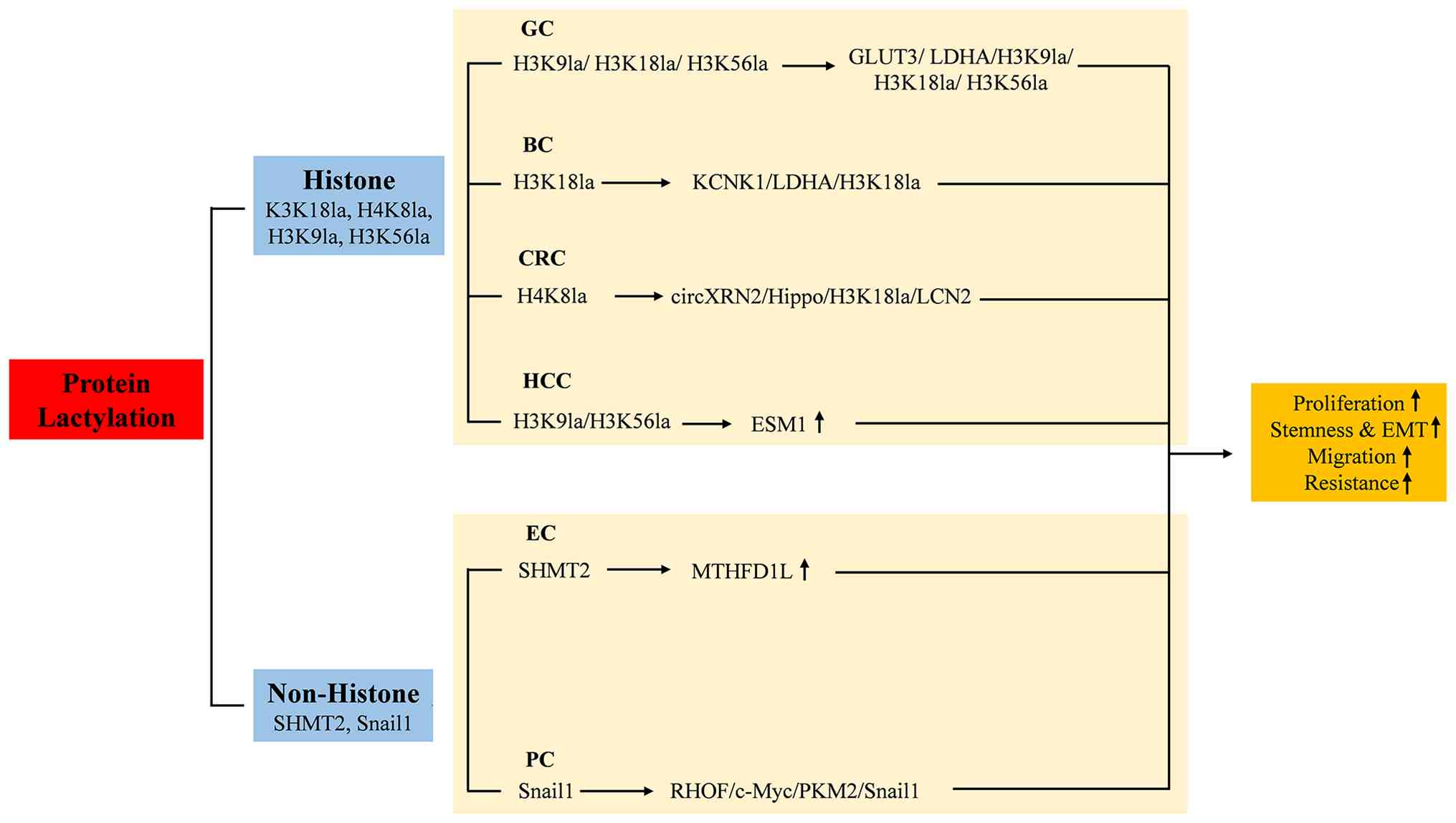 | Figure 5Histone and non-histone lactylation
in tumor cell metastasis. Protein lactylation enhances tumor cell
metastasis by mediating several pathways such as
GLUT3/LDHA/H3K9la/H3K18la/H3K56la, KCNK1/LDHA/H3K18la, and
RHOF/c-Myc/PKM2/Snail1. GLUT3, glucose transporter protein 3; LDHA,
lactate dehydrogenase A; H3K9, histone H3 lysine 9; H3K18, histone
H3 lysine 18; H3K56, histone H3 lysine 56; H4K8, histone H4 lysine
8; SHMT2, serine hydroxymethyltransferase-2; MTHFD1L,
methylenetetrahydrofolate dehydrogenase 1 like; KCNK1, potassium
two pore domain channel subfamily K member 1; LCN2, lipocalin 2;
RHOF, Rho GTPase Rif; ESM1, endothelial cell-specific molecule 1;
EMT, epithelial-mesenchymal transition; GC, gastric cancer; BC,
bladder cancer; CRC, colorectal cancer; HCC, hepatocellular
carcinoma; PC, pancreatic cancer; EC, esophageal cancer. |
6. Acetylation and lactylation
As two notable PTMs, acetylation and lactylation
play distinct but interrelated roles in cellular metabolism, gene
expression regulation and tumor biology. Compared with acetylation,
lactylation is more prominent in the TME, especially under hypoxia
and high glycolytic conditions, where it increases glycolytic
activity by upregulating the expression of key enzymes (such as
LDHA) and promotes tumor cell proliferation, migration and invasion
(11,21). Although both types of modification
regulate gene expression by altering the chemistry of lysine
residues, lactylation is more directly associated with cellular
metabolic states, whereas acetylation is more involved in a broad
network of gene regulation, such as protein stability,
liquid-liquid phase separation and enzyme activity (64).
Studies have also shown that specific enzymes such
as KAT8 catalyze not only acetylation but also lactylation
reactions, indicating a potential cross-regulatory mechanism
between these modifications (54).
Elucidating the differences between acetylation and lactylation and
their potential synergistic effects is required to fully resolve
the complex intracellular regulatory network, and this
understanding should provide a theoretical basis for the
development of new anticancer therapies.
7. Summary and discussion
As an emerging PTM of proteins, lactate modification
overturns the traditional perception of lactate as a metabolic
waste product only, revealing its central regulatory role in
tumorigenesis and development. Histone lactylation (such as H3K18la
and H3K9la) induces an immunosuppressive microenvironment by
activating the transcription of genes such as PD-L1 and IL-11 or by
recruiting immunosuppressive cells such as M2 macrophages and
Tregs, whereas non-histone lactylation (such as APOC2) promotes
immune escape by enhancing the signaling pathway interactions or
metabolic reprogramming. Histone lactylation (for example H3K18la
activates TTK/BUB1B in PDAC) and non-histone lactylation (for
example NUSAP1 and XRCC1 stabilize metabolism-critical enzymes)
work together to increase glycolysis, resulting in the formation of
a ‘lactate production-lactylation’ positive feedback loop that
provides an energetic advantage to tumor cells.
In terms of mechanistic depth, although multiple
studies have revealed the lactylation sites of specific proteins
and their functions, the specific lactylation transferases
(writers) and delactylation enzymes (erasers) that catalyze these
modifications have mostly not been identified. Furthermore, the
interplay between lactylation and other PTMs (such as acetylation,
phosphorylation and ubiquitination), for example, competing for the
same lysine residue or synergistically regulating protein function,
remains largely unexplored. At the level of modeling and
validation, numerous studies rely on in vitro cell
experiments and mouse xenograft models but lack validation in
spontaneous tumor models or gene knock-in/knockout animal models.
In terms of clinical translation, the development of drugs
targeting lactylation is still in its early stages. Although some
studies have demonstrated the efficacy of small-molecule inhibitors
or specific antibodies in vitro and in vivo, the
specificity, pharmacokinetic properties and long-term toxicity of
these compounds have not yet been systematically evaluated.
Furthermore, although lactylation modification has shown potential
as a biomarker for predicting response to immunotherapy in some
studies (65,66), it lacks validation in prospective
clinical trials, and its combined application value in combination
with other known biomarkers remains unexplored.
The discovery of lactylation has opened up a new
dimension of metabolism-epigenetic intersection for tumor research,
and its key role in tumor immunity, metabolism, metastasis and
other aspects makes it a promising therapeutic target. In the
future, it needs to be synergistically promoted through mechanistic
research, technological innovation and clinical translation, which
is expected to overcome the bottleneck of traditional treatment and
provide a brand-new strategy for precision tumor medicine.
Acknowledgements
Not applicable.
Funding
Funding: The present review was funded by the Xingdian Talent
Support Program of Yunnan Province (grant no.
XDYC-QNRC-2022-0264).
Availability of data and materials
Not applicable.
Authors' contributions
SCF was responsible for topic selection, literature
search and manuscript writing. ZZS conceptualized the review,
confirmed the final draft and supervised the work. Both authors
have read and approved the final manuscript. Data authentication is
not applicable.
Ethics approval and consent to
participate
Not applicable.
Patient consent for publication
Not applicable.
Competing interests
The authors declare that they have no competing
interests.
References
|
1
|
Wang JH, Mao L, Wang J, Zhang X, Wu M, Wen
Q and Yu SC: Beyond metabolic waste: Lysine lactylation and its
potential roles in cancer progression and cell fate determination.
Cell Oncol (Dordr). 46:465–480. 2023.PubMed/NCBI View Article : Google Scholar
|
|
2
|
Yang H, Zou X, Yang S, Zhang A, Li N and
Ma Z: Identification of lactylation related model to predict
prognostic, tumor infiltrating immunocytes and response of
immunotherapy in gastric cancer. Front Immunol.
14(1149989)2023.PubMed/NCBI View Article : Google Scholar
|
|
3
|
Zhang D, Tang Z, Huang H, Zhou G, Cui C,
Weng Y, Liu W, Kim S, Lee S, Perez-Neut M, et al: Metabolic
regulation of gene expression by histone lactylation. Nature.
574:575–580. 2019.PubMed/NCBI View Article : Google Scholar
|
|
4
|
Wang J, Wang Z, Wang Q, Li X and Guo Y:
Ubiquitous protein lactylation in health and diseases. Cell Mol
Biol Lett. 29(23)2024.PubMed/NCBI View Article : Google Scholar
|
|
5
|
Li H, Liu C, Li R, Zhou L, Ran Y, Yang Q,
Huang H, Lu H, Song H, Yang B, et al: AARS1 and AARS2 sense
L-lactate to regulate cGAS as global lysine lactyltransferases.
Nature. 634:1229–1237. 2024.PubMed/NCBI View Article : Google Scholar
|
|
6
|
Zhu R, Ye X, Lu X, Xiao L, Yuan M, Zhao H,
Guo D, Meng Y, Han H, Luo S, et al: ACSS2 acts as a lactyl-CoA
synthetase and couples KAT2A to function as a lactyltransferase for
histone lactylation and tumor immune evasion. Cell Metab.
37:361–376.e7. 2025.PubMed/NCBI View Article : Google Scholar
|
|
7
|
Qu J, Li P and Sun Z: Histone lactylation
regulates cancer progression by reshaping the tumor
microenvironment. Front Immunol. 14(1284344)2023.PubMed/NCBI View Article : Google Scholar
|
|
8
|
Liu X, Zhang Y, Li W and Zhou X:
Lactylation, an emerging hallmark of metabolic reprogramming:
Current progress and open challenges. Front Cell Dev Biol.
10(972020)2022.PubMed/NCBI View Article : Google Scholar
|
|
9
|
Chen J, Zhao D, Wang Y, Liu M, Zhang Y,
Feng T, Xiao C, Song H, Miao R, Xu L, et al: Lactylated
apolipoprotein C-II induces immunotherapy resistance by promoting
extracellular lipolysis. Adv Sci (Weinh).
11(e2406333)2024.PubMed/NCBI View Article : Google Scholar
|
|
10
|
Gu J, Zhou J, Chen Q, Xu X, Gao J, Li X,
Shao Q, Zhou B, Zhou H, Wei S, et al: Tumor metabolite lactate
promotes tumorigenesis by modulating MOESIN lactylation and
enhancing TGF-β signaling in regulatory T cells. Cell Rep.
39(110986)2022.PubMed/NCBI View Article : Google Scholar
|
|
11
|
Li G, Wang D, Zhai Y, Pan C, Zhang J, Wang
C, Huang R, Yu M, Li Y, Liu X, et al: Glycometabolic
reprogramming-induced XRCC1 lactylation confers therapeutic
resistance in ALDH1A3-overexpressing glioblastoma. Cell Metab.
36:1696–1710.e10. 2024.PubMed/NCBI View Article : Google Scholar
|
|
12
|
Wang M, He T, Meng D, Lv W, Ye J, Cheng L
and Hu J: BZW2 modulates lung adenocarcinoma progression through
Glycolysis-mediated IDH3G lactylation modification. J Proteome Res.
22:3854–3865. 2023.PubMed/NCBI View Article : Google Scholar
|
|
13
|
Fu C, Jiang W, Wang C, Song SJ, Tao H,
Zhang XG, Li WT, Jin X, Yu BB, Hao JJ, et al: AP001885.4 promotes
the proliferation of esophageal squamous cell carcinoma cells by
histone lactylation- and NF-κB (p65)-dependent transcription
activation and METTL3-mediated mRNA stability of c-myc. Anim Cells
Syst (Seoul). 28:536–550. 2024.PubMed/NCBI View Article : Google Scholar
|
|
14
|
Liao J, Chen Z, Chang R, Yuan T, Li G, Zhu
C, Wen J, Wei Y, Huang Z, Ding Z, et al: CENPA functions as a
transcriptional regulator to promote hepatocellular carcinoma
progression via cooperating with YY1. Int J Biol Sci. 19:5218–5232.
2023.PubMed/NCBI View Article : Google Scholar
|
|
15
|
Yue Q, Wang Z, Shen Y, Lan Y, Zhong X, Luo
X, Yang T, Zhang M, Zuo B, Zeng T, et al: Histone H3K9 lactylation
confers temozolomide resistance in glioblastoma via LUC7L2-mediated
MLH1 intron retention. Adv Sci (Weinh). 11(e2309290)2024.PubMed/NCBI View Article : Google Scholar
|
|
16
|
Wang T, Ye Z, Li Z, Jing DS, Fan GX, Liu
MQ, Zhuo QF, Ji SR, Yu XJ, Xu XW and Qin Y: Lactate-induced protein
lactylation: A bridge between epigenetics and metabolic
reprogramming in cancer. Cell Prolif. 56(e13478)2023.PubMed/NCBI View Article : Google Scholar
|
|
17
|
Liberti MV and Locasale JW: The warburg
effect: How does it benefit cancer cells? Trends Biochem Sci.
41:211–218. 2016.PubMed/NCBI View Article : Google Scholar
|
|
18
|
Pucino V, Cucchi D and Mauro C: Lactate
transporters as therapeutic targets in cancer and inflammatory
diseases. Expert Opin Ther Targets. 22:735–743. 2018.PubMed/NCBI View Article : Google Scholar
|
|
19
|
Böttcher M, Baur R, Stoll A, Mackensen A
and Mougiakakos D: Linking immunoevasion and metabolic
reprogramming in B-Cell-derived lymphomas. Front Oncol.
10(594782)2020.PubMed/NCBI View Article : Google Scholar
|
|
20
|
Xiong J, He J, Zhu J, Pan J, Liao W, Ye H,
Wang H, Song Y, Du Y, Cui B, et al: Lactylation-driven
METTL3-mediated RNA m6A modification promotes immunosuppression of
tumor-infiltrating myeloid cells. Mol Cell. 82:1660–1677.e10.
2022.PubMed/NCBI View Article : Google Scholar
|
|
21
|
Zhang C, Zhou L, Zhang M, Du Y, Li C, Ren
H and Zheng L: H3K18 lactylation potentiates immune escape of
Non-small cell lung cancer. Cancer Res. 84:3589–3601.
2024.PubMed/NCBI View Article : Google Scholar
|
|
22
|
Zhao Y, Jiang J, Zhou P, Deng K, Liu Z,
Yang M, Yang X, Li J, Li R and Xia J: H3K18 lactylation-mediated
VCAM1 expression promotes gastric cancer progression and metastasis
via AKT-mTOR-CXCL1 axis. Biochem Pharmacol.
222(116120)2024.PubMed/NCBI View Article : Google Scholar
|
|
23
|
Li M, Sun P, Tu B, Deng G, Li D and He W:
Hypoxia conduces the glioma progression by inducing M2 macrophage
polarization via elevating TNFSF9 level in a
histone-lactylation-dependent manner. Am J Physiol Cell Physiol.
327:C487–C504. 2024.PubMed/NCBI View Article : Google Scholar
|
|
24
|
Hu X, Huang Z and Li L: LDHB mediates
histone lactylation to activate PD-L1 and promote ovarian cancer
immune escape. Cancer Invest. 43:70–79. 2025.PubMed/NCBI View Article : Google Scholar
|
|
25
|
Huang C, Xue L, Lin X, Shen Y and Wang X:
Histone lactylation-driven GPD2 mediates M2 macrophage polarization
to promote malignant transformation of cervical cancer progression.
DNA Cell Biol. 43:605–618. 2024.PubMed/NCBI View Article : Google Scholar
|
|
26
|
Li XM, Yang Y, Jiang FQ, Hu G, Wan S, Yan
WY, He XS, Xiao F, Yang XM, Guo X, et al: Histone lactylation
inhibits RARγ expression in macrophages to promote colorectal
tumorigenesis through activation of TRAF6-IL-6-STAT3 signaling.
Cell Rep. 43(113688)2024.PubMed/NCBI View Article : Google Scholar
|
|
27
|
Sun T, Liu B, Li Y, Wu J, Cao Y, Yang S,
Tan H, Cai L, Zhang S, Qi X, et al: Oxamate enhances the efficacy
of CAR-T therapy against glioblastoma via suppressing
ectonucleotidases and CCR8 lactylation. J Exp Clin Cancer Res.
42(253)2023.PubMed/NCBI View Article : Google Scholar
|
|
28
|
Wang R, Li C, Cheng Z, Li M, Shi J, Zhang
Z, Jin S and Ma H: H3K9 lactylation in malignant cells facilitates
CD8+ T cell dysfunction and poor immunotherapy response. Cell Rep.
43(114686)2024.PubMed/NCBI View Article : Google Scholar
|
|
29
|
Huang ZW, Zhang XN, Zhang L, Liu LL, Zhang
JW, Sun YX, Xu JQ, Liu Q and Long ZJ: STAT5 promotes PD-L1
expression by facilitating histone lactylation to drive
immunosuppression in acute myeloid leukemia. Signal Transduct
Target Ther. 8(391)2023.PubMed/NCBI View Article : Google Scholar
|
|
30
|
Liu Y, Liu P, Duan S, Lin J, Qi W, Yu Z,
Gao X, Sun X, Liu J, Lin J, et al: CTCF enhances pancreatic cancer
progression via FLG-AS1-dependent epigenetic regulation and
macrophage polarization. Cell Death Differ. 32:745–762.
2024.PubMed/NCBI View Article : Google Scholar
|
|
31
|
Li Z, Liang P, Chen Z, Chen Z, Jin T, He
F, Chen X and Yang K: CAF-secreted LOX promotes PD-L1 expression
via histone Lactylation and regulates tumor EMT through TGFβ/IGF1
signaling in gastric cancer. Cell Signal.
124(111462)2024.PubMed/NCBI View Article : Google Scholar
|
|
32
|
Gu J, Xu X, Li X, Yue L, Zhu X, Chen Q,
Gao J, Takashi M, Zhao W, Zhao B, et al: Tumor-resident microbiota
contributes to colorectal cancer liver metastasis by lactylation
and immune modulation. Oncogene. 43:2389–2404. 2024.PubMed/NCBI View Article : Google Scholar
|
|
33
|
Hanahan D and Weinberg RA: Hallmarks of
cancer: The next generation. Cell. 144:646–674. 2011.PubMed/NCBI View Article : Google Scholar
|
|
34
|
Shapira SN and Christofk HR: Metabolic
regulation of tissue stem cells. Trends Cell Biol. 30:566–576.
2020.PubMed/NCBI View Article : Google Scholar
|
|
35
|
Chen Y, Wu J, Zhai L, Zhang T, Yin H, Gao
H, Zhao F, Wang Z, Yang X, Jin M, et al: Metabolic regulation of
homologous recombination repair by MRE11 lactylation. Cell.
187:294–311.e21. 2024.PubMed/NCBI View Article : Google Scholar
|
|
36
|
He Y, Song T, Ning J, Wang Z, Yin Z, Jiang
P, Yuan Q, Yu W and Cheng F: Lactylation in cancer: Mechanisms in
tumour biology and therapeutic potentials. Clin Transl Med.
14(e70070)2024.PubMed/NCBI View Article : Google Scholar
|
|
37
|
Li F, Si W, Xia L, Yin D, Wei T, Tao M,
Cui X, Yang J, Hong T and Wei R: Positive feedback regulation
between glycolysis and histone lactylation drives oncogenesis in
pancreatic ductal adenocarcinoma. Mol Cancer. 23(90)2024.PubMed/NCBI View Article : Google Scholar
|
|
38
|
Zhou J, Xu W, Wu Y, Wang M, Zhang N, Wang
L, Feng Y, Zhang T, Wang L and Mao A: GPR37 promotes colorectal
cancer liver metastases by enhancing the glycolysis and histone
lactylation via Hippo pathway. Oncogene. 42:3319–3330.
2023.PubMed/NCBI View Article : Google Scholar
|
|
39
|
Chen B, Deng Y, Hong Y, Fan L, Zhai X, Hu
H, Yin S, Chen Q, Xie X, Ren X, et al: Metabolic recoding of
NSUN2-Mediated m5C modification promotes the progression of
colorectal cancer via the NSUN2/YBX1/m5C-ENO1 positive feedback
loop. Adv Sci (Weinh). 11(e2309840)2024.PubMed/NCBI View Article : Google Scholar
|
|
40
|
Wang X, Ying T, Yuan J, Wang Y, Su X, Chen
S, Zhao Y, Zhao Y, Sheng J, Teng L, et al: BRAFV600E restructures
cellular lactylation to promote anaplastic thyroid cancer
proliferation. Endocr Relat Cancer. 30(e220344)2023.PubMed/NCBI View Article : Google Scholar
|
|
41
|
Wei S, Zhang J, Zhao R, Shi R, An L, Yu Z,
Zhang Q, Zhang J, Yao Y, Li H, et al: Histone lactylation promotes
malignant progression by facilitating USP39 expression to target
PI3K/AKT/HIF-1α signal pathway in endometrial carcinoma. Cell Death
Discov. 10(121)2024.PubMed/NCBI View Article : Google Scholar
|
|
42
|
Pandkar MR, Sinha S, Samaiya A and Shukla
S: Oncometabolite lactate enhances breast cancer progression by
orchestrating histone lactylation-dependent c-Myc expression.
Transl Oncol. 37(101758)2023.PubMed/NCBI View Article : Google Scholar
|
|
43
|
Li L, Li Z, Meng X, Wang X, Song D, Liu Y,
Xu T, Qin J, Sun N, Tian K, et al: Histone lactylation-derived
LINC01127 promotes the self-renewal of glioblastoma stem cells via
the cis-regulating the MAP4K4 to activate JNK pathway. Cancer Lett.
579(216467)2023.PubMed/NCBI View Article : Google Scholar
|
|
44
|
Tian L, Zhou N, Zhao N, Qiao M, He M, Mao
Z, Xu W, Xu D, Wang Y, Xu Y and Chen T: Low level exposure to
BDE-47 facilitates the development of prostate cancer through
TOP2A/LDHA/lactylation positive feedback circuit. Environ Res.
263(120094)2024.PubMed/NCBI View Article : Google Scholar
|
|
45
|
Chen M, Cen K, Song Y, Zhang X, Liou YC,
Liu P, Huang J, Ruan J, He J, Ye W, et al:
NUSAP1-LDHA-Glycolysis-Lactate feedforward loop promotes Warburg
effect and metastasis in pancreatic ductal adenocarcinoma. Cancer
Lett. 567(216285)2023.PubMed/NCBI View Article : Google Scholar
|
|
46
|
Ju J, Zhang H, Lin M, Yan Z, An L, Cao Z,
Geng D, Yue J, Tang Y, Tian L, et al: The alanyl-tRNA synthetase
AARS1 moonlights as a lactyltransferase to promote YAP signaling in
gastric cancer. J Clin Invest. 134(e174587)2024.PubMed/NCBI View Article : Google Scholar
|
|
47
|
Yang L, Niu K, Wang J, Shen W, Jiang R,
Liu L, Song W, Wang X, Zhang X, Zhang R, et al: Nucleolin
lactylation contributes to intrahepatic cholangiocarcinoma
pathogenesis via RNA splicing regulation of MADD. J Hepatol.
81:651–666. 2024.PubMed/NCBI View Article : Google Scholar
|
|
48
|
Huang H, Wang S, Xia H, Zhao X, Chen K,
Jin G, Zhou S, Lu Z, Chen T, Yu H, et al: Lactate enhances NMNAT1
lactylation to sustain nuclear NAD+ salvage pathway and promote
survival of pancreatic adenocarcinoma cells under glucose-deprived
conditions. Cancer Lett. 588(216806)2024.PubMed/NCBI View Article : Google Scholar
|
|
49
|
Zhou Z, Yin X, Sun H, Lu J, Li Y, Fan Y,
Lv P, Han M, Wu J, Li S, et al: PTBP1 lactylation promotes glioma
stem cell maintenance through PFKFB4-driven glycolysis. Cancer Res.
85:739–757. 2025.PubMed/NCBI View Article : Google Scholar
|
|
50
|
Yao G and Yang Z: Glypican-3 knockdown
inhibits the cell growth, stemness, and glycolysis development of
hepatocellular carcinoma cells under hypoxic microenvironment
through lactylation. Arch Physiol Biochem. 130:546–554.
2024.PubMed/NCBI View Article : Google Scholar
|
|
51
|
Mi J, Zhao L, Shen Y, Mo S and Kuang Y:
PFKP lactylation promotes the ovarian cancer progression through
targeting PTEN. Biochem Genet. 63:5294–5311. 2024.PubMed/NCBI View Article : Google Scholar
|
|
52
|
Li Q, Lin G, Zhang K, Liu X, Li Z, Bing X,
Nie Z, Jin S, Guo J and Min X: Hypoxia exposure induces lactylation
of Axin1 protein to promote glycolysis of esophageal carcinoma
cells. Biochem Pharmacol. 226(116415)2024.PubMed/NCBI View Article : Google Scholar
|
|
53
|
Zang Y, Wang A, Zhang J, Xia M, Jiang Z,
Jia B, Lu C, Chen C, Wang S, Zhang Y, et al: Hypoxia promotes
histone H3K9 lactylation to enhance LAMC2 transcription in
esophageal squamous cell carcinoma. iScience.
27(110188)2024.PubMed/NCBI View Article : Google Scholar
|
|
54
|
Xie B, Zhang M, Li J, Cui J, Zhang P, Liu
F, Wu Y, Deng W, Ma J, Li X, et al: KAT8-catalyzed lactylation
promotes eEF1A2-mediated protein synthesis and colorectal
carcinogenesis. Proc Natl Acad Sci USA.
121(e2314128121)2024.PubMed/NCBI View Article : Google Scholar
|
|
55
|
Xie B, Lin J, Chen X, Zhou X, Zhang Y, Fan
M, Xiang J, He N, Hu Z and Wang F: CircXRN2 suppresses tumor
progression driven by histone lactylation through activating the
Hippo pathway in human bladder cancer. Mol Cancer.
22(151)2023.PubMed/NCBI View Article : Google Scholar
|
|
56
|
Wang R, Xu F, Yang Z, Cao J, Hu L and She
Y: The mechanism of PFK-1 in the occurrence and development of
bladder cancer by regulating ZEB1 lactylation. BMC Urol.
24(59)2024.PubMed/NCBI View Article : Google Scholar
|
|
57
|
Hou X, Ouyang J, Tang L, Wu P, Deng X, Yan
Q, Shi L, Fan S, Fan C, Guo C, et al: KCNK1 promotes proliferation
and metastasis of breast cancer cells by activating lactate
dehydrogenase A (LDHA) and up-regulating H3K18 lactylation. PLoS
Biol. 22(e3002666)2024.PubMed/NCBI View Article : Google Scholar
|
|
58
|
Yang J, Luo L, Zhao C, Li X, Wang Z, Zeng
Z, Yang X, Zheng X, Jie H, Kang L, et al: A positive feedback loop
between inactive VHL-Triggered histone lactylation and PDGFRβ
signaling drives clear cell renal cell carcinoma progression. Int J
Biol Sci. 18:3470–3483. 2022.PubMed/NCBI View Article : Google Scholar
|
|
59
|
Wang J, Liu Z, Xu Y, Wang Y, Wang F, Zhang
Q, Ni C, Zhen Y, Xu R, Liu Q, et al: Enterobacterial LPS-inducible
LINC00152 is regulated by histone lactylation and promotes cancer
cells invasion and migration. Front Cell Infect Microbiol.
12(913815)2022.PubMed/NCBI View Article : Google Scholar
|
|
60
|
Yang H, Yang S, He J, Li W, Zhang A, Li N,
Zhou G and Sun B: Glucose transporter 3 (GLUT3) promotes
lactylation modifications by regulating lactate dehydrogenase A
(LDHA) in gastric cancer. Cancer Cell Int. 23(303)2023.PubMed/NCBI View Article : Google Scholar
|
|
61
|
Qiao Z, Li Y, Li S, Liu S and Cheng Y:
Hypoxia-induced SHMT2 protein lactylation facilitates glycolysis
and stemness of esophageal cancer cells. Mol Cell Biochem.
479:3063–3076. 2024.PubMed/NCBI View Article : Google Scholar
|
|
62
|
Zhao R, Yi Y, Liu H, Xu J, Chen S, Wu D,
Wang L and Li F: RHOF promotes Snail1 lactylation by enhancing
PKM2-mediated glycolysis to induce pancreatic cancer cell
endothelial-mesenchymal transition. Cancer Metab.
12(32)2024.PubMed/NCBI View Article : Google Scholar
|
|
63
|
Zhao P, Qiao C, Wang J, Zhou Y and Zhang
C: Histone lactylation facilitates hepatocellular carcinoma
progression by upregulating endothelial cell-specific molecule 1
expression. Mol Carcinog. 63:2078–2089. 2024.PubMed/NCBI View Article : Google Scholar
|
|
64
|
Jiang W, Chen Y, Lin S, Liu Y and Liao X:
Lactylation and acetylation: Parallel paths, divergent deeds, and
research dilemmas. J Transl Med. 24(617)2026.PubMed/NCBI View Article : Google Scholar
|
|
65
|
Li G, Chen Y, Liu Y, Liu L, Chen S, Sun W,
Pang T, Fang C, Wei B, Zhang X, et al: Integrated proteogenomic
characterization identifies breast cancer immune subgroups and
Subtype-specific therapeutic strategies. Research (Wash D C).
9(1271)2026.PubMed/NCBI View Article : Google Scholar
|
|
66
|
Lin Z, Chen Y, You L, Wang Y, Wang X and
Chi L: Identification of a lactylation-related gene signature in
microsatellite stable gastric cancer based on bulk and single-cell
RNA-seq. Front Immunol. 17(1838771)2026.PubMed/NCBI View Article : Google Scholar
|

















