1. Introduction
The kidneys are among the most structurally and
functionally complex organs in the human body, with highly
specialized nephron segments and vascular networks that coordinate
filtration, reabsorption, secretion and endocrine functions
Composed of millions of filtration units and a vascular network
spanning several kilometers, they undertake a variety of
physiological functions (1,2). Due
to their intricate structure and complex functions, the kidneys are
also prone to various abnormal conditions, including imbalances in
the transport of sodium ions and other electrolytes mediated by the
renal tubules, as well as congenital defects in tissue structure
resulting from abnormal growth (3,4).
When a single gene, or several genes, undergo mutations or
deletions, this may trigger kidney disease, thereby disrupting the
normal physiological balance of the body (3,5,6).
Occasionally, patients with hereditary kidney diseases are
diagnosed in infancy or childhood, often with a family history of
inheritance, and the same gene mutation may occur in multiple
relatives. However, a larger number of patients are diagnosed only
after they have reached adulthood, by which time the kidneys often
have suffered irreversible damage, and the condition becomes more
complicated (5-7).
Chronic kidney disease (CKD) is a common kidney
disease that endangers human health. Although diabetes,
hypertension and glomerulonephritis are the primary factors that
induce CKD (8,9), genetic and congenital kidney diseases
are also notable causative factors of this condition. According to
the Global Burden of Disease 2023 study, CKD affected ~788 million
adults aged ≥20 years worldwide in 2023, increasing from 378
million in 1990, and is ranked as the ninth leading cause of death
globally, accounting for 1.48 million mortalities in 2023 (95%
uncertainty interval, 1.30-1.65 million) (10). In addition, an analysis of the
Global Burden of Disease 2021 data reported that CKD caused
1,527,639 deaths worldwide in 2021, corresponding to a mortality
rate of 18.5 per 100,000 population, and that the CKD incidence
rate was 233.6 per 100,000 population, with both incidence and
mortality showing increasing trends from 1990 to 2021(11). The World Health Organization has
also recognized kidney disease as an increasing global priority
among non-communicable diseases, noting that ~674 million
individuals live with CKD, accounting for ~9% of the global
population. Eventually, CKD develops into end-stage renal disease,
and patients must undergo hemodialysis or kidney transplantation to
survive. However, both of these treatment methods have drawbacks.
For example, hemodialysis can only partially restore kidney
function, but not the function of entire kidney, and causes
discomfort to certain patients. The shortage of donor organs and a
lifelong use of immunosuppressants following transplantation is a
notable limitation of kidney transplants. Therefore, it is
necessary to identify more effective treatment options to address
the continually rising prevalence and annual incidence of CKD
worldwide (8-11).
Research on organoids dates back to 1907, when
Wilson (12) mechanically
separated sponge cells and allowed them to form functional
organisms in vitro. In the following decades,
separation-recombination experiments were performed using embryonic
tissues from amphibians and chickens, generating organ-like or
tissue-like structures, including amphibian pronephric kidney
structures and chick limb-bud-derived mesenchymal/cartilaginous
structures, thereby demonstrating that embryonic cells are able to
self-organize in vitro (13,14).
In 1975, a study by Rheinwald and Green generated a stratified
squamous epithelial community resembling human epidermis by
culturing primary human keratinocytes together with 3T3 fibroblasts
(15). By the 1980s, pluripotent
stem cells (PSCs) were first isolated from mouse embryos, and
subsequently mesenchymal stem cells (MSCs), human embryonic stem
cells (ESCs) and induced PSCs (iPSCs) were revealed (16). Subsequently, the development and
progress of stem cell technology has provided novel insights to the
field of tissue engineering. In 2009, a study by Sato et al
(17) revealed that adult
intestinal stem cells form small intestinal tissue bodies with
crypt-villus structures in vitro. This represented a
milestone in the field of tissue engineering, and demonstrated that
stem cells have the potential to differentiate into spatial
structures similar to those of internal organs. Since then,
organoid cultivation technology has flourished, and an increasing
number of organoids have been developed, including brain, retina,
lung, stomach, liver, bile duct, pancreas and kidney (18).
In the present review, the literature on human iPSC
(hiPSC)-induced kidney organoids that are used to model
experimental diseases associated with gene mutations or deletions
are discussed. Furthermore, the application of this methodology in
building experimental models and treatment protocols is summarized.
Finally, the present review assesses the current understanding of
the role of hiPSCs-induced organoids in disease modeling.
2. From kidney development to kidney
organoid cultivation
Nephrons are formed during pregnancy. Although there
are variations among individuals, on average, each kidney generates
~1 million units. The kidneys originate from the ureteric bud (UB),
which is derived from the anterior mesoderm, and the metanephric
mesenchyme (MM), which is derived from the posterior mesoderm
(19,20). The nephric duct (or Wolffian duct)
grows outward by extending processes, under the main influence of
glial cell line-derived neurotrophic factor (GDNF) from the
surrounding MM. This protrusion forms the UB, which then begins to
develop into MM. UB branching morphogenesis is regulated by
reciprocal epithelial-mesenchymal interactions, particularly a
positive feedback loop between cap mesenchyme (CM)-derived GDNF and
UB-derived Wnt family member 11(21). The establishment of this signaling
loop depends on the condensation of the MM around the UB tips,
giving rise to the SIX homeobox 2-positive (SIX2+) CM
(22). This iterative signaling
exchange ensures repeated dichotomous branching, patterning the
collecting ducts (CDs) and ureters. The UB-derived signaling
protein Wnt9b instructs adjacent CM cells to form a pretubular
aggregate. The cells in this aggregate subsequently respond to
local fibroblast growth factor (FGF) and Wnt4 signals, which act
together to sustain the expression of LIM homeobox 1 [LHX1
(previously Lim1)], a transcription factor required for early
nephron patterning and renal vesicle maturation (23,24).
The onset of this genetic program initiates the
mesenchymal-to-epithelial transition, a fundamental process that
generates a polarized epithelium with a central lumen, which
establishes the renal vesicle. The renal vesicle then undergoes a
series of morphological changes, including bending, elongation and
patterning, to first form the comma-shaped body, and subsequently
the S-shaped body (25,26). This patterning process is regulated
by multiple signaling pathways, including Wnt4, FGF and Notch
signaling pathways (23,26,27).
Within the S-shaped body, vascular endothelial growth factor-A
(VEGF-A) isoforms are produced by podocyte precursors in the
proximal region, which recruits endothelial cells that contribute
to glomerular formation (28,29).
These endothelial cells subsequently secrete platelet-derived
growth factor subunit B, which attracts mesangial progenitors and
supports the organization of glomerular capillary loops (29,30).
As development continues, the S-shaped body further differentiates
into a mature nephron (29,31-33).
The renal stroma is derived predominantly from Forkhead box
D1-positive (FOXD1+) stromal progenitors located at the
periphery of the MM, with additional contributions from invading
T-box transcription factor 18-positive (TBX18+) ureteric
stromal progenitor. These FOXD1+ and TBX18+
progenitors differentiate into fibroblasts, smooth muscle cells,
pericytes and mesangial cells. Furthermore, stromal cells provide
regulatory signals, such as GDNF, which influence UB branching and
CM differentiation (34-41).
Notably, nephrogenesis concludes permanently at approximately
gestational week 36.
In 2015, two studies established the modern kidney
organoid research field by demonstrating that human PSCs (hPSCs)
can be directed to form kidney organoids containing nephron-like
structures and multiple renal lineages (42,43).
The core principle of this strategy is the stepwise recapitulation
of in vivo nephrogenesis (33,42-44).
Through the precise temporal activation and inhibition of key
signaling pathways, primarily the Wnt, FGF and bone morphogenetic
protein (BMP) signaling pathways, hPSCs are directed sequentially
through primordial mesoderm, intermediate mesoderm, and finally, a
self-organizing kidney progenitor population (expressing markers
such as the zinc-finger transcription factor odd-skipped related 1
and SIX2) that forms 3D organoids containing glomeruli, proximal
tubules, distal tubules and stromal elements (42-44).
This classic protocol has been applied to different hPSC lines,
including the H9 human embryonic stem cell line and the BJFF.6
human induced pluripotent stem cell line; nevertheless, kidney
organoids generated from these lines showed substantial variation
in nephron segment ratios, with a coefficient of variation of ~30%,
as reported in a follow-up study by Wu et al (44).
Building upon the classic protocol, subsequent
efforts have focused on streamlining the process to reduce cost and
complexity. An example of this is provided by the protocol
developed in the study by Freedman et al (45), which minimizes the number of
recombinant growth factors required. Following initial mesoderm
induction, the protocol relies predominantly on FGF9 to support the
expansion of nephron progenitor cells and their subsequent
self-organization. This approach offers notable advantages in
cost-effectiveness and simplicity, and its robustness is
demonstrated across multiple cell lines, making it suitable for
large-scale drug screening initiatives, and also as a starting
point for disease modeling. However, this protocol is not able to
resolve the inherent heterogeneity of the resulting organoids.
To obtain a purer population of kidney progenitors,
alternative strategies have been sought after, which are aimed at
specifying the lineage earlier in the differentiation process. A
study by Taguchi et al (20) demonstrates that, through modulating
factors such as BMP4, hPSCs could first be directed toward a
ventral mesoderm fate, a precursor population that gives rise to
the kidney lineage. This method aims to enhance the purity of the
resulting kidney progenitors by reducing contamination from
non-renal mesodermal lineages (for example, skeletal muscle and
bone). The trade-off is that this approach often requires more
precise signaling modulation, which potentially increases the
complexity of the protocol.
Unlike conventional kidney organoid protocols that
primarily rely on spontaneous self-organization after kidney
progenitor induction, nephron subtype induction strategies use
additional signaling cues to enrich specific nephron segments..
Subtype patterning aims to bias differentiation toward specific
nephron segments. For example, sustained Wnt or BMP signaling
following nephron progenitor aggregation favors proximal tubule
formation (46), whereas VEGF
supplementation supports glomerular maturation. This provides more
precise models for segment-specific studies.
However, assembly or co-culture strategies take a
‘bottom-up’ approach. This involves separately differentiating
distinct kidney progenitor populations (for example, nephron
progenitors and UB progenitor cells), and then combining them in a
3D environment to promote tissue-tissue interactions (47). A key example of this is provided in
the study by van den Berg et al (48), which demonstrates that
transplanting pre-formed organoids under the renal capsule of mice
leads to enhanced vascularization and maturation as part of an
in vivo assembly process. The subcapsular transplantation
model is associated with improved vascularization and glomerular
maturation, including the formation of fenestrated endothelium and
podocyte foot processes. However, the procedure is also
low-throughput, surgically invasive and not amenable to
high-throughput drug screening. True in vitro assembly is
still challenging, although it represents the frontier of
generating organoids with higher-order architectural features, such
as branched collecting systems. A recent study by Shi et al
(49) at Cincinnati Children's
Hospital Medical Center describes the development of a system of
CDs by inducing the assembly of induced nephrogenic mesenchyme with
UB progenitors. This leads to a CD network that is functionally
integrated in kidney organoids through fusion with the distal
tubule. This provides an important step toward functional renal
tissue regeneration (Table I).
 | Table IDifferentiation strategies for kidney
organoid generation. |
Table I
Differentiation strategies for kidney
organoid generation.
| Type of
protocol | Core principle | Key signaling
manipulation | Advantages | Limitations | (Refs.) |
|---|
| Classic
multi-stage | Stepwise
recapitulation of nephrogenesis via temporal modulation of Wnt,
FGF9 and BMP | Mesoderm induction:
High-concentration CHIR99021; kidney lineage induction: CHIR99021
withdrawal with FGF9 supplementation; aggregation/early nephron
induction: Transient low-concentration CHIR99021 pulse, achieved by
short-term CHIR99021 exposure followed by withdrawal; vesicle
formation: Continued culture after CHIR99021 withdrawal. | Standardized;
highly reproducible; generates complex organoids with glomeruli and
proximal/distal tubules | Efficiency varies
across cell lines; heterogeneous nephron segments; limited
vascularization; Overall differentiation period of 20-30 days from
hPSCs to analyzable kidney organoids. | (42-44) |
|
Optimized/simplified | Reduces the use of
exogenous recombinant growth factors, such as BMP4 and activin A,
and relies mainly on FGF9; relies on cellular
self-organization | Mesoderm: CHIR;
kidney lineage and aggregation: FGF9 only; vesicle formation: CHIR
pulse | Lower cost and
simpler operation than classic multi-stage protocols; robust
efficiency across multiple cell lines | Limited control
over nephron subtype specification | (45) |
| Ventral
mesoderm | Enhances purity of
kidney progenitors via early ventral mesoderm induction | Posterior/ventral
mesoderm induction: 3 µM CHIR99021, 3 ng/ml BMP4 and 0.1 µM
retinoic acid; nephron progenitor induction: 1 µM CHIR99021 and 5
ng/ml FGF9; later stages: Nephron progenitor aggregation and 3D
culture for renal vesicle, tubule and glomerular-like
differentiation. | Reduces
contamination from non-renal lineages (such as muscle and
bone) | Requires precise
signaling control; increased protocol complexity | (20) |
| Nephron subtype
induction | Biases
differentiation toward specific segments (such as the proximal
tubule or glomerulus) | PT enrichment:
Sustained Wnt agonist or BMP7 after vesicle stage; glomerular
enrichment: Addition of VEGF | High purity for
target segment; precise model for segment-specific diseases, such
as podocytopathies, Alport syndrome, Gitelman syndrome and proximal
tubule disorders | Sacrifices overall
organoid complexity; less mature phenotypes | (46) |
| Assembly and
co-culture | ‘Bottom-up’
combination of separately differentiated kidney progenitors | Differentiate
nephron progenitors and UB cells separately; combine in 3D
co-culture (such as in transplantation) | Enables study of
tissue-tissue interactions; potential for branched collecting duct
structures | Technically
challenging; low throughput; not yet widely used | (47-49) |
The utility of these differentiation strategies is
through their integration with disease modeling. Either by
utilizing patient-specific iPSCs or introducing known pathogenic
mutations into wild-type hPSCs using CRISPR/Cas9 gene editing,
studies have phenocopied a range of genetic kidney disorders,
including polycystic kidney disease (PKD) and podocytopathies, in
organoids (50,51). A particularly powerful application
is the use of isogenic controls, specifically in cases in which
iPSCs of a patient have had the disease-causing mutation corrected,
which provide an unmatched control for validating disease
phenotypes and conducting drug screens. This synergy enables kidney
organoids to serve as a platform for personalized medicine and
investigation into mechanistic disease and drug discovery.
In biomedical research, the kidney organoid system
is well suited for studying the underlying mechanisms of renal
diseases, drug discovery and toxicology (42,43,45,46,50,52).
Especially in hereditary kidney diseases, with the advent of genome
engineering technologies such as CRISPR/Cas9, it is possible to
modify hiPSCs, either to introduce or correct disease-specific
mutations, or to introduce reporter genes for drug screening. This
provides a favorable approach to investigate genetic diseases
(Fig. 1). At present, the interest
in disease modeling using kidney organoids continues to grow as
their potential is progressively investigated and a large number of
human diseases are successfully modeled.
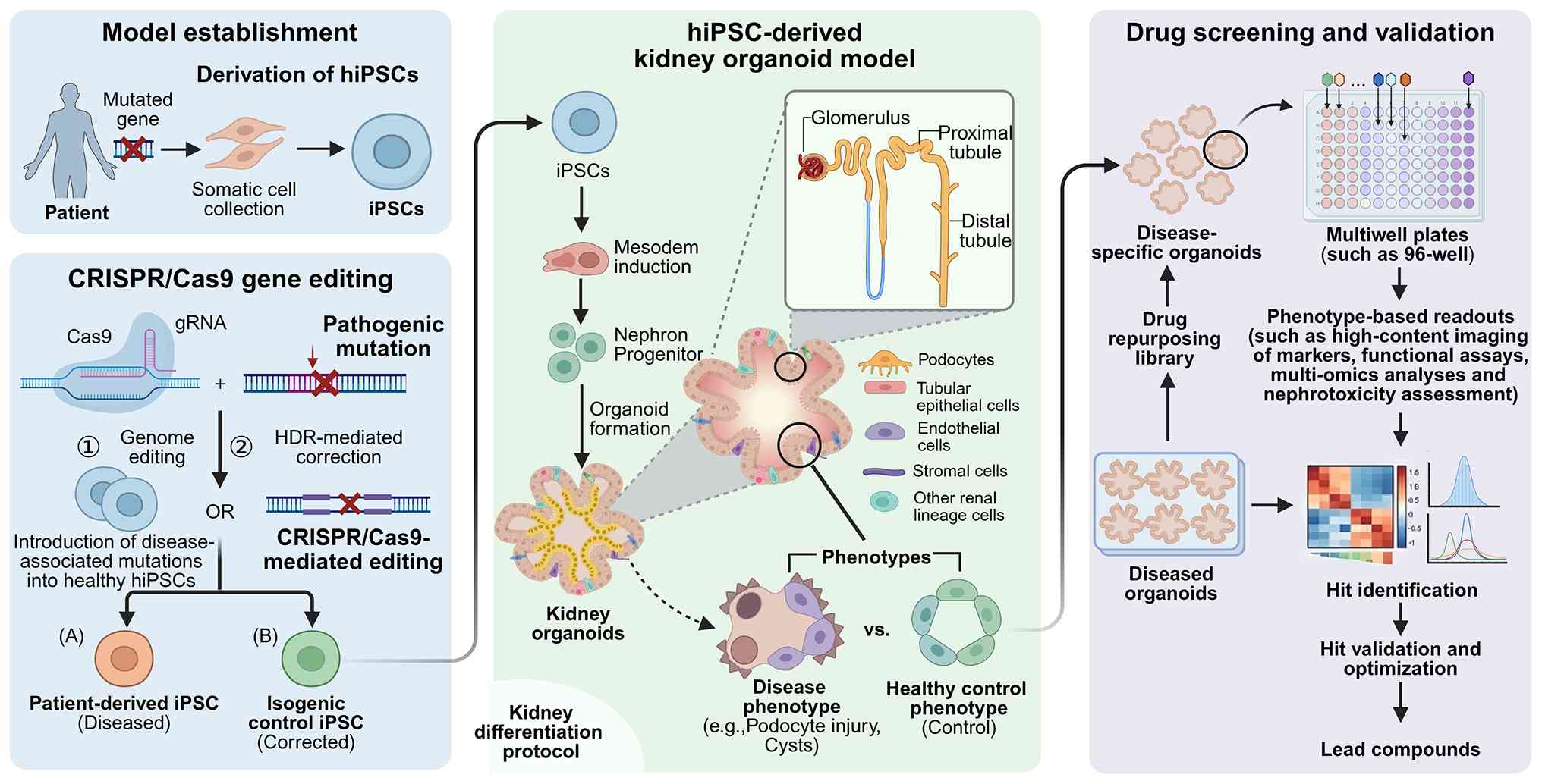 | Figure 1Establishment of hiPSC-derived
genetic kidney disease organoid models and their application in
drug screening and validation. The schematic illustrates the
workflow for generating kidney organoid models of hereditary kidney
diseases using patient-derived hiPSCs and CRISPR/Cas9-mediated
genome editing. Somatic cells obtained from patients carrying
disease-associated mutations are reprogrammed into hiPSCs. In
parallel, CRISPR/Cas9 gene editing can be used either to introduce
pathogenic mutations into healthy hiPSCs or to correct
disease-associated mutations through HDR, thereby generating
patient-derived disease hiPSC lines and isogenic control hiPSC
lines. These hiPSCs are subsequently subjected to a kidney
differentiation protocol involving mesoderm induction, nephron
progenitor specification and organoid formation to generate
hiPSC-derived kidney organoids containing nephron-like structures,
including glomeruli, proximal tubules and distal tubules, as well
as podocytes, tubular epithelial cells, endothelial cells, stromal
cells and other renal lineage cells. Disease and control organoids
can then be compared to identify pathological phenotypes, such as
podocyte injury and cyst formation. Diseased organoids may further
be applied in drug repurposing and multiwell plate-based drug
screening, together with phenotype-based readouts, such as
high-content imaging, functional assays, multi-omics analyses and
toxicity assessment, thereby facilitating hit identification, hit
validation, optimization and lead compound selection. hiPSC, human
induced pluripotent stem cell; CRISPR, clustered regularly
interspaced short palindromic repeats; Cas9, CRISPR-associated
protein 9; gRNA, guide RNA; HDR, homology-directed repair. |
3. Disease modeling
Autosomal dominant polycystic kidney
disease (ADPKD)
ADPKD is genetically heterogeneous, dominated by two
genes, PKD1 (located on chromosome 16.p13.3) affects ~78% of
families and PKD2 (located on chromosome 4p21) affects ~15%
of families. In 2016, a rare (affecting ~0.3% of families) third
locus, the glucosidase II α subunit gene (on chromosome 11q12.3)
was revealed (53). A study by
Freedman et al (45)
generated CRISPR-mutant kidney organoids from hPSCs and demonstrate
that knocking out PKD1 or PKD2 induces cyst formation in kidney
tubules. The subsequent study by Xu et al (54) reveals a distinct population of
CD24+ renal epithelial cells with unique metabolic and
gene regulatory programs and generates adult human kidney organoids
from these cells. By combining CD24+ cell-derived
tubule-like structures with multiplexed CRISPR-Cas9 gene editing,
an ADPKD organoid model that enables rapid cyst induction was
established, highlighting the potential of lineage-specific
differentiation strategies for improved disease modeling (54).
Building on this foundation, a study by Vishy et
al (55) develops base-edited
PKD kidney organoids that have four clinical non-sense mutations in
PKD1 and PKD2, which enables allele-specific modeling
of cystogenesis and therapeutic testing. A key finding is that
heterozygous mutated organoids do not spontaneously form cysts,
suggesting a potential therapeutic window for intervention.
Furthermore, from a translational perspective, the study by Vishy
et al (55) identifies
eukaryotic ribosome-selective glycosides as candidate therapeutics,
demonstrating that these compounds are able to mediate non-sense
mutation readthrough, which is potentially a targeted treatment
strategy for patients with such mutations.
Autosomal recessive polycystic kidney
disease (ARPKD)
ARPKD is a kidney disease that differs from ADPKD.
Although both disorders are classified as ciliopathies, sharing
core pathological pathways such as ciliary dysfunction and abnormal
epithelial cell proliferation, ARPKD has unique clinical and
genetic characteristics (56). It
has an autosomal recessive inheritance pattern, primarily caused by
mutations in the polycystic kidney and hepatic disease 1
(PKHD1) gene (and less commonly in the DAZ-interacting zinc
finger protein 1-like gene) (57,58),
with the onset of the disorder typically occurring in neonates or
childhood. The hallmark features of ARPKD, namely fusiform
dilatation of the renal CDs accompanied by congenital hepatic
fibrosis (59,60), differ to the widespread cyst
formation that occurs throughout the kidneys and adult-onset
presentation that are characteristic of ADPKD.
A study by Low et al (46) established an ARPKD model using
patient-derived iPSCs harboring biallelic PKHD1 frameshift
mutations. The aforementioned study demonstrates that cAMP
activation via the natural compound forskolin induces extensive
cystogenesis specifically in the PKHD1-/-
organoids, a response that is markedly attenuated in heterozygous
and wild-type controls (46).
Furthermore, a study by Hiratsuka et al (61) integrates ARPKD organoids with an
organ-on-chip platform. This revealed a novel pathogenic mechanism,
demonstrating that fluid flow-induced mechanical stress, mediated
by the mechanosensors Ras-related C3 botulinum toxin substrate 1
(RAC1) and Fos proto-oncogene (c-Fos), promotes cyst formation
(61).
Fabry's disease (FD)
The original animal model of FD, developed in a
study by Ohshima et al (62), used a targeting construct to knock
out the α-galactosidase A (α-Gal A) gene in C57BL/6 male mice,
which were then mated with normal C57BL/6 females to obtain
heterozygous mice lacking the α-Gal A gene, generating α-Gal
A+/- mice. The construction of the animal model was
completed by mating α-Gal A+/- female mice with C57BL/6
male mice to obtain α-Gal A-/0 mutant male mice
(62).
Several years later, a study by Porto et al
(63) carried out pharmacological
investigations using fibroblasts from patients with FD. After the
study by Freedman et al (45) that first applied CRISPR/Cas9 gene
editing technology to kidney organoid disease models, several
studies have applied CRISPR/Cas9 or related genome-editing
approaches to kidney organoid models by disrupting, introducing or
correcting disease-associated genes (45,54,55,64-68).
A 2021 study by Kim et al (69) confirmed that, in kidney organoids
derived from hiPSCs carrying mutations in GLA, the gene
encoding α-Gal A, the deposition of the glycosphingolipid
globotriaosylceramide (Gb3) led to structural deformation of
podocytes and renal tubular cells, thereby exacerbating oxidative
stress and apoptosis. The aforementioned study further evaluates
two therapeutic strategies, namely enzyme replacement therapy
(using recombinant human α-Gal A), which is shown to
alleviate oxidative stress and repair cell structure by eliminating
Gb3, and antioxidant therapy (glutathione replacement), which is
shown to directly reduce oxidative stress, thereby alleviating
structural damage to organoids (69).
A study by Cui et al (65) differentiated FD model kidney
organoids using patient-derived and gene-edited hiPSCs. Kidney
organoids were cultivated from hiPSCs of two male patients with
different types of GLA mutations who had FD. Compared with
the wild-type organoids, the patient-derived organoids had
decreased α-Gal A activity and increased Gb3 lipid deposition.
These abnormalities were more pronounced in organoids carrying the
classic Fabry disease-associated GLA mutation, which was
associated with markedly reduced α-Gal A activity and a more severe
Gb3 accumulation phenotype. Using electron microscopy,
multilamellar lysosomal inclusion bodies, also known as zebra
bodies, were observed in the mutant organoids, thereby reproducing
a characteristic pathological feature of Fabry nephropathy.
To further investigate treatment strategies,
CRISPR/Cas9 was used to knock out the α-1,4-galactosyltransferase
(A4GALT) gene, which encodes Gb3 synthase. The results
demonstrate that, in the GLA mutant kidney organoids,
A4GALT knockout reduces Gb3 deposition, and lysosome
inclusion bodies are not observed (64).
Collectively, these findings not only confirm the
potential of glutathione and A4GALT as possible new
therapeutic targets for FD, but they also provide a reliable in
vitro model platform for drug development (64).
Karyomegalic interstitial nephritis
(KIN)
KIN is an extremely rare hereditary form of chronic
interstitial nephritis. Although a reliable population-based
incidence rate has not been established, its estimated prevalence
is <1 per 1,000,000 individuals, it accounts for <1% of
kidney biopsies, and <100 native-kidney cases have been reported
in the literature (70,71). In order to identify the causative
gene for nephronophthisis (NPHP)-like ciliopathy, a study by Zhou
et al (72) evaluates two
affected siblings of Maori descent in New Zealand. Renal
histopathology analysis reveals enlarged nuclei (nucleolar
enlargement) in the renal cells, and a diagnosis of KIN is
suggested. Subsequent pure-synteny localization and exome
sequencing reveals a pure-synteny non-sense mutation in
FAN1, the gene that encodes Fanconi anemia-associated
nuclease 1. Additionally, through sequencing FAN1 exons from
the DNA samples of 10 families with KIN, it is revealed that a
recessive mutation in FAN1 is the important etiologic factor
in KIN. Another study by Lim et al (73) generated kidney organoids using
FAN1-mutant and wild-type hiPSCs. After inducing DNA damage
via mitomycin C treatment, the FAN1-mutant organoids demonstrated
increased expression of the DNA damage-associated marker H2A.X
compared with their wild-type counterparts; Ki67 was also assessed
to evaluate cell proliferation and viability within the organoids.
Taken together, these findings demonstrate that
FAN1-deficient kidney organoids successfully recapitulate
the KIN phenotype, suggesting a potential model platform for
investigating the mechanisms through which defective DNA repair
contributes to the development of CKD.
Gitleman syndrome (GS)
GS (online mendelian inheritance in man: 263800),
also known as familial hypokalemia-hypomagnesemia, is a salt-losing
tubulopathy that is characterized by hypomagnesiuria, hypocalciuria
and secondary aldosteronism, leading to hypokalemia and metabolic
alkalosis with autosomal recessive inheritance (74). GS is associated with mutations in
the solute carrier family 12 member 3 (SLC12A3) gene, which
encodes the thiazide-sensitive sodium chloride cotransporter (NCCT)
in the distal convoluted tubule (75). A study by Lim et al
(66) prepared general hiPSCs and
gene-modified hiPSCs by sequencing the SLC12A3 gene using
CRISPR/Cas9 technology with peripheral blood mononuclear cells from
patients with GS. Furthermore, in a subsequent study (68), the SLC12A3 gene mutation was
corrected using CRISPR-Cas9 technology, yielding genetically
repaired hiPSCs. When both the mutant and corrected hiPSCs are
differentiated into kidney organoids, the SLC12A3-mutant
tissues exhibit reduced mRNA and protein levels of NCCT compared
with wild-type controls. However, these levels are restored in the
genetically corrected organoids. Although the aforementioned study
models GS in kidney organoids for the first time and demonstrates
reduced NCCT expression levels in E-cadherin-positive epithelial
cells, as well as its rescue by genetic correction, it did not
directly demonstrate the defective electrolyte transport associated
with disease pathogenesis. Overcoming this limitation may require
more advanced kidney organoid platforms that incorporate
vascularization, tubular fluid flow and functional assays capable
of assessing solute transport.
Alport syndrome (AS)
AS is a one of the most common hereditary glomerular
diseases. Its main clinical features include progressive glomerular
injury and extrarenal manifestations, particularly sensorineural
hearing loss and ocular abnormalities and it may eventually
progress to end-stage renal disease (76). The causative genes for AS are
COL4A3-5, which encode the α3, α4 and α5 chains of type IV
collagen, respectively. Mutations in these genes lead to abnormal
type IV collagen structure in the glomerular basement membrane (BM)
(77-79).
Given the severity of the condition and the current lack of
curative options, developing a preclinical platform that accurately
recapitulates the disease phenotype is challenging.
Using kidney organoids derived from hiPSCs, studies
have successfully constructed disease models that simulate AS
(80,81). A study by Hirayama et al
(80) reports on hiPSCs derived
from two male patients with AS and differentiates them into kidney
organoids. The organoids derived from these patients successfully
reproduces the key disease phenotypes, including abnormal
expression of the type IV collagen α5 chain, α5(IV). The
aforementioned study confirms that the severity of the phenotype is
associated with the type of genetic mutation, and that the normal
expression of α5 (IV) can be restored through genetic correction.
Furthermore, the chemical chaperone 4-phenylbutyric acid improves
BM defects in mild-phenotype organoids, although it is ineffective
in severe-phenotype organoids. These findings suggest the necessity
of personalized treatment based on genetic typing (80).
A study by Morais et al (81) reports the BM pathology in AS. It is
revealed that, although kidney organoids derived from iPS cells
from patients with AS form normal-appearing glomeruli and tubules
under a light microscope, their molecular composition is already
abnormal, manifesting as an increased deposition of laminin β2,
specifically in the extraglomerular BM. Furthermore, the
aforementioned study reveals that BM composition is regulated from
development to adulthood, a process that is disrupted by pathogenic
COL4A5 variants. Consequently, the model successfully
recapitulates the laminin dysregulation in patients with AS,
suggesting that kidney organoids may serve as a potential platform
for studying aberrant BM assembly in human developmental diseases
(81).
Nephrotic syndrome
The normal function of glomerular podocytes relies
on the slit diaphragm, a protein complex composed of molecules
including nephrin (NPHS1) and podocin (NPHS2). Mutations in either
NPHS1 or 2 disrupt the formation of the slit diaphragm, leading to
congenital nephrotic syndrome (82,83).
Several studies validate this pathogenic mechanism using
patient-specific iPSC-derived kidney organoid models (84-87).
Two studies demonstrate that a NPHS1 missense mutation results in
abnormal NPHS1 localization and impairs slit diaphragm formation in
podocytes (84,85). However, a study by Jansen et
al (86) reports that NPHS2
mutations lead to both a loss of NPHS2 expression levels and the
mislocalization of NPHS1, which is a reversible phenotype upon
genetic correction. Furthermore, a study by Majmundar et al
(87) models glomerular
developmental abnormalities and an increased rate of apoptosis by
introducing a patient-derived nitric oxide synthase 1 adaptor
protein variant into human kidney organoids. Collectively, the
aforementioned studies highlight the possible utility of kidney
organoids in terms of recapitulating the pathological mechanisms of
podocytopathies.
Autosomal dominant tubulointerstitial
kidney disease (ADTKD)
ADTKD, the third most common monogenic kidney
disease, causes progressive renal failure via tubular/interstitial
injury, and it is attributed to mutations in five genes, namely
uromodulin, mucin 1 (MUC1), renin (REN), SEC61
translocon subunit α 1 and hepatocyte nuclear factor 1-β
(HNF1β) (88,89).
Kidney organoid models provide a platform for
investigating the functional roles of causative genes in ADTKD. A
study by Przepiorski et al (68), which investigates the role of the
HNF1β gene, uses CRISPR/Cas9 technology to knock out
HNF1β in order to observe any downregulation in the
expression of markers associated with proximal tubules and thick
ascending limbs in the organoids. This demonstrates the regulatory
role of genes in nephron segment patterning. Another study by Mae
et al (90) investigates UB
organoids, further revealing that the heterozygous loss of
HNF1β leads to impaired branching morphogenesis and loss of
apicobasal polarity. By contrast, a study by Dvela-Levitt et
al (91) generates kidney
organoids from iPSCs derived from patients with MUC1
mutations. This recapitulates the pathological hallmark of aberrant
mutant protein accumulation, and use this model to identify a
small-molecule compound, BRD4780, which directs the mutant protein
for degradation via the lysosomal pathway, demonstrating the
application of organoids in targeted drug discovery (91).
Autosomal recessive-renal tubular
dysgenesis (AR-RTD)
AR-RTD is a lethal genetic disorder characterized by
the complete absence or severe hypoplasia of proximal tubules,
which results from pathogenic mutations in key genes of the
renin-angiotensin-aldosterone system (RAAS), including
angiotensin-converting enzyme (ACE), angiotensin II receptor
type 1 (AGTR1), autophagy-related and REN (92). A study by Pode-Shakked et al
(93) generated RAAS-deficient
kidney organoids by differentiating ACE-/- and
AGTR1-/- iPSCs, as well as patient-derived iPSCs
with AR-RTD, into kidney organoids. This demonstrates that
RAAS-deficient organoids form proximal tubules in vitro;
however, AGTR1-/- organoids display impaired
engraftment at the renal vesicle stage due to insufficient VEGF-A
expression levels, suggesting that delayed angiogenesis is a
possible mechanism that underlies autosomal recessive renal tubular
dysgenesis (93) Additionally, the
aforementioned study reveals that, under conventional or hypoxic
culture conditions, the loss of RAAS genes does not directly affect
proximal tubule patterning in organoids. Following transplantation
under the renal capsule of immunodeficient mice, renal
vesicle-stage AGTR1-/- organoids have impaired
engraftment due to insufficient VEGF-A expression levels and
delayed angiogenesis. Hypoxic culture induces the expression of
VEGF-A and rescues the engraftment of AGTR1-/-
organoids (93). These findings
suggest that proximal tubule dysgenesis in AR-RTD is not primarily
induced by cell-autonomous tubular defects, but is induced by
delayed angiogenesis as a non-cell-autonomous consequence of
impaired RAAS signaling.
Nephronophthisis (NPHP)
NPHP is an autosomal recessive disorder that is
characterized pathologically by disruption of the tubular BM,
tubular atrophy, interstitial fibrosis and progression to end-stage
kidney disease (94). To
investigate its underlying mechanisms, studies focus on the
intraflagellar transport 140 (IFT140) gene, which encodes a
core component of the intraflagellar transport complex A involved
in retrograde ciliary transport (95,96).
In a study by Forbes et al (51), kidney organoids are generated using
patient-derived IFT140-mutant iPSCs alongside isogenic
gene-corrected control iPSCs. This reveals that mutant organoids
have shortened, malformed primary cilia and defects in cellular
polarization, whereas genetic correction successfully rescues these
abnormal phenotypes, confirming the central role of IFT140
mutations in NPHP pathogenesis (51).
Cystinosis
Cystinosis is an autosomal recessive lysosomal
storage disorder caused by mutations in the cystinosin, lysosomal
cystine transporter gene, which encodes the protein cystinosin
(97). The major characteristic of
cystinosis is the accumulation of cystine in lysosomes, which leads
to progressive renal tubular damage and multiple organ dysfunction
(98).
A study by Hollywood et al (98) differentiates kidney organoids from
iPSCs derived from patients with cystinosis, recapitulating key
disease phenotypes. For example, cystine crystal accumulation was
observed in the proximal tubular cells of the organoids,
accompanied by lysosomal dysfunction, elevated oxidative stress and
impaired levels of autophagy. Electron microscopy and molecular
analyses further revealed features of mitochondrial dysfunction and
increased rates of apoptosis of renal tubular epithelial cells in
the organoids, demonstrating the successful modeling of progressive
injury processes observed in patient kidneys (98) (Table
II).
| Table IIGuidance for model selection:
Matching differentiation strategy to disease application. |
Table II
Guidance for model selection:
Matching differentiation strategy to disease application.
| Disease
category | Representative
diseases | Recommended
differentiation strategy | Key model
characteristics | (Refs.) |
|---|
| Ciliopathies with
cystogenesis | ADPKD and
ARPKD | Classic
multi-stage; assembly/co-culture strategies to model collecting
duct-derived cystogenesis | Cyst formation upon
cAMP induction; collecting duct integration for ARPKD | (45,46,50,54,55,61) |
| Glomerular basement
membrane diseases | Alport
syndrome | Classic
multi-stage; nephron subtype induction (glomerular enrichment) | Abnormal type IV
collagen expression levels; podocyte foot process effacement | (80,81) |
|
Podocytopathies/slit diaphragm
disorders | Congenital
nephrotic syndrome (NPHS1 and 2) | Classic
multi-stage; ventral mesoderm (for increased podocyte purity) | Nephrin/podocin
mislocalization; impaired slit diaphragm formation | (84-87) |
| Tubular transport
disorders | Gitelman syndrome
(SLC12A3) |
Optimized/simplified; nephron subtype
induction (PT enrichment) | Reduced NCCT
expression; electrolyte transport defects requiring functional
validation, such as thiazide-sensitive NaCl uptake, ion flux and
transepithelial transport assays | (66,67) |
| DNA
repair-associated tubulointerstitial disease | Karyomegalic
interstitial nephritis (FAN1) | Classic or
optimized protocol | Increased H2A.X
expression after mitomycin C treatment; Ki67 was assessed to
evaluate cell proliferation and viability | (73) |
| Lysosomal storage
disorders | Fabry disease and
cystinosis | Classic or
optimized protocol | Gb3 deposition
(Fabry); cystine crystals (cystinosis); oxidative stress | (64,65,69,98) |
| Nephron segment
patterning disorders | ADTKD (HNF1B and
MUC1) | Classic
multi-stage; ureteric bud organoid assembly | Reduced proximal
tubule markers, such as LTL, CUBN and AQP1, and thick ascending
limb markers, such as uromodulin and SLC12A1/NKCC2; branching
defects | (68,90,91) |
| RAAS-related
developmental defects | AR-RTD (ACE and
AGTR1) | Classic protocol
followed by transplantation | Delayed
angiogenesis; impaired engraftment after transplantation | (93) |
| Ciliopathy with
tubular atrophy | Nephronophthisis
(IFT140) | Classic or
optimized protocol | Shortened primary
cilia; defective cellular polarization | (51) |
4. Emerging applications, limitations and
translational challenges
Overview of emerging applications
Kidney organoid technology demonstrates
multifaceted application potential, including disease modeling,
toxicological safety assessment, personalized medicine, organ
transplantation, organ-on-chip development and extracellular
vesicle (EV)-associated research (99).
Two transplantation models promote vascularization
and maturation of kidney organoids. A study by van den Berg et
al (48) first transplanted
organoids under the renal capsule of immunodeficient mice,.
Subsequently, a study by Koning et al (100) used intracoelomic transplantation
into chicken embryos to study vasculogenesis in kidney organoids.
These transplantation studies demonstrated that in vivo
engraftment promotes the vascularization and maturation of kidney
organoids. In the renal subcapsular transplantation model, van den
Berg et al (48) showed
that transplanted organoids developed host-derived vascularization,
fenestrated endothelial cells and podocyte foot processes, and
exhibited selective permeability to dextran and albumin, suggesting
the formation of a functional glomerular filtration barrier. In the
intracoelomic transplantation model, Koning et al (100) demonstrated vascular integration
and improved maturation of kidney organoids in the chicken embryo
environment. Together, these findings indicate that transplantation
can enhance vascularization, glomerular maturation and epithelial
organization in kidney organoids.
The study by Lim et al (101) was the first to combine collagen
scaffold encapsulation with minimally invasive intrarenal
injection, overcoming the shortcomings of traditional subcapsular
or abdominal transplantation that require surgical exposure.
Furthermore, the method avoids the problems of uneven distribution
and low survival rate that occurs with simple cell injection. This
study (101) demonstrates that
this method not only preserves the structural integrity of
organoids, but it also promotes their functional integration into
the host kidney, providing a feasible preclinical model for future
organoid-based kidney regeneration therapy.
Microfluidic technology, via the construction of
microphysiological systems with dynamic fluidic microenvironments,
provides kidney organoids with a culture platform that more closely
mimics in vivo conditions, notably enhancing organoid
vascularization, maturation and functional simulation capabilities.
This technology is able to simulate fluid shear stress and
intraluminal pressure within nephrons, promoting the polarization
and functional maturation of renal tubular epithelial cells
(102). Through multi-chamber
designs, it enables spatial coupling of the glomerular filtration
barrier with tubular structures, simulating the urine-formation
process (103). Additionally, in
disease modeling, microfluidic systems successfully recapitulate
drug-induced nephrotoxicity (104) and the cyst-expansion mechanisms
in genetic diseases such as ADPKD (105). In drug screening, its integrated
high-throughput detection systems allow real-time monitoring of
organoid responses to drugs, which notably improves the precision
of nephrotoxicity assessments (106). Furthermore, connecting kidney
organoid chips with other organ chips provides a novel platform for
studying systemic diseases and multi-organ interactions (107). However, despite their advantages,
current microfluidic platforms have several technical bottlenecks
that limit their broader application, including: i) Chip material
(polydimethylsiloxane) adsorption of hydrophobic drugs causing
dosing inaccuracy; ii) inconsistent flow shear stress across
replicates due to bubble formation; iii) limited imaging depth
(typically <200 µm) for thick organoids; and iv) a lack of
standardized protocols for organoid loading and perfusion.
Moreover, common failure points involve organoid detachment,
leakage at chip interfaces and microbial contamination during
long-term culture (>7 days). Therefore, these issues must be
addressed prior to the widespread adoption of this technology.
EVs are heterogeneous membrane-enclosed
nanoparticles that mediate intercellular communication and
participate in both physiological and pathophysiological conditions
in the kidney (108). EVs derived
from stem cells, especially MSCs, which are adult stem cells found
in tissues such as bone marrow, adipose tissue, umbilical cord,
placenta and dental pulp, have demonstrated notable regenerative
potential in various models of acute and chronic kidney injury
(109,110).
Critical appraisal of kidney organoid
models
Kidney organoids have notably revolutionized renal
research and, when combined with gene-editing techniques, are a
valuable research model. This model uses genome-wide screening to
investigate the underlying mechanisms of genetic diseases,
facilitates drug and toxicity screening on personalized
human-derived platforms, and serves as a key tool for deciphering
renal morphogenesis and functional maintenance (111,112). However, several constraints limit
clinical translation. Compared with conventional animal models,
kidney organoids provide a human genetic background and greater
compatibility with high-throughput screening. However, they lack
key aspects of systemic physiology, including hemodynamic forces,
endocrine regulation and immune interactions (111,112) For drug screening, organoids
enable rapid phenotypic readouts such as cyst growth and protein
mislocalization, but have lower scalability compared with 2D
cultures. Reproducibility remains a major concern. Independent
differentiations of the same hiPSC line yield organoids with up to
30-40% variability in nephron progenitor numbers (44). In addition, scalability for drug
screening is limited by manual handling; bioreactor-based expansion
does improve yield, although this process also introduces shear
stress artifacts.
Key limitations and challenges
Firstly, the cells generated by kidney organoids
are mostly immature, possessing embryonic or fetal characteristics,
and fail to form the mature cell types that are unique to adult
kidneys (112,113). The epithelial cells within
organoids primarily express development-associated genes and often
show limited expression or maturation of functional proteins
associated with kidney diseases, such as NPHS1, NPHS2, COL4A3-5,
SLC12A3 and uromodulin. Targeted induction protocols are required
to promote the further maturation of organoids, which enables the
construction of a research model that more closely mimics the
physiological characteristics of adult kidneys (44).
Secondly, batch-to-batch variations, residual
off-target cells and individual variability during maturation can
lead to misleading conclusions when comparing patient-derived
organoids with isogenic control organoids (44,114). This heterogeneity notably
undermines the reliability of research results and presents a
critical factor limiting their precise application.
Thirdly, key cell types, such as vascular
endothelial cells and immune cells, are absent in kidney organoids
derived from hPSCs; however, these cells perform crucial roles in
the maintenance of renal physiological functions and pathological
processes (for example, inflammation and injury repair) (114-116).
This deficiency has notable limitations for investigations into the
interactions between renal cells and immune cells, which especially
hinders the in vitro modeling of complex diseases such as
infectious and autoimmune nephropathies (114,116).
Fourthly, kidney organoids lack the macroanatomical
structures that are similar to those of natural kidneys and more
complex organ tissue architectures. This prevents them from
simulating core renal functions, such as tubular reabsorption and
renal filtration (114), making
it difficult to recapitulate the physiological functional
characteristics of adult kidneys.
The functional validation of kidney organoids has
been insufficiently addressed in numerous studies (44,111-113,116,117).
Beyond morphological assessment, critical parameters also require
systematic evaluation. Electrophysiological properties, including
transepithelial resistance and ion channel activity, remain largely
uncharacterized. Electrolyte transport could be measured via an
assessment of Na+/K+-ATPase activity (namely,
ouabain-sensitive ATP hydrolysis) (117) and by evaluating glucose uptake or
reabsorption (105).
Vascularization could be assessed morphologically by calculating
the CD31+ vascular area fraction and functionally by
evaluating fluorescent tracer perfusion. Transcriptomic maturity
should be benchmarked using principal component analysis, comparing
organoid transcriptomes to both fetal (12-20 weeks) and adult
kidney reference datasets; a maturity score closer to that of the
adult kidney indicates an improved differentiation (44). Long-term stability over weeks to
months in culture or after transplantation also requires systematic
reporting. These functional benchmarks are essential for validating
organoid utility in disease modeling and drug screening (Table III).
| Table IIIProposed assessment metrics for
kidney organoid quality evaluation. |
Table III
Proposed assessment metrics for
kidney organoid quality evaluation.
| Metric
category | Specific assay | Target | (Refs.) |
|---|
| Transcriptomic
maturity | Principal component
analysis comparing organoid transcriptome of fetal (12-20 weeks of
age) vs. adult kidney | Closer clustering
with adult human kidney transcriptomic profiles indicates greater
transcriptomic maturity | (44) |
| Glomerular
filtration proxy | FITC-inulin
clearance in microfluidic device | Detectable
selective filtration/permeability assessed using FITC-inulin,
dextran or albumin tracer assays in organoid-on-chip or
transplantation-based models | (103) |
| Transporter
activity |
Na+/K+-ATPase
activity; organic cation transporter 2/multidrug and toxin
extrusion 2-K uptake/efflux assays | Expression of a
functional transporter and polarity; inhibitor-sensitive
activity | (104-106,117) |
| Vascularization
efficiency | CD31+
area fraction quantified using confocal microscopy | Increased
CD31+ vascular area fraction compared with static
controls | (46,48,100,102) |
| Long-term
stability | Histology,
immunofluorescence staining, vascular perfusion/tracer assays and
graft retention analysis after transplantation | Maintained graft
survival, vascular integration and tissue architecture at
representative follow-up points, such as 2-4 weeks and, when
applicable, up to 12 weeks after transplantation | (48,100,101) |
Bioengineering solutions to overcome
limitations
Organoids-on-chip are able to optimize nutrient
exchange via shear stress, and compared with static culture, they
are more likely to induce organoids to form structures resembling
mature kidneys (102,118). This technology has a wide range
of applications, for example, it can be used to study
vascularization (providing key support for drug testing), and can
simulate glomerular filtration function through the co-culture of
podocytes and endothelial cells (103,104).
Although organoids-on-chip offer notable advantages
over traditional research platforms, they are still not able to
fully replicate the physiological complexity of human kidneys and
currently face several challenges, including limited
standardization, poor reproducibility, chip material-related
limitations and microfluidic designs that may not fully accommodate
the size, 3D architecture and long-term perfusion requirements of
kidney organoids. These limitations complicate organoid
integration, stable perfusion, long-term culture and imaging-based
analysis at different tissue depths (119).
As a cutting-edge technology, 3D bioprinting has a
potential for developing kidney organoids. Through enabling the
precise positioning of cells and biomaterials, 3D bioprinting may
construct larger-scale, more complex kidney-like tissues,
suggesting a potential for the development of preclinical models
(120). A key advantage of this
method is its capacity for high-throughput production, which
ensures consistent cell numbers, high viability and reduced
inter-organoid variability (121). By leveraging iPSCs, bioprinting
platforms have been demonstrated to generate kidney constructs with
improved uniformity and expression of lineage-specific markers,
including NPHS1, NPHS2 and WT1 for podocytes; LTL, E-cadherin and
AQP1 for tubular epithelial cells; and CD31/PECAM1 and
VE-cadherin/CDH5 for endothelial cells (122). Furthermore, the technology allows
the modulation of biophysical parameters, such as cell number and
tissue size, to improve the modelling of renal functional and
physiological characteristics (121). This strategy not only mitigates
patient-derived heterogeneity, but it also establishes stable,
reproducible models that are suitable for high-throughput screening
and comparative studies (118).
The integration of patient-derived iPSCs, genome
editing, reporter lines, microfluidic devices and 3D bioengineering
approaches is expanding the utility of kidney organoids for disease
modeling and preclinical drug evaluation (107,111-114,117-124).
Reporter lines can facilitate lineage tracing, cell-type
identification and real-time monitoring of organoid
differentiation; examples include SIX2-based reporters for nephron
progenitor cells, WT1- or NPHS1-based reporters for
podocyte-lineage cells, and PECAM1/CD31- or CDH5-based reporters
for endothelial cells. In parallel, patient-derived iPSCs and
gene-edited isogenic controls allow disease-associated phenotypes
to be modeled in a human genetic background, whereas microfluidic
and bioengineering platforms can provide dynamic culture conditions
and more standardized tissue construction (107,111-114,117-124).
Together, these approaches may improve the analysis of pathological
endpoints related to drug efficacy and toxicity and support more
individualized disease modeling.
Translational challenges and
standardization
Translating kidney organoids to clinical
applications requires overcoming several hurdles. Good
Manufacturing Practice compliance demands xeno-free, defined media
and stringent quality control release criteria. Regulatory barriers
are substantial, since guidance from the Food and Drug
Administration on organoid-based products remains in its early
phase. Furthermore, scalability is limited by manual handling; in
addition, although bioreactor-based expansion improves yield, it
also introduces shear stress artifacts. Quantitative benchmarking
is suggested to be mandated for preclinical studies. Proposed
metrics include organoid size distribution (target coefficient of
variation <20%), cell type proportions by single-cell
RNA-sequencing (>80% kidney lineage) and functional assays, such
as FITC-inulin clearance for glomerular filtration or
Na+/K+-ATPase activity for proximal tubule
function. These benchmarks may facilitate cross-laboratory
comparisons and accelerate clinical translation.
Emerging applications: modeling viral
kidney disease
As well as hereditary disorders, kidney organoids
are also applied to model viral nephropathies. SARS-CoV-2 infection
of kidney organoids recapitulates viral entry, replication and
cytopathic effects, primarily targeting proximal tubular cells via
ACE2, with enhanced organoid maturation improving viral replication
modeling (123). Similarly, BK
polyomavirus, a major cause of nephropathy in transplant
recipients, induces nuclear enlargement characteristic of BK virus
nephropathy in kidney tubuloids, and such models enable antiviral
compound testing (124). These
applications expand the use of kidney organoids beyond genetic
disorders, highlighting their potential in infectious disease
modeling and drug development.
5. Conclusion
Despite challenges in standardization, maturity and
scalability, kidney organoids have the potential to support
personalized medicine by enabling patient-specific disease
modeling, genotype-informed therapeutic testing and individualized
drug-response assessment. From ‘modeling diseases in a dish’ to
‘designing tailored therapies for patients’, kidney organoids may
improve the understanding of genetic kidney diseases and support
the development of individualized therapeutic interventions.
Acknowledgements
Not applicable.
Funding
Funding: No funding was received.
Availability of data and materials
Not applicable.
Authors' contributions
SC, TC, YZ, ML and HZ were responsible for writing
the original draft. JJ and MY were responsible for reviewing and
editing the article. Data authentication is not applicable. All
authors read and approved the final version of the manuscript.
Ethics approval and consent to
participate
Not applicable.
Patient consent for publication
Not applicable.
Competing interests
The authors declare that they have no competing
interests.
References
|
1
|
Haruhara K, Kanzaki G and Tsuboi N:
Nephrons, podocytes and chronic kidney disease: Strategic
antihypertensive therapy for renoprotection. Hypertens Res.
46:299–310. 2023.PubMed/NCBI View Article : Google Scholar
|
|
2
|
Bertram JF, Douglas-Denton RN, Diouf B,
Hughson MD and Hoy WE: Human nephron number: Implications for
health and disease. Pediatr Nephrol. 26:1529–1533. 2011.PubMed/NCBI View Article : Google Scholar
|
|
3
|
Vivante A and Hildebrandt F: Exploring the
genetic basis of early-onset chronic kidney disease. Nat Rev
Nephrol. 12:133–146. 2016.PubMed/NCBI View Article : Google Scholar
|
|
4
|
Mount DB: Thick ascending limb of the loop
of Henle. Clin J Am Soc Nephrol. 9:1974–1986. 2014.PubMed/NCBI View Article : Google Scholar
|
|
5
|
Groopman EE, Marasa M, Cameron-Christie S,
Petrovski S, Aggarwal VS, Milo-Rasouly H, Li Y, Zhang J, Nestor J,
Krithivasan P, et al: Diagnostic utility of exome sequencing for
kidney disease. N Engl J Med. 380:142–151. 2019.PubMed/NCBI View Article : Google Scholar
|
|
6
|
KDIGO Conference Participants. Genetics in
chronic kidney disease: Conclusions from a Kidney Disease:
Improving Global Outcomes (KDIGO) Controversies Conference. Kidney
Int. 101:1126–1141. 2022.PubMed/NCBI View Article : Google Scholar
|
|
7
|
Knoers N, Antignac C, Bergmann C, Dahan K,
Giglio S, Heidet L, Lipska-Ziętkiewicz BS, Noris M, Remuzzi G,
Vargas-Poussou R and Schaefer F: Genetic testing in the diagnosis
of chronic kidney disease: Recommendations for clinical practice.
Nephrol Dial Transplant. 37:239–254. 2022.PubMed/NCBI View Article : Google Scholar
|
|
8
|
Jha V, Garcia-Garcia G, Iseki K, Li Z,
Naicker S, Plattner B, Saran R, Wang AY and Yang CW: Chronic kidney
disease: Global dimension and perspectives. Lancet. 382:260–272.
2013.PubMed/NCBI View Article : Google Scholar
|
|
9
|
Kovesdy CP: Epidemiology of chronic kidney
disease: An update 2022. Kidney Int Suppl (2011). 12:7–11.
2022.PubMed/NCBI View Article : Google Scholar
|
|
10
|
GBD 2023 Chronic Kidney Disease
Collaborators. Global, regional, and national burden of chronic
kidney disease in adults, 1990-2023, and its Attributable risk
factors: A systematic analysis for the Global burden of disease
study 2023. Lancet. 406:2461–2482. 2025.PubMed/NCBI View Article : Google Scholar
|
|
11
|
Li Z, He R, Wang Y, Qu Z, Liu J, Yu R and
Yang S: Global trends of chronic kidney disease from 1990 to 2021:
A systematic analysis for the global burden of disease study 2021.
BMC Nephrol. 26(385)2025.PubMed/NCBI View Article : Google Scholar
|
|
12
|
Wilson HV: A new method by which sponges
may be artificially reared. Science. 25:912–915. 1907.PubMed/NCBI View Article : Google Scholar
|
|
13
|
Zwilling E: Development of fragmented and
of dissociated limb bud mesoderm. Dev Biol. 9:20–37.
1964.PubMed/NCBI View Article : Google Scholar
|
|
14
|
Okabayashi K and Asashima M: In vitro
organogenesis using amphibian pluripotent cells. Proc Jpn Acad Ser
B Phys Biol Sci. 82:197–207. 2006.PubMed/NCBI View Article : Google Scholar
|
|
15
|
Rheinwald JG and Green H: Serial
cultivation of strains of human epidermal keratinocytes: The
formation of keratinizing colonies from single cells. Cell.
6:331–343. 1975.PubMed/NCBI View Article : Google Scholar
|
|
16
|
Yu J, Vodyanik MA, Smuga-Otto K,
Antosiewicz-Bourget J, Frane JL, Tian S, Nie J, Jonsdottir GA,
Ruotti V, Stewart R, et al: Induced pluripotent stem cell lines
derived from human somatic cells. Science. 318:1917–1920.
2007.PubMed/NCBI View Article : Google Scholar
|
|
17
|
Sato T, Vries RG, Snippert HJ, van de
Wetering M, Barker N, Stange DE, van Es JH, Abo A, Kujala P, Peters
PJ and Clevers H: Single Lgr5 stem cells build crypt-villus
structures in vitro without a mesenchymal niche. Nature.
459:262–265. 2009.PubMed/NCBI View Article : Google Scholar
|
|
18
|
Han X, Cai C, Deng W, Shi Y, Li L, Wang C,
Zhang J, Rong M, Liu J, Fang B, et al: Landscape of human
organoids: Ideal model in clinics and research. Innovation (Camb).
5(100620)2024.PubMed/NCBI View Article : Google Scholar
|
|
19
|
Barak H, Rosenfelder L, Schultheiss TM and
Reshef R: Cell fate specification along the anterior-posterior axis
of the intermediate mesoderm. Dev Dyn. 232:901–914. 2005.PubMed/NCBI View Article : Google Scholar
|
|
20
|
Taguchi A, Kaku Y, Ohmori T, Sharmin S,
Ogawa M, Sasaki H and Nishinakamura R: Redefining the in vivo
origin of metanephric nephron progenitors enables generation of
complex kidney structures from pluripotent stem cells. Cell Stem
Cell. 14:53–67. 2014.PubMed/NCBI View Article : Google Scholar
|
|
21
|
Majumdar A, Vainio S, Kispert A, McMahon J
and McMahon AP: Wnt11 and Ret/Gdnf pathways cooperate in regulating
ureteric branching during metanephric kidney development.
Development. 130:3175–3185. 2003.PubMed/NCBI View Article : Google Scholar
|
|
22
|
Kobayashi A, Valerius MT, Mugford JW,
Carroll TJ, Self M, Oliver G and McMahon AP: Six2 defines and
regulates a multipotent self-renewing nephron progenitor population
throughout mammalian kidney development. Cell Stem Cell. 3:169–181.
2008.PubMed/NCBI View Article : Google Scholar
|
|
23
|
Grieshammer U, Cebrián C, Ilagan R, Meyers
E, Herzlinger D and Martin GR: FGF8 is required for cell survival
at distinct stages of nephrogenesis and for regulation of gene
expression in nascent nephrons. Development. 132:3847–3857.
2005.PubMed/NCBI View Article : Google Scholar
|
|
24
|
Perantoni AO, Timofeeva O, Naillat F,
Richman C, Pajni-Underwood S, Wilson C, Vainio S, Dove LF and
Lewandoski M: Inactivation of FGF8 in early mesoderm reveals an
essential role in kidney development. Development. 132:3859–3871.
2005.PubMed/NCBI View Article : Google Scholar
|
|
25
|
Georgas K, Rumballe B, Valerius MT, Chiu
HS, Thiagarajan RD, Lesieur E, Aronow BJ, Brunskill EW, Combes AN,
Tang D, et al: Analysis of early nephron patterning reveals a role
for distal RV proliferation in fusion to the ureteric tip via a cap
mesenchyme-derived connecting segment. Dev Biol. 332:273–286.
2009.PubMed/NCBI View Article : Google Scholar
|
|
26
|
Halt K and Vainio S: Coordination of
kidney organogenesis by Wnt signaling. Pediatr Nephrol. 29:737–744.
2014.PubMed/NCBI View Article : Google Scholar
|
|
27
|
Cheng HT, Kim M, Valerius MT, Surendran K,
Schuster-Gossler K, Gossler A, McMahon AP and Kopan R: Notch2, but
not Notch1, is required for proximal fate acquisition in the
mammalian nephron. Development. 134:801–811. 2007.PubMed/NCBI View Article : Google Scholar
|
|
28
|
Kretzler M, Schröppel B, Merkle M, Huber
S, Mundel P, Horster M and Schlöndorff D: Detection of multiple
vascular endothelial growth factor splice isoforms in single
glomerular podocytes. Kidney Int Suppl. 67 (Suppl 1):S159–S161.
1998.PubMed/NCBI View Article : Google Scholar
|
|
29
|
Schell C, Wanner N and Huber TB:
Glomerular development-shaping the multi-cellular filtration unit.
Semin Cell Dev Biol. 36:39–49. 2014.PubMed/NCBI View Article : Google Scholar
|
|
30
|
Lindahl P, Hellström M, Kalén M, Karlsson
L, Pekny M, Pekna M, Soriano P and Betsholtz C: Paracrine
PDGF-B/PDGF-Rbeta signaling controls mesangial cell development in
kidney glomeruli. Development. 125:3313–3322. 1998.PubMed/NCBI View Article : Google Scholar
|
|
31
|
Little M, Georgas K, Pennisi D and
Wilkinson L: Kidney development: Two tales of tubulogenesis. Curr
Top Dev Biol. 90:193–229. 2010.PubMed/NCBI View Article : Google Scholar
|
|
32
|
Faa G, Gerosa C, Fanni D, Monga G,
Zaffanello M, Van Eyken P and Fanos V: Morphogenesis and molecular
mechanisms involved in human kidney development. J Cell Physiol.
227:1257–1268. 2012.PubMed/NCBI View Article : Google Scholar
|
|
33
|
Little MH, Kumar SV and Forbes T:
Recapitulating kidney development: Progress and challenges. Semin
Cell Dev Biol. 91:153–168. 2019.PubMed/NCBI View Article : Google Scholar
|
|
34
|
Humphreys BD, Lin SL, Kobayashi A, Hudson
TE, Nowlin BT, Bonventre JV, Valerius MT, McMahon AP and Duffield
JS: Fate tracing reveals the pericyte and not epithelial origin of
myofibroblasts in kidney fibrosis. Am J Pathol. 176:85–97.
2010.PubMed/NCBI View Article : Google Scholar
|
|
35
|
Bohnenpoll T, Bettenhausen E, Weiss AC,
Foik AB, Trowe MO, Blank P, Airik R and Kispert A: Tbx18 expression
demarcates multipotent precursor populations in the developing
urogenital system but is exclusively required within the ureteric
mesenchymal lineage to suppress a renal stromal fate. Dev Biol.
380:25–36. 2013.PubMed/NCBI View Article : Google Scholar
|
|
36
|
Li W, Hartwig S and Rosenblum ND:
Developmental origins and functions of stromal cells in the normal
and diseased mammalian kidney. Dev Dyn. 243:853–863.
2014.PubMed/NCBI View Article : Google Scholar
|
|
37
|
Kobayashi A, Mugford JW, Krautzberger AM,
Naiman N, Liao J and McMahon AP: Identification of a multipotent
self-renewing stromal progenitor population during mammalian kidney
organogenesis. Stem Cell Rep. 3:650–662. 2014.PubMed/NCBI View Article : Google Scholar
|
|
38
|
McMahon AP: Development of the mammalian
kidney. Curr Top Dev Biol. 117:31–64. 2016.PubMed/NCBI View Article : Google Scholar
|
|
39
|
Rowan CJ, Sheybani-Deloui S and Rosenblum
ND: Origin and function of the renal stroma in health and disease.
Results Probl Cell Differ. 60:205–229. 2017.PubMed/NCBI View Article : Google Scholar
|
|
40
|
Fusco AN, Oxburgh L and Carroll TJ: The
kidney stroma in development and disease. Nat Rev Nephrol.
21:756–777. 2025.PubMed/NCBI View Article : Google Scholar
|
|
41
|
Magella B, Adam M, Potter AS,
Venkatasubramanian M, Chetal K, Hay SB, Salomonis N and Potter SS:
Cross-platform single cell analysis of kidney development shows
stromal cells express Gdnf. Dev Biol. 434:36–47. 2018.PubMed/NCBI View Article : Google Scholar
|
|
42
|
Morizane R, Lam AQ, Freedman BS, Kishi S,
Valerius MT and Bonventre JV: Nephron organoids derived from human
pluripotent stem cells model kidney development and injury. Nat
Biotechnol. 33:1193–1200. 2015.PubMed/NCBI View Article : Google Scholar
|
|
43
|
Takasato M, Er PX, Chiu HS, Maier B,
Baillie GJ, Ferguson C, Parton RG, Wolvetang EJ, Roost MS, Chuva de
Sousa Lopes SM and Little MH: Kidney organoids from human iPS cells
contain multiple lineages and model human nephrogenesis. Nature.
526:564–568. 2015.PubMed/NCBI View Article : Google Scholar
|
|
44
|
Wu H, Uchimura K, Donnelly EL, Kirita Y,
Morris SA and Humphreys BD: Comparative analysis and refinement of
human PSC-Derived kidney organoid differentiation with Single-cell
transcriptomics. Cell Stem Cell. 23:869–881.e8. 2018.PubMed/NCBI View Article : Google Scholar
|
|
45
|
Freedman BS, Brooks CR, Lam AQ, Fu H,
Morizane R, Agrawal V, Saad AF, Li MK, Hughes MR, Werff RV, et al:
Modelling kidney disease with CRISPR-mutant kidney organoids
derived from human pluripotent epiblast spheroids. Nat Commun.
6(8715)2015.PubMed/NCBI View Article : Google Scholar
|
|
46
|
Low JH, Li P, Chew EGY, Zhou B, Suzuki K,
Zhang T, Lian MM, Liu M, Aizawa E, Rodriguez Esteban C, et al:
Generation of human PSC-derived kidney organoids with patterned
nephron segments and a de novo vascular network. Cell Stem Cell.
25:373–387.e9. 2019.PubMed/NCBI View Article : Google Scholar
|
|
47
|
Taguchi A and Nishinakamura R:
Higher-order kidney organogenesis from pluripotent stem cells. Cell
Stem Cell. 21:730–746.e6. 2017.PubMed/NCBI View Article : Google Scholar
|
|
48
|
van den Berg CW, Ritsma L, Avramut MC,
Wiersma LE, van den Berg BM, Leuning DG, Lievers E, Koning M,
Vanslambrouck JM, Koster AJ, et al: Renal subcapsular
transplantation of PSC-Derived kidney organoids induces
Neo-vasculogenesis and significant glomerular and tubular
maturation in vivo. Stem Cell Reports. 10:751–765. 2018.PubMed/NCBI View Article : Google Scholar
|
|
49
|
Shi M, Crouse B, Sundaram N, Pode Shakked
N, Thorner K, King NM, Dutta P, Ester L, Zhang W, Govindarajah V,
et al: Integrating collecting systems in human kidney organoids
through fusion of distal nephron to ureteric bud. Cell Stem Cell.
32:1055–1070.e8. 2025.PubMed/NCBI View Article : Google Scholar
|
|
50
|
Cruz NM, Song X, Czerniecki SM, Gulieva
RE, Churchill AJ, Kim YK, Winston K, Tran LM, Diaz MA, Fu H, et al:
Organoid cystogenesis reveals a critical role of microenvironment
in human polycystic kidney disease. Nat Mater. 16:1112–1119.
2017.PubMed/NCBI View Article : Google Scholar
|
|
51
|
Forbes TA, Howden SE, Lawlor K, Phipson B,
Maksimovic J, Hale L, Wilson S, Quinlan C, Ho G, Holman K, et al:
Patient-iPSC-derived kidney organoids show functional validation of
a ciliopathic renal phenotype and reveal underlying pathogenetic
mechanisms. Am J Hum Genet. 102:816–831. 2018.PubMed/NCBI View Article : Google Scholar
|
|
52
|
Czerniecki SM, Cruz NM, Harder JL, Menon
R, Annis J, Otto EA, Gulieva RE, Islas LV, Kim YK, Tran LM, et al:
High-throughput screening enhances kidney organoid differentiation
from human pluripotent stem cells and enables automated
multidimensional phenotyping. Cell Stem Cell. 22:929–940.e4.
2018.PubMed/NCBI View Article : Google Scholar
|
|
53
|
Cornec-Le Gall E, Torres VE and Harris PC:
Genetic complexity of autosomal dominant polycystic kidney and
liver diseases. J Am Soc Nephrol. 29:13–23. 2018.PubMed/NCBI View Article : Google Scholar
|
|
54
|
Xu Y, Kuppe C, Perales-Patón J, Hayat S,
Kranz J, Abdallah AT, Nagai J, Li Z, Peisker F, Saritas T, et al:
Adult human kidney organoids originate from CD24+ cells and
represent an advanced model for adult polycystic kidney disease.
Nat Genet. 54:1690–1701. 2022.PubMed/NCBI View Article : Google Scholar
|
|
55
|
Vishy CE, Thomas C, Vincent T, Crawford
DK, Goddeeris MM and Freedman BS: Genetics of cystogenesis in
base-edited human organoids reveal therapeutic strategies for
polycystic kidney disease. Cell Stem Cell. 31:537–553.e5.
2024.PubMed/NCBI View Article : Google Scholar
|
|
56
|
Cordido A, Vizoso-Gonzalez M and
Garcia-Gonzalez MA: Molecular pathophysiology of autosomal
recessive polycystic kidney disease. Int J Mol Sci.
22(6523)2021.PubMed/NCBI View Article : Google Scholar
|
|
57
|
Lu H, Galeano MCR, Ott E, Kaeslin G,
Kausalya PJ, Kramer C, Ortiz-Brüchle N, Hilger N, Metzis V,
Hiersche M, et al: Mutations in DZIP1L, which encodes a
ciliary-transition-zone protein, cause autosomal recessive
polycystic kidney disease. Nat Genet. 49:1025–1034. 2017.PubMed/NCBI View Article : Google Scholar
|
|
58
|
Goggolidou P and Richards T: The genetics
of autosomal recessive polycystic kidney disease (ARPKD). Biochim
Biophys Acta Mol Basis Dis. 1868(166348)2022.PubMed/NCBI View Article : Google Scholar
|
|
59
|
Bergmann C, Guay-Woodford LM, Harris PC,
Horie S, Peters DJM and Torres VE: Polycystic kidney disease. Nat
Rev Dis Primers. 4(50)2018.PubMed/NCBI View Article : Google Scholar
|
|
60
|
Guay-Woodford LM, Muecher G, Hopkins SD,
Avner ED, Germino GG, Guillot AP, Herrin J, Holleman R, Irons DA,
Primack W, et al: The severe perinatal form of autosomal recessive
polycystic kidney disease maps to chromosome 6p21.1-p12:
Implications for genetic counseling. Am J Hum Genet. 56:1101–1107.
1995.PubMed/NCBI
|
|
61
|
Hiratsuka K, Miyoshi T, Kroll KT, Gupta
NR, Valerius MT, Ferrante T, Yamashita M, Lewis JA and Morizane R:
Organoid-on-a-chip model of human ARPKD reveals mechanosensing
pathomechanisms for drug discovery. Sci Adv.
8(eabq0866)2022.PubMed/NCBI View Article : Google Scholar
|
|
62
|
Ohshima T, Murray GJ, Swaim WD,
Longenecker G, Quirk JM, Cardarelli CO, Sugimoto Y, Pastan I,
Gottesman MM, Brady RO and Kulkarni AB: alpha-Galactosidase A
deficient mice: A model of Fabry disease. Proc Natl Acad Sci USA.
94:2540–2544. 1997.PubMed/NCBI View Article : Google Scholar
|
|
63
|
Porto C, Pisani A, Rosa M, Acampora E,
Avolio V, Tuzzi MR, Visciano B, Gagliardo C, Materazzi S, la Marca
G, et al: Synergy between the pharmacological chaperone
1-deoxygalactonojirimycin and the human recombinant
alpha-galactosidase A in cultured fibroblasts from patients with
Fabry disease. J Inherit Metab Dis. 35:513–520. 2012.PubMed/NCBI View Article : Google Scholar
|
|
64
|
Cui S, Shin YJ, Fang X, Lee H, Eum SH, Ko
EJ, Lim SW, Shin E, Lee KI, Lee JY, et al: CRISPR/Cas9-mediated
A4GALT suppression rescues Fabry disease phenotypes in a kidney
organoid model. Transl Res. 258:35–46. 2023.PubMed/NCBI View Article : Google Scholar
|
|
65
|
Cui S, Fang X, Lee H, Shin YJ, Koh ES,
Chung S, Park HS, Lim SW, Lee KI, Lee JY, et al: Modeling of Fabry
disease nephropathy using patient derived human induced pluripotent
stem cells and kidney organoid system. J Transl Med.
21(138)2023.PubMed/NCBI View Article : Google Scholar
|
|
66
|
Lim SW, Shin YJ, Cui S, Ko EJ, Lee KI, Lee
JY, Chung BH and Yang CW: Generation of a human induced pluripotent
stem cell line (CMCi002-A) from a patient with Gitelman's syndrome.
Stem Cell Res. 49(102110)2020.PubMed/NCBI View Article : Google Scholar
|
|
67
|
Lim SW, Fang X, Cui S, Lee H, Shin YJ, Ko
EJ, Lee KI, Lee JY, Chung BH and Yang CW: CRISPR-Cas9-Mediated
correction of SLC12A3 gene mutation rescues the Gitelman's disease
phenotype in a Patient-derived kidney organoid system. Int J Mol
Sci. 24(3019)2023.PubMed/NCBI View Article : Google Scholar
|
|
68
|
Przepiorski A, Sander V, Tran T, Hollywood
JA, Sorrenson B, Shih JH, Wolvetang EJ, McMahon AP, Holm TM and
Davidson AJ: A simple Bioreactor-based method to generate kidney
organoids from pluripotent stem cells. Stem Cell Reports.
11:470–484. 2018.PubMed/NCBI View Article : Google Scholar
|
|
69
|
Kim JW, Kim HW, Nam SA, Lee JY, Cho HJ,
Kim TM and Kim YK: Human kidney organoids reveal the role of
glutathione in Fabry disease. Exp Mol Med. 53:1580–1591.
2021.PubMed/NCBI View Article : Google Scholar
|
|
70
|
Burry AF: Extreme dysplasia in renal
epithelium of a young woman dying from hepatocarcinoma. J Pathol.
113:147–150. 1974.PubMed/NCBI View Article : Google Scholar
|
|
71
|
El-Husseiny Moustafa F, Nagy E, Elwasif SM
and Sobh M: Karyomegalic interstitial nephritis as a rare cause of
kidney graft dysfunction: Case report and review of literature. BMC
Nephrol. 24(137)2023.PubMed/NCBI View Article : Google Scholar
|
|
72
|
Zhou W, Otto EA, Cluckey A, Airik R, Hurd
TW, Chaki M, Diaz K, Lach FP, Bennett GR, Gee HY, et al: FAN1
mutations cause karyomegalic interstitial nephritis, linking
chronic kidney failure to defective DNA damage repair. Nat Genet.
44:910–915. 2012.PubMed/NCBI View Article : Google Scholar
|
|
73
|
Lim SW, Na D, Lee H, Fang X, Cui S, Shin
YJ, Lee KI, Lee JY, Yang CW and Chung BH: Modeling of
FAN1-Deficient kidney disease using a human induced pluripotent
stem Cell-derived kidney organoid system. Cells.
12(2319)2023.PubMed/NCBI View Article : Google Scholar
|
|
74
|
Gitelman HJ, Graham JB and Welt LG: A new
familial disorder characterized by hypokalemia and hypomagnesemia.
Trans Assoc Am Physicians. 79:221–235. 1966.PubMed/NCBI
|
|
75
|
Blanchard A, Bockenhauer D, Bolignano D,
Calò LA, Cosyns E, Devuyst O, Ellison DH, Karet Frankl FE, Knoers
NV, Konrad M, et al: Gitelman syndrome: Consensus and guidance from
a Kidney Disease: Improving Global Outcomes (KDIGO) Controversies
Conference. Kidney Int. 91:24–33. 2017.PubMed/NCBI View Article : Google Scholar
|
|
76
|
Kashtan CE: Alport syndrome: Achieving
early diagnosis and treatment. Am J Kidney Dis. 77:272–279.
2021.PubMed/NCBI View Article : Google Scholar
|
|
77
|
Jefferson JA, Lemmink HH, Hughes AE, Hill
CM, Smeets HJ, Doherty CC and Maxwell AP: Autosomal dominant Alport
syndrome linked to the type IV collage alpha 3 and alpha 4 genes
(COL4A3 and COL4A4). Nephrol Dial Transplant. 12:1595–1599.
1997.PubMed/NCBI View Article : Google Scholar
|
|
78
|
Bekheirnia MR, Reed B, Gregory MC, McFann
K, Shamshirsaz AA, Masoumi A and Schrier RW: Genotype-phenotype
correlation in X-linked Alport syndrome. J Am Soc Nephrol.
21:876–883. 2010.PubMed/NCBI View Article : Google Scholar
|
|
79
|
Lennon R, Byron A, Humphries JD, Randles
MJ, Carisey A, Murphy S, Knight D, Brenchley PE, Zent R and
Humphries MJ: Global analysis reveals the complexity of the human
glomerular extracellular matrix. J Am Soc Nephrol. 25:939–951.
2014.PubMed/NCBI View Article : Google Scholar
|
|
80
|
Hirayama R, Toyohara K, Watanabe K, Otsuki
T, Araoka T, Mae SI, Horinouchi T, Yamamura T, Okita K, Hotta A, et
al: iPSC-derived type IV collagen α5-expressing kidney organoids
model Alport syndrome. Commun Biol. 6(854)2023.PubMed/NCBI View Article : Google Scholar
|
|
81
|
Morais M, Tian P, Lawless C, Murtuza-Baker
S, Hopkinson L, Woods S, Mironov A, Long DA, Gale DP, Zorn TMT, et
al: Kidney organoids recapitulate human basement membrane assembly
in health and disease. Elife. 11(e73486)2022.PubMed/NCBI View Article : Google Scholar
|
|
82
|
Veissi S, Smeets B, van den Heuvel LP,
Schreuder MF and Jansen J: Nephrotic syndrome in a dish: Recent
developments in modeling in vitro. Pediatr Nephrol. 35:1363–1372.
2020.PubMed/NCBI View Article : Google Scholar
|
|
83
|
Shabaka A, Tato Ribera A and
Fernández-Juárez G: Focal segmental glomerulosclerosis:
State-of-the-Art and clinical perspective. Nephron. 144:413–427.
2020.PubMed/NCBI View Article : Google Scholar
|
|
84
|
Tanigawa S, Islam M, Sharmin S, Naganuma
H, Yoshimura Y, Haque F, Era T, Nakazato H, Nakanishi K, Sakuma T,
et al: Organoids from nephrotic Disease-derived iPSCs identify
impaired NEPHRIN localization and slit diaphragm formation in
kidney podocytes. Stem Cell Reports. 11:727–740. 2018.PubMed/NCBI View Article : Google Scholar
|
|
85
|
Ohmori T, De S, Tanigawa S, Miike K, Islam
M, Soga M, Era T, Shiona S, Nakanishi K, Nakazato H and
Nishinakamura R: Impaired NEPHRIN localization in kidney organoids
derived from nephrotic patient iPS cells. Sci Rep.
11(3982)2021.PubMed/NCBI View Article : Google Scholar
|
|
86
|
Jansen J, van den Berge BT, van den Broek
M, Maas RJ, Daviran D, Willemsen B, Roverts R, van der Kruit M,
Kuppe C, Reimer KC, et al: Human pluripotent stem cell-derived
kidney organoids for personalized congenital and idiopathic
nephrotic syndrome modeling. Development.
149(dev200198)2022.PubMed/NCBI View Article : Google Scholar
|
|
87
|
Majmundar AJ, Buerger F, Forbes TA, Klämbt
V, Schneider R, Deutsch K, Kitzler TM, Howden SE, Scurr M, Tan KS,
et al: Recessive NOS1AP variants impair actin remodeling and cause
glomerulopathy in humans and mice. Sci Adv.
7(eabe1386)2021.PubMed/NCBI View Article : Google Scholar
|
|
88
|
Devuyst O, Olinger E, Weber S, Eckardt KU,
Kmoch S, Rampoldi L and Bleyer AJ: Autosomal dominant
tubulointerstitial kidney disease. Nat Rev Dis Primers.
5(60)2019.PubMed/NCBI View Article : Google Scholar
|
|
89
|
Eckardt KU, Alper SL, Antignac C, Bleyer
AJ, Chauveau D, Dahan K, Deltas C, Hosking A, Kmoch S, Rampoldi L,
et al: Autosomal dominant tubulointerstitial kidney disease:
Diagnosis, classification, and management-A KDIGO consensus report.
Kidney Int. 88:676–683. 2015.PubMed/NCBI View Article : Google Scholar
|
|
90
|
Mae SI, Ryosaka M, Sakamoto S, Matsuse K,
Nozaki A, Igami M, Kabai R, Watanabe A and Osafune K: Expansion of
Human iPSC-derived ureteric bud organoids with repeated branching
potential. Cell Rep. 32(107963)2020.PubMed/NCBI View Article : Google Scholar
|
|
91
|
Dvela-Levitt M, Kost-Alimova M, Emani M,
Kohnert E, Thompson R, Sidhom EH, Rivadeneira A, Sahakian N,
Roignot J, Papagregoriou G, et al: Small molecule targets TMED9 and
promotes lysosomal degradation to reverse proteinopathy. Cell.
178:521–535.e23. 2019.PubMed/NCBI View Article : Google Scholar
|
|
92
|
Gubler MC and Antignac C:
Renin-angiotensin system in kidney development: Renal tubular
dysgenesis. Kidney Int. 77:400–406. 2010.PubMed/NCBI View Article : Google Scholar
|
|
93
|
Pode-Shakked N, Slack M, Sundaram N,
Schreiber R, McCracken KW, Dekel B, Helmrath M and Kopan R:
RAAS-deficient organoids indicate delayed angiogenesis as a
possible cause for autosomal recessive renal tubular dysgenesis.
Nat Commun. 14(8159)2023.PubMed/NCBI View Article : Google Scholar
|
|
94
|
Wolf MTF, Bonsib SM, Larsen CP and
Hildebrandt F: Nephronophthisis: A pathological and genetic
perspective. Pediatr Nephrol. 39:1977–2000. 2024.PubMed/NCBI View Article : Google Scholar
|
|
95
|
Perrault I, Saunier S, Hanein S, Filhol E,
Bizet AA, Collins F, Salih MA, Gerber S, Delphin N, Bigot K, et al:
Mainzer-Saldino syndrome is a ciliopathy caused by IFT140
mutations. Am J Hum Genet. 90:864–870. 2012.PubMed/NCBI View Article : Google Scholar
|
|
96
|
Schmidts M, Frank V, Eisenberger T, Al
Turki S, Bizet AA, Antony D, Rix S, Decker C, Bachmann N, Bald M,
et al: Combined NGS approaches identify mutations in the
intraflagellar transport gene IFT140 in skeletal ciliopathies with
early progressive kidney disease. Hum Mutat. 34:714–724.
2013.PubMed/NCBI View Article : Google Scholar
|
|
97
|
Nesterova G and Gahl WA: Cystinosis: The
evolution of a treatable disease. Pediatr Nephrol. 28:51–59.
2013.PubMed/NCBI View Article : Google Scholar
|
|
98
|
Hollywood JA, Przepiorski A, D'Souza RF,
Sreebhavan S, Wolvetang EJ, Harrison PT, Davidson AJ and Holm TM:
Use of human induced pluripotent stem cells and kidney organoids to
develop a Cysteamine/mTOR inhibition combination therapy for
cystinosis. J Am Soc Nephrol. 31:962–982. 2020.PubMed/NCBI View Article : Google Scholar
|
|
99
|
Ceccotti E, Semnani A, Bussolati B and
Bruno S: Human kidney organoids for modeling the development of
different diseases. Curr Top Dev Biol. 163:364–393. 2025.PubMed/NCBI View Article : Google Scholar
|
|
100
|
Koning M, Dumas SJ, Avramut MC, Koning RI,
Meta E, Lievers E, Wiersma LE, Borri M, Liang X, Xie L, et al:
Vasculogenesis in kidney organoids upon transplantation. NPJ Regen
Med. 7(40)2022.PubMed/NCBI View Article : Google Scholar
|
|
101
|
Lim D, Kim I, Song Q, Kim JH, Atala A,
Jackson JD and Yoo JJ: Development and intra-renal delivery of
renal progenitor organoids for effective integration in vivo. Stem
Cells Transl Med. 14(szae078)2025.PubMed/NCBI View Article : Google Scholar
|
|
102
|
Homan KA, Gupta N, Kroll KT, Kolesky DB,
Skylar-Scott M, Miyoshi T, Mau D, Valerius MT, Ferrante T,
Bonventre JV, et al: Flow-enhanced vascularization and maturation
of kidney organoids in vitro. Nat Methods. 16:255–262.
2019.PubMed/NCBI View Article : Google Scholar
|
|
103
|
Petrosyan A, Cravedi P, Villani V,
Angeletti A, Manrique J, Renieri A, De Filippo RE, Perin L and Da
Sacco S: A glomerulus-on-a-chip to recapitulate the human
glomerular filtration barrier. Nat Commun. 10(3656)2019.PubMed/NCBI View Article : Google Scholar
|
|
104
|
Weber EJ, Chapron A, Chapron BD,
Voellinger JL, Lidberg KA, Yeung CK, Wang Z, Yamaura Y, Hailey DW,
Neumann T, et al: Development of a microphysiological model of
human kidney proximal tubule function. Kidney Int. 90:627–637.
2016.PubMed/NCBI View Article : Google Scholar
|
|
105
|
Li SR, Gulieva RE, Helms L, Cruz NM,
Vincent T, Fu H, Himmelfarb J and Freedman BS: Glucose absorption
drives cystogenesis in a human organoid-on-chip model of polycystic
kidney disease. Nat Commun. 13(7918)2022.PubMed/NCBI View Article : Google Scholar
|
|
106
|
Vormann MK, Gijzen L, Hutter S, Boot L,
Nicolas A, van den Heuvel A, Vriend J, Ng CP, Nieskens TTG, van
Duinen V, et al: Nephrotoxicity and kidney transport assessment on
3D perfused proximal tubules. AAPS J. 20(90)2018.PubMed/NCBI View Article : Google Scholar
|
|
107
|
Ingber DE: Human organs-on-chips for
disease modelling, drug development and personalized medicine. Nat
Rev Genet. 23:467–491. 2022.PubMed/NCBI View Article : Google Scholar
|
|
108
|
Grange C and Bussolati B: Extracellular
vesicles in kidney disease. Nat Rev Nephrol. 18:499–513.
2022.PubMed/NCBI View Article : Google Scholar
|
|
109
|
Grange C, Skovronova R, Marabese F and
Bussolati B: Stem Cell-derived extracellular vesicles and kidney
regeneration. Cells. 8(1240)2019.PubMed/NCBI View Article : Google Scholar
|
|
110
|
Bruno S, Grange C, Deregibus MC, Calogero
RA, Saviozzi S, Collino F, Morando L, Busca A, Falda M, Bussolati
B, et al: Mesenchymal stem cell-derived microvesicles protect
against acute tubular injury. J Am Soc Nephrol. 20:1053–1067.
2009.PubMed/NCBI View Article : Google Scholar
|
|
111
|
Trush O and Takasato M: Kidney organoid
research: Current status and applications. Curr Opin Genet Dev.
75(101944)2022.PubMed/NCBI View Article : Google Scholar
|
|
112
|
Karp S, Pollak MR and Subramanian B:
Disease modeling with kidney organoids. Micromachines (Basel).
13(1384)2022.PubMed/NCBI View Article : Google Scholar
|
|
113
|
Tekguc M, Gaal RCV, Uzel SGM, Gupta N,
Riella LV, Lewis JA and Morizane R: Kidney organoids: A pioneering
model for kidney diseases. Transl Res. 250:1–17. 2022.PubMed/NCBI View Article : Google Scholar
|
|
114
|
Liu M, Cardilla A, Ngeow J, Gong X and Xia
Y: Studying kidney diseases using organoid models. Front Cell Dev
Biol. 10(845401)2022.PubMed/NCBI View Article : Google Scholar
|
|
115
|
England AR, Chaney CP, Das A, Patel M,
Malewska A, Armendariz D, Hon GC, Strand DW, Drake KA and Carroll
TJ: Identification and characterization of cellular heterogeneity
within the developing renal interstitium. Development.
147(dev190108)2020.PubMed/NCBI View Article : Google Scholar
|
|
116
|
Shankar AS, Tejeda-Mora H, Du Z, Nlandu Q,
Palomares-Cabeza V, van den Bosch TPP, Korevaar SS, Da Costa
Gonçalves F, Bindels EMJ, Kramann R, et al: Interactions of the
immune system with human kidney organoids. Transpl Int.
37(12468)2024.PubMed/NCBI View Article : Google Scholar
|
|
117
|
Rizki-Safitri A, Traitteur T and Morizane
R: Bioengineered kidney models: Methods and functional assessments.
Function (Oxf). 2(zqab026)2021.PubMed/NCBI View Article : Google Scholar
|
|
118
|
Romero-Guevara R, Ioannides A and Xinaris
C: Kidney organoids as disease models: Strengths, weaknesses and
perspectives. Front Physiol. 11(563981)2020.PubMed/NCBI View Article : Google Scholar
|
|
119
|
Hu C, Yang S, Zhang T, Ge Y, Chen Z, Zhang
J, Pu Y and Liang G: Organoids and organoids-on-a-chip as the new
testing strategies for environmental toxicology-applications &
advantages. Environ Int. 184(108415)2024.PubMed/NCBI View Article : Google Scholar
|
|
120
|
Song SS, Park HJ, Kim YK and Kang SW:
Revolutionizing biomedical research: The imperative need for
heart-kidney-connected organoids. APL Bioeng.
8(010902)2024.PubMed/NCBI View Article : Google Scholar
|
|
121
|
Lawlor KT, Vanslambrouck JM, Higgins JW,
Chambon A, Bishard K, Arndt D, Er PX, Wilson SB, Howden SE, Tan KS,
et al: Cellular extrusion bioprinting improves kidney organoid
reproducibility and conformation. Nat Mater. 20:260–271.
2021.PubMed/NCBI View Article : Google Scholar
|
|
122
|
Shin J, Chung H, Kumar H, Meadows K, Park
S, Chun J and Kim K: 3D bioprinting of human iPSC-derived kidney
organoids using a low-cost, high-throughput customizable 3D
bioprinting system. Bioprinting. 38(e00337)2024.
|
|
123
|
Vanslambrouck JM, Wilson SB, Tan KS,
Groenewegen E, Rudraraju R, Neil J, Lawlor KT, Mah S, Scurr M,
Howden SE, et al: Enhanced metanephric specification to functional
proximal tubule enables toxicity screening and infectious disease
modelling in kidney organoids. Nat Commun. 13(5943)2022.PubMed/NCBI View Article : Google Scholar
|
|
124
|
Schutgens F, Rookmaaker MB, Margaritis T,
Rios A, Ammerlaan C, Jansen J, Gijzen L, Vormann M, Vonk A, Viveen
M, et al: Tubuloids derived from human adult kidney and urine for
personalized disease modeling. Nat Biotechnol. 37:303–313.
2019.PubMed/NCBI View Article : Google Scholar
|

















