Introduction
Salivary gland tumors account for ~3% of all head
and neck neoplasms, the vast majority (>90%) of which are of
epithelial origin (1). Despite
advances in imaging techniques, no single modality provides
reliable differentiation between benign and malignant salivary
gland lesions. Although features such as irregular tumor margins
may suggest malignancy, imaging findings alone are typically
insufficient for definitive diagnosis (2).
Fine-needle aspiration cytology (FNAC) is a
minimally invasive diagnostic tool in the evaluation of salivary
gland lesions, used due to the superficial location and
accessibility of these glands (3).
FNAC serves a key role in distinguishing between inflammatory,
cystic and neoplastic processes and contributes to preoperative
decision-making. However, its diagnostic accuracy depends not only
on sampling quality and cytopathological expertise but also on the
integration of clinical findings, patient history and anatomical
localization (4-7).
A challenge in salivary gland cytology lies in the
marked morphological diversity of both benign and malignant
lesions, as well as the overlapping cytological features between
different tumor entities. This heterogeneity complicates not only
the differentiation between benign and malignant processes but also
the determination of tumor histogenesis. While characteristic
cytomorphological patterns have been described for numerous
entities, their interpretation requires experience and remains
prone to diagnostic pitfalls (8).
In recent years, molecular pathological findings
have increasingly contributed to the classification and diagnosis
of salivary gland tumors, particularly in malignant cases.
Nevertheless, cytomorphology remains the cornerstone of initial
diagnostic assessment, especially in routine clinical practice
where molecular testing is not always readily available or
feasible. Given these challenges, a comprehensive evaluation of the
diagnostic value of FNAC, particularly with regard to
cytomorphological criteria and their role in determining
histogenesis, is of considerable clinical relevance (9).
There are three major salivary glands: Parotid, the
sublingual and the submandibular glands. The parotid gland, a
serous gland, is composed of acini, intercalated ducts, striated
ducts and larger excretory ducts. It contains flat to highly
prismatic epithelium, basal cells, myoepithelial cells, fat cells
and connective tissue rich in collagen fibres (10).
Acute sialadenitis may be caused by bacteria,
viruses (mumps), stasis, calculus or poor oral hygiene. In FNAC
specimens, it may show inflammatory cells such as granulocytes and
histiocytes, which can overlap with other inflammatory or
neoplastic processes, making cytological interpretation challenging
(10)
Chronic sialadenitis is characterized by the
presence of numerous lymphocytes, plasma cells and fibroblasts.
Epithelioid cell granulomas may be seen in diseases such as
tuberculosis or sarcoidosis. Other causes of inflammation include
Sjögren's syndrome, Heerfordt's syndrome and radiation or
radioiodine therapy. Sjögren's syndrome manifests as notable
atrophy of the parotid gland with signs of lymphocytic
inflammation. Several small and partly budding globules of
connective tissue are seen, surrounded by uniformly sized
epithelial cells with normal-appearing nuclei. In the background
there is blood and normal lymphocytes. Because Sjögren's syndrome
is rare and can be diagnosed without FNA based on the patient
history and associated clinical symptoms such as dry mouth and
eyes, there are few descriptions of the cytological findings. An
important feature is the proliferation of lymphocytes, which is
observed in ~20% of FNA from the salivary glands (10,11).
A total of ~20% of all salivary gland lesions is
cystic, and cystic components are often seen in local recurrence or
metastases (10). Accurate
classification of cysts can be challenging due to the wide spectrum
of underlying benign and malignant entities, frequent cystic
degeneration of solid tumors, and variable cellularity of
aspirates, which may result in nondiagnostic or misleading
cytological findings that can be challenging (12). Cytologically, fluid is seen in the
background of the specimen with intercrystalline spaces. In
addition, scattered inflammatory cells are typically present, along
with possible evidence of previous hemorrhage or pathogens
(12).
Pleomorphic adenoma (PA) is a benign tumor that most
commonly occurs in the parotid gland, but can also be found in
other salivary glands. It is the most common benign neoplasm of the
salivary glands, accounting for 85% of cases and approximately half
of all salivary gland tumors. The peak incidence is in the 4th to
6th decade of life, with a reported age range of 10-80 years and
with a mean age of 43 years. Females are slightly more affected
than males (3:2) (11,13). Cytologically, the tumor presents
with a pink myxoid ground substance and characteristic stromal
fissures. Chondroid cells, myoepithelial squamous metaplasia and
columnar epithelial cells may also be present, forming isomorphic
cell clusters. In addition, there are scattered cells with round
nuclei and moderately wide cyanophilic cytoplasm. Identification of
epithelial and myoepithelial/stoma components is key for the
diagnosis of PA (12,14). PA has a tendency to recur, and in
~5% of cases, carcinoma may develop at an advanced age. Molecular
genetic studies show a translocation at 8q12 and 12q14-15 in 70% of
cases. Although PA is usually readily recognizable, its
morphological heterogeneity may result in diagnostic challenges.
Differential diagnostic considerations include carcinomas,
myoepithelial neoplasms, and basal cell adenoma or carcinoma
(11,13).
First described in 1929, Warthin's tumor (WT) is
also known as cystadenolymphoma. The peak incidence occurs in the
6th to 7th decade of life (age range of 40-90 years). WT is
typically found in the parotid gland (>90% of cases), although
bilateral involvement may occur. Cytologically, WT presents with
abundant lymphocytes with chromatin segregation, oncocytic cell
clusters and occasionally cystic structures with crystal lacunae,
macrophages and cell debris. Most prominent are the orange
elongated rectangular crystalline or proteinaceous objects,
representing degenerating ductal and/or oncocytic epithelial cell
aggregates in a background of cellular debris, blood and focal
regressive metaplastic changes. The cytological findings are not
typical of WT. These crystalline or proteinaceous crystal-like
objects appear deep blue (11,14).
These structures are non-birefringent and are considered to
represent condensed salivary secretory proteins, most probably
α-amylase derived from salivary gland epithelial cells (14). The differential diagnosis includes
oncocytoma or malignant lymphoma (11).
Basal cell adenoma accounts for 2% of all salivary
gland tumors and typically presents in the 6th decade of life, an
age range of 30-80 years (11).
Cytologically, a cell-rich punctate pattern is observed with
uniform small basaloid cells forming cohesive clusters with a
palisade-like nuclear arrangement. A hyaline stroma is visible in
the background. Immunohistochemistry shows positivity for
pancytokeratin in all tumor cells and positivity for p63, smooth
muscle actin (SMA) and calponin in the peripheral palisade-like
cells (15,16). Differential diagnoses include
adenoid cystic carcinoma or basal cell carcinoma, where more cell
dissociation and coarser features are observed (11).
Cytologically, oncocytoma is a benign salivary gland
tumor composed of oncocytes. The cells show granular eosinophilic
cytoplasm in sheets, papillary structures and single cells.
Cytological atypia is minimal, characterized by small, uniform
nuclei with smooth contours, fine chromatin, inconspicuous
nucleoli, and absence of significant pleomorphism or mitotic
activity (11).
Squamous cell carcinoma is rare as a primary tumor
in the salivary glands and is more likely to occur as part of the
squamous differentiation of another salivary gland carcinoma or as
a metastasis. Cytologically, cells with basaloid morphology or
keratinized cells are found individually or in clusters.
Characteristic features include polygonal and tail-like cells, also
called snake cells (14). The
cytoplasm is stratified and orangophilic. The background often
includes detritus, necrosis and inflammation. The differential
diagnosis includes necrotizing sialometaplasia (salivary gland
infarction) (17).
Adenoid cystic carcinoma (ACC) is a malignant
salivary gland tumor with myoepithelial and ductal differentiation,
characterized by an indolent but aggressive clinical course, with
local recurrence rates of 16-67% and distant metastases in 20-64%
of cases, most commonly affecting the lungs (18). The growth pattern may be
glandular-cribriform, tubular or solid basaloid. Cytologically,
hyaline PAS-positive globules are typical, round to irregular,
glassy (hyaline) eosinophilic extracellular or intracellular
structures that stain positively with the PAS reaction, indicating
a high content of carbohydrate-rich material. In addition, small
uniform basaloid cells with minimal cell dissociation and cystic
changes may be observed, typically associated with metachromatic
spheres or cylinders of cellular hyaline stoma. Neither cytological
features of high-grade malignancy nor keratinization are seen
(Fig. 1J and K). Immunohistochemically, the inner
epithelial cells are positive for CD117, while the myoepithelial
cells are positive for SMA and p63. Molecular genetic analysis
reveals t(6;9) translocations and, less frequently, t(8;9)
translocation and fusions of the MYB or MYBL1 oncogenes in >80%
of cases (11,18). The main entities to be
distinguished from adenoid cystic carcinoma are pleomorphic
adenoma, polymorphic adenocarcinoma, epithelial-myoepithelial
carcinoma and basal cell adenocarcinoma (19).
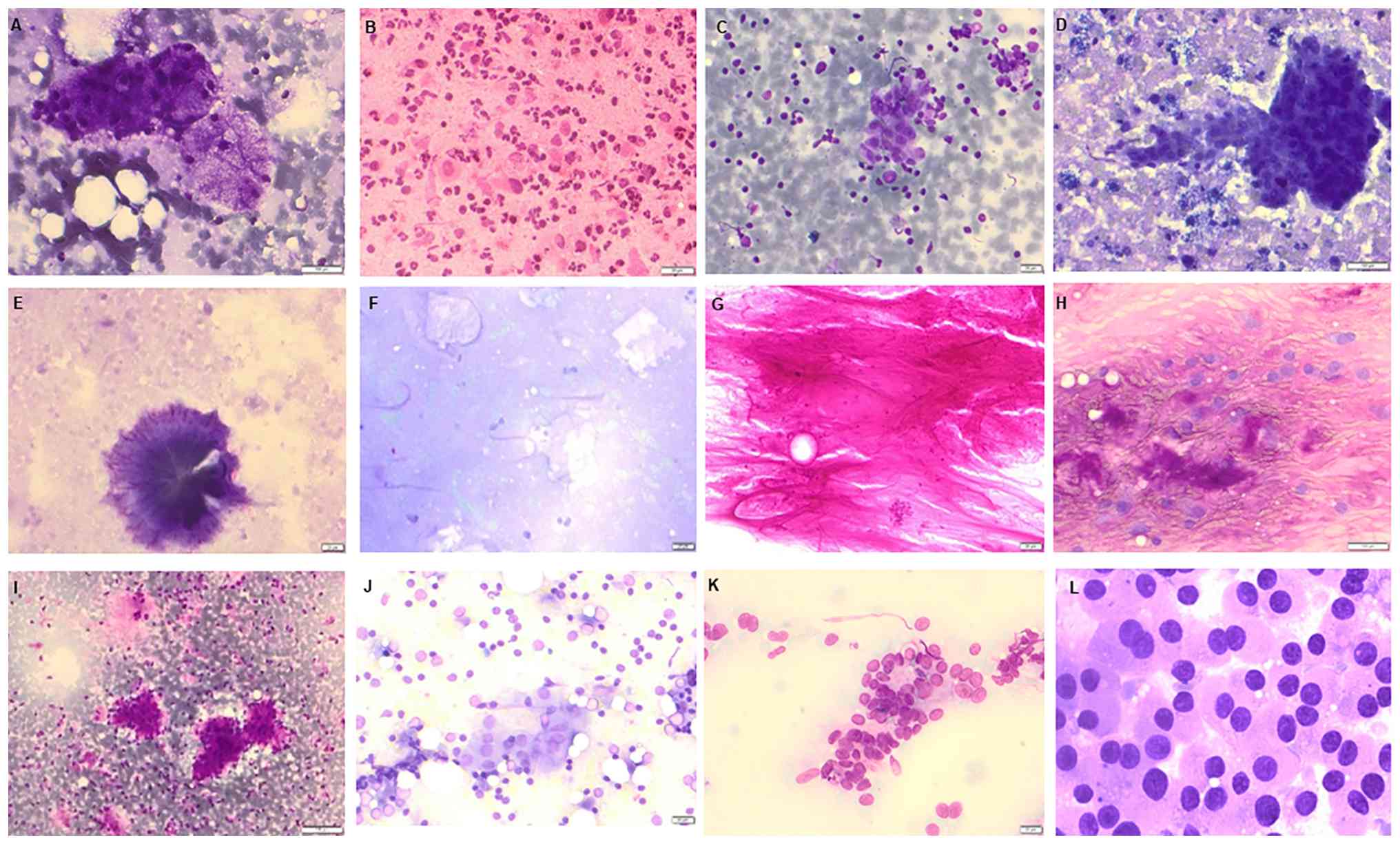 | Figure 1(A) Normal cells of the salivary
gland (magnification, x25). (B) Acute sialadenitis (magnification,
x25). (C) Chronic sialadenitis (magnification, x20). (D) Cyst with
actinomyces (magnification, x20), (E) a cystic cavity or dilated
duct, colonized by Actinomyces species, and often associated
with chronic suppurative inflammation (magnification, x20), and (F)
a cyst with actinomyces and crystal gaps (magnification, x20). (G)
Previous or resolved bleeding (magnification x20). (H) Pleomorphic
adenoma with myxoid background (magnification, x20). (I) Myxoid
background, epithelial and myoepithelial cell groups in pleomorphic
adenoma (magnification, x20). (J) Warthin's tumor, Lymphocytes and
oncocytic cells (magnification, x25). (K) Basal cell adenoma
(magnification, x20). (L) Oncocytic cells from oncocytoma
(magnification, x25). |
Mucoepidermoid carcinoma (MEC) is a malignant
salivary gland tumor composed of three distinct cell types:
Mucinous, intermediate and squamous. Tumor cells are classified as
either low- or high-grade lesions (11). Immunohistochemically, the
epidermoid cells are positive for CK5/6 and p63. Cytologically, MEC
may appear cystic and acellular with mucin in the background.
Clusters of pale intermediate or epithelial cells with
mucus-secreting cells are typically observed (11). Molecular genetics typically show a
t(11;19) translocation and a CRTC1-MAML2 gene fusion (20,21).
A rare translocation t(6;22) (p21; q12) is also found. CRTC1-MAML2
gene fusion is a chimeric (fusion) gene created by a chromosomal
translocation, most commonly t(11;19) (q21; p13), in which parts of
two separate genes become abnormally joined (11).
Adenocarcinoma, not otherwise specified, is the
third most common carcinoma of the salivary gland and lacks
features that allow classification into a specific tumor entity,
included in the figures is the histologic sample of this case
(11).
Acinar cell carcinoma is common in the parotid gland
(80% of cases). It can occur at any age, with a reported mean age
of diagnosis of 50 years. Cytologically, acinar cell carcinoma
presents with cell-rich preparations with a monomorphic pattern and
a clear background. Acinar cell clusters with fine granules in the
cytoplasm show typical PAS positivity. The nuclei do not show
atypia. Immunohistochemically, it is positive for discovered on
GIST-1 (DOG-1) and weakly positive for CK7 (22,23).
Secretory carcinoma (new in the 2017 World Health
Organization classification) is analogous to mammary carcinoma and
is considered a low-grade malignancy typically found in the parotid
gland (5). Cytologically,
papillary structures with epithelial cells showing vacuoles and
metachromatic granules are observed. Immunohistochemically, the
tumor is positive for S100 and mammaglobulin but negative for
DOG-1(11). Molecular cytogenetics
shows an ETV6-NTRK3 fusion (24).
Clear cell carcinoma aspirates show groups of
cohesive small and large epithelial cells with prominent cell
borders and uniform, round to ovoid nuclei with granular chromatin,
small nucleoli and abundant cleaved cytoplasm (14). PAS staining is positive. Clear cell
carcinoma shows consistent EWRS1-ATF1 gene fusion, which is a
chimeric oncogenic fusion gene formed by a chromosomal
translocation, most commonly t(12;22)(q13;q12), in which parts of
two genes become abnormally joined (25).
Basal cell adenocarcinoma smears are cellular and
show basaloid clusters of monomorphic small cells with scant
cytoplasm and frequently naked nuclei. The stroma is sparse to
absent: In the membranous type, it consists mainly of hyaline
matrix (11). A subset of these
tumors, mainly of the membranous type, is associated with
multicentricity and a higher recurrence rate (26).
Salivary duct carcinoma smears are cellular and
typically contain 3D clusters, sometimes with papillary
configurations and flat sheets of epithelial tumor cells with
necrotic backgrounds. The tumor cells are large and polygonal with
abundant cytoplasm. The nuclei are pleomorphic with prominent
nucleoli (11). Salivary duct
carcinoma is typically characterized by ERBB2 (HER2) amplification
and androgen receptor expression rather than pleomorphic adenoma
gene 1 (PLAG1) or high mobility group AT-hook 2 (HMGA2)
rearrangements (11). PLAG1 (a
transcription factor gene that becomes oncogenic when rearranged,
leading to overexpression and promotion of tumor growth) and/or
HMGA2 (a chromatin remodeling gene involved in regulation of
proliferation; rearrangements lead to abnormal expression)
rearrangements are identified in most cases of salivary duct
carcinoma arising in pleomorphic adenoma (27).
Myoepithelial carcinoma aspirate smears show a
mixture of spindled and/or plasmacytoid cells in small groups or
large fragments. The nuclei may be round or oval with variable
cytoplasmic features (11). The
immunohistochemical expression of S-100 and SOX10 is an important
diagnostic feature in myoepithelial carcinoma. EWSRI gene
rearrangement has been described in myoepithelial carcinomas with
aggressive features and frequent necrosis (28).
Carcinoma ex pleomorphic adenoma smears typically
show features of pleomorphic adenoma or high-grade adenocarcinoma;
rarely, both components are defined (11). Carcinoma ex pleomorphic adenoma
often harbors PLAG1 and HMGA2 rearrangements, reflecting its origin
from pleomorphic adenoma (29).
Although molecular pathology is increasingly relevant in salivary
gland tumors, its use remains largely confined to diagnostic
support (30).
The primary aim of the present study was to evaluate
the diagnostic performance of FNAC in determining the histogenesis
of salivary gland lesions based on cytomorphological features in a
single-center cohort. Secondary objectives included comparison of
cytological findings with histopathological outcomes,
identification of diagnostic pitfalls and comparison with the
existing literature. By integrating these aspects, the present
study aimed to refine diagnostic criteria and contribute to
improved diagnostic accuracy in salivary gland cytopathology.
Materials and methods
Patients
A total of 544 patients, including 283 females (52%)
and 261 males (48%), between 18 and 86 years old. FNAC specimens
from salivary gland lesions and other head and neck swellings were
analyzed at the Institute of Pathology, Hannover Medical School
(Hannover, Germany), between January 2000 and December 2024. The
criteria for inclusion in the study were as follows: i) Presence of
a clinically and/or radiologically confirmed salivary gland lesion
(parotid, submandibular, sublingual or minor salivary glands); ii)
performance of FNAC of the lesion; and iii) availability of
adequate cytological material for evaluation. The exclusion
criteria were as follows: i) Inadequate or non-diagnostic aspirates
(e.g., blood only, insufficient cellularity); ii) previously
treated lesions (for example, after radiotherapy or chemotherapy)
that could affect cytological interpretation; and iii) recurrent
tumors (depending on study design, often excluded or analyzed
separately).
Cytological samples were obtained using standard
FNAC techniques. After clinical and radiological assessment,
lesions were aspirated using a 23-25-gauge needle attached to a
syringe under manual negative pressure. Multiple passes were
performed when necessary to ensure adequate cellularity. Smears
were prepared and routinely stained using May-Grünwald-Giemsa
(MGG). In selected cases, with diagnostic uncertainty or suspicion
of mucinous, glycogen-rich, or extracellular matrix-producing
lesions, additional PAS staining was performed to evaluate
cytoplasmic and extracellular components. When available, cell
blocks were prepared from cytological material and processed
according to standard histological protocols (31).
Cytological diagnoses were associated with
corresponding histopathological findings where surgical specimens
were available. Immunohistochemical analyses were performed on
selected cases with diagnostic uncertainty, indeterminate
cytomorphological features, or when further tumor classification
was required, using cell block material, cytospin preparations or
alcohol-fixed smears. Alcohol-fixed smears were prepared using
standard laboratory conditions (ethanol fixation at room
temperature for routine diagnostic processing), according to
established cytology protocols (32). Staining was performed using an
automated immunostaining system (BenchMark, Ventana/Roche
Diagnostics) following the manufacturer's protocols.
MGG staining
Air-dried smears were fixed in MG (in methanol) for
3-5 min, then stained with 1:1 mixture of MG and buffer (pH 6.8)
for 5 min and Giemsa stain (1:20-1:30) for 10-20 min. All steps
were performed at room temperature (20-25˚C). Samples were rinsed
with water and examined, using a bright-field light microscope
(Zeiss-Microscope).
PAS smears were fixed in 95% ethanol (or 10% neutral
buffered formalin) for 10-15 min at room temperature (20-25˚C),
rinsed in distilled water, oxidized with 0.5-1.0% periodic acid for
5.0-10.0 min, rinsed in distilled water, stained with Schiff
reagent for 10.0-20.0 min, washed in running tap water for 10.0
min, counterstained with hematoxylin for 1.0-2.0 min, then rinsed
in water, dehydrated through graded alcohols, cleared in xylene,
and mounted.
Immunohistochemistry
Cytological material (cell blocks, cytospin
preparations, or direct smears) was fixed using either formalin- or
alcohol-based fixatives at room temperature. The specimens were
prepared on positively charged glass slides, with cell block
sections cut at a thickness of 3-4 µm. Air-dried smears were
rehydrated and post-fixed in 10% formalin for 10 min. Slides were
baked at 60˚C for 30-60 min. On the BenchMark stainer the slides
were deparaffinized, rehydrated, and subjected to heat-induced
antigen retrieval according to the standard automated
immunohistochemistry (33).
Antigen retrieval was carried out using CC1 (pH 8.5) or CC2 (pH 6)
for 32-64 min at 95˚C. Endogenous peroxidase was blocked using
H2O2. The primary antibody was then applied
for 16-32 min, followed by detection with the ultraView or OptiView
DAB system and counterstaining with hematoxylin at room
temperature. The immunohistochemical stains were performed using
ready-to-use primary antibodies (Roche Tissue Diagnostics) on an
automated staining platform (BenchMark ULTRA; Roche Tissue
Diagnostics). The exact antibody concentrations are not disclosed
by the manufacturer for these pre-diluted reagents. Staining was
performed according to the manufacturer's optimized protocols.
Primary antibodies were applied at
manufacturer-recommended dilutions (catalogue number and supplier
specified for each antibody, Roche Tissue Diagnostics) and
incubated for 30-60 min at room temperature (or overnight at 4˚C
when indicated). After washing in phosphate-buffered saline, slides
were incubated with appropriate secondary antibodies
(enzyme-conjugated, typically horseradish peroxidase-labelled
systems; catalogue number and supplier specified, Roche Tissue
Diagnostics) for 20-30 min at room temperature.
Immunohistochemistry was performed using a panel of primary
antibodies, including: CK7 (clone OV-TL 12/30), AE1/AE3
(pan-cytokeratin cocktail AE1/AE3/PCK26), CK5/6 (clone D5/16 B4),
p63 (clone 4A4), DOG1 (clone SP31), WT1 (clone 6F-H2), smooth
muscle actin (SMA; clone 1A4), mammaglobin (clone 31A5), HER2/neu
(clone 4B5), S-100 protein [clone (if available)], SOX10 [clone (if
available)], and Ki-67 (clone MIB-1). All antibodies were applied
as ready-to-use reagents on an automated Ventana BenchMark ULTRA
staining platform (Roche Tissue Diagnostics) according to the
manufacturer's standardized protocols. As these are proprietary,
pre-optimized diagnostic reagents, exact antibody concentrations,
working dilutions, and catalogue numbers are not disclosed by the
manufacturer. Visualization was performed using diaminobenzidine as
a chromogen. Sections were counterstained with hematoxylin for 1-2
min at room temperature, followed by dehydration through graded
alcohols, clearing in xylene, and mounting. Stained slides were
evaluated using a bright-field light microscope.
Results
Patients
A total of 544 salivary gland FNAC specimens
(Table I) were evaluated. Patients
included 283 females (52%) and 261 males (48%). FNAC demonstrated a
relatively high diagnostic value in cases with representative
material, particularly in distinguishing benign and malignant
lesions, as evidenced by comparison with the corresponding
histopathological diagnoses in cases where surgical follow-up was
available. Of all cases (Table I),
422 (77.6%) were benign, 59 (10.80%) were malignant, 26 (4.80%)
were normal (Fig. 1A) and 37
(6.80%) were non-representative (the sample did not adequately
reflect the lesion that was targeted, so it is not suitable for
diagnosis). Among 422 benign lesions (Table II), 174 (41.20%) were inflammatory
lesions (Fig. 1B and C), 108 (25.60%) were cysts (Fig. 1D-F), 72 (17.10%) were pleomorphic
adenoma (Fig. 1H and I), 64 (15.20%) were Warthin's tumor
(Fig. 1J), (0.50%) were basal cell
adenoma (Fig. 1K) and 2 (0.50%)
were oncocytoma (Fig. 1L). Among
59 malignant lesions (Table
III), 13 (18.64%) were adenocarcinoma (Fig. 2A and B), 3 (5.09%) were poorly differentiated
carcinoma, 2 (3.40%) were acinar cell carcinoma (Fig. 2C and D), 1 (1.69%) was basal cell carcinoma, 1
(1.69%) was secretory carcinoma (Fig.
2E and F), 13 (22.03%) were
squamous cell carcinoma (Fig. 2G
and H), 1 (1.69%) was adenoid
cystic carcinoma (Fig. 2I and
J), 1 (1.69%) was ductal
carcinoma, 1 (1.69%) was mucoepidermoid carcinoma (Fig. 2K and L), 1 (1.69%) was myoepithepithelial
carcinoma, 20 (33.92%) were non-Hodgkin lymphoma (NHL), 1 (1.69%)
was malignant melanoma and 3 (5.09%) were renal cell carcinoma
metastasis.
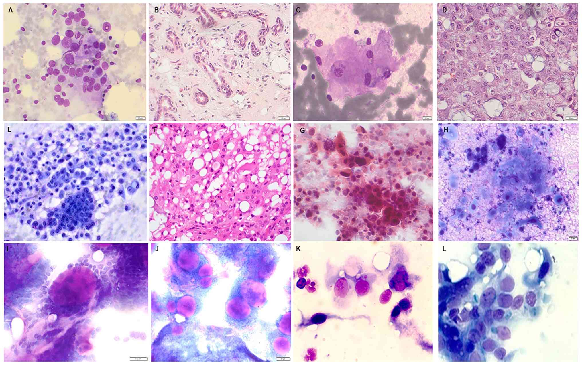 | Figure 2(A) Adenocarcinoma NOS (MGG;
magnification, x25); (B) adenocarcinoma NOS of the salivary gland
(H&E; magnification, x20); (C) acinus cell carcinoma (MGG;
magnification, x25); (D) acinus cell carcinoma (H&E;
magnification, x20); (E) secretory carcinoma (MGG; magnification,
x25); (F) secretory carcinoma (H&E; magnification, x20). (G)
Squamous cell carcinoma (magnification, x40); (H) inflammatory
cells and necrotic cells in squamous cell carcinoma (magnification,
x40); (I) adenoid cystic ca. with globules (magnification, x40);
(J) adenoid cystic carcinoma (magnification, x25); (K)
mucoepidermoid carcinoma (magnification, x40); (L) malignant cells
of mucoepidermoid carcinoma (magnification, x40). NOS, not
otherwise specified; H&E, hematoxylin and eosin; MGG,
May-Grünwald-Giemsa. |
 | Table ITotal number of cases with different
salivary gland lesions. |
Table I
Total number of cases with different
salivary gland lesions.
| Diagnosis | Number of
cases | % |
|---|
| Benign | 422 | 77.57 |
| Malignant | 59 | 10.85 |
| Normal | 26 | 4.78 |
|
Non-representative | 37 | 6.80 |
| Table IIBenign lesions of the salivary gland
(n=422). |
Table II
Benign lesions of the salivary gland
(n=422).
| Diagnosis | Number of
cases | % |
|---|
| Inflammation | 174 | 41.23 |
| Cyst | 108 | 25.60 |
| Pleomophic
adenoma | 72 | 17.06 |
| Warthin's
tumor | 64 | 15.17 |
| Basal cell
adenoma | 2 | 0.47 |
| Oncocytoma | 2 | 0.47 |
| Table IIIMalignant tumors of the salivary
gland (n=59). |
Table III
Malignant tumors of the salivary
gland (n=59).
| Diagnosis | Number of
cases | % |
|---|
| Squamous cell
carcinoma | 13 | 22.03 |
| Adenocarcinoma | 11 | 18.64 |
| Poorly
differentiated carcinoma | 3 | 5.09 |
| Acinar cell
carcinoma | 2 | 3.40 |
| Basal cell
carcinoma | 1 | 1.69 |
| Ductal cell
carcinoma | 1 | 1.69 |
| Myoepithelial
carcinoma | 1 | 1.69 |
| Mucoepidermoid
carcinoma | 1 | 1.69 |
| Adenoid cystic
carcinoma | 1 | 1.69 |
| Secretory
carcinoma | 1 | 1.69 |
| Non-Hodgkin
lymphoma | 20 | 33.92 |
| Malignant
melanoma | 1 | 1.69 |
| Renal cell
carcinoma | 3 | 5.09 |
Diagnostic accuracy
Based on available histopathology (n=481 cases;
excluding normal and non-representative aspirates), FNAC diagnostic
performance for distinguishing benign vs. malignant lesions
[Table IV: True positives
(TP)=56, false negatives (FN)=3, false positives (FP)=2, true
negatives (TN)=420, total=481] yielded sensitivity of 94.9%
(sensitivity=TP/FN+TP=56/3+56=56/59), specificity of 99.5%
(TN/TN+FP=420/420+2=420/422), positive predictive value of 96.6%
(TP/TP+FP=56/56+2=56/58), negative predictive value of 99.3%
(TN/TN+FN=420/420+3=420/423) and overall accuracy of 98.1%
(TP+TN/total=56+420/481). False-negative cases (n=3) included
low-grade mucoepidermoid carcinoma, small acinar cell carcinoma and
poorly differentiated carcinoma where cellularity was insufficient.
There were two false-positives: Reactive lymphoid hyperplasia
mimicking lymphoma and severely inflamed Warthin's tumor
misinterpreted as malignant.
| Table IVFine-needle aspiration cytology
diagnostic performance for distinguishing benign vs. malignant
lesions. |
Table IV
Fine-needle aspiration cytology
diagnostic performance for distinguishing benign vs. malignant
lesions.
| | FNAC diagnosis | |
|---|
| Histopathology | Malignant | Benign | Total |
|---|
| Malignant | 56 (TP) | 3 (FN) | 59 |
| Benign | 2 (FP) | 420 (TN) | 422 |
Certain tumor entities, such as adenoid cystic
carcinoma, presented greater diagnostic challenges, with lower
detection rates in the range of 13 to 50%. In these cases,
overlapping cytomorphological features and limited cellularity
contributed to diagnostic uncertainty. Overall, FNAC findings were
most reliable when correlated with histopathological results and
supported by clinical and radiological information. The 5-year
survival rate (Table V) varies
depending on the tumor type, and late recurrence is common
(35-79%).
| Table VOverall, 5-year survival of patients
with salivary gland tumors. |
Table V
Overall, 5-year survival of patients
with salivary gland tumors.
| Salivary gland
tumor | 5-year survival
rate, % |
|---|
| Acinar cell
carcinoma | 80 |
| Mucoepidermoid
carcinoma | |
|
Low-grade | <70 |
|
High-grade | <50 |
| Adenocarcinoma, not
otherwise specified | 50-70 |
| Squamous cell
carcinoma | 40-55 |
| Undifferentiated
Carcinoma | 30-40 |
| Carcinoma in
pleomorphic adenoma | 50-70 |
Discussion
The present study highlighted the diagnostic utility
of FNAC in the evaluation of salivary gland lesions, while also
emphasizing its limitations. The present series confirmed the
predominance of benign (77.6%) over malignant lesions (10.8%),
consistent with previous reports (1,11,15).
Inflammatory lesions and cysts were the most frequent benign
lesions, while NHL represented a notable portion of malignancies.
FNAC demonstrated high diagnostic accuracy (overall, 98.1%),
specificity (99.5%) and sensitivity (94.9%). False-negative results
occurred primarily in low-cellularity tumors or cystic lesions,
emphasizing the need for adequate sampling. False positives were
rare but highlight potential pitfalls in inflammatory or reactive
lesions.
The relatively wide range of sensitivity and
specificity observed between the present and previous studies
reflects the inherent complexity and morphological diversity of
salivary gland pathology (11,15,34).
However, FNAC accuracy is dependent on several factors, including
sampling technique, preparation quality and the experience of the
cytopathologist. In addition, the integration of clinical history
and imaging findings serves a crucial role in accurate
interpretation.
FNAC is used as a preoperative diagnostic modality
for salivary gland lesions due to its minimal invasiveness, rapid
turnaround and relatively low cost. Numerous studies (11,15)
have demonstrated its overall high diagnostic performance,
especially for distinguishing benign from malignant processes,
although sensitivity and specificity vary substantially across
patient populations and institutional settings. For example,
systematic reviews have shown that sensitivity in identifying
malignancy ranges widely and that FNAC generally achieves high
specificity in salivary gland lesions, although notable
heterogeneity exists among studies (34,35).
The reported sensitivity and specificity of FNAC in
salivary gland lesions shows variability, ranging from 60.0 to
97.5%, depending on lesion type and sample quality (15,34,35).
In a clinical series comparing FNAC with
histopathology, sensitivity and specificity values have ranged from
50 to >90 %, depending on lesion type, sampling adequacy and
interpretive criteria (15,33,34).
These findings are consistent with the present study, where FNAC
demonstrated a relatively high diagnostic value in representative
material with variable performance metrics (including sensitivity,
specificity, positive predictive value and negative predictive
value). Factors contributing to this variability include the
intrinsic morphological diversity of salivary gland tumors,
sampling error in cystic or hypocellular lesions and interpretive
challenges posed by tumors with overlapping cytomorphological
features (11,15,34).
The cytological interpretation of certain entities,
such as adenoid cystic carcinoma and other types of basaloid
neoplasm, remains particularly challenging due to subtle
morphological features that may be underrepresented in aspirate.
Moreover, low-grade malignancies, cystic tumors, and heterogeneous
tumors such as pleomorphic adenomas can yield hypocellular or
misleading cytological specimens, further decreasing sensitivity
(11,15,34).
These limitations underscore the importance of associating FNAC
findings with clinical presentation and imaging studies to optimize
diagnostic accuracy (35).
Several authors (11,15)
have also highlighted the role of adjunctive techniques such as
immunocytochemistry and molecular diagnostics in improving
diagnostic specificity and subclassification, particularly when
cytomorphology is equivocal. However, these techniques are
typically limited by sample quantity and are not necessary in
straightforward cases (36).
Therefore, although ancillary studies contribute valuable
information, cytomorphological assessment remains the cornerstone
of FNAC interpretation.
In recent years (11,30,37,38),
cytology specimens have increasingly been used for molecular
testing to complement conventional cytomorphological assessment.
FNA material, particularly cell blocks, is used for
immunohistochemistry, fluorescence in situ hybridization and
NGS to detect gene fusions, mutations and other molecular
alterations relevant for tumor classification and, in certain
cases, targeted therapy. For example, the TS variant transcription
factor 6-eurotrophic receptor tyrosine kinase 3 fusion (ETV6-NTRK3;
ETV6-NTRK3 fusion is a gene rearrangement resulting from a
translocation between chromosomes 12 and 15, leading to the
formation of a constitutively active tyrosine kinase fusion protein
that drives oncogenic signaling. It is the defining molecular
alteration of secretory carcinoma of the salivary gland) or
MYB/MYBL1 rearrangements (MYB or MYBL1 rearrangements refer to gene
fusions or translocations involving the MYB or MYBL1 transcription
factor genes, resulting in overexpression of oncogenic
transcriptional programs. These alterations are characteristic
molecular events in adenoid cystic carcinoma and are associated
with tumor development and progression) are reliably identified in
FNA-derived material, enabling more precise subclassification and
improved diagnostic confidence (11,18,20).
However, the use of cytology samples for molecular analysis has
limitations. Adequate cellularity is key and cystic or
low-cellularity lesions may yield insufficient DNA/RNA. Fixatives
such as alcohol or formalin, commonly used for smears and cell
blocks, may also affect nucleic acid quality (30,36,38).
Nevertheless, when sufficient material is available, molecular
testing on cytology specimens provides key diagnostic and
prognostic information, especially in cases where surgical tissue
is unavailable or limited. Integrating molecular testing with
conventional cytology thus enhances diagnostic yield and supports
personalized patient management (11,30,38).
Here, NHL accounted for approximately one-third of
malignant salivary gland lesions, making it the most frequent
malignancy in the present cohort. This high prevalence contrasts
with published reports (39,40),
where epithelial malignancies such as mucoepidermoid carcinoma,
adenoid cystic carcinoma and adenocarcinoma typically predominate.
The elevated proportion of NHL in the present study may reflect the
referral patterns to our tertiary center (Institute for Pathology,
Hannover medical School, Hannover, Germany), which frequently
receives cases of lymphoproliferative disorders requiring
cytological evaluation. Cytologically, NHL poses diagnostic
challenges, as cellular smears can mimic reactive lymphoid
hyperplasia or other small round cell tumors, increasing the risk
of false-negative or ambiguous results. FNAC, combined with
immunophenotyping on cell blocks (CD20, CD3, Ki-67), allows
reliable subclassification and differentiation from benign lymphoid
proliferations, guiding management. Molecular investigations and
clonality testing performed on the cell block support the diagnosis
of malignant lymphoma. These findings underscore the importance of
considering lymphoid malignancy in the differential diagnosis of
salivary gland masses, particularly in older adults, where primary
salivary gland lymphomas are more prevalent, and in patients with
systemic (B) symptoms suggestive of hematologic disease. This
highlights the role of FNAC as a first-line, minimally invasive
diagnostic tool (39,40).
In summary, the literature (11,15,28)
supports the use of FNAC as a valuable component of the diagnostic
pathway for salivary gland lesions, particularly when integrated
within a multidisciplinary framework including clinical evaluation
and imaging. Such an integrated approach improves diagnostic yield
and decreases limitations of cytological interpretation alone.
In the present study, FNAC, when combined with
cytomorphological assessment and clinical-radiological findings,
allowed reliable histogenetic interpretation in a notable
proportion of cases.
FNAC is a useful first-line diagnostic tool in the
evaluation of salivary gland lesions, particularly for guiding the
distinction between benign and malignant entities. Its diagnostic
performance is influenced by lesion type, specimen adequacy and
interpretative expertise, which accounts for variability in
accuracy across different diagnostic categories. For optimal
diagnostic reliability, FNAC should be applied within a structured
multidisciplinary framework that integrates cytological, clinical
and radiological findings.
Acknowledgements
The authors would like to thank Mrs Petra Abbas
(Freelancer; Greven, Germany) for proofreading.
Funding
Funding: No funding was received.
Availability of data and materials
The data generated in the present study may be
requested from the corresponding author.
Authors' contributions
BS contributed to the conception and design of the
study. BS developed the methodological framework, including
cytological, histopathological and molecular procedures. BS
performed and/or reviewed the cytological and histopathological
assessments. BS supervised molecular pathological analyses,
including NGS and related techniques. MA contributed to the
conception and design of the study. MA performed and/or reviewed
the cytological and histopathological assessments. MA drafted the
manuscript. BS and MA were responsible for collection and
processing of clinical, cytological, and histopathological data. BS
and MA analyzed and interpreted the data and contributed to the
evaluation of diagnostic performance. All authors have read and
approved the final manuscript. BS and MA confirm the authenticity
of all the raw data.
Ethics approval and consent to
participate
The study was reviewed by the Ethics Committee of
Hannover Medical School (approval no. 18.02.2025; Hannover,
Germany), which waived the requirement for ethical approval and
written informed consent due to the retrospective and fully
anonymized nature of the data.
Patient consent for publication
Not applicable.
Competing interests
The authors declare that they have no competing
interests.
References
|
1
|
Eveson JW and Cawson RA: Salivary gland
tumours. A review of 2,410 cases with particular reference to
histological types, site, age and sex distribution. J Pathol.
146:51–58. 1985.PubMed/NCBI View Article : Google Scholar
|
|
2
|
Aher TS, Ekhar VR, Shelkar RN and Vedi JN:
Salivary gland lesions: A clinico-pathological correlation. Indian
J Otolaryngol Head Neck Surg. 75:1665–1669. 2023.PubMed/NCBI View Article : Google Scholar
|
|
3
|
Venkatesh S, Srinivas T and Hariprasad S:
Parotid gland tumors: 2-year prospective clinicopathological study.
Ann Maxillofac Surg. 9:103–109. 2019.PubMed/NCBI View Article : Google Scholar
|
|
4
|
Tani EM and Skoog L: Salivary glands and
rare head and neck lesions. In: Comprehensive Cytopathology. Bibbo
M and Wilbur DC (eds). 3rd edition. Saunders Elsevier,
Philadelphia, PA, pp607-632, 2008.
|
|
5
|
El-Naggar AK: WHO classification of tumors
of salivary glands. In: WHO Classification of Head and Neck
Tumours. El-Naggar AK, Chan JKC, Grandis JR, Takata T and Slootweg
PJ (eds). 4th edition. IARC, Lyon, p160, 2017.
|
|
6
|
Klijanienko J, Vielh P, Batsakis JD,
el-Naggar AK, Jelen M and Piekarski JD: Salivary gland tumours.
Monogr Clin Cytol. 15:III–XII. 2005.
|
|
7
|
Faur AC, Buzaș R, Lăzărescu AE and Ghenciu
LA: Current developments in diagnosis of salivary gland tumors:
From structure to artificial intelligence. Life (Basel).
14(727)2024.PubMed/NCBI View Article : Google Scholar
|
|
8
|
Saoud C, Bailey GE, Graham A, Bonilla LM,
Sanchez SI and Maleki Z: Pitfalls in salivary gland cytology. Acta
Cytol. 68:194–205. 2024.PubMed/NCBI View Article : Google Scholar
|
|
9
|
Carillo AM, DeLuca C, Pisapia P, Vigliar
E, Ikenberg K, Freiberger SN, Troncone G, Rupp NJ and Bellevicine
C: Molecular testing in salivary gland cytopathology: A practical
overview in conjunction with the Milan system. Cytopathology.
35:330–343. 2024.PubMed/NCBI View Article : Google Scholar
|
|
10
|
Kessler AT and Bhatt AA: Review of the
major and minor salivary glands, Part 1: Anatomy, infectious, and
inflammatory processes. J Clin Imaging Sci. 8(47)2018.PubMed/NCBI View Article : Google Scholar
|
|
11
|
El-Naggar AK, Chan JKC, Grandis JR, Takata
T and Slootweg PJ (eds): WHO Classification of Head and Neck
Tumours. 5th edition. IARC, Lyon, 2022.
|
|
12
|
Pantanowitz L, Thompson LDR and Rossi ED:
Diagnostic approach to fine needle aspirations of cystic lesions of
the salivary gland. Head Neck Pathol. 12:548–561. 2018.PubMed/NCBI View Article : Google Scholar
|
|
13
|
Ersöz C, Cetik F, Aydin O, Cosar EF and
Talas DU: Salivary duct carcinoma ex pleomorphic adenoma: Analysis
of the findings in fine-needle aspiration cytology and histology.
Diagn Cytopathol. 19:201–204. 1998.PubMed/NCBI View Article : Google Scholar
|
|
14
|
Sáenz-Santamaría J, Catalina-Fernandéz I
and Fernandez-Mera JJ: Sialadenitis with crystalloid formation.
Fine needle aspiration cytodiagnosis of 15 cases. Acta Cytol.
47:1–4. 2003.PubMed/NCBI View Article : Google Scholar
|
|
15
|
Faquin WC and Rossi ED (eds): The Milan
System for Reporting Salivary Gland Cytopathology. Springer
International Publishing, Cham, 2018.
|
|
16
|
Bhat A, Rao M, Geethamani V and Shetty AC:
Basal cell adenoma of the parotid gland: Cytological diagnosis of
an uncommon tumor. J Oral Maxillofac Pathol. 19(106)2015.PubMed/NCBI View Article : Google Scholar
|
|
17
|
Li S, Baloch ZW, Tomaszewski JE and
LiVolsi VA: Worrisome histologic alterations following fine-needle
aspiration of benign parotid lesions. Arch Pathol Lab Med.
124:87–91. 2000.PubMed/NCBI View Article : Google Scholar
|
|
18
|
Xu B, Drill E, Ho A, Ho A, Dunn L,
Prieto-Granada CN, Chan T, Ganly I, Ghossein R and Katabi N: .
Predictors of outcome in adenoid cystic carcinoma of salivary
glands: A clinicopathologic study with correlation between MYB
fusion and protein expression. Am J Surg Pathol. 41:1422–1432.
2017.PubMed/NCBI View Article : Google Scholar
|
|
19
|
Mukunyadzi P: Review of fine-needle
aspiration cytology of salivary gland neoplasms, with emphasis on
differential diagnosis. Am J Clin Pathol. 118:S100–S115.
2002.PubMed/NCBI View Article : Google Scholar
|
|
20
|
Toper MH and Sarioglu S: Molecular
pathology of salivary gland neoplasms: Diagnostic, prognostic, and
predictive perspective. Adv Anat Pathol. 28:81–93. 2021.PubMed/NCBI View Article : Google Scholar
|
|
21
|
Wakely PE Jr: Mucoepidermoid carcinoma:
Analysis of 76 cytologic cases and correlation with histologic
grade. Cancer Cytopathol. 130:783–799. 2022.PubMed/NCBI View Article : Google Scholar
|
|
22
|
Griffith CC, Siddiqui MT and Schmitt AC:
Ancillary testing strategies in salivary gland aspiration cytology:
A practical pattern-based approach. Diagn Cytopathol. 45:808–819.
2017.PubMed/NCBI View
Article : Google Scholar
|
|
23
|
Meiklejohn K, Hrones M, Wang M, Prasad ML,
Cai G, Adeniran A and Gilani SM: Diagnosis of acinic cell carcinoma
of the salivary gland on cytology specimens: Role of NOR-1 (NR4A3)
immunohistochemistry. Cytopathology. 34:219–224. 2023.PubMed/NCBI View Article : Google Scholar
|
|
24
|
Agarwal C, Jain M, Pujani M, Rajpoot J,
Goel H, Sarohi M and Sasidharan J: Secretory carcinoma of salivary
gland: A rare tumor masquerading as acinic cell carcinoma on
cytology. Diagn Cytopathol. 52:E19–E22. 2024.PubMed/NCBI View
Article : Google Scholar
|
|
25
|
Hirose K, Usami Y, Kohara M, Sato S,
Iwamoto Y, Murakami S, Uchihashi T, Oya K, Fukuda Y, Hori Y, et al:
Clear cell carcinoma of palatal minor salivary gland harboring a
novel EWSR1-ATF1 fusion gene: Report of a case and review of the
literature. Head Neck Pathol. 15:676–681. 2021.PubMed/NCBI View Article : Google Scholar
|
|
26
|
Robinson RA: Basal cell adenoma and basal
cell adenocarcinoma. Surg Pathol Clin. 14:25–42. 2021.PubMed/NCBI View Article : Google Scholar
|
|
27
|
Wakely PE: Salivary duct carcinoma: A
report of 70 FNA cases and review of the literature. Cancer
Cytopathol. 130:595–608. 2022.PubMed/NCBI View Article : Google Scholar
|
|
28
|
Argyris PP and Wakely PE Jr: Cytopathology
of salivary gland myoepithelial carcinoma: A study of 13 cases and
review of the literature. J Am Soc Cytopathol. 12:461–468.
2023.PubMed/NCBI View Article : Google Scholar
|
|
29
|
Bahrami A and Perez-Ordoñez B: Carcinoma
ex pleomorphic adenoma: Pathogenesis and molecular genetics. Head
Neck Pathol. 8:30–36. 2014.
|
|
30
|
Pusztaszeri M, Rossi ED and Faquin WC: Is
molecular testing of salivary gland FNA specimens ready for prime
time? Cancer Cytopathol. 132:393–395. 2024.PubMed/NCBI View Article : Google Scholar
|
|
31
|
Kiernan JA: Histological and histochemical
methods: Theory and practice. 5th edition. Scion Publishing,
Banbury (UK), 2015.
|
|
32
|
Koss LG and Melamed MR: Koss' diagnostic
cytology and its histopathologic bases. 5th ed. Philadelphia:
Lippincott Williams & Wilkins; 2006.
|
|
33
|
Dabbs DJ: Diagnostic immunohistochemistry:
Theranostic and genomic applications. 6th ed. Philadelphia:
Elsevier; 2022.
|
|
34
|
Schmidt RL, Hall BJ, Wilson AR and
Layfield LJ: A systematic review and meta-analysis of the
diagnostic accuracy of fine-needle aspiration cytology for parotid
gland lesions. Am J Clin Pathol. 136:45–59. 2011.PubMed/NCBI View Article : Google Scholar
|
|
35
|
Fouladpour N, Ali SZ and Olson MT:
Accuracy of fine-needle aspiration cytology in salivary gland
lesions: A systematic review and meta-analysis. Cancer Cytopathol.
124:695–709. 2016.
|
|
36
|
Cibas ES and Ducatman BS: Cytology:
Diagnostic principles and clinical correlates. 5th ed. Elsevier;
2020.
|
|
37
|
-Gargano SM, Senarathne W, Feldman R,
Florento E, Stafford P, Swensen J, Vranic S and Gatalica Z: Novel
therapeutic targets in salivary duct carcinoma uncovered by
comprehensive molecular profiling. Cancer Med. 8:7322–7329.
2019.PubMed/NCBI View Article : Google Scholar
|
|
38
|
Rossi ED and Faquin WC: Molecular testing
in cytopathology: Current applications and future perspectives.
Cancer Cytopathol. 131:289–303. 2023.
|
|
39
|
Van Wal JE, Wolvius EB, Huijgens PC and
van der Waal I: Malignant lymphomas of the salivary glands: A
clinicopathological study. Oral Oncol. 38:403–409. 2002.
|
|
40
|
Ellis GL and Auclair PL: Tumors of the
salivary glands. AFIP Atlas of Tumor Pathology, Series 4, Fascicle
9. Washington, DC: American Registry of Pathology; 2008.
|

















