Introduction
Spontaneous esophageal rupture, also known as
Boerhaave syndrome, is a rare and potentially fatal condition
characterized by a full-thickness tear of the esophageal wall.
Boerhaave syndrome typically results from a sudden rise in
intraesophageal pressure against a closed glottis, most commonly
triggered by forceful vomiting after excessive eating or alcohol
consumption (1). The perforation
most frequently occurs in the left posterolateral aspect of the
distal esophagus, which represents a region of anatomical weakness
(2).
Despite advances in diagnostic imaging, critical
care and minimally invasive techniques, Boerhaave syndrome
continues to carry a high mortality rate, often exceeding 30-50%
when diagnosis and treatment are delayed beyond 24-48 h (3,4). Due
to its rarity and nonspecific symptomatology, Boerhaave syndrome is
frequently misdiagnosed as acute myocardial infarction, aortic
dissection or acute pancreatitis, leading to critical delays in
appropriate management (5).
Diagnosis is primarily established through
contrast-enhanced computed tomography (CT), with characteristic
findings including pneumomediastinum, pleural effusion and
esophageal wall discontinuity (6).
Notably, upper gastrointestinal endoscopy has evolved into a
valuable adjunctive diagnostic and therapeutic modality, and novel
endoscopic techniques have revolutionized the management of
esophageal perforations, allowing for organ-preserving treatment
(7). However, direct endoscopic
visualization of vital mediastinal structures through an esophageal
perforation remains a rare clinical observation.
The present study reports a case of spontaneous
esophageal rupture complicated by severe mediastinitis, in which
bedside gastroscopy served a pivotal role in both diagnosis and
initial therapeutic intervention, revealing direct visualization of
the pulsating heart through the perforation.
Case report
A 54-year-old man presented to the Emergency
Department of Qilu Hospital of Shandong University (Jinan, China)
in March 2024 with sudden-onset severe vomiting following excessive
alcohol consumption and overeating, accompanied by intense
retrosternal chest pain radiating to the back and fever. The past
medical history of the patient was unremarkable, with no known
history of esophageal disease, peptic ulcer or prior
gastrointestinal procedures. On initial physical examination, the
patient appeared acutely ill. Vital signs were notable for
tachycardia (heart rate, 122 beats/min; normal range, 60-100
beats/min) and a borderline blood pressure of 108/70 mmHg (normal
range, 90-139/60-89 mmHg; mean arterial pressure, 83 mmHg) with a
temperature of 37.3˚C (within the normal range of 36.0-37.5˚C
measured at the axilla; the patient developed a documented fever of
38.9˚C 6 h after admission). No subcutaneous emphysema was detected
on palpation of the neck or chest wall. Abdominal examination
revealed mild epigastric tenderness without signs of
peritonitis.
Within 24 h of symptom onset, the hemodynamic status
of the patient deteriorated, progressing to septic shock requiring
vasopressor support. The patient was admitted to the intensive care
unit (ICU) for aggressive fluid resuscitation and hemodynamic
stabilization.
The initial diagnostic workup included
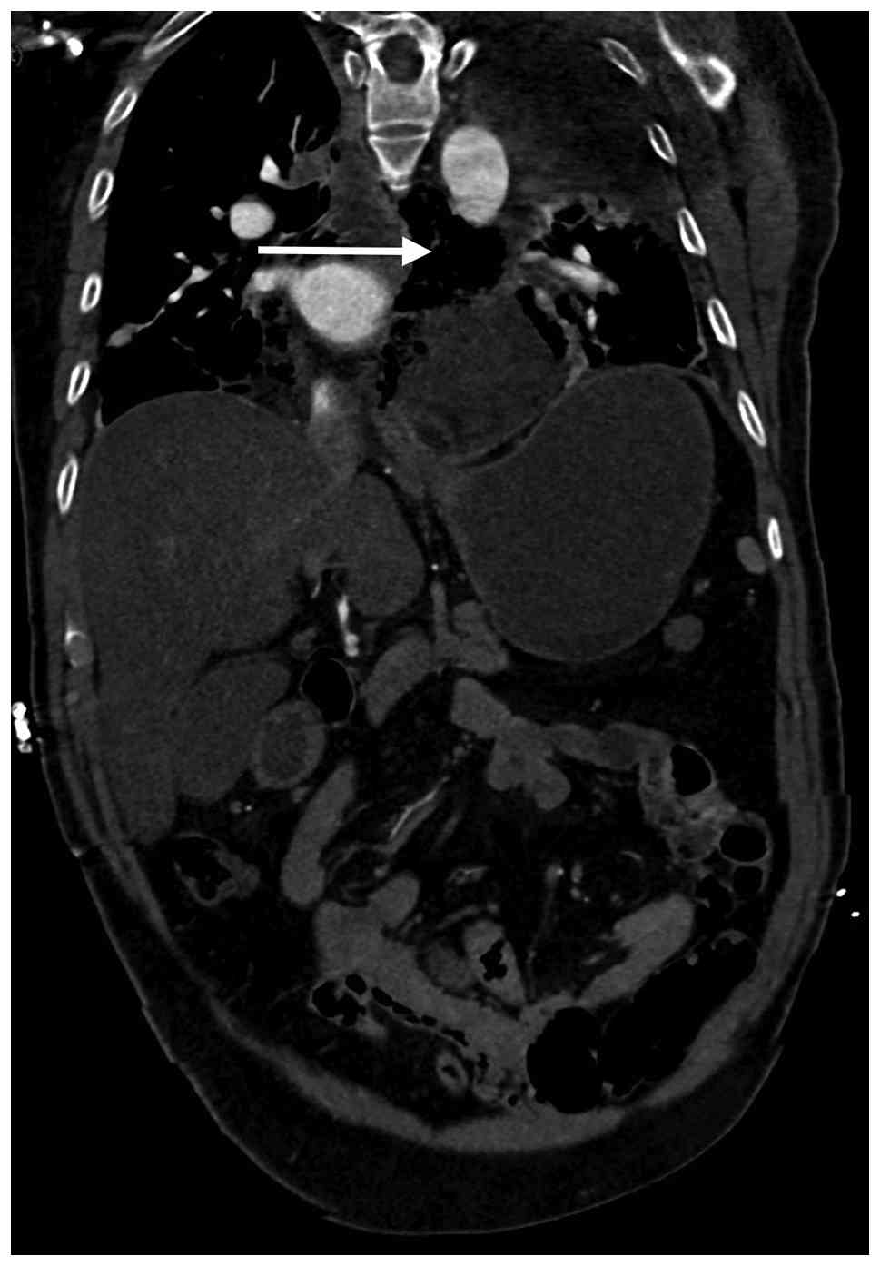
contrast-enhanced CT of the chest and abdomen, which demonstrated
high-density soft tissue surrounding the lower esophagus within the
posterior mediastinum, raising immediate suspicion for esophageal
rupture with mediastinal contamination (Fig. 1). Laboratory investigations on day
1 revealed severe infection and organ dysfunction: White blood cell
(WBC) count, 17.79x10⁹/l (normal range,
3.5-9.5x10⁹/l); C-reactive protein (CRP), 309.5 mg/l
(normal value, <8 mg/l); procalcitonin, 12.5 ng/ml (normal
value, <0.05 ng/ml); lactate, 4.8 mmol/l (normal range, 0.5-2.2
mmol/l); and albumin, 32.1 g/l (normal range, 40-55 g/l). The
patient required continuous intravenous norepinephrine (initiated
at 0.5 µg/kg/min and titrated up to 0.8 µg/kg/min) combined with
vasopressin (0.03 U/min, intravenous), with an initial Sequential
Organ Failure Assessment (SOFA) score of 11, which corresponds to a
predicted in-hospital mortality of ~50% and indicates severe
multi-organ dysfunction (8). Blood
cultures were obtained, with subsequent results on day 3
demonstrating growth of Streptococcus anginosus in two out
of two aerobic bottles, sensitive to penicillin, ceftriaxone,
vancomycin and meropenem, and so empiric broad-spectrum intravenous
antibiotic therapy with meropenem (1 g q8h) and linezolid (600 mg
q12h) was initiated. Total parenteral nutrition was also
commenced.
Although esophageal rupture was suspected early, the
profound hemodynamic instability of the patient precluded immediate
surgical or endoscopic intervention. From day 1 to 4, the patient
was managed conservatively in the ICU. By day 5 of illness, the
hemodynamic status had partially stabilized; the patient required
only low-dose vasopressor norepinephrine (0.05 µg/kg/min) and their
SOFA score improved to 6. Laboratory parameters showed slight
improvement but indicated an ongoing infection: WBC count,
14.20x10⁹/l; CRP, 185.2 mg/l; procalcitonin, 8.4 ng/ml;
lactate, 2.1 mmol/l; and albumin, 28.5 g/l. At this time, bedside
gastroscopy was performed to confirm the diagnosis and achieve
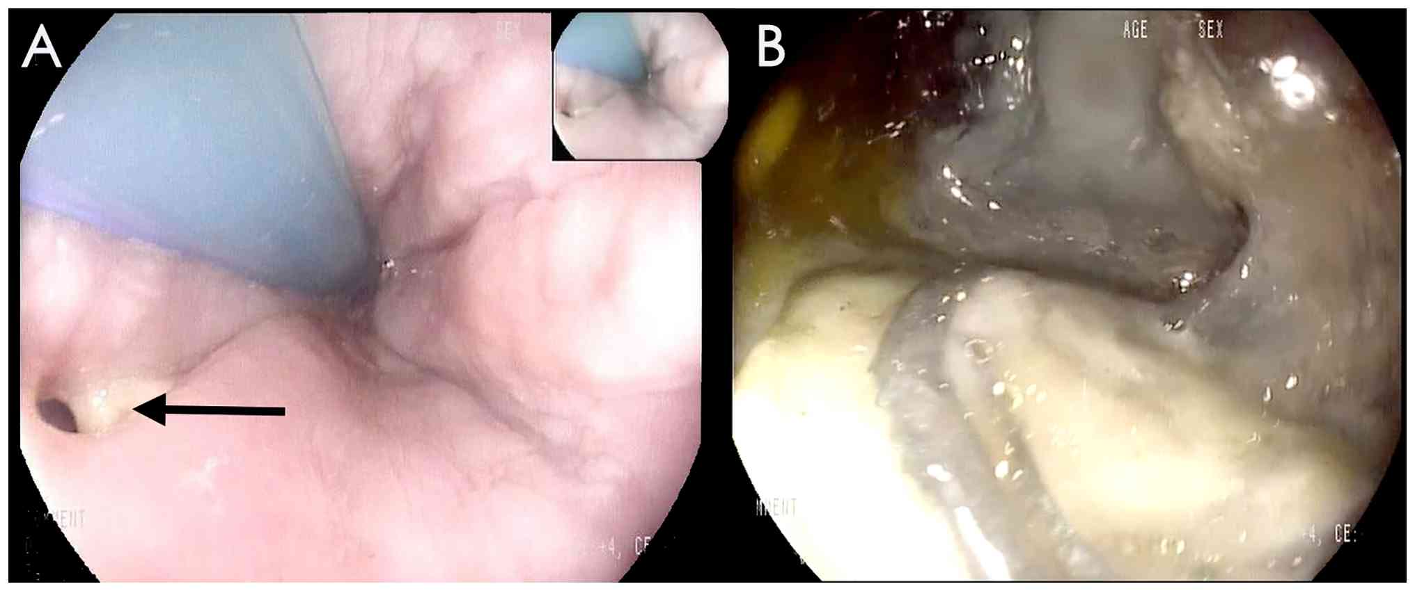
initial source control. Endoscopy revealed a large transmural
perforation in the lower esophagus. The endoscope was advanced
through the perforation into the mediastinal cavity, where
extensive necrotic tissue, exposed blood vessels and a pulsatile
pericardial structure were directly visualized (Fig. 2). Endoscopic debridement of
necrotic tissue and thorough irrigation were performed. A
transesophageal drainage tube was placed through the perforation
site to facilitate continuous drainage.
On day 6, blood and mediastinal fluid cultures
returned positive for Streptococcus anginosus and Candida
albicans. Consequently, intravenous fluconazole (400 mg daily)
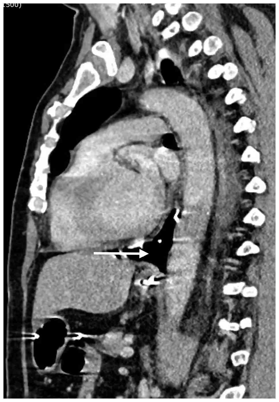
was added to the antimicrobial regimen. Follow-up CT imaging on day
7 demonstrated a residual mediastinal abscess with gas shadows
(Fig. 3). Given the massive size
of the defect and ongoing mediastinal infection, the patient was
referred for definitive surgical treatment.
On day 8 of illness (3 days post-endoscopy), the
patient was successfully weaned off vasopressors (SOFA score, 3),
with further decreasing inflammatory markers (WBC count,
11.50x10⁹/l; CRP, 110.4 mg/l; procalcitonin, 3.2 ng/ml;
lactate, 1.5 mmol/l; and albumin, 30.2 g/l). The patient then
underwent video-assisted thoracoscopic surgery (VATS). The
procedure included extensive debridement of the mediastinal
abscess, decortication and primary surgical repair of the
esophageal perforation, which was reinforced with a pleural flap.
Two thoracic drains were placed. The transesophageal drainage tube
placed during endoscopy was left in situ.
Postoperatively, the patient exhibited steady
clinical and laboratory improvement. By day 15 of illness
(postoperative day 7), the SOFA score had dropped to 1 and
inflammatory marker levels had normalized (WBC count,
8.10x10⁹/l; CRP, 45.6 mg/l; procalcitonin, 0.8 ng/ml;
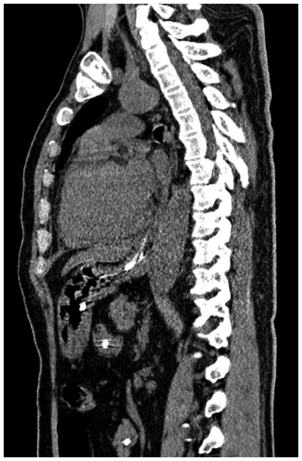
lactate, 1.1 mmol/l; and albumin, 35.4 g/). On day 22 of illness
(postoperative day 14), a contrast esophagogram confirmed complete
healing of the perforation with no evidence of leakage (Fig. 4). The transesophageal tube was
subsequently removed and enteral feeding via a nasojejunal tube was
initiated. Oral intake was successfully resumed on day 25.
The clinical and laboratory parameters of the
patient progressively normalized (Tables I and II) and they were discharged 1 month
after admission. At a 6-month follow-up, the patient reported
normal swallowing function with no stricture formation or
recurrence of infection.
 | Table ISerial laboratory and clinical
parameters during hospitalization of the patient. |
Table I
Serial laboratory and clinical
parameters during hospitalization of the patient.
| Parameter | Admission (day
1) | Pre-endoscopy (day
5) | Pre-VATS (day 8) | Post-VATS (day
15) | Discharge |
|---|
| WBC,
x109/l | 17.79 | 14.20 | 11.50 | 8.10 | 6.50 |
| CRP, mg/l | 309.5 | 185.2 | 110.4 | 45.6 | 8.2 |
| Procalcitonin,
ng/ml | 12.5 | 8.4 | 3.2 | 0.8 | <0.1 |
| Lactate, mmol/l | 4.8 | 2.1 | 1.5 | 1.1 | 0.9 |
| Albumin, g/l | 32.1 | 28.5 | 30.2 | 35.4 | 39.1 |
| Vasopressor use | Yes (high dose) | Yes (low dose) | No | No | No |
| SOFA score | 11 | 6 | 3 | 1 | 0 |
| Table IITimeline of clinical events and
interventions. |
Table II
Timeline of clinical events and
interventions.
| Day of illness | Clinical
event/intervention |
|---|
| Day 1 | Symptom onset
(vomiting and chest pain); admission to ED; initial CT scan;
progression to septic shock; ICU admission; empiric antibiotics
started. |
| Days 1-4 | Aggressive fluid
resuscitation and vasopressor support in ICU; TPN initiated. |
| Day 5 | Hemodynamic
stabilization; bedside gastroscopy performed (debridement,
irrigation, drainage tube placement). |
| Day 6 | Culture results
returned; antifungal therapy (fluconazole) added. |
| Day 7 | Follow-up CT showed
residual mediastinal abscess. |
| Day 8 | Definitive surgery:
VATS debridement and primary esophageal repair with pleural
flap. |
| Day 22 | Contrast esophagogram
confirmed no leakage; transesophageal drainage tube removed. |
| Days 22-24 | Enteral feeding via
nasojejunal tube. |
| Day 25 | Resumption of oral
intake. |
| Day 30 | Patient
discharged. |
Discussion
The present case illustrates several important
clinical aspects regarding the contemporary diagnosis and
management of Boerhaave syndrome. The CT finding of high-density
soft tissue surrounding the lower esophagus served as the critical
diagnostic evidence, underscoring the importance of a high index of
clinical suspicion (6).
The initial presentation with severe chest and back
pain following forceful vomiting, combined with rapidly progressive
septic shock, posed a challenging differential diagnosis. Aortic
dissection, acute pancreatitis and myocardial infarction were
appropriately considered and excluded through initial imaging
(2,5). This underscores the importance of
careful CT interpretation and the need to maintain a high index of
clinical suspicion for Boerhaave syndrome, as delayed diagnosis
notably exacerbates the risk of severe complications such as
mediastinitis and sepsis (6).
The present case demonstrates the unique diagnostic
and therapeutic utility of bedside gastroscopy in suspected
esophageal perforation. Historically, endoscopic evaluation was
avoided due to concerns about exacerbating the injury through
insufflation. However, accumulating evidence has suggested that
careful endoscopic assessment can be performed safely and provides
invaluable diagnostic information (8,9). In
the present case, bedside gastroscopy not only confirmed the
diagnosis but also enabled direct visualization of the mediastinal
cavity, debridement of necrotic tissue and placement of a drainage
tube, thereby serving as an effective therapeutic bridge to
definitive surgical repair. The endoscopic finding of exposed blood
vessels and a pulsating heart visible through the esophageal
perforation is particularly noteworthy and has only been described
in a small number of previous case reports (9). This finding serves as an illustration
of the anatomical proximity of the esophagus to vital mediastinal
structures and underscores the severity of the mediastinal
contamination present in this case.
The management of Boerhaave syndrome necessitates a
highly individualized, multidisciplinary approach (6,10).
The current World Society of Emergency Surgery clinical practice
guidelines recommend treatment strategies tailored to the
hemodynamic stability of the patient, the timing of presentation
and the extent of contamination (10). While early surgical intervention
(within 24 h) remains the gold standard for large perforations,
studies have highlighted the growing success of minimally invasive
surgical techniques, such as VATS, and advanced endoscopic
interventions (4,7). Notably, endoscopic vacuum therapy has
emerged as a highly effective, organ-sparing therapeutic option for
esophageal perforations, showing success rates of ≤89% in
multicenter cohorts (7,11).
In the present case, the combination of initial ICU
stabilization, endoscopic debridement and drainage as a bridging
intervention, followed by definitive VATS-assisted surgical repair,
exemplifies a modern, stepwise management strategy for Boerhaave
syndrome. Notably, mortality increases substantially when treatment
is delayed beyond 48 h from symptom onset (3,4).
Despite the delayed presentation in the present case (day 5 of
illness), the combined endoscopic and minimally invasive surgical
approach mitigated the adverse prognostic impact and ultimately
achieved a successful outcome.
The present case has certain limitations. First, it
was managed at a single institution and the generalizability of
this specific endoscopic bridging approach requires validation in
larger case series. Second, long-term follow-up data beyond the
initial recovery period were not available.
In conclusion, the present case highlights the
importance of maintaining a high index of suspicion for spontaneous
esophageal rupture in patients presenting with vomiting, chest pain
and sepsis. Bedside gastroscopy can serve as a valuable diagnostic
and therapeutic tool. A stepwise, combined endoscopic-surgical
approach, incorporating modern minimally invasive techniques, is
essential for achieving optimal outcomes, even in delayed
presentations with severe mediastinal contamination.
Acknowledgements
Not applicable.
Funding
Funding: No funding was received.
Availability of data and materials
The data generated in the present study are included
in the figures and/or tables of this article.
Authors' contributions
YZ collected the clinical data and drafted the
manuscript. YY performed the radiological image interpretation and
analysis. JC contributed to the analysis and interpretation of the
clinical and laboratory data, participated in the multidisciplinary
discussion regarding the timing and selection of endoscopic and
surgical interventions, performed the literature review and
critically revised the manuscript for important intellectual
content. NZ was responsible for clinical management,
conceptualization, manuscript supervision and final approval. YZ
and NZ confirm the authenticity of all the raw data. All authors
read and approved the final manuscript.
Ethics approval and consent to
participate
Not applicable.
Patient consent for publication
Written informed consent was obtained from the
patient for the publication of any associated data and accompanying
images.
Competing interests
The authors declare that they have no competing
interests.
References
|
1
|
Sdralis EIK, Petousis S, Rashid F, Lorenzi
B and Charalabopoulos A: Epidemiology, diagnosis, and management of
esophageal perforations: Systematic review. Dis Esophagus. 30:1–6.
2017.PubMed/NCBI View Article : Google Scholar
|
|
2
|
Tayyib M, Latif M, Sakr A and Mahmoud A:
Chest pain after vomiting: Recognizing boerhaave syndrome in the
emergency department. Cureus. 17(e97219)2025.PubMed/NCBI View Article : Google Scholar
|
|
3
|
Sherrin S, Kochhar JK, Mustafa W and
Batham K: Management of Boerhaave's syndrome in the intensive care
unit. Int J Crit Illn Inj Sci. 15:132–135. 2025.PubMed/NCBI View Article : Google Scholar
|
|
4
|
Aiolfi A, Micheletto G, Guerrazzi G,
Bonitta G, Campanelli G and Bona D: Minimally invasive surgical
management of Boerhaave's syndrome: A narrative literature review.
J Thorac Dis. 12:4411–4417. 2020.PubMed/NCBI View Article : Google Scholar
|
|
5
|
Predescu D, Achim F, Socea B, Rotariu A,
Moraru AC, Rasuceanu A, Constantin C, Rosianu CG and Constantin A:
Boerhaave syndrome-narrative review. Diagnostics (Basel).
15(2463)2025.PubMed/NCBI View Article : Google Scholar
|
|
6
|
Godinho M, Wiezel EH, Marchi E, Módena SF
and Paula RA: Spontaneous rupture of the esophagus: Boerhaave's
syndrome. Rev Col Bras Cir. 39:83–84. 2012.PubMed/NCBI View Article : Google Scholar
|
|
7
|
Luttikhold J, Pattynama LMD, Seewald S,
Groth S, Morell BK, Gutschow CA, Ida S, Nilsson M, Eshuis WJ and
Pouw RE: Endoscopic vacuum therapy for esophageal perforation: A
multicenter retrospective cohort study. Endoscopy. 55:859–864.
2023.PubMed/NCBI View Article : Google Scholar
|
|
8
|
Vincent JL, Moreno R, Takala J, Willatts
S, De Mendonça A, Bruining H, Reinhart CK, Suter PM and Thijs LG:
The SOFA (Sepsis-related Organ Failure Assessment) score to
describe organ dysfunction/failure. On behalf of the Working Group
on Sepsis-related problems of the European society of intensive
care medicine. Intensive Care Med. 22:707–710. 1996.PubMed/NCBI View Article : Google Scholar
|
|
9
|
Tellechea JI, Gonzalez JM, Miranda-García
P, Culetto A, Barbier-Pariente Z and Barthet M: Role of endoscopy
in the management of Boerhaave syndrome. Clin Endosc. 51:186–191.
2018.PubMed/NCBI View Article : Google Scholar
|
|
10
|
Chirica M, Kelly MD, Siboni S, Aiolfi A,
Riva CG, Asti E, Ferrari D, Leppäniemi A, Ten Broek RPG, Brichon
PY, et al: Esophageal emergencies: WSES clinical practice
guidelines. World J Emerg Surg. 14(26)2019.PubMed/NCBI View Article : Google Scholar
|
|
11
|
Chon SH, Scherdel J, Rieck I, Lorenz F,
Dratsch T, Kleinert R, Gebauer F, Fuchs HF, Goeser T and Bruns CJ:
A new hybrid stent using endoscopic vacuum therapy in treating
esophageal leaks: A prospective single-center experience of its
safety and feasibility with mid-term follow-up. Dis Esophagus.
35(doab067)2022.PubMed/NCBI View Article : Google Scholar
|