Introduction
Paragonimiasis, also known as lung fluke disease, is
a foodborne parasitic zoonosis caused by trematodes of the genus
Paragonimus (1,2). Infection in humans typically results
from the consumption of raw or undercooked freshwater crabs,
crayfish, shrimp or snails contaminated with metacercariae
(2,3). According to estimates from the World
Health Organization and multiple epidemiological studies, ~20
million people are infected worldwide (1,4). The
disease is mainly distributed in specific regions of Asia, Africa
and the Americas where raw or undercooked freshwater products are
consumed (1,4). In China, the prevalence of
paragonimiasis is 1.7%, and the general population has no innate
immunity, meaning that anyone exposed to the infectious
metacercariae is susceptible to infection (5). Pleural effusion may occur as the sole
or predominant manifestation of paragonimiasis, and the true
incidence is likely underestimated due to frequent misdiagnosis
(6,7).
While many patients present with cough, chest pain,
fever, and hemoptysis, a significant subset develops pleural
effusion (PE) when the parasites remain in the pleural cavity or
enter the pleura after failing to migrate to the lungs (6). The clinical and radiological
manifestations of paragonimiasis induced PE are nonspecific and
mimic those of tuberculous pleurisy, especially in
tuberculosis-endemic regions, leading to frequent misdiagnosis
(8-23);
patients may also be misdiagnosed with malignant PE (24,25).
The consequences of misdiagnosis include unnecessary
anti-tuberculosis therapy (which exposes patients to drug toxicity
without clinical benefit), disease progression (from isolated
effusion to parenchymal cavitation or extrapulmonary
dissemination), and prolonged impairment of quality of life.
Conventional diagnostic methods for paragonimiasis
have low sensitivity: sputum smear, culture, and even metagenomic
next-generation sequencing (mNGS) often fail to detect the
parasite, particularly in early-stage disease or when the infection
is limited to the pleural space (7,26).
Therefore, serological tests, including ELISA for
Paragonimus-specific IgG antibody and the IDTPA test for
Paragonimus-specific antigen, play a crucial role in diagnosis
(27-29).
The standard treatment for paragonimiasis is
praziquantel, administered at 25 mg/kg three times daily for 3
consecutive days. One to two courses are usually sufficient, but
more courses may be needed in severe or complicated cases (5,7,30).
An alternative agent is triclabendazole, which has shown comparable
efficacy and better tolerability in clinical trials (31,32).
Early diagnosis and prompt antiparasitic treatment are essential to
prevent chronic disease and irreversible organ damage.
The present study reports a patient with no definite
history of raw freshwater crab, shrimp or snail ingestion but with
longterm occupational exposure to freshwater aquatic products, who
was misdiagnosed with TB pleurisy for 8 months. The present study
also aimed to review the relevant literature to provide a reference
for the clinical diagnosis and management of paragonimiasis.
Case report
A 41yearold male patient living in a rural area
presented to the Department of Respiratory and Critical Care
Medicine at Gucheng County People's Hospital (Xiangyang, China) in
October 2024, with a 10 day history of cough, sputum production,
fever and shortness of breath. The patient reported no night
sweats, chills, chest pain or hemoptysis, had no known history of
TB, no contact with patients with TB and no family members with
similar respiratory symptoms. Physical examination revealed clear
percussion notes bilaterally, coarse breath sounds in the left lung
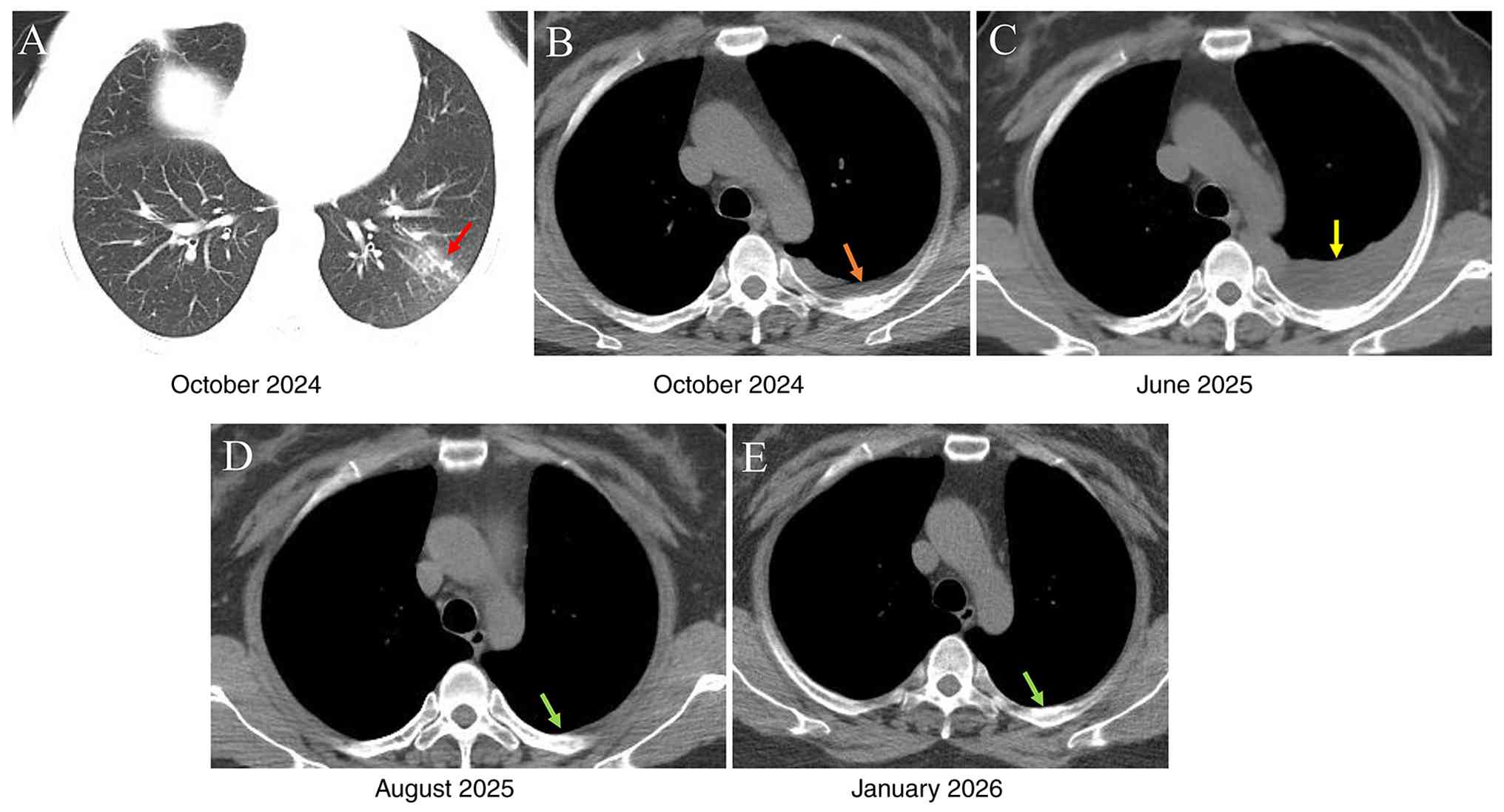
and a few fine crackles in the left lower lobe. Chest CT showed an
infectious lesion in the left lower lobe (Fig. 1A). Laboratory findings on admission
were as follows: Highsensitivity Creactive protein (hs-CRP), 1
µg/ml (normal range: 0-10 mg/l); CRP, <5.0 µg/ml (normal range:
0-10 µg/ml); procalcitonin, 0.05 ng/ml (normal range: 0-0.5 ng/ml);
erythrocyte sedimentation rate (ESR), 23 mm/h (normal range: 0-15
mm/h); negative respiratory virus antibody detection; positive TB
antibody (TBAb) detection; and normal carcinoembryonic antigen
(CEA) (normal range: 0-5 µg/l). Routine urinalysis was
unremarkable. Complete blood count showed the following results:
White blood cells, 12.5x10⁹/l (normal range:
3.5-9.5x10⁹/l); neutrophils, 50.3%; lymphocytes, 33.6%;
monocytes, 5.2%; eosinophils, 15.2% (absolute count,
1.9x10⁹/l) and basophils, 0.7%. Liver and kidney
function, electrolytes and lipids were normal. Based on these
findings, the patient was diagnosed with left lung infection and
treated with ceftezole (antibiotic), doxofylline (bronchodilator)
and nebulized inhalation.
At 9 days later, repeat chest CT revealed new
leftsided, small PE (Fig. 1B).
Ultrasoundguided thoracentesis was attempted but yielded little
fluid. Given the clinical course, imaging findings, laboratory
results (positive TBAb, elevated ESR) and poor response to 1 week
of antibiotics, the patient consented to diagnostic antiTB therapy
with isoniazid, rifampin, pyrazinamide and ethambutol. The patient
took these medications at home for 7 months, but his clinical
symptoms did not improve. The patient did not attend follow-up
during the 8 month anti-TB therapy.
At 8 months after the initial presentation, the
patient was admitted to the Department of Respiratory and Critical
Care Medicine at Taihe Hospital, Shiyan, China, in June 2025, with
persistent cough, sputum production, dyspnea on exertion and
intermittent fever. Physical examination was unremarkable except
for dullness to percussion and diminished breath sounds in the left
lower lobe, along with a few crackles in the same area. No
peripheral edema was noted. The patient had lost 5 kg since initial
presentation. Chest CT showed progression of the left lower lobe
lesion with increased left PE (Fig.
1C). Laboratory tests revealed the following results: ESR, 34
mm/h; hs-CRP, 15.7 mg/l; and peripheral blood eosinophil count,
2.61x10⁹/l (18.0%). Pleural fluid analysis showed
yellow, turbid fluid with a positive Rivalta test (indicating an
exudative effusion). The total nucleated cell count was
91,763x106/l, with 10% mononuclear and 90%
polymorphonuclear cells. Biochemical analysis revealed total
protein, 55.22 g/l; glucose, 2.07 mmol/l; total cholesterol, 3.44
mmol/l; lactate dehydrogenase, 1,111.8 U/l; adenosine deaminase,
38.7 U/l; and CEA, 1.17 µg/l. No bacteria, fungi or acidfast
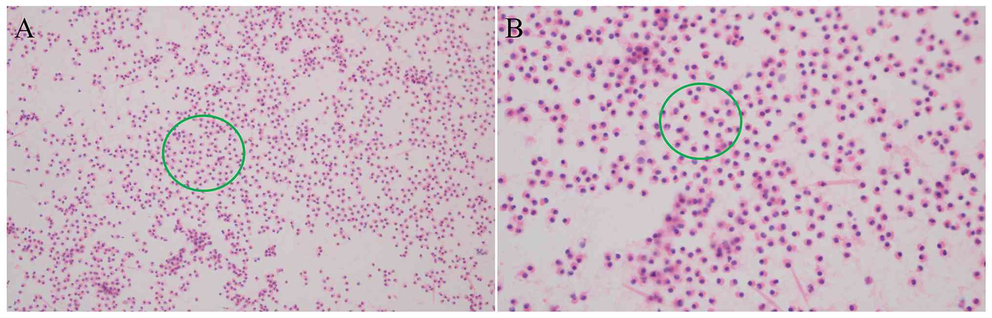
bacilli were detected on smear or culture after 7 days. Pleural
fluid cytology showed numerous eosinophils, with no malignant cells
(Fig. 2). Pleural fluid cytology
was performed using liquidbased cytology (LBC) with hematoxylin and
eosin staining. For specimen preparation, the pleural fluid was
fixed in 95% ethanol at room temperature for 10 min. For
hematoxylin staining, the slides were immersed in Harris
hematoxylin solution for 5 min at room temperature, then briefly
differentiated with 1-2 dips in 0.3% acid alcohol, followed by
bluing in tap water (or Scott's tap water substitute) for 5 min.
Counterstaining was performed using 1% eosin Y solution for 1-2 min
at room temperature. The slides were then dehydrated through graded
alcohols (70, 95, and 100%), cleared in xylene, and mounted with
coverslips. Finally, the stained slides were examined under a
standard light microscope (Olympus BX43) with brightfield
illumination at x200 and x400 magnifications, and representative
images were captured using a digital camera. Bronchoscopy was
normal and bronchoalveolar lavage fluid (BALF) culture and smear
were negative. Metagenomic nextgeneration sequencing (mNGS) of BALF
also failed to identify any pathogen; quality control parameters of
mNGS of BALF are shown in Table
SI. For mNGS of the BALF, nucleic acid extraction and library
preparation were performed using the PMseq® workflow.
For the DNA workflow, the MGIEasy Fast FS Library Prep Set V2.0
(MGI Tech Co., Ltd., cat. No. 1000006987 for 16 reactions, and Cat.
No. 1000006988 for 96 reactions) was used, while the RNA workflow
employed the MGIEasy Microorganism Fast RNA Library Prep Set (MGI
Tech Co., Ltd.; Cat. No. 940-000107-00 for 16 reactions, and Cat.
No. 940-000108-00 for 96 reactions). The library concentration was
measured using a Qubit™ 4.0 fluorometer (Thermo Fisher Scientific,
MA, USA) with the Qubit dsDNA HS Assay Kit (Cat. No. Q32854), and
the acceptable concentration after purification was set at ≥5
ng/µl. The size distribution of the libraries was assessed using an
Agilent 2100 Bioanalyzer (Agilent Technologies, CA, USA) with the
High Sensitivity DNA Kit (Cat. No. 5067-4626). The quality
acceptance criteria were defined as a DNA Integrity Number (DIN) ≥6
for DNA samples, an RNA Integrity Number (RIN) ≥6 for RNA samples,
and a minimum concentration of ≥1 ng/µl for both. Sequencing was
performed on the DNBSEQ G99 platform (MGI Tech Co., Ltd.) using a
single-end 50-bp (SE50) read length. For the RNA workflow, the
DNBSEQ G99S High-throughput Sequencing Set (G99 SM FCL SE100/PE50;
Cat. No. 940-000409-00; MGI Tech Co., Ltd.) was used. For the DNA
workflow, the MGISEQ 200RS High-throughput (Rapid) Sequencing
Reagent kit (cat. no. 1000019846; MGI Tech Co., Ltd.) was used. The
loading concentration of the final pooled library was determined by
Qubit 4.0 fluorometer. The actual loading concentration was 1.8 pM,
as measured by the Qubit 4.0 fluorometer and further verified with
the KAPA Library Quantification Kit. Primary base-calling analysis
was performed using the built-in sequencing instrument software
provided by MGI Tech Co., Ltd. Subsequent bioinformatics analysis
was conducted using the HALOS PMseq Gene Data Analysis Workstation
(Version V3.0, Shenzhen Huada Gene Technology Co., Ltd., China),
which aligned the sequencing reads against the PMDB clinical-grade
microbial database and the NCBI NT database (v2024.03; ftp.ncbi.nlm.nih.gov/blast/db/)
(33). The analytical workflow
comprised quality filtering, removal of host reads by alignment to
the human reference genome (hg38), and taxonomic classification
using a k-mer-based algorithm with a minimum confidence threshold
of 50 RPhK (normalized reads per 100,000 total sequencing reads)
for parasite detection. Quality assessment of the raw data was
performed using FastQC v0.11.9 (bioinformatics.babraham.ac.uk/projects/fastqc/;
), and multi-QC reports were automatically generated within the
HALOS system.
As the cause of PE and systemic symptoms remained
undiagnosed despite comprehensive investigations, medical
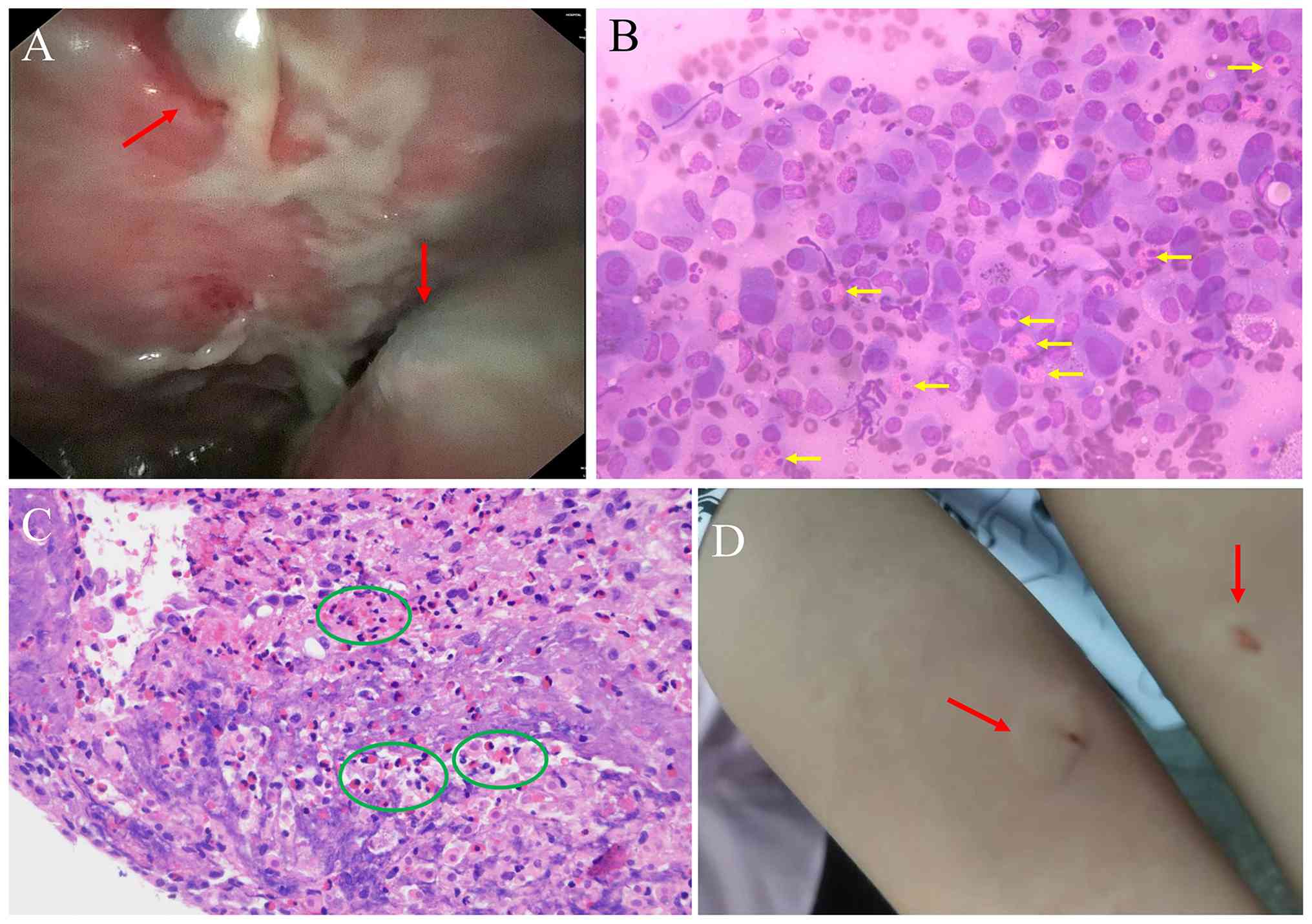
thoracoscopy was performed, revealing ~1,000 ml yellow pleural
fluid. The parietal and diaphragmatic pleura were congested and
thickened, with white patchy material adherent (Fig. 3A). Multiple biopsies were taken
from the parietal pleura. Rapid onsite evaluation showed numerous
mesothelial cells and eosinophils (Fig. 3B). Histopathological examination of
the pleural biopsy specimens revealed fibroadipose tissue with
areas of exudation and necrosis, mesothelial cell hyperplasia,
histiocytic proliferation and dense infiltration of lymphocytes,
eosinophils and neutrophils (Fig.
3C). Stains, including periodic acid-Schiff (PAS), acid-fast
and Gomori methenamine silver, were negative for specific
microorganisms. Immunohistochemistry was positive for CK(P)
(cytokeratin, pan-specific; a marker of mesothelial cells), CD68
(histiocytes) and WT1/calretinin (Wilms tumor 1, markers of
mesothelial cells) and negative for SOX10, S100, TTF1 (Thyroid
Transcription Factor-1; a marker of lung adenocarcinoma and thyroid
origin), napsin A, langerin, CD1a and P40. Special stains including
periodic acid-Schiff, acidfast and silver stain were negative. PCR
for Mycobacterium tuberculosis was negative. The pleural
biopsy specimens were fixed in 10% neutral buffered formalin at
room temperature for 24-48 h. Tissues were dehydrated through
graded ethanol solutions (70, 80, 95, and 100%, each for 1 hour),
cleared in xylene (two changes, each for 1 h), and embedded in
paraffin. The resulting paraffin blocks were sectioned at a
thickness of 4 µm. Sections were first deparaffinized in xylene
(two changes, 5 min each) and rehydrated through graded ethanol
solutions (100, 95, 80, and 70%, 3 min each). The sections were
stained with Harris hematoxylin for 5 min at room temperature,
rinsed in tap water for 5 min, differentiated with 1 to 2 dips in
0.3% acid alcohol, and blued in Scott's tap water for 2 min.
Counterstaining was performed with 1% eosin Y solution for 2 min at
room temperature. Finally, the sections were dehydrated, cleared,
and mounted with coverslips using synthetic resin. The stained
sections were examined under a standard light microscope.
Immunohistochemical staining was performed on
formalin-fixed, paraffin-embedded sections at 4 µm thickness using
a polymer peroxidase-based detection system (EnVision FLEX, Dako).
Following deparaffinization and rehydration as aforementioned,
heat-induced antigen retrieval was carried out using Target
Retrieval Solution (at pH 6.0 or 9.0, depending on the primary
antibody) in a pressure cooker at 120˚C for 3 min. Endogenous
peroxidase activity was blocked with 3% hydrogen peroxide for 10
min. The sections were then incubated with primary antibodies at
4˚C overnight (or at room temperature for 1 h). The panel of
primary antibodies used included: CK(P) (clone AE1/AE3; Dako;
1:200), CD68 (clone KP1; Dako; 1:100), WT1 (clone 6F-H2; Dako;
1:50), calretinin (clone DAK-Calret1; Dako; 1:100), SOX10 (clone
EP268; Zytomed; 1:100), S100 (polyclonal; Dako; 1:200), TTF-1
(clone 8G7G3/1; Dako; 1:100), napsin A (clone MRQ-60; Roche;
ready-to-use), langerin (clone 12D6; Novocastra; 1:50), CD1a (clone
O10; Dako; 1:50), and P40 (clone BC28; Zytomed; 1:100). After
washing, the sections were incubated with a horseradish
peroxidase-labeled polymer (EnVision FLEX, Dako) for 30 min at room
temperature. Diaminobenzidine (DAB) was used as the chromogen for 5
min, followed by counterstaining with Harris hematoxylin. The
sections were then dehydrated, cleared, and mounted. Positive and
negative controls (by omission of the primary antibody) were run in
parallel for each staining run. All sections were evaluated under
bright-field light microscopy (Olympus BX43).
DNA was extracted from the pleural biopsy tissue
using the QIAamp DNA Mini Kit (Qiagen GmbH; cat. No. 51304)
strictly according to the manufacturer's protocol. Real-time PCR
was performed using the TB PCR Kit (Da An Gene Co., Ltd.,
Guangzhou, China; cat. No. DA-TB-001). Amplification was carried
out with an initial denaturation step at 95˚C for 5 min, followed
by 40 cycles of denaturation at 95˚C for 15 sec and
annealing/extension at 60˚C for 1 min. A FAM-labeled probe was used
for specific detection. A positive control (genomic DNA from M.
tuberculosis H37Rv) and a negative control (nuclease-free water)
were included in each run. According to the manufacturer's
instructions, Cq >38 was interpreted as negative. The
amplification and detection were performed on the Applied
Biosystems 7500 Real-Time PCR System (Thermo Fisher
Scientific).
Given the marked eosinophilia in peripheral blood
and pleural fluid and tissue, the patient was requestioned about
his exposure history. The patient reported longterm occupational
contact with freshwater aquatic products as a fish farmer, with no
history of raw seafood ingestion. On day 6 of admission, he
underwent intradermal test for Paragonimus-specific antigen
(IDTPA) at the Department of Parasitology, Basic Medicine School,
Hubei University of Medicine (Shiyan, China). The test was positive
within 15 min (Fig. 3D). In
accordance with standard clinical practice for IDTPA (7), the result was read at 15 min
(immediate reaction); delayed readings at 24-48 h were not
performed. Serum Paragonimus-specific IgG antibody was
detected using a Paragonimus IgG ELISA kit (Eiger Diagnostics,
Inc.; cat. number: EG-Par-G) according to the manufacturer's
instructions, with a positive result defined as optical density
>0.23. A positive result was obtained. This ELISA kit uses
Paragonimus antigen-coated plates and anti-human IgG
conjugate; it has been reported to show no or limited
cross-reactivity with sera from patients with clonorchiasis or
schistosomiasis (27). CT of the
abdomen, and pelvis were normal, and brain magnetic resonance
imaging were normal. A diagnosis of PE caused by paragonimiasis was
made.
Because the patient had not been receiving antiTB
medication for >2 weeks, empirical antiparasitic therapy with
praziquantel was initiated at a dose of 25 mg/kg three times daily
for 3 consecutive days. The course was repeated after a 1week
interval. Given the prolonged diagnostic delay (8 months) and the
presence of massive PE with marked peripheral eosinophilia (18.0%),
a more extended regimen was adopted to ensure complete parasite
eradication. The first course was administered during
hospitalization and the subsequent three courses were completed in
the outpatient setting. Following the first course, the patient
reported partial improvement in cough and dyspnea, but peripheral
blood eosinophils remained elevated (9.2%, absolute count
1.35x10⁹/l). After the second course, eosinophils
decreased to 5.1% (0.71x10⁹/l), and chest CT showed
reduction but not complete resolution of PE. Therefore, two
additional courses were administered. After four courses, the
patient achieved clinical cure with no adverse effects. At 1 month
later (August 2025), chest CT showed normal lungs (Fig. 1D), and peripheral blood eosinophil
count decreased to 0.46x10⁹/l (6.2%). The patient
remained well at followup in January 2026, with a normal chest CT
and no PE (Fig. 1E), however
follow-up serological testing (ELISA or IDTPA) was not performed
because the patient was asymptomatic and declined further blood
tests. The patient did not attend scheduled follow-ups after that
time.
Literature review
A comprehensive search of the PubMed (pubmed.ncbi.nlm.nih.gov/), Google Scholar
(scholar.nq69.top/) and Web of Science databases
(ycfw.hbmu.edu.cn:9000/https/28jS5Bg4AKhLUpVG9oah1DPUUo1YgpmgrnPgYfOr2yqbl5IiX/wos/?Init=Yes&SrcApp=CR&SID=USW2EC0F3EzPK3iv6njUeKoAZ4ePS)
was conducted to identify relevant studies published from January
1, 2000 to January 1, 2026, using the following terms:
(paragonimiasis OR lung fluke disease OR Paragonimus) AND
(pleural effusion OR pleurisy OR pleuropulmonary) AND (misdiagnosis
OR misdiagnosed OR diagnostic delay OR confusion) AND (tuberculosis
OR tuberculous pleurisy OR tuberculous pleural effusion).
Additionally, references from retrieved articles meeting the
inclusion criteria [studies written in English; original case
reports or case series or article (not reviews, systematic reviews,
or meta-analyses); sufficient clinical or therapeutic details to
allow data extraction; confirmed microbiological diagnosis of
Paragonimus infection (including serology, IDTPA test, egg
detection, mNGS, or histopathology); and pleural effusion as the
primary or a major manifestation (i.e., not pericardial effusion as
the primary presentation)] were manually searched. Exclusion
criteria included studies not written in English, reviews without
original case data, systematic reviews or metaanalyses, reports
with insufficient clinical or therapeutic detail or cases with
pericardial effusion as the primary manifestation. The extracted
data included geographic region, age, sex, epidemiological history,
diagnostic modalities, radiological findings, treatment details and
clinical outcome.
In total, 53 published cases of pleuropulmonary
paragonimiasis misdiagnosed as TB pleurisy or TPE were identified
across 16 Englishlanguage publications (including 11 case reports
and five case series; Table I).
After excluding two cases with incomplete data, 51 unique published
cases met the criteria for analysis. These cases were reported from
2001 to 2026 (8-23),
with the following geographic distribution: China (n=14), Nepal
(n=17), India (n=9), South Korea (n=5), Japan (n=4), Cambodia
(n=1), Ecuador (n=1) and Peru (n=1). The patients included 32 males
and 19 females. Age ranged from 6 to 68 years, with eight cases
lacking age data. A total of 47 patients (92.2%) had a confirmed
history of consuming raw or undercooked freshwater crabs, crayfish
or other aquatic products (8-23).
Among the 14 Chinese cases, the majority originated from
paragonimiasisendemic provinces, including Yunnan, Sichuan,
Chongqing, Zhejiang and Fujian (5,34).
 | Table IClinical data of previously reported
cases of paragonimiasis with pleural effusion misdiagnosed as
tuberculous pleurisy/pleural effusion (n=51). |
Table I
Clinical data of previously reported
cases of paragonimiasis with pleural effusion misdiagnosed as
tuberculous pleurisy/pleural effusion (n=51).
| First author/s,
year | Country | No. of cases | Age, years | Sex | Raw consumption
history | Clinical
manifestations | Peripheral blood
eosinophilia | Pleural fluid
eosinophilia | Imaging
findings | Diagnostic
methods | Treatment | Outcome | (Refs.) |
|---|
| Li et al,
2026 | China | 1 | 18 | F | Yes | Cough, sputum,
hemoptysis for 4 years | Yes | NR | Pulmonary
lesions | mNGS (+87 reads),
ELISA (+), bronchial biopsy (-), GeneXpert (-), culture (-) | Praziquantel (1.2 g
tid x 3 d) | Hemoptysis resolved
in 15 d; CT lesion notably reduced at 2 months | (8) |
| Yang et al,
2024 | China | 1 | NR | NR | NR | Isolated pleural
effusion | NR | NR | Pleural
effusion | Immunology,
serology | Praziquantel | Cured | (9) |
| Villanueva-Villegas
et al, 2023 | Peru | 1 | 29 | M | Yes | Cough, chest pain,
fever, hemoptysis for 3 years | NR | NR | Pulmonary
lesions | Sputum smear
positive for eggs, TB test (-) |
Triclabendazole | Clinical and
radiological improvement | (10) |
| Poudyal et
al, 2022 | Nepal | 20 | Median, 34 | NR | 100% (20/20) | Fever 80%, cough
90%, pleural effusion 100% | Mean absolute count
16,678/ml | 100% (>10%) | GGO 65% | Sputum microscopy
(5 cases), pleural fluid analysis | Praziquantel (19
effective) | 95% effective | (22) |
| Calvopina et
al, 2017 | Ecuador | 1 | 30 | M | NR | Cough, rusty
sputum, chest pain, dyspnea for 5 years | NR | NR | Multiple small
bilateral lung nodules | Sputum smear
positive for eggs (P. mexicanus by molecular ID), AFB
(-) | Praziquantel | Cough and sputum
resolved | (11) |
| Luo et al,
2016 | China | 1 | 43 | M | NR | Cough, bloody
sputum, chest pain | NR | NR | Patchy
consolidation, pleural effusion | ELISA (+),
thoracoscopy showing Charcot-Leyden crystals | NR | Cured | (12) |
| Sah et al,
2017 | Nepal | 3 | NR | NR | 100% | Cough, hemoptysis,
dyspnea, chest pain, fever | Yes | NR | TB-like | Sputum smear
positive for eggs, TB test (-) | Praziquantel | Improved | (17) |
| Singh et al,
2005 | India | 3 | NR | NR | 100% | TB-like | Markedly
elevated | Markedly
elevated | TB-like | Sputum smear
positive for eggs | Praziquantel | Improved | (23) |
| Prasad et
al, 2015 | India | 1 | NR | NR | Yes | TB-like | NR | NR | NR | Failed anti-TB
treatment | Antiparasitic | Improved | (13) |
| Lee et al,
2014 | China | 1 | 53 | F | NR | Cough,
foul-smelling sputum, bloody sputum | NR | NR | Subpleural cavitary
consolidation, right upper lobe | VATS wedge
resection pathology (+), bronchoscopy (-), cytology (-) | Anti-TB then
recurrence, pathological confirmation | Cured | (15) |
| Song et al,
2014 | South Korea | 1 | 38 | M | NR | Low-grade fever,
pleuritic chest pain | NR | NR | Right pleural
effusion, pleural thickening, subpleural consolidation | VATS pathology (+),
ADA 85.3 U/l, TB test (-) | Anti-TB for 2
months ineffective | Cured | (14) |
| Nagakura et
al, 2002 | Japan | 1 | NR | NR | Yes | X-ray mis-diagnosed
as TB | Yes | NR | X-ray | Sputum smear: eggs,
abundant eosinophils, Charcot-Leyden crystals | NR | NR | (16) |
| Sharma et
al, 2024 | Cambodia | 1 | 62 | M | Yes | Cough, dyspnea,
recurrent hemoptysis | Yes | NR | Pleural thickening,
subpleural nodules | ELISA (+) | Praziquantel | Improved | (19) |
| Yin et al,
2025 | China | 2 | 40 / 60 | F | Case 1, NR; case 2,
yes | Case 1: Cough,
dyspnea for 1 week; Case 2: Cough, hemoptysis, chest pain, wheezing
>1 month | NR | NR | Case 1: Right
pleural effusion, lung lesion; Case 2: Cavitary lung lesion,
pleural involvement | Case 1: ELISA (+).
Case 2: ELISA (+), elevated serum IgG4 | Antiparasitic (2
months for case 1, 1 week for case 2) | Case 1, improved;
Case 2 improved, IgG normalized | (20) |
| Das et al,
2024 | India | 3 | Children | NR | NR | TB-like | NR | NR | TB-like | NR | NR | NR | (21) |
| Hwang et al,
2015 | South Korea | 10 | NR | NR | NR | Pleural fluid
analysis | NR | Eosinophilic
pleural effusion | NR | Pleural fluid
analysis, ELISA | Praziquantel | Improved | (18) |
The most common clinical manifestations included
cough (88.2%) (8,10,12-15,17-23),
chest pain (60.8%) (10,12,14,15,17,19,22,23),
fever (54.9%) (8,10-12,14,15,17,20,22,23),
hemoptysis (47.1%) (8,10,12,14-16,20,22),
expectoration (43.1%) (8,12,14,15,19,22)
and dyspnea (31.4%) (10,14,15,17-19,22).
Radiologically, PE was the predominant finding (100%) (8-23),
followed by pulmonary consolidation/nodules (64.7%) (8-10,12-15,17-20,22,23)
and pneumothorax (7.8%) (14,22).
Peripheral blood eosinophilia (eosinophils >5% or absolute count
>0.5x10⁹/l) was present in 48/51 patients (94.1%)
(8-10,12-16,18-23).
Pleural fluid analysis, available for 36 patients, revealed
exudative effusion in all cases (100%) (8-10,12-15,17-20,22,23),
with eosinophilic PE (EPE; eosinophils >10%) documented in 30/36
patients (83.3%) (8,12-15,17,18,20,22,23).
The most common diagnostic method was serological testing (ELISA
for Paragonimus-specific IgG antibody), which was positive
in 45/48 tested patients (93.8%) (8-12,14-20,22,23).
Direct visualization of Paragonimus eggs in sputum was
achieved in 14 patients (27.5%) (8,10,12,14-16,22,23).
Thoracoscopy with pleural biopsy was performed in 12 patients,
revealing eosinophilic infiltration or parasite eggs in pleural
tissue in 10 cases (83.3%) (12,14,15,18,20,22,23).
mNGS of bronchoalveolar lavage fluid was employed in two cases,
both with positive detection of Paragonimus sequences
(8,23).
All patients received antiparasitic therapy, with
praziquantel (standard regimen of 25 mg/kg three times daily for 3
consecutive days or equivalent dosing) as the primary treatment
(8-23).
Anti-TB treatment was discontinued following confirmation of
paragonimiasis. Among the 51 patients, followup duration ranged
from 1 to 24 months. Outcome analysis revealed clinical improvement
or cure in all 51 patients (100%) (8-23),
with resolution or marked decrease of PE and normalization of
peripheral blood eosinophil counts. No deaths were reported.
Discussion
A systematic review and metaanalysis covering the
period from 1954 to 2024 showed that in China, paragonimiasis is
primarily caused by Paragonimus westermani and
Paragonimus skrjabini, with cases predominantly distributed
in provinces along the Yangtze River basin: Chongqing (20.85%),
Zhejiang (18.39%), Hubei (17.08%), Sichuan (10.00%), and Hunan
(4.88%), accounting for 71.21% of the national total (4,7,34).
The present patient came from a rural mountainous area of Hubei
Province, which is consistent with an endemic region for
paragonimiasis. paragonimiasis affects all age groups, and the mean
age of patients with paragonimiasis induced PE is 46.5±5.1 years,
as reported by Wang et al (7). The clinical manifestations of
paragonimiasis are predominantly respiratory, including cough,
sputum production, chest pain and dyspnea, often accompanied by
nonspecific symptoms such as lowgrade fever, night sweats, weight
loss and fatigue (7,35). The present patient presented with
cough, sputum and fever. Chest radiography or CT findings in
paragonimiasis may include PE, hydropneumothorax, pulmonary
nodules, cavities, and cystic opacities (15,30);
among these, cavitary lesions are considered characteristic. In the
present case, however, imaging only revealed PE without specific
features.
Although the present patient had no history of
ingesting raw freshwater crabs, shrimp or snails, he had longterm
occupational exposure to freshwater crabs and crayfish. The present
clinical course was characterized by cough, sputum, intermittent
fever, poor response to initial antibiotics and empirical antiTB
therapy. Peripheral blood eosinophil counts were markedly elevated
on both admissions (1.9 and 2.61x10⁹/l, respectively)
and abundant eosinophils were observed in pleural fluid and biopsy
tissue. IDTPA and serum Paragonimus IgG antibody were
positive, and the patient responded well to praziquantel.
Therefore, the diagnosis of paragonimiasisinduced PE was
established.
Because PE as the sole imaging feature of
paragonimiasis is nonspecific, misdiagnosis and diagnostic delay
are common. A study by Ahn et al (36) analyzing 685 patients with
pleuropulmonary paragonimiasis over 22 years found that 119
patients experienced diagnostic delays ≥25 weeks, mainly due to
misdiagnosis as TB, malignancy or chronic obstructive pulmonary
disease, with an odds ratio of 111.75 (95% CI: 43.25-288.74),
indicating high risk of misdiagnosis. Paragonimiasis is often
misdiagnosed as TPE (8-23),
parapneumonic PE (7) or malignant
PE (24,25). The present patient was misdiagnosed
with TPE for 8 months. Misdiagnosis of paragonimiasis as
tuberculous pleurisy carries clinical and economic consequences. In
the 51 reviewed cases, the average duration of unnecessary anti-TB
therapy before correct diagnosis was 4.2 months (range, 1-11
months), exposing patients to drug toxicity and financial burden
without clinical benefit. Diagnostic delay also permits disease
progression (from isolated effusion to parenchymal cavitation or
extrapulmonary dissemination) and prolongs symptom-associated
impairment of quality of life. Moreover, misclassification as TB
may trigger unnecessary contact tracing, diverting public health
resources from TB control efforts.
Paragonimiasis infection leads to increased absolute
or relative eosinophil counts in peripheral blood or pleural biopsy
tissue (3). However, eosinophilia
is also common in allergic disease, parasitic infections,
eosinophilic gastritis, hypereosinophilic syndrome and fungal
infections, but is uncommon in TB (13). In addition, patients with TB
pleurisy typically have a history of TB exposure and present with
toxic symptoms such as prolonged lowgrade fever, fatigue and night
sweats, and the tuberculin skin test (TST) is usually positive
(8). Definitive diagnosis of
paragonimiasis relies on the identification of parasite eggs,
however, the sensitivity of detecting eggs in sputum, feces or
pleural fluid is low (36). Thus,
direct pathogen detection is difficult, and serological methods
based on immunological tests are the most effective laboratory
diagnostic approach (9). The
primary immunological tests include IDTPA test, double agar gel
diffusion test and immunoenzymatic assays (10,11).
The IDTPA test has a positive rate of up to 95% (12,13).
It has been reported that serum Paragonimus antibody ELISA
has a sensitivity of 90.2% and specificity of 100.0% (26). However, ELISA is relatively complex
and has a long turnaround time, whereas IDTPA is more convenient
and rapid, yielding results within 15 min. Du et al
(29) demonstrated that the IDTPA
has good sensitivity in diagnosing cerebral paragonimiasis in 12
children. Xia et al (28)
and Paranjape et al (29)
also indicated that laboratory evaluation of cerebral
paragonimiasis includes eosinophil count, IDTPA and ELISA. The
present case showed a strong positive IDTPA reaction. These
findings collectively underscore the importance of IDTPA in the
diagnosis of paragonimiasis.
In recent years, molecular techniques such as mNGS
and multiplex PCRbased targeted NGS (tNGS) have been widely used to
directly detect target organisms in respiratory samples, with a
detectable pathogen spectrum covering >95% of clinical
infectious cases (37-39).
To the best of our knowledge, however, reports on the use of these
molecular techniques for diagnosing paragonimiasis are rare. Li
et al (8) reported a case
of paragonimiasis confirmed by mNGS and serological testing; thus,
mNGS and tNGS serve as auxiliary diagnostic methods for
paragonimiasis. Nevertheless, their clinical application has
limitations, including high cost and long turnaround time (40). The positivity rate of NGS for
detecting pathogens such as Paragonimus is low (41). Although mNGS theoretically detects
all microorganisms, its sensitivity for parasite DNA in practice is
generally poor. This is because, in the early stage of infection or
under low parasite burden, the nucleic acid content is low and can
be easily overwhelmed by host cell DNA or other microorganisms
(42). Clinical studies (41,43,44)have shown that the detection rate of
mNGS for thickwalled microorganisms (including certain parasites)
is notably lower than that for bacteria, viruses and fungi. For
example, in a study of 45 patients with pulmonary infection, the
detection rates of mNGS for Mycobacterium tuberculosis and
fungi were 70.6 and 100.0%, respectively, while the detection rate
for parasites (such as Paragonimus) was typically <20.0%
(41). Taken together, mNGS and
tNGS are not allpowerful in the field of parasite detection
(31,32,45).
Their limitations stem from technical shortcomings (host background
interference, lack of reference databases) and the biological
characteristics of parasites (low abundance, difficulty in nucleic
acid extraction). In the present case, several factors limited the
sensitivity of mNGS for detecting Paragonimus. First, no
host DNA depletion was performed and the human cell concentration
was 4x10⁶ cells/ml, resulting in a high human background
(>95% of total reads). Second, the patient presented with
isolated eosinophilic PE without parenchymal lung lesions,
indicating a very low parasite burden in the bronchoalveolar space.
In the present literature review of 51 cases of paragonimiasis with
PE, two (3.9%) had positive mNGS results from BALF. Third, the
proportion of microbial reads was low (14,674 out of 29.7 million
reads), illustrating the difficulty of detecting a low-abundance
parasite without targeted enrichment. Therefore, a negative mNGS
result does not rule out paragonimiasis. The present diagnosis was
established on the basis of peripheral blood eosinophilia (18.0%,
absolute count, 2.61x10⁹/l), EPE and pleural tissue
infiltration, positive IDTPA and Paragonimus IgG ELISA and
the excellent response to praziquantel. In clinical diagnosis,
sequencing results should be considered as an important reference,
but traditional methods such as serology and microscopy should be
used in combination to improve diagnostic accuracy.
Although misdiagnosis of paragonimiasis as TB
pleurisy is well documented (8,9,22,36),
the present case possessed distinctive features. Unlike most
previously reported cases in which patients had a definite history
of ingesting raw or undercooked freshwater crabs or crayfish (92.2%
of 51 reviewed cases) (8-23),
the present patient had no dietary exposure but instead had
long-term occupational contact with freshwater aquatic products as
a fish farmer. The patient specifically reported wading in fish
ponds, cleaning them and handling bycatch (freshwater crabs and
crayfish) without gloves, sustaining minor hand injuries. This
observation expands the spectrum of at-risk populations and
emphasizes the importance of inquiring about occupational and
environmental exposures in endemic regions. The diagnosis was
rapidly established using the IDTPA, which yielded a positive
result within 15 min; this bedside tool is particularly valuable in
resource-limited settings where ELISA may be unavailable or
delayed. Notably, mNGS of BALF failed to detect any pathogen,
despite the high parasite burden suggested by extreme eosinophilia.
Finally, despite an 8-month diagnostic delay and ineffective
anti-TB treatment, the patient achieved complete clinical cure
following four courses of praziquantel without any adverse effects,
reinforcing the efficacy and safety profile of praziquantel, as
consistently demonstrated in the reviewed literature (8-23).
Apart from thoracentesis for PE drainage, the firstline treatment
for paragonimiasis induced PE is oral praziquantel (2,5,6).
Case series and reports have demonstrated the effectiveness of
praziquantel in achieving clinical cure, including cases
complicated by PE (7,12,14,15,18,30,46).
In China, praziquantel remains the standard antiparasitic therapy
for paragonimiasis (5,30). In addition, triclabendazole is an
alternative treatment option for paragonimiasis (47). Calvopiña et al (32) conducted a randomized clinical trial
involving 62 patients with paragonimiasis and found that
triclabendazole had comparable efficacy to praziquantel with better
patient tolerance. A systematic review by Keiser et al
(31): indicated that
triclabendazole is a promising alternative to praziquantel.
Furthermore, Hu et al (46)
confirmed that triclabendazole is highly effective against P.
skrjabini infection, with a favorable safety and tolerability
profile.
The present case, together with the 51 previously
reported cases (8-23),
illustrates that paragonimiasis with PE is typically misdiagnosed
as TB pleurisy, with an initial misdiagnosis rate approaching 100%
and diagnostic delays ranging from weeks to years. Several factors
contribute to this clinical pitfall. First, the clinical and
radiological manifestations are highly nonspecific: Symptoms such
as cough, sputum, fever, and dyspnea are indistinguishable from
those of pulmonary TB and isolated PE without characteristic
parenchymal lesions, present in all 51 reviewed cases, typically
mimics tuberculous pleurisy, especially in TBendemic regions
(8-23).
Second, overreliance on positive TBassociated tests (TBAb, TST or
IFNγ release assays) creates a cognitive bias toward TB (13,14,22),
leading clinicians to overlook alternative diagnoses. TBAb test
used has low sensitivity and specificity and is not recommended by
the World Health Organization for the diagnosis of TB;
nevertheless, its positive result contributed to the initial
misdiagnosis in the present case. Third, the diagnostic value of
eosinophilia is often ignored: Peripheral blood eosinophilia was
present in 94.1% of reviewed cases, yet in the present case an
initial eosinophil percentage of 15.2% was not followed up until
the second admission 8 months later; similarly, EPE (observed in
83.3% of cases with available data) is typically overlooked.
Fourth, inadequate epidemiological history taking is common:
Although 92.2% of reviewed patients had a confirmed history of raw
or undercooked freshwater product ingestion (8-23),
the present longterm occupational exposure to freshwater crabs and
crayfish was not elicited during the first admission, highlighting
the need to inquire not only about dietary habits but also about
occupational and environmental exposures in endemic areas (such as
the Yangtze River basin provinces in China). Fifth, conventional
pathogen detection methods have low sensitivity: Sputum smear,
culture and mNGS failed to detect Paragonimus in the present
case, and direct visualization of eggs in sputum was achieved in
27.5% of reviewed cases (8-23),
underscoring the essential role of immunological tests (ELISA and
IDTPA), which demonstrate high positivity rates (93.8 and 100.0%,
respectively). Sixth, diagnostic delay and therapeutic inertia are
pervasive: The median time to correct diagnosis ranged from 2 to 8
months, and the present patient received empirical antiTB therapy
for 7 months without improvement. This pattern of ‘diagnostic
momentum’, continuing a treatment despite lack of response, is a
common pitfall; clinicians should reassess the diagnosis if a
patient with suspected TB shows no clinical or radiological
improvement following 2 months of appropriate therapy, especially
when eosinophilia or a relevant exposure history is present. Based
on these observations, practical strategies to avoid misdiagnosis
include maintaining a high index of suspicion for paragonimiasis in
patients with unexplained exudative PE from endemic areas;
routinely ordering a complete blood count with differential and, if
available, pleural fluid eosinophil count; taking a detailed
dietary and occupational history, including indirect exposure to
freshwater crustaceans; when eosinophilia is present, performing
serological testing (ELISA) and/or IDTPA without delay; not relying
solely on mNGS to rule out paragonimiasis; and considering a
therapeutic trial of praziquantel in suspicious cases after
excluding TB and other contraindications.
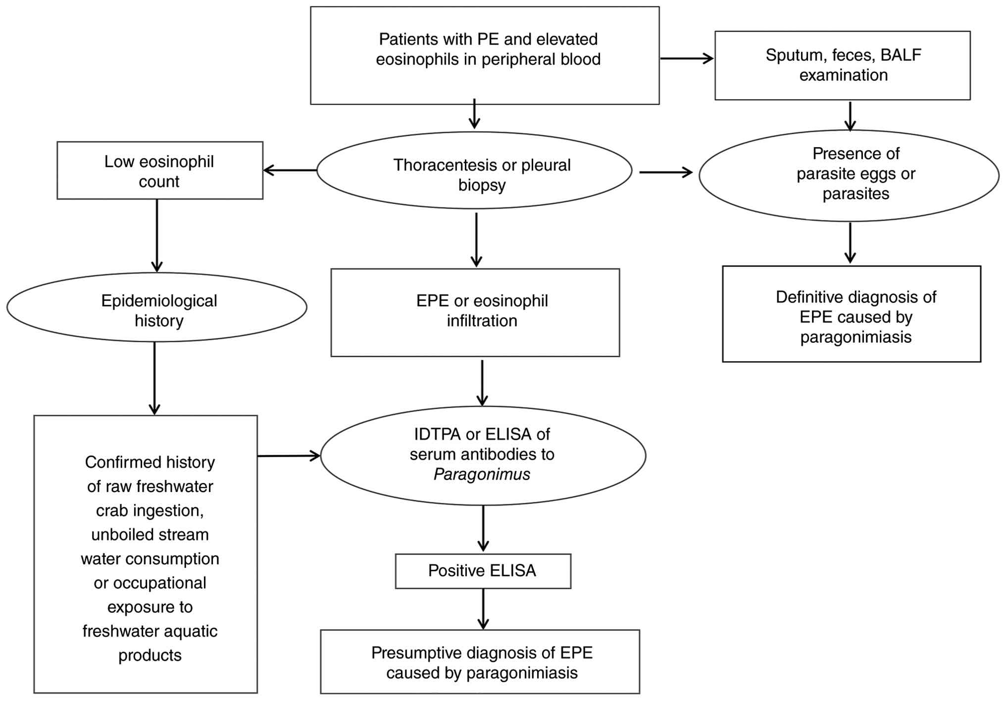
Based on the findings of the present case and the
51 reviewed cases, a stepwise diagnostic approach may be proposed
(Fig. 4). In any patient with
unexplained EPE from an endemic area, Paragonimus serology
(ELISA) or IDTPA should be performed regardless of raw seafood
ingestion history; occupational exposure alone should be
sufficient. Negative mNGS does not exclude paragonimiasis;
traditional immunological tests (ELISA, IDTPA) are key. Lack of
response to anti-TB therapy after 2 months warrants diagnostic
reassessment; praziquantel is effective even after long diagnostic
delays. As a single case report, the present findings may lack
generalizability. However, the systematic review of 51 additional
cases strengthens the evidence base. Other limitations include
potential publication bias inherent to case series and the
restriction to English-language publications, which may have
excluded relevant cases reported in other journals, thereby
introducing a languagebased selection bias. However, English is the
most widely accepted language for international scientific
communication; using Englishonly publications ensures that the
reviewed data are accessible to the broadest possible readership,
decreasing language barriers for global readers. Another limitation
is the lack of interim follow-up data during the 8-month period of
empirical anti-TB therapy. The present patient did not attend
scheduled follow-ups during that time, which reflects real-world
challenges in rural healthcare settings, but the eventual
re-presentation with persistent symptoms supports the conclusion
that no clinical improvement occurred. Additionally, follow-up
duration was limited to 7 months and serological confirmation of
cure (ELISA or IDTPA negativity) was not obtained. However,
clinical and radiographic resolution together with normalization of
peripheral eosinophil counts are widely accepted criteria for cure
in paragonimiasis (9,11,12)
and the patient remained asymptomatic with no evidence of relapse
at the last follow-up. Clinicians should not rely on this test for
clinical decisions, and a positive result alone does not confirm
TB. Praziquantel is the treatment of choice; triclabendazole is an
effective alternative. Heightened clinical awareness is key to
reduce misdiagnosis and improve outcomes.
Supplementary Material
Quality control parameters of
metagenomic next-generation sequencing of bronchoalveolar lavage
fluid.
Acknowledgements
Not applicable.
Funding
Funding: No funding was received.
Availability of data and materials
The data generated in the present study may be
found in the National Center for Biotechnology Information Sequence
Read Archive under accession number PRJNA1476379 or at the
following URL: ncbi.nlm.nih.gov/bioproject/1476379.
Authors' contributions
XL and JL conceived and designed the study. XL and
PW drafted the manuscript and analyzed and interpreted data. DC and
XP interpreted data and revised the manuscript. LX, PW and JW
interpreted data. XL and PW performed patient follow-up and the
literature review. XL, JW and JL confirm the authenticity of all
the raw data. All authors have read and approved the final
manuscript.
Ethics approval and consent to
participate
The present retrospective case report was reviewed
and approved by the Institutional Review Board of Taihe Hospital,
Hubei University of Medicine (Shiyan, China), which granted a
waiver of formal ethical approval for single case reports. Written
informed consent was obtained from the patient for participation in
this study.
Patient consent for publication
Written informed consent was obtained from the
patient for publication of the present cases report and any
accompanying images.
Competing interests
The authors declare that they have no competing
interests.
References
|
1
|
Lu YB, Hu K, Mao ML, Shi LB, Zhu CC and
Zou JX: The global distribution of Paragonimus species. Infect Dis
Poverty. 15(27)2026.PubMed/NCBI View Article : Google Scholar
|
|
2
|
Blair D: Paragonimiasis. Adv Exp Med Biol.
1454:203–238. 2024.PubMed/NCBI View Article : Google Scholar
|
|
3
|
Yoshida A, Doanh PN and Maruyama H:
Paragonimus and paragonimiasis in Asia: An update. Acta Trop.
199(105074)2019.PubMed/NCBI View Article : Google Scholar
|
|
4
|
Fürst T, Keiser J and Utzinger J: Global
burden of human food-borne trematodiasis: A systematic review and
meta-analysis. Lancet Infect Dis. 12:210–221. 2012.PubMed/NCBI View Article : Google Scholar
|
|
5
|
Liu Q, Wei F, Liu W, Yang S and Zhang X:
Paragonimiasis: An important food-borne zoonosis in China. Trends
Parasitol. 24:318–323. 2008.PubMed/NCBI View Article : Google Scholar
|
|
6
|
Chai JY: Paragonimiasis. Handb Clin
Neurol. 114:283–296. 2013.PubMed/NCBI View Article : Google Scholar
|
|
7
|
Wang H, Wang L, Yu D, Luo G, Yang S, Liu
Y, Ren T and Wang M: Exudative pleural effusion caused by lung
fluke infection: A practical diagnostic approach. Int J Infect Dis.
135:8–11. 2023.PubMed/NCBI View Article : Google Scholar
|
|
8
|
Li R, Liu X, Song G, Zhang CQ, Shen LJ, Li
WX and Bai ZP: Misdiagnosis of pulmonary paragonimiasis as
tuberculosis: A case report. Medicine (Baltimore).
105(e47555)2026.PubMed/NCBI View Article : Google Scholar
|
|
9
|
Yang QS, Qi RJ, Wang N and Ruan SS:
Pleural parasite infection presenting with an isolated pleural
effusion misdiagnosed as tuberculosis: A case report. Diagn
Microbiol Infect Dis. 110(116515)2024.PubMed/NCBI View Article : Google Scholar
|
|
10
|
Villanueva-Villegas R, Diaz-Mendoza J,
Salas-Lopez J and Espiche C: Paragonimiasis misdiagnosed as
pulmonary tuberculosis: A case report. Cureus.
15(e36169)2023.PubMed/NCBI View Article : Google Scholar
|
|
11
|
Calvopina M, Romero-Alvarez D, Macias R
and Sugiyama H: Severe pleuropulmonary paragonimiasis caused by
paragonimus mexicanus treated as tuberculosis in ecuador. Am J Trop
Med Hyg. 96:97–99. 2017.PubMed/NCBI View Article : Google Scholar
|
|
12
|
Luo J, Wang MY, Liu D, Zhu H, Yang S,
Liang BM and Liang Z: Pulmonary paragonimiasis mimicking
tuberculous pleuritis: A case report. Medicine (Baltimore).
95(e3436)2016.PubMed/NCBI View Article : Google Scholar
|
|
13
|
Prasad K, Basu A, Khana S and Wattal C:
Pulmonary paragonimiasis mimicking tuberculosis. J Assoc Physicians
India. 63:82–83. 2015.PubMed/NCBI
|
|
14
|
Song J, Hong G, Song JU, Kim W, Han SG, Ko
Y, Chang B, Jeong BH, Eom JS, Lee JH, et al: A case of pleural
paragonimiasis confused with tuberculous pleurisy. Tuberc Respir
Dis (Seoul). 76:175–178. 2014.PubMed/NCBI View Article : Google Scholar
|
|
15
|
Lee S, Yu Y, An J, Lee J, Son JS, Lee YK,
Song S, Kim H and Kim S: A case of delayed diagnosis of pulmonary
paragonimiasis due to improvement after anti-tuberculosis therapy.
Tuberc Respir Dis (Seoul). 77:178–183. 2014.PubMed/NCBI View Article : Google Scholar
|
|
16
|
Nagakura K, Oouchi M, Abe K and Araki K:
Pulmonary paragonimiasis misdiagnosed as tuberculosis: with special
references on paragonimiasis. Tokai J Exp Clin Med. 27:97–100.
2002.PubMed/NCBI
|
|
17
|
Sah R and Khadka S: Case series of
paragonimiasis from Nepal. Oxf Med Case Reports.
2017(omx083)2017.PubMed/NCBI View Article : Google Scholar
|
|
18
|
Hwang KE, Song HY, Jung JW, Oh SJ, Yoon
KH, Park DS, Jeong ET and Kim HR: Pleural fluid characteristics of
pleuropulmonary paragonimiasis masquerading as pleural
tuberculosis. Korean J Intern Med. 30:56–61. 2015.PubMed/NCBI View Article : Google Scholar
|
|
19
|
Sharma S, Agrawal G, Labru V, Bhatt AP,
Ahmed S and Bhatt R: Pulmonary paragonimiasis: Close mimic to
pulmonary tuberculosis in endemic regions. Indian Journal of
Medical Sciences. 2024; 76:45–47. 2024.
|
|
20
|
Yin P, Duan M, Deng L, Li T, Huang X, Fang
R, Li H and Bai B: Initial misdiagnosis to a revised diagnosis of
pulmonary paragonimiasis with elevated serum immunoglobulin G4
concentrations: A case report. J Int Med Res.
53(3000605251317980)2025.PubMed/NCBI View Article : Google Scholar
|
|
21
|
Das P: Childhood pulmonary
paragonimiasis-a diagnostic conundrum: A case series from a
tertiary care hospital in Kohima District of Northeast India.
Ethiopian Journal of Pediatrics and Child Health. 2025;19:179–184.
2025.
|
|
22
|
Poudyal BS, Paudel B, Bista B, Shrestha GS
and Pudasaini P: Clinical, laboratory and radiological features of
paragonimiasis misdiagnosed as pulmonary tuberculosis. Iran J
Parasitol. 17:410–414. 2022.PubMed/NCBI View Article : Google Scholar
|
|
23
|
Singh TN, Kananbala S and Devi KS:
Pleuropulmonary paragonimiasis mimicking pulmonary tuberculosis-a
report of three cases. Indian J Med Microbiol. 23:131–134.
2005.PubMed/NCBI View Article : Google Scholar
|
|
24
|
Im JG, Whang HY, Kim WS, Han MC, Shim YS
and Cho SY: Pleuropulmonary paragonimiasis: Radiologic findings in
71 patients. AJR Am J Roentgenol. 159:39–43. 1992.PubMed/NCBI View Article : Google Scholar
|
|
25
|
Zhou R, Zhang M, Cheng N and Zhou Y:
Paragonimiasis mimicking chest cancer and abdominal wall
metastaisis: A case report. Oncol Lett. 11:3769–3771.
2016.PubMed/NCBI View Article : Google Scholar
|
|
26
|
Lee JS, Lee J, Kim SH and Yong TS:
Molecular cloning and characterization of a major egg antigen in
Paragonimus westermani and its use in ELISA for the immunodiagnosis
of paragonimiasis. Parasitol Res. 100:677–681. 2007.PubMed/NCBI View Article : Google Scholar
|
|
27
|
Imai J: Evaluation of ELISA for the
diagnosis of paragonimiasis westermani. Trans R Soc Trop Med Hyg.
81:3–6. 1987.PubMed/NCBI View Article : Google Scholar
|
|
28
|
Xia Y, Chen J, Ju Y and You C:
Characteristic CT and MR imaging findings of cerebral
paragonimiasis. J Neuroradiol. 43:200–206. 2016.PubMed/NCBI View Article : Google Scholar
|
|
29
|
Paranjape N, Nunez V and Dicks D:
Paragonimiasis presenting as an acute hemorrhagic stroke. IDCases.
26(e01287)2021.PubMed/NCBI View Article : Google Scholar
|
|
30
|
Lan H and Song-Ping W: Clinical analysis
of 50 cases of serous cavity effusion related with paragonimiasis.
Zhongguo Xue Xi Chong Bing Fang Zhi Za Zhi. 30:452–454.
2018.PubMed/NCBI View Article : Google Scholar : (In Chinese).
|
|
31
|
Simner PJ, Miller S and Carroll KC:
Understanding the promises and hurdles of metagenomic
next-generation sequencing as a diagnostic tool for infectious
diseases. Clin Infect Dis. 66:778–788. 2018.PubMed/NCBI View Article : Google Scholar
|
|
32
|
Elbehiry A and Abalkhail A: Metagenomic
next-generation sequencing in infectious diseases: Clinical
applications, translational challenges, and future directions.
Diagnostics (Basel). 15(1991)2025.PubMed/NCBI View Article : Google Scholar
|
|
33
|
Sayers EW, Bolton EE, Brister JR, Canese
K, Chan J, Comeau DC, Connor R, Funk K, Kelly C, Kim S, et al:
Database resources of the national center for biotechnology
information. Nucleic Acids Res. 50(D1):D20–D26. 2022.PubMed/NCBI View Article : Google Scholar
|
|
34
|
Liu K, Sun YC, Pan RT, Xu AL, Xue H, Tian
N, Zheng JX, Shi FY, Lu Y and Li LH: Infection and biogeographical
characteristics of Paragonimus westermani and P. skrjabini in
humans and animal hosts in China: A systematic review and
meta-analysis. PLoS Negl Trop Dis. 18(e0012366)2024.PubMed/NCBI View Article : Google Scholar
|
|
35
|
Ai L, Li J, Ye T, Wang W and Li Y:
Exudative pleural effusion caused by lung fluke infection: Case
report. Int J Infect Dis. 114:175–177. 2022.PubMed/NCBI View Article : Google Scholar
|
|
36
|
Mukae H, Taniguchi H, Matsumoto N, Iiboshi
H, Ashitani J, Matsukura S and Nawa Y: Clinicoradiologic features
of pleuropulmonary Paragonimus westermani on Kyusyu Island, Japan.
Chest. 120:514–520. 2001.PubMed/NCBI View Article : Google Scholar
|
|
37
|
Yin Y, Zhu P, Guo Y, Li Y, Chen H, Liu J,
Sun L, Ma S, Hu C and Wang H: Enhancing lower respiratory tract
infection diagnosis: Implementation and clinical assessment of
multiplex PCR-based and hybrid capture-based targeted
next-generation sequencing. EBioMedicine.
107(105307)2024.PubMed/NCBI View Article : Google Scholar
|
|
38
|
Chen Q, Yi J, Liu Y, Yang C, Sun Y, Du J,
Liu Y, Gu D, Liu H, Xu Y and Chen Y: Clinical diagnostic value of
targeted next-generation sequencing for infectious diseases
(Review). Mol Med Rep. 30(153)2024.PubMed/NCBI View Article : Google Scholar
|
|
39
|
Ma H, Wang H, Han X and Fei J: Efficacy of
targeted next generation sequencing for pathogen detection in lower
respiratory tract infections. Am J Transl Res. 16:3637–3645.
2024.PubMed/NCBI View Article : Google Scholar
|
|
40
|
Miller S, Naccache SN, Samayoa E, Messacar
K, Arevalo S, Federman S, Stryke D, Pham E, Fung B, Bolosky WJ, et
al: Laboratory validation of a clinical metagenomic sequencing
assay for pathogen detection in cerebrospinal fluid. Genome Res.
29:831–842. 2019.PubMed/NCBI View Article : Google Scholar
|
|
41
|
Chen W, Liu R, Qi Q, Xu L and Sun G:
Comparative evaluation of mNGS and traditional culture methods in
pathogen detection for pulmonary infections. Infect Drug Resist.
18:6991–6998. 2025.PubMed/NCBI View Article : Google Scholar
|
|
42
|
Ding H, Huang J, Lin L, Chen Y, Wang Q, Li
W, Huang Y, Fang X and Zhang W: Shedding light on negative cultures
in osteoarticular infections: Leveraging mNGS to unravel risk
factors and microbial profiles. Front Cell Infect Microbiol.
14(1457639)2024.PubMed/NCBI View Article : Google Scholar
|
|
43
|
Zhou Y, Shi W, Wen Y, Mao E and Ni T:
Comparison of pathogen detection consistency between metagenomic
next-generation sequencing and blood culture in patients with
suspected bloodstream infection. Sci Rep. 13(9460)2023.PubMed/NCBI View Article : Google Scholar
|
|
44
|
Wilson MR, Sample HA, Zorn KC, Arevalo S,
Yu G, Neuhaus J, Federman S, Stryke D, Briggs B, Langelier C, et
al: Clinical metagenomic sequencing for diagnosis of meningitis and
encephalitis. N Engl J Med. 380:2327–2340. 2019.PubMed/NCBI View Article : Google Scholar
|
|
45
|
Zhao Y, Zhang W and Zhang X: Application
of metagenomic next-generation sequencing in the diagnosis of
infectious diseases. Front Cell Infect Microbiol.
14(1458316)2024.PubMed/NCBI View Article : Google Scholar
|
|
46
|
Chen J, Qin L and Tang LF: Case report:
Intermittent chest pain with cough, fever, and pericardial effusion
over the course of 1 year in an 11-year-old girl. Front Pediatr.
10(896824)2022.PubMed/NCBI View Article : Google Scholar
|
|
47
|
Keiser J, Engels D, Büscher G and Utzinger
J: Triclabendazole for the treatment of fascioliasis and
paragonimiasis. Expert Opin Investig Drugs. 14:1513–1526.
2005.PubMed/NCBI View Article : Google Scholar
|