Introduction
Patients with malignancy have an increased risk of
both venous and arterial thrombosis. The risk of venous
thromboembolism in cancer patients has been reported to be
approximately ninefold higher than that in the general population
(1). Similarly, patients with
cancer are at an elevated risk of arterial thromboembolism
(2). Retrospective studies have
shown that aortic thrombosis is associated with malignancy in
approximately 20% of cases (3).
Aortic thrombosis can lead to a wide range of
embolic events, among which spinal cord infarction (SCI) is rare
but potentially devastating. SCI often results in irreversible
neurological deficits, making prompt recognition and management
crucial. Clinically, presentations such as bilateral lower limb
weakness and bladder-rectal dysfunction can mimic spinal cord
compression-a recognized oncologic emergency, making differential
diagnosis particularly challenging.
To our knowledge, SCI caused by aortic mural
thrombosis in patients with malignancy is exceedingly rare. Herein,
we report a case of SCI secondary to aortic mural thrombosis in a
patient receiving chemotherapy for cancer of unknown primary (CUP).
This case highlights the diverse clinical manifestations of
arterial thrombosis in cancer patients and underscores the
importance of including SCI in the differential diagnosis of acute
neurological symptoms resembling spinal cord compression.
Case report
A 75-year-old man was diagnosed with CUP with
multiple bone metastases involving the scapula, clavicle, sternum,
ribs, ilium, ischium, and lumbar vertebrae in June 2024. He was
diagnosed and treated at the Department of Medical Oncology, Nara
Medical University Hospital (Kashihara, Japan). The Department of
Medical Oncology is clinically and organizationally integrated with
the Department of Cancer Genomics and Medical Oncology, Nara
Medical University, where the treating physicians are primarily
affiliated. Bone biopsy revealed a malignant lesion composed in
part of loosely arranged bundles of spindle-shaped cells. Although
consistent with malignancy, no definite line of differentiation was
identified. Sarcomatoid carcinoma and soft tissue sarcoma were
considered in the differential diagnosis; however, a definitive
diagnosis could not be established based on histology alone. Given
the possibility that the tumor represented either a sarcoma or CUP,
treatment with doxorubicin was initiated, and the patient received
six courses beginning in June 2024. As the disease progressed,
nivolumab therapy, approved in Japan for CUP, was initiated in
December 2024.
In August 2025, the patient presented urgently with
acute bilateral lower limb weakness and inability to walk. He was
admitted to the same institution. On admission, manual muscle
testing showed 3/5 strength in the iliopsoas, hamstring, and
quadriceps muscles bilaterally, while the tibialis anterior and
gastrocnemius muscles were preserved at 5/5. Deep tendon reflexes
of the lower limbs were decreased. He was unable to urinate or
defecate independently, indicating bladder and rectal
dysfunction.
Given his known bone metastases, spinal cord
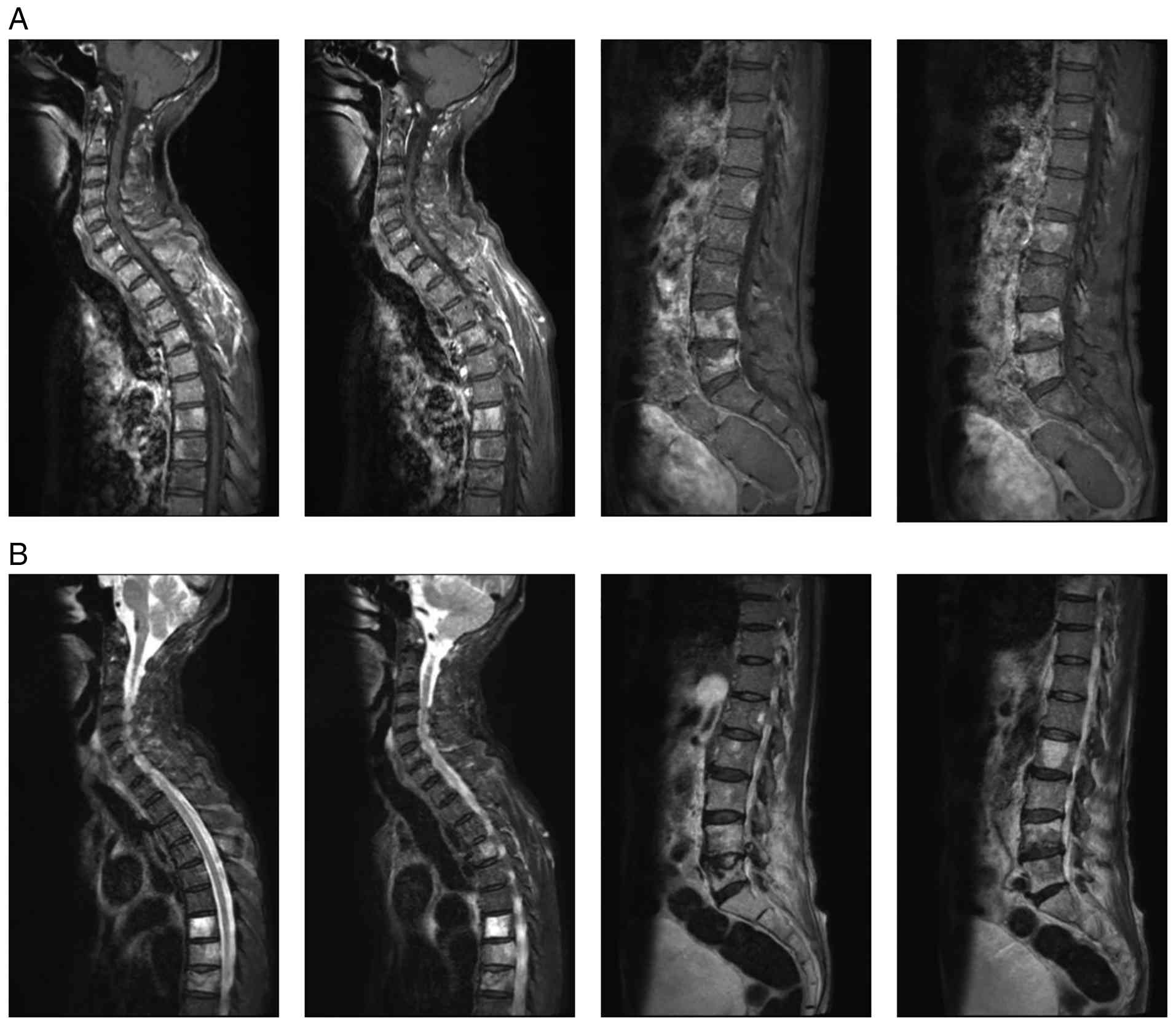
compression was initially suspected. However, whole-spine
contrast-enhanced magnetic resonance imaging (MRI) revealed
multiple vertebral metastases but no evidence of cord compression
or leptomeningeal dissemination (Fig.
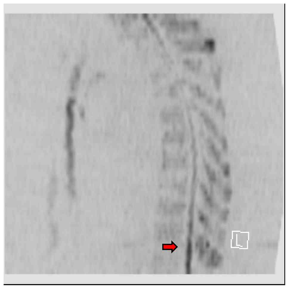
1). Diffusion-weighted imaging showed high signal intensity
with reduced apparent diffusion coefficient values in the spinal
cord from thoracic vertebrae 11 to 12 (Th11-Th12), raising
suspicion of SCI (Fig. 2). Because
he was receiving nivolumab, immune-related adverse event (irAE)
myelitis was also considered. Cerebrospinal fluid analysis,
however, showed no pleocytosis, no protein elevation, normal
glucose, and normal opening pressure, making irAE myelitis
unlikely. Based on these findings, SCI was diagnosed.
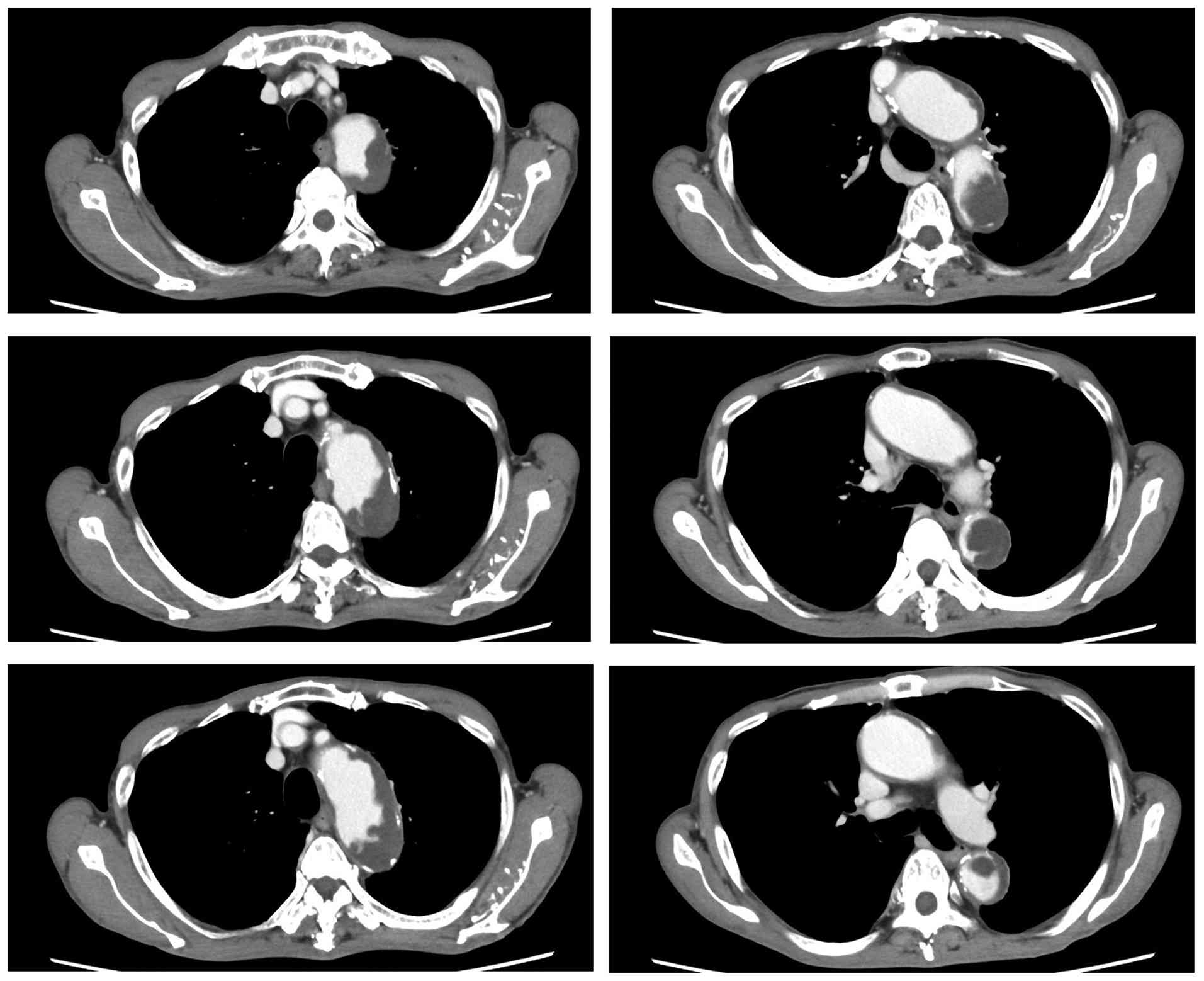
Subsequent contrast-enhanced computed tomography
(CT) revealed a large aortic mural thrombus (Fig. 3), which was considered the most
likely etiology of SCI after exclusion of alternative causes. The
D-dimer level at presentation was 1.6 µg/ml (reference range, 0-1.0
µg/ml); previous measurements ranged from 0.8 to 1.8 µg/ml, showing
no significant elevation from baseline. Routine coagulation studies
showed a prothrombin time-international normalized ratio (PT-INR)
of 1.10 (reference range, 0.8-1.15) and an activated partial
thromboplastin time (APTT) of 40 sec (reference range, 24-39 sec),
without clinically significant abnormalities. Fibrinogen was mildly
elevated at 473 mg/dl (reference range, 200-400 mg/dl). Heparin
anticoagulation therapy was initiated, and early rehabilitation
began during hospitalization. By hospital Day 3, lower limb motor
function had improved sufficiently to allow ambulation with a
walker. However, by hospital Day 9, he developed difficulty
maintaining a kneeling posture, and no further neurological
improvement was observed. A repeat spinal MRI performed following
this deterioration demonstrated no apparent extension of the
infarcted area or new findings suggestive of progressive spinal
cord injury. Although infarct progression was considered unlikely,
the patient's overall condition deteriorated because of advanced
malignancy and declining performance status. Therefore, the
decision to transition to best supportive care was based on his
overall clinical condition rather than neurological status alone.
He was discharged home on hospital Day 19 under best supportive
care. The latest follow-up was performed in September 2025 at the
time of discharge, when persistent lower extremity neurological
deficits remained without further improvement.
Discussion
The most frequent causes of SCI are idiopathic or
related to aortic surgery. Other reported etiologies include aortic
dissection, fibrocartilaginous embolism, systemic atherosclerosis,
systemic hypotension, hypercoagulable states, cardioembolism, and
vasculitis (4-6).
In the present case, imaging suggested that the SCI
was caused by an aortic mural thrombus. However, transthoracic and
transesophageal echocardiography were not performed; therefore,
occult cardioembolic sources such as nonbacterial thrombotic
endocarditis, intracardiac thrombus, or patent foramen ovale could
not be completely excluded. Consequently, the causal relationship
between the aortic mural thrombus and SCI cannot be definitively
established and should be regarded as a clinical inference based on
imaging findings and exclusion of alternative etiologies.
Alternative etiologies were also considered. The patient had no
documented episodes of systemic hypotension or hemodynamic
instability preceding symptom onset, making spinal cord ischemia
due to hypoperfusion unlikely. In addition, routine coagulation
studies did not support disseminated intravascular coagulation
(DIC), with no clinically significant abnormalities in PT-INR or
APTT and only mild elevation of fibrinogen, which was considered
attributable to the underlying malignancy and inflammatory status.
Although a cancer-associated hypercoagulable state may have
contributed to thrombus formation, there was no clinical or
radiological evidence specifically suggesting overt DIC.
Furthermore, tumor embolism was considered unlikely because
contrast-enhanced CT demonstrated a large aortic mural thrombus
that provided a plausible embolic source, and no pathological or
imaging findings suggested tumor embolization. Although
small-vessel disease and other embolic sources cannot be completely
excluded, the imaging findings and the presence of a large aortic
mural thrombus were considered more consistent with an embolic
mechanism originating from the aorta. Nevertheless,
contrast-enhanced CT demonstrated a large mural thrombus extending
from the aortic arch to the descending thoracic aorta. Furthermore,
the anatomical distribution of the thrombus was considered
compatible with a potential source of embolization to the spinal
cord circulation, supporting aortic mural thrombosis as the most
likely etiology in this case. Patients with malignancy are known to
have an increased risk of arterial thrombosis compared with the
general population (2). Although
additional testing for lupus anticoagulant and anticardiolipin
antibodies was not performed, routine coagulation studies showed no
clinically significant abnormalities in PT-INR or APTT. Fibrinogen
was mildly elevated (473 mg/dl), which was considered attributable
to the underlying malignancy and inflammatory status rather than a
specific hypercoagulable disorder. Notably, the D-dimer level at
SCI onset was not substantially elevated compared with previous
measurements. This may reflect the patient's pre-existing
cancer-associated hypercoagulable state, as D-dimer levels had
remained persistently elevated before SCI onset. Consequently, any
additional increase associated with SCI may have been relatively
small and difficult to distinguish from baseline variation.
Furthermore, D-dimer levels do not necessarily correlate with the
extent or clinical severity of arterial thrombosis. Furthermore,
emerging evidence indicates that patients receiving immune
checkpoint inhibitors (ICIs) may have a higher risk of arterial
thrombosis than those not treated with ICIs (7). The exact mechanisms underlying
ICI-associated thrombosis remain incompletely understood. However,
several potential mechanisms have been proposed, including enhanced
T-cell activation, endothelial inflammation, accelerated
atherosclerosis, and neutrophil extracellular trap (NET)-mediated
thrombogenesis (8). In particular,
PD-1 plays an important role in suppressing pro-atherogenic T-cell
responses, and PD-1 blockade may exacerbate vascular inflammation
by releasing these inhibitory signals, thereby promoting thrombus
formation (8). Although causality
cannot be established in the present case, these mechanisms may
have contributed to the development and progressive enlargement of
the aortic mural thrombus observed after nivolumab initiation.
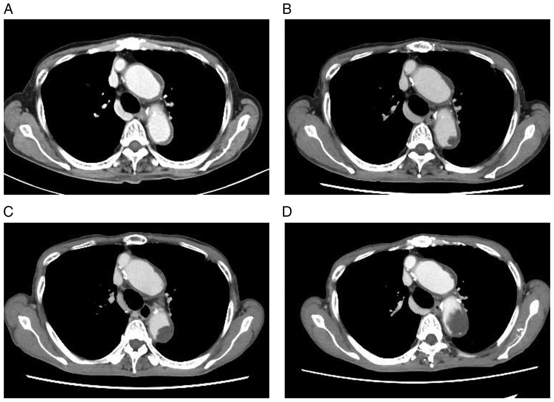
Although a definitive causal relationship cannot be established in
this case, follow-up contrast-enhanced CT after initiation of
nivolumab demonstrated the emergence and progressive enlargement of
an aortic mural thrombus over time (Fig. 4). Evidence regarding aortic
thrombosis in cancer patients remains limited; a small
retrospective study reported that 3 of 13 patients with primary
aortic mural thrombus had concomitant malignancy (3). While several cases of SCI due to
aortic thrombosis have been reported (9,10),
SCI caused by aortic mural thrombosis in patients with malignancy
is exceedingly rare. Reports on cancer-associated SCI are limited,
but a recent systematic review indicated a poor prognosis, with a
median survival of approximately 4 months (11). In our patient, early initiation of
physical therapy led to rapid neurological recovery, allowing
ambulation with a walker within several days. However, the patient
subsequently developed recurrent generalized weakness and a decline
in performance status, ultimately necessitating a transition to
best supportive care. Repeat spinal MRI was performed after
neurological deterioration following the initial recovery and
demonstrated no apparent extension of the infarcted area or other
radiological findings suggestive of progressive spinal cord injury.
Therefore, infarct progression was considered unlikely to explain
the subsequent neurological deterioration. Nevertheless, secondary
neuronal injury or other factors contributing to the recurrent
neurological deficits cannot be completely excluded. Another
limitation of the present study is the incomplete imaging
characterization of the aortic mural thrombus. Although
contrast-enhanced CT demonstrated a large mural thrombus extending
from the aortic arch to the descending thoracic aorta, precise
measurement of its maximum diameter and length was difficult
because the lesion exhibited an elongated mural configuration with
irregular and continuous extension along the aortic wall rather
than a discrete mass. In addition, thrombus mobility and
classification as pedunculated or sessile could not be reliably
assessed using static contrast-enhanced CT alone, and dynamic
imaging modalities such as transesophageal echocardiography or cine
imaging were not performed. Furthermore, the relationship between
the thrombus and the ostia of spinal feeding arteries, including
the artery of Adamkiewicz, could not be evaluated because these
vessels were not clearly visualized on routine contrast-enhanced
CT. Therefore, interpretation of the embolic potential and
anatomical relationship of the thrombus is limited by the available
imaging data. Importantly, the decision to transition to best
supportive care was based not only on neurological status but also
on the patient's overall clinical deterioration and progressive
decline in performance status associated with advanced malignancy.
Treatment options for aortic mural thrombus include anticoagulation
therapy, stent graft placement, and surgical intervention (3). In this case, stent graft insertion or
surgery was deemed high risk due to the potential for further
embolic events, and conservative management with heparin alone was
chosen. Given the rarity of asymptomatic aortic mural thrombosis in
patients with malignancy receiving ICIs and the limited available
evidence, no established guidelines currently exist regarding
screening or prophylactic treatment in this setting. Therefore,
routine prophylactic anticoagulation solely on the basis of ICI
therapy cannot be recommended. If an asymptomatic aortic mural
thrombus is incidentally identified, management should be
individualized based on thromboembolic risk, bleeding risk,
thrombus characteristics, cancer status, and overall prognosis.
Although anticoagulation therapy may be considered in selected
high-risk patients, the optimal agent and treatment duration remain
uncertain and warrant further investigation. SCI in cancer patients
may therefore severely limit further oncologic treatment and
negatively impact overall prognosis.
In this case, SCI occurred during nivolumab therapy
for CUP. Given the presence of vertebral metastases, spinal cord
compression was initially suspected. After imaging excluded
compression and leptomeningeal dissemination, irAE myelitis was
also considered. Cerebrospinal fluid analysis, however, revealed
normal cell counts, protein, and glucose, effectively ruling out
irAE myelitis. The final diagnosis of SCI was established based on
diffusion-weighted imaging findings on MRI, neurological
examination, and identification of an aortic mural thrombus on
contrast-enhanced CT.
Although SCI is rare, it can mimic other urgent
neurological conditions. Clinicians evaluating acute neurological
deficits in patients receiving ICIs should consider not only spinal
cord compression and irAE-related myelitis but also SCI in the
differential diagnosis to ensure timely recognition and
management.
Acknowledgements
Not applicable.
Funding
Funding: No funding was received.
Availability of data and materials
The data generated in the present study may be
requested from the corresponding author.
Authors' contributions
NH conceptualized the study. NH, SO, RS, YK, YY, MO,
MK, HN, MI, TM, YN, MaiT and MasT participated in the diagnosis and
clinical management of the patient. Acquisition and interpretation
of data were performed by NH, SO, RS, YK, YY, MO, MK, HN, MI, TM,
YN, MaiT and MasT. Writing the original draft was undertaken by NH,
and writing, reviewing and editing of the manuscript were carried
out by NH and MasT. NH and MasT confirm the authenticity of all the
raw data. All authors read and approved the final manuscript, and
agreed to be accountable for all aspects of the research in
ensuring that the accuracy or integrity of any part of the work is
appropriately investigated and resolved.
Ethics approval and consent to
participate
Not applicable.
Patient consent for publication
Written informed consent was obtained from the
patient for the publication of this case report and the
accompanying images. The patient was informed that all identifying
information would be removed to ensure anonymity.
Competing interests
The authors declare that they have no competing
interests.
References
|
1
|
Mulder FI, Horváth-Puhó E, van Es N, van
Laarhoven HWM, Pedersen L, Moik F, Ay C, Büller HR and Sørensen HT:
Venous thromboembolism in cancer patients: A population-based
cohort study. Blood. 137:1959–1969. 2021.PubMed/NCBI View Article : Google Scholar
|
|
2
|
Navi BB, Reiner AS, Kamel H, Iadecola C,
Okin PM, Elkind MSV, Panageas KS and DeAngelis LM: Risk of arterial
thromboembolism in patients with cancer. J Am Coll Cardiol.
70:926–938. 2017.PubMed/NCBI View Article : Google Scholar
|
|
3
|
Mendes D, Veiga C, Machado R, Sá-Pinto P
and Almeida R: The challenge of managing a primary aortic mural
thrombus: Outcomes and technical considerations. Int J Angiol.
33:156–164. 2024.PubMed/NCBI View Article : Google Scholar
|
|
4
|
Zalewski NL, Rabinstein AA, Krecke KN,
Brown RD Jr, Wijdicks EFM, Weinshenker BG, Kaufmann TJ, Morris JM,
Aksamit AJ, Bartleson JD, et al: Characteristics of spontaneous
spinal cord infarction and proposed diagnostic criteria. JAMA
Neurol. 76:56–63. 2019.PubMed/NCBI View Article : Google Scholar
|
|
5
|
Ros Castelló V, Sánchez Sánchez A, Natera
Villalba E, Gómez López A, Parra P, Rodríguez Jorge F, Buisán
Catevilla J, García Barragán N, Masjuan J and Corral I: Spinal cord
infarction: Aetiology, imaging findings, and prognostic factors in
a series of 41 patients. Neurología (Engl Ed). 38:391–398.
2023.PubMed/NCBI View Article : Google Scholar
|
|
6
|
Salvador de la Barrera S, Barca-Buyo A,
Montoto-Marqués A, Ferreiro-Velasco ME, Cidoncha-Dans M and
Rodriguez-Sotillo A: Spinal cord infarction: Prognosis and recovery
in a series of 36 patients. Spinal Cord. 39:520–525.
2001.PubMed/NCBI View Article : Google Scholar
|
|
7
|
Zhu J, Chen Y, Zhang Y, Wang W, Wang Y, Lu
Z, Zhang Y, Lei H, Li D, Long B and Liu H: Association of immune
checkpoint inhibitors therapy with arterial thromboembolic events
in cancer patients: A retrospective cohort study. Cancer Med.
12:18531–18541. 2023.PubMed/NCBI View Article : Google Scholar
|
|
8
|
Wang TF and Carrier M: Immune checkpoint
inhibitors-associated thrombosis: Incidence, risk factors and
management. Curr Oncol. 30:3032–3046. 2023.PubMed/NCBI View Article : Google Scholar
|
|
9
|
Yogendranathan N, Herath HMMTB, Jayamali
WD, Matthias AT, Pallewatte A and Kulatunga A: A case of anterior
spinal cord syndrome in a patient with unruptured thoracic aortic
aneurysm with a mural thrombus. BMC Cardiovasc Disord.
18(48)2018.PubMed/NCBI View Article : Google Scholar
|
|
10
|
Giammello F, Gardin A, Brizzi T, Casella
C, Fazio MC, Galletta K, Mormina E, Vinci SL, Musolino RF, La Spina
P and Toscano A: Unilateral posterior spinal cord ischemia due to a
floating aortic thrombus: A case report. Clin Transl Neurosci.
7(26)2023.
|
|
11
|
Puente-Hernandez M, Rivero-de-Aguilar A
and Varela-Lema L: Cancer-associated spinal cord infarction: A
systematic review and analysis of survival predictors. J Neurol
Sci. 446(120580)2023.PubMed/NCBI View Article : Google Scholar
|