Introduction
Ischemic heart disease, including myocardial
infarction, has remained the leading cause of mortality worldwide.
Driven by population aging and increasing cardiovascular risk
factors, the absolute global incidence and mortality of myocardial
infarction continue to rise, and this upward trend is particularly
pronounced in China (1-3).
Thrombolysis or percutaneous coronary intervention is an effective
treatment for myocardial infarction, but it often results in
myocardial ischemia-reperfusion injury (MIRI), which can markedly
hinder patient recovery (1,2). At
present, the main strategies for the prevention and treatment of
MIRI include ischemic preconditioning, ischemic post-adaptation,
drug preconditioning and drug post-processing. Commonly used
clinical agents, such as vitamin E, cariporide and allopurinol,
often yield suboptimal therapeutic outcomes due to the complex
pathophysiology of MIRI (3).
Unlike single-target pharmacological agents, traditional Chinese
medicine (TCM) compound prescriptions exert their therapeutic
effects through a synergistic ‘multi-component, multi-target, and
multi-pathway’ mechanism. This enables the various active
ingredients within a single formula to simultaneously modulate
interconnected pathological networks, such as oxidative stress,
inflammation and apoptosis, thereby offering comprehensive
protection against MIRI (4).
Buyang Huanwu Decoction (BYHWD) is a representative
prescription of the traditional Yiqi Huoxue formula and a classical
treatment for cardio-cerebrovascular ischemic diseases in TCM
(٤-٨). BYHWD originates from ‘Yi Lin Gai Cuo’ (meaning ‘Correcting
the Errors of Medical Works’ in English), authored by Qingren (٩)
during the Qing Dynasty, and is composed of Radix Astragali,
Radix Angelicae Sinensis, Radix Paeoniae Rubra, Lumbricus, Rhizoma
Chuanxiong, Flos Carthami and Semen Persicae. Modern
studies have shown that BYHWD can effectively improve myocardial
function in patients with MIRI by reducing the level of serum
inflammatory cytokines, including TNF-α, IL-6 and IL-1β. In
addition, it can scavenge oxygen free radicals, inhibit calcium
overload and improve vascular microcirculation, with beneficial
effects having been demonstrated in numerous cardiovascular
diseases, including myocardial infarction, MIRI, angina pectoris,
heart failure and arrhythmia (5,6). In
previous years, active-component extracts of TCM prescriptions have
gradually become a key direction in compound research, establishing
an experimental foundation for clarifying the material basis of TCM
compound prescriptions (١٠). Previous studies have also shown that
glycosides are among the main active components of BYHWD in the
treatment of cardio-cerebrovascular ischemia-associated diseases
(٤,7). Fu et al (٨) found that the glycosides of BYHWD can
alleviate inflammatory reactions in vivo and in vitro
by downregulating the JAK/STAT signaling pathway, thereby exerting
an anti-atherosclerotic effect. This is similar to the overall
effect of BYHWD, suggesting that glycosides are important active
components of BYHWD. The main active glycoside-associated
components are astragaloside IV, paeoniflorin and amygdalin (٨).
However, there is still a lack of studies specifically
investigating the effects of the total glycosides of BYHWD on MIRI.
This is primarily as previous pharmacological research has
predominantly focused either on the crude aqueous decoction to
reflect holistic properties, or on single isolated monomers (such
as pure astragaloside IV). Furthermore, BYHWD has traditionally
been studied more extensively in the context of cerebrovascular
ischemic diseases. Consequently, the synergistic protective
potential of its glycoside-enriched fraction in myocardial
ischemia-reperfusion injury remains relatively underexplored.
Mitochondria, as organelles that generate energy,
serve an important role in cardiomyocytes, participating in a
number of cellular functions, such as the synthesis of metabolites,
calcium storage, reactive oxygen species (ROS) production and
inflammation. When reperfusion occurs, more damaged mitochondria
are produced and an increase in ROS, released after mitochondrial
damage, is observed. Higher levels of ROS can cause cell
homeostasis imbalance, resulting in severe oxidative stress, which
subsequently triggers lipid peroxidation, protein oxidation, DNA
damage, and ultimately apoptotic or necrotic cell death (1). Mitochondrial autophagy can clear the
damaged mitochondria in time, thus maintaining the normal function
and number of cardiomyocytes (١١). Therefore, the maintenance of
normal mitochondrial function by regulating mitochondrial autophagy
is markedly important for the maintenance of myocardial homeostasis
during reperfusion injury (12,13).
The PTEN-induced kinase 1 (PINK1)/Parkin pathway is one of the
classical mitochondrial autophagy pathways, capable of rapidly
responding to mitochondrial damage and serving a key role in
maintaining mitochondrial network integrity and promoting cell
survival (14).
Overall, prescriptions for replenishing qi and
activating blood circulation have been widely used in the clinical
treatment of MIRI in TCM. In recent years, research regarding TCM
compound prescriptions has increasingly focused on their active
components and the total glycosides in BYHWD have been indicated to
be major active components involved in improving ischemic
myocardial injury. It should be noted that the extract used is a
glycoside-enriched fraction, in which astragaloside IV,
paeoniflorin and amygdalin are the three major quantified
components, rather than a pure mixture of these three compounds.
However, the efficacy and mechanism of BYHWD glycosides against
MIRI remain unclear. Mitochondrial autophagy is an important
component of MIRI. Therefore, investigating the regulation of
mitochondrial autophagy by TCM compound prescriptions is of marked
importance. Accordingly, the present study focused on investigating
the effects of BYHWD glycosides on MIRI, potentially through the
regulation of mitochondrial autophagy.
Materials and methods
Reagents
Standard paeoniflorin (batch no. R002155; purity,
98%) was obtained from RHAWN; Shanghai Saihan Technology Co., Ltd.
Amygdalin reference compound (batch no. BMMI20687; purity ≥98%) was
supplied by Shanghai Yuanye Bio-Technology Co., Ltd. Astragaloside
IV standard (batch no. A800922; purity ≥98.5%) was purchased from
Shanghai Macklin Biochemical Co., Ltd. Primary antibodies against
PTEN-induced kinase 1 (PINK1; cat. no. YN2037), Parkin (cat. no.
YT3593), p62 (cat. no. YT7058) and silent mating type information
regulation 2 homolog-1 (SIRT1; cat. no. YT4302) were obtained from
ImmunoWay Biotechnology Company. Antibodies against LC3 (cat. no.
14600-1) and Beclin-1 (cat. no. PD017) were purchased from
Proteintech Group, Inc. and MBL International Co., respectively.
Anti-β-actin antibody (cat. no. DW9562), HRP-conjugated anti-rabbit
secondary antibody (cat. no. WD-GAR007) and anti-mouse secondary
antibody (cat. no. WD0990) were provided by Hangzhou Dawen Biotech
Co., Ltd.
RNA extraction was performed using an RNA Quick
Purification Kit (cat. no. RN001; Shanghai Yishan Biotechnology
Co., Ltd.) and complementary DNA (cDNA) synthesis was conducted
with the BeyoRT™ II First Strand cDNA Synthesis Kit (cat. no.
D7170M; Beyotime Biotechnology). Quantitative PCR (qPCR) reactions
employed PowerUp™ SYBR™ Green Master Mix (Thermo Fisher Scientific,
Inc.). Assay kits for reactive oxygen species (ROS; cat. no.
S0033), JC-1 mitochondrial membrane potential (MMP) detection (cat.
no. M8650), malondialdehyde (MDA; cat. no. S0131M),
monodansylcadaverine (MDC; cat. no. C3018S) and superoxide
dismutase (SOD; cat. no. S0101S) were obtained from Beyotime
Biotechnology or Beijing Solarbio Science & Technology Co.,
Ltd., through local suppliers. High-performance liquid
chromatography (HPLC)-grade acetonitrile (cat. no. 75-05-8) was
purchased from Merck KGaA.
Preparation of BYHWD glycosides
BYHWD, which is composed of Radix Astragali
(Huang Qi), Semen Persicae (Tao Ren), Flos Carthami
(Hong Hua), Radix Paeoniae Rubra (Chi Shao), Radix
Angelicae Sinensis (Dang Gui), Rhizoma Chuanxiong (Chuan
Xiong) and Lumbricus (Di Long) were supplied by The
Affiliated Hospital of Zhejiang Chinese Medical University
(Hangzhou, China) and identified by the co-author, Professor Ding
Zhishan from Zhejiang Chinese Medicine University (Hangzhou,
China). The proportions of each aforementioned medicinal material
were 20:3:3:3:3:2:3. The detailed composition and sources of BYHWD
are summarized in Table I. A
packet of proportionally compatible compound samples was taken,
powdered and sifted through 60 mesh. The compound crude powder was
accurately weighed (50 g) and 11 volumes of distilled water were
added. Next, the sample was boiled at 82˚C for 75 min and filtered.
The process was repeated three times, combining the filtrate each
time, followed by centrifugation at 3,000 x g for 10 min at room
temperature to remove the precipitate. The filtrate was
concentrated by rotary evaporation. Finally, the concentrated
liquid reached ~150 ml. Then, 95% ethanol was gradually added to
the aqueous extract until the final ethanol concentration reached
85% (v/v) and the mixture was sealed and kept at 4˚C overnight (≥12
h) for alcohol precipitation. The supernatant was then concentrated
to ~20 ml. Extraction with petroleum ether was performed three
times and the aqueous layer was retained, followed by extraction
with ethyl acetate three times. Next, the ethyl acetate layer was
reserved and the aqueous layer was extracted with n-butanol
solution three times. Finally, the n-butanol and ethyl acetate
layers were combined in an evaporation dish and dried under vacuum
to obtain crude glycosides. Notably, the extract used was a
glycoside-enriched fraction, in which astragaloside IV,
paeoniflorin and amygdalin were the three major quantified
components, rather than a pure mixture of these three
compounds.
 | Table IComposition and source of BYHWD. |
Table I
Composition and source of BYHWD.
| Source | Chinese herbal
name | Latin name | Species name | Weight ratio |
|---|
| BYHWD prepared | Huang Qi | Radix
Astragali | Astragalus
membranaceus (Fisch.) Bge.var. Mongholicus (Bge.)
Hsiao | 20 |
| by the
affiliated | Tao Ren | Semen Persicae | Amygdalus
persica L. | 3 |
| Hospital of
Zhejiang | Hong Hua | Flos Carthami | Carthamus
tinctorius L. | 3 |
| Chinese
Medical | Chi Shao | Radix Paeoniae
Rubra | Paeonia
lactiflora Pall. | 3 |
| University | Dang Gui | Radix Angelicae
Sinensis | Angelica
sinensis (Oliv.) Diels | 3 |
| (batch no. 2001621)
in 10 g/bags | Chuan Xiong | Rhizoma
Chuanxiong | Ligusticum
chuanxiong. Hort | 2 |
| | Di Long | Lumbricus | Pheretima
aspergillum (E. Perrier) | 3 |
Qualitative and quantitative analyses
of glycosides
Chromatographic separation was carried out using a
reversed-phase C18 analytical column (250 mm in length) maintained
at 30˚C. The mobile phase consisted of acetonitrile (solvent A) and
purified water (solvent B), delivered at a constant flow rate of
0.8 ml/min. Sample injection volume was set at 10 µl. Detection
wavelengths were adjusted to 230 nm for paeoniflorin and 210 nm for
amygdalin. A gradient elution program was applied as follows: 8-10%
solvent A for 0-15 min, 10-15% solvent A for 15-25 min, 15-18%
solvent A for 25-35 min and 18-21% solvent A for 35-40 min. The
system operated under a column pressure of 0.71±0.41 MPa.
Quantification of astragaloside IV in BYHWD total glycosides was
performed using a vanillin-sulfuric acid colorimetric assay.
Briefly, the colorimetric reaction was conducted at 60˚C for 15
min. The standard calibration curve was constructed based on the
modified protocol described by Le et al (١٥) and the
astragaloside IV content was calculated accordingly. Preparation of
the astragaloside IV standard curve, calculation formula and
determination of the results are provided in Fig. S1 and Tables SI and SII.
Cell culture and model
establishment
H9C2, the rat cardiomyoblast cell line was obtained
from the China Center for Type Culture Collection. Cells were
maintained in DMEM (Gibco; Thermo Fisher Scientific, Inc.)
supplemented with 10% FBS (CellMax Technologies AB) and 1%
penicillin-streptomycin (Northrend) at 37˚C in a humidified
incubator containing 5% CO2. Cells were passaged and
used for subsequent experiments upon reaching 80-90%
confluence.
H9C2 cells were randomly assigned to three groups: A
control group, an oxygen-glucose deprivation/reoxygenation (OGD/R)
group and a BYHWD total glycosides pre-treatment group. The OGD/R
model was established (as detailed below) to simulate
ischemia-reperfusion injury in vitro. For the pre-treatment,
cells in the BYHWD group were incubated with the BYHWD
glycoside-enriched fraction at 37˚C for 12 h prior to OGD/R
induction. Specifically, the glycosides were prepared in complete
culture medium and added to the cells, while the model and control
groups received an equal volume (100 µl/well) of normal complete
medium. Next, with the exception of the control group, complete
culture medium was replaced with minimum essential medium without
FBS in the other groups, followed by exposure to an airtight
chamber (cat. no. MIC-101; Embrient, Inc.) containing a mixture of
95% N2 and 5% CO2 anoxic culture at 37˚C for
21 h. The supernatant was replaced with complete medium and
cultured in a normal cell incubator for 6 h to induce reperfusion
injury in H9C2 cells.
Cell viability assay
H9C2 cells were seeded into 96-well culture plates
at a density of 5,000 cells per well and allowed to adhere prior to
experimental treatment. Following the indicated interventions,
cellular viability was assessed using an MTT assay (BioFroxx;
neoFroxx GmbH). Briefly, 10 µl MTT working solution was added to
each well and the plates were incubated at 37˚C for 4 h to allow
for formazan crystal formation. The culture supernatant was
subsequently removed and the resulting formazan precipitate was
solubilized by adding 200 µl dimethyl sulfoxide to each well.
Absorbance was recorded at 560 nm using the BioTek Epoch 2
microplate reader (Agilent Technologies, Inc.).
Detection of oxidative stress in H9C2
cells
After treatment, the cells in each group were
collected. Subsequently, ROS (cat. no. S0033), MDA (cat. no.
S0131M) and SOD (cat. no. S0101S) assay kits obtained from Beyotime
Biotechnology or Beijing Solarbio Science & Technology Co.,
Ltd., through local suppliers were used to detect the changes in
cellular ROS levels, MDA content and SOD activity, respectively,
strictly according to the manufacturers' protocols.
Detection of autophagy by the MDC
method
Upon treatment, the culture medium was removed and
the cells were rinsed three times with PBS. Cells were then exposed
to 200 µl trypsin per well for ~2 min at 37˚C to achieve
detachment. Enzymatic digestion was terminated by adding 1 ml
complete culture medium. Subsequently, the cell suspension was
gently pipetted and transferred into 1.5 ml centrifuge tubes.
Samples were centrifuged at 250 x g for 5 min at room temperature
and the supernatant was discarded. The cell pellets were washed
three times with assay buffer. Subsequently, the cells were
incubated with 1 ml MDC staining solution for 30 min at room
temperature in the dark. After staining, cells were centrifuged
again at 250 x g for 5 min at room temperature, washed three times
with assay buffer and finally subjected to flow cytometric analysis
to evaluate autophagic activity.
Flow cytometric analysis
As MDC is a live-cell tracking dye, no fixation,
blocking or antibodies were used in this assay. Fluorescence
signals of the MDC-stained autophagic vacuoles were acquired using
an Accuri C6 flow cytometer (Becton, Dickinson and Company). Data
analysis was subsequently performed using FlowJo v10 (BD
Biosciences).
Detection of MMP by JC-1
MMP was assessed using the JC-1 fluorescent probe
according to the manufacturer's protocol. Briefly, JC-1 staining
solution and working buffer were freshly prepared prior to use.
Treated cells were incubated with the JC-1 dye at 37˚C for 20 min,
followed by removal of the staining solution. Cells were then
washed with JC-1 buffer to eliminate excess dye. Fluorescence
signals were observed using a fluorescence microscope. JC-1
monomers were detected with excitation at 490 nm and emission at
530 nm, exhibiting green fluorescence, whereas JC-1 aggregates were
visualized at an excitation wavelength of 530 nm and emission at
590 nm, producing red fluorescence. MMP is expressed as the ratio
of red to green fluorescence intensity and was calculated as
follows: MMP (%)=(red fluorescence intensity/xgreen fluorescence
intensity) x100%.
Reverse transcription (RT)-qPCR
Total RNA was isolated from treated cells using the
RNA Quick Purification Kit and stored at -80˚C until further
analysis. cDNA was synthesized using the BeyoRT™ II First Strand
cDNA Synthesis Kit according to the manufacturer's instructions.
RT-qPCR was performed using the PowerUp™ SYBR™ Green Master Mix.
Amplification and fluorescence signal acquisition were conducted on
a StepOnePlus™ Real-Time PCR System (Applied Biosystems; Thermo
Fisher Scientific, Inc.). The thermocycling conditions were as
follows: Initial UDG activation at 50˚C for 2 min, initial
denaturation at 95˚C for 2 min, followed by 40 cycles of
denaturation at 95˚C for 15 sec and annealing/extension at 60˚C for
1 min. Relative gene expression levels were normalized to GAPDH and
calculated using the 2-ΔΔCq method (16). The primer sequences used in the
present study are listed in Table
II.
| Table IISequences of primers used in reverse
transcriptase-quantitative PCR. |
Table II
Sequences of primers used in reverse
transcriptase-quantitative PCR.
| Gene name | Sequence
(5'-3') | Product size,
bp |
|---|
| SIRT1 | Forward:
CCAGATCCTCAAGCCATG | 18 |
| | Reverse:
TTGGATTCCTGCAACCTG | 18 |
| Beclin-1 | Forward:
GAATGGAGGGGTCTAAGGCG | 20 |
| | Reverse:
CTTCCTCCTGGCTCTCTCCT | 20 |
| PINK1 | Forward:
ACTACCTATGCCCATCCATCTA | 22 |
| | Reverse:
CTCGGTGACAGCTAAGTCATC | 21 |
| Parkin | Forward:
CAGCTAAACCCACCTACCACAG | 22 |
| | Reverse:
CATCCGGTTTGGAATTAAGACA | 22 |
| p62 | Forward:
CTGCTCCATCAGAGGATCCCAA | 22 |
| | Reverse:
GGTCTAGAGAGCTTGGCCCT | 20 |
| GAPDH | Forward:
AGGTCGGTGTGAACGGATTTG | 21 |
| | Reverse:
TGTAGACCATGTAGTTGAGGTCA | 23 |
Western blotting
After the experimental treatment, cells were lysed
in RIPA buffer (Beyotime Biotechnology) supplemented with a
protease inhibitor cocktail (including pepstatin, leupeptin and
aprotinin). Lysates were centrifuged at 14,000 x g for 15 min at
4˚C and the supernatants were collected for protein analysis.
Protein concentrations were determined using a BCA assay. Equal
amounts of protein (20 µg per lane) were mixed with loading buffer
and denatured by heating at 95˚C for 10 min, followed by separation
on 10% SDS-PAGE. Proteins were transferred onto PVDF membranes,
which were subsequently blocked with 5% BSA (Beijing Solarbio
Science & Technology Co., Ltd.) in TBS-Tween-20 for 1 h at room
temperature. Membranes were then incubated overnight at 4˚C with
the appropriate primary antibodies (diluted at 1:1,000). After
washing with TBS-T, membranes were incubated with HRP-conjugated
secondary antibodies (diluted at 1:5,000) for 2 h at 37˚C. Protein
bands were visualized using an ECL detection system (Shenyang Wanli
Biotechnology Co., Ltd.). Band intensities were quantified using a
C-DiGit Blot Scanner (LI-COR Biotech, LLC) and normalized to
β-actin expression.
Statistical analysis
Statistical analyses were conducted using GraphPad
Prism software (version 6.02; GraphPad; Dotmatics). Experimental
data are presented as the mean ± SD from three independent
experiments. Differences among multiple groups were analyzed using
one-way ANOVA tests followed by Tukey's post hoc test for multiple
comparisons between two specific groups. P<0.05 was considered
to indicate a statistically significant difference.
Results
Quantitative analysis of the main
effective components of BYHWD glycosides
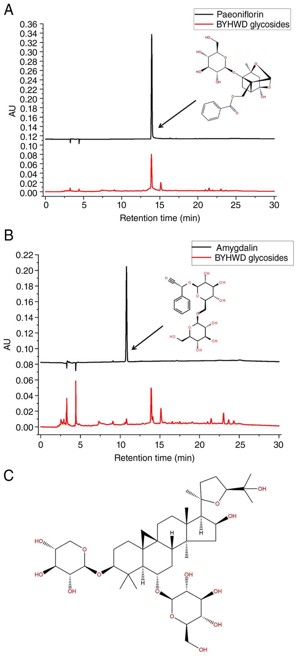
Paeoniflorin and amygdalin in BYHWD glycosides were
detected by HPLC and the two main components were quantitatively
analyzed by the external standard method as previously described
(١٧). The results are presented in Fig. 1A and B. In addition, since astragaloside IV
could not be well separated by HPLC, a standard curve of
astragaloside IV was established by improving the vanillin-sulfuric
acid colorimetric method previously described (17) and the content of astragaloside IV
in three types of compound was determined. The chemical structure
formula of astragaloside IV is shown in Fig. 1C. Astragaloside IV exhibited the
highest content, accounting for 12.61% of the total glycosides,
followed by paeoniflorin (6.61%) and amygdalin (1.04%). The
quantitative data of the main glycosides is summarized in Table III.
| Table IIIQuantitative analysis of main active
components of BYHWD glycosides |
Table III
Quantitative analysis of main active
components of BYHWD glycosides
| Component | BYHWD
glycosides |
|---|
| Astragaloside
IV | 12.61% |
| Paeoniflorin | 6.61% |
| Amygdalin | 1.04% |
Screening of safe and effective
concentrations of BYHWD glycosides
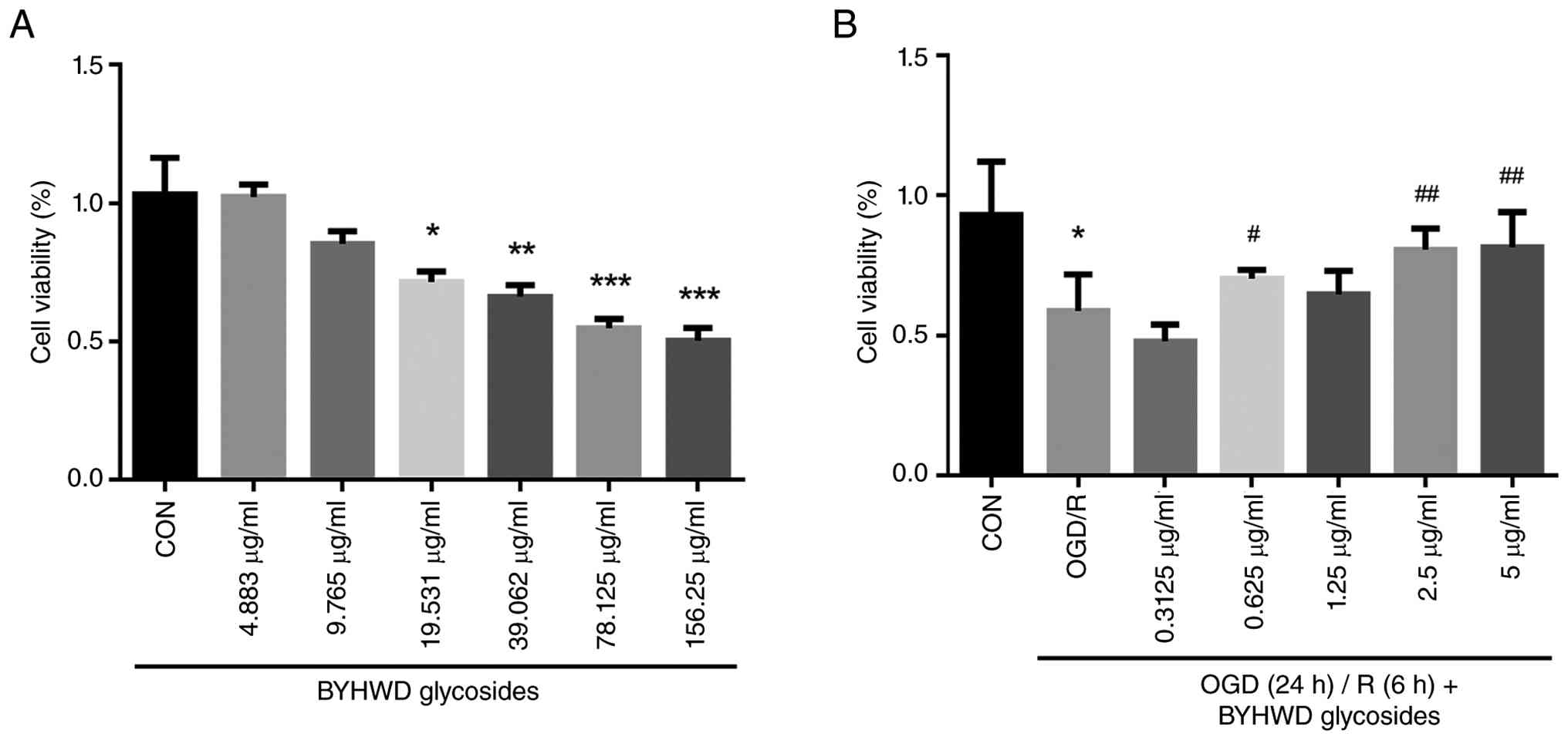
As shown in Fig.
2A, BYHWD glycosides showed significant dose-dependent toxicity
on H9C2 cells and significantly inhibited the viability of H9C2
cells when the concentration was >9.765 µg/ml. In addition, the
protective effects of BYHWD glycosides on the survival rate of H9C2
induced by OGD/R were evaluated. As shown in Fig. 2B, the survival rate of H9C2 cells
subjected to OGD/R was significantly lower compared with that of
the untreated group. However, BYHWD glycosides (0.625-5 µg/ml)
significantly increased the activity of H9C2 cells. These results
suggest that, in a specific concentration range, BYHWD glycosides
can promote the viability of H9C2 cells after OGD/R injury. This
provided a concentration reference for subsequent experiments.
Effects of BYHWD glycosides on the
oxidative stress level of H9C2 cells induced by OGD/R
Cardiomyocytes injured by ischemia-reperfusion are
regulated by processes such as autophagy and oxidative stress. ROS,
MDA and SOD are all important indicators to assess the level of
oxidative stress. Thus, these three indicators were measured in the
present study to assess the effects of BYHWD glycosides on
OGD/R-induced oxidative stress in H9C2 cells. The results are shown
in Fig. 3A-D. The contents of ROS
and MDA in the model group were significantly higher compared with
those in the untreated group, while the level of SOD in the model
group was significantly lower compared with that in the untreated
group. However, pre-administration of BYHWD glycosides
significantly inhibited the increase in ROS levels after modeling,
exhibiting dose-dependent effects. Compared with the model group,
BYHWD glycosides pre-treatment significantly reduced the elevated
MDA levels and significantly restored the depleted SOD activity
(Fig. 3).
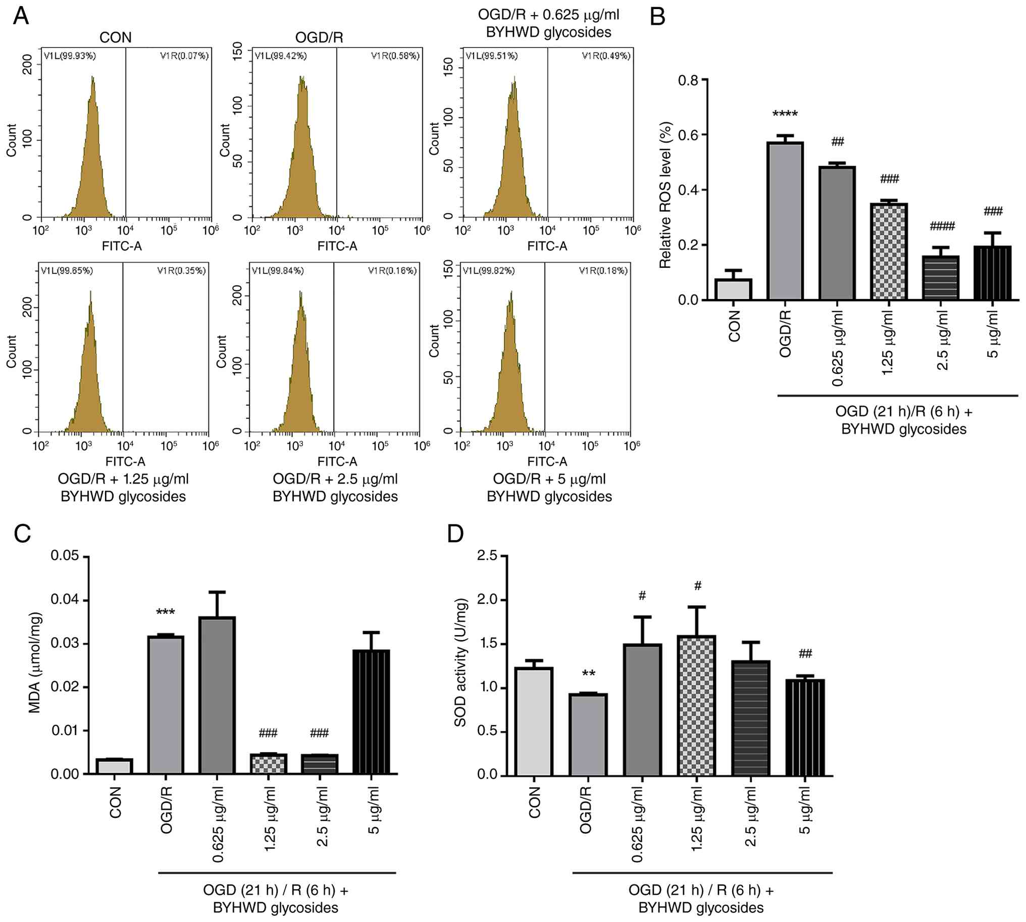 | Figure 3Effects of BYHWD glycosides on OGD/R
induced oxidative damage of H9C2 cells. (A) The level of ROS in
each group was detected by flow cytometry. (B) Quantitative
analysis of ROS by flow detection. (C) Detection of the MDA levels
in each group. (D) Detection results of the SOD levels in each
group. Data are expressed as the mean±SD (n=3).
**P<0.01, ***P<0.001,
****P<0.0001 vs. control, #P<0.05,
##P<0.01, ###P<0.001 and
####P<0.0001 vs. OGD/R group. OGD/R, oxygen-glucose
deprivation/reoxygenation; ROS, reactive oxygen species; BYHWD,
Buyang Huanwu Decoction; MDA, malondialdehyde; SOD, superoxide
dismutase; CON, control. |
Effects of BYHWD glycosides on the
autophagy of H9C2 cells induced by OGD/R
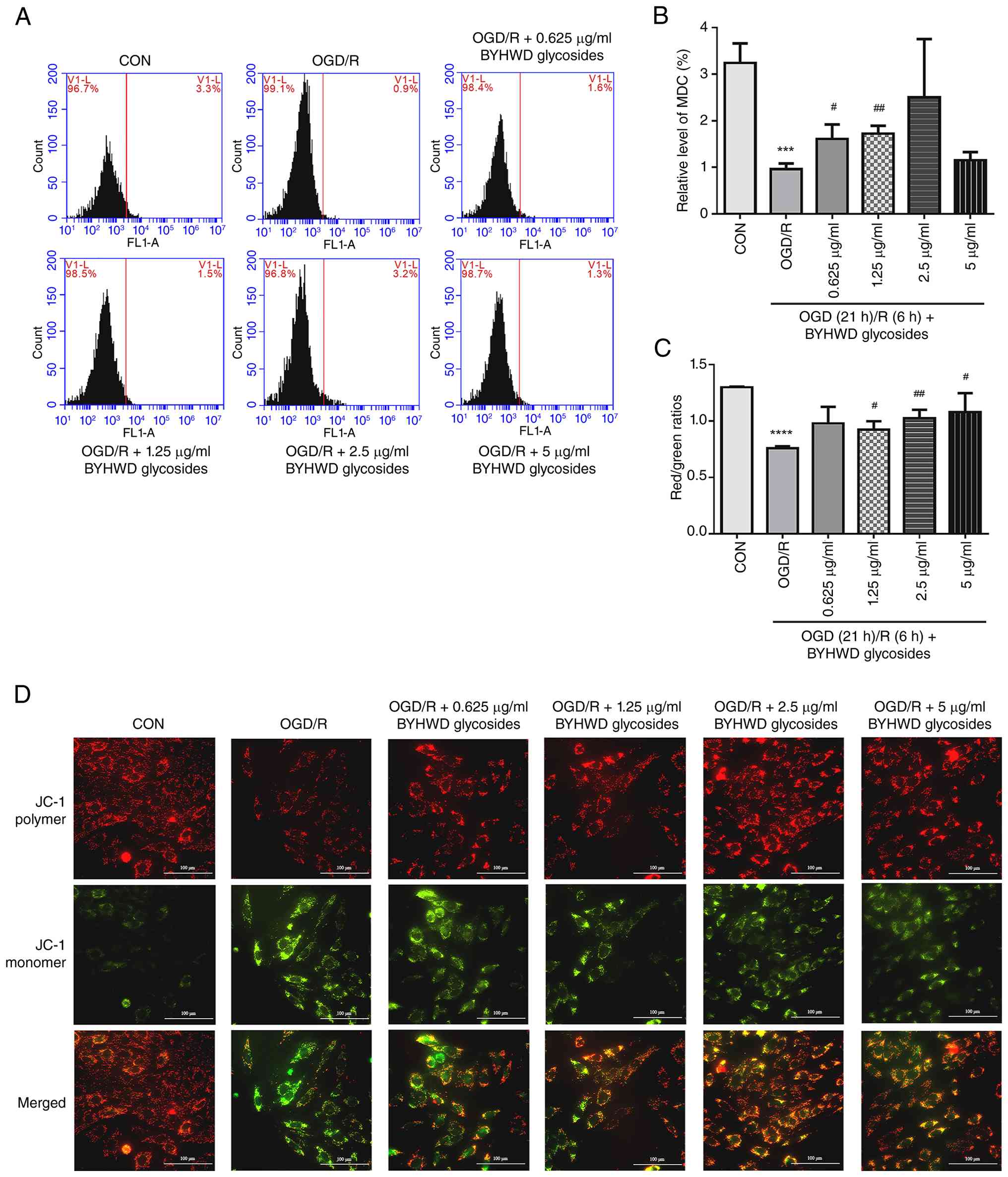
To detect the effects of BYHWD glycosides on
autophagy in H9C2 cells induced by OGD/R, MDC autophagy
fluorescence labeling was used and the autophagy levels in
cardiomyocytes were analyzed by flow cytometry. As shown in
Fig. 4A and B, the relative level of MDC in the OGD/R
model group was significantly lower compared with that in the
control group. This finding suggests that OGD/R-induced reperfusion
injury in H9C2 cardiomyocytes can inhibit the level of autophagy
(11,18). Pretreatment with BYHWD glycosides
significantly increased the relative levels of MDC-labeled
autophagic vacuoles specifically at doses of 0.625 and 1.25 µg/ml
compared with the OGD/R model group, indicating a potent promotion
of autophagic activity at these concentrations.
Effects of BYHWD glycosides on the MMP
of H9C2 cells induced by OGD/R
ROS production and mitochondrial dysfunction are
associated with mitophagy during reperfusion injury (11,13,18).
A normal membrane potential is key in maintaining proper
mitochondrial function. JC-1 is a widely used fluorescent probe
(١٩,20) for detecting MMP. In the present study, MMP was observed
using a fluorescence inverted microscope. The results of the JC-1
assay are shown in Fig. 4C and
D. In the control group, the cells
maintained a highly polarized MMP, indicating that JC-1 accumulated
in the matrix of mitochondria to form polymers, thus producing
strong red fluorescence. However, it appeared difficult for JC-1 to
accumulate in mitochondrial matrix in the OGD/R group; thus, the
MMP was observed to be lower, demonstrating a stronger green
fluorescence signal. Compared with the findings in the model group,
BYHWD glycosides were shown to promote the aggregation of JC-1 in
mitochondrial matrix and thus resulted in an increase of red/green
fluorescence intensity.
Effects of BYHWD glycosides on
mitochondrial autophagy-associated mRNA in H9C2 cells induced by
OGD/R
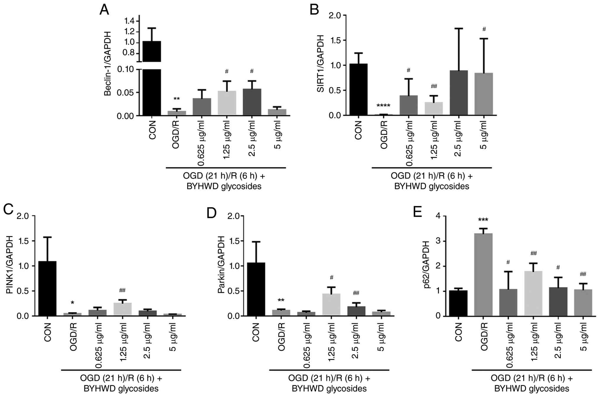
PINK1/Parkin-mediated activation of mitochondrial
autophagy helps to protect mitochondria from hypoxia-induced
decreases in MMP. As shown in Fig.
5A-E, the level of p62 mRNA in the model group was
significantly higher compared with that in the control group. The
expression levels of Beclin-1, SIRT1, PINK1 and Parkin were
significantly decreased in the model group, compared with the
control group . The majority of concentrations of BYHWD glycosides
were shown to significantly reverse the abnormal expression of
autophagy-associated mRNA after OGD/R injury. Among them, the
regulatory effect of 1.25 µg/ml BYHWD glycosides was found to be
the most notable.
Effects of BYHWD glycosides on
mitochondrial autophagy-associated proteins induced by OGD/R in
H9C2 cells
To further determine the effects of BYHWD glycosides
on the aforementioned mRNA levels, proteins associated with the
PINK1/Parkin mitochondrial autophagy pathway were detected using
western blotting analysis. As shown in Fig. 6A-G, compared with the untreated
group, the expression of Beclin-1, SIRT1, PINK1, Parkin and LC3II/I
protein in the OGD/R group decreased significantly, while the
expression of p62 protein increased significantly. In addition,
compared with the model group, the majority of BYHWD glycosides
were shown to significantly reverse the abnormal expression of
these proteins. The trends observed in these western blotting
results was found to be consistent with that of the present RT-qPCR
results, further demonstrating that OGD/R inhibited mitochondrial
autophagy in H9C2 cells. This suggested that treatment with BYHWD
glycosides may promote the autophagy level of H9C2 cells to varying
degrees and its mechanism of action may involve the PINK1/Parkin
mitochondrial autophagy pathway.
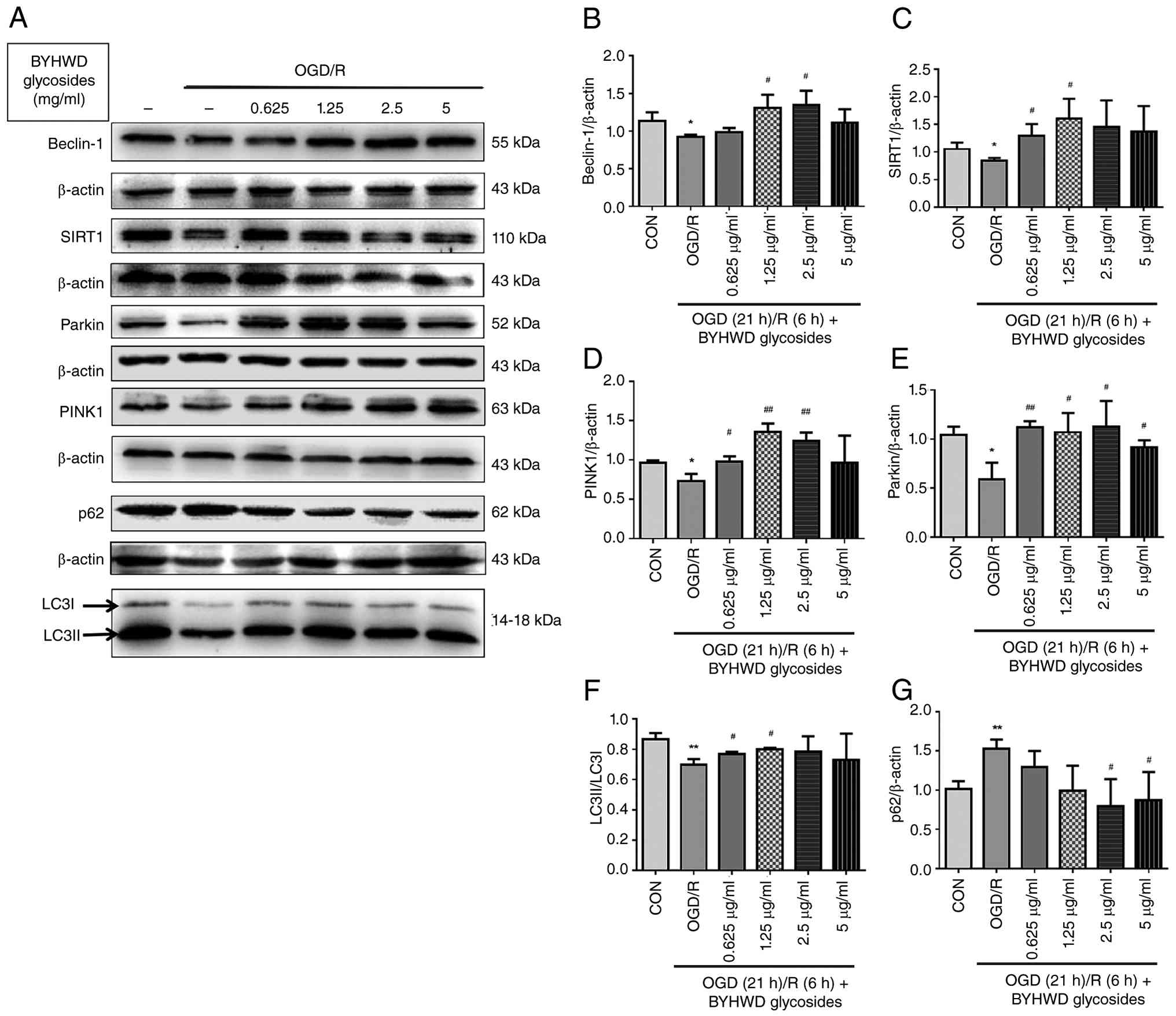 | Figure 6Effect of BYHWD glycosides on the
expression levels of Beclin-1, SIRT1, PINK1, Parkin, LC3 and p62 in
OGD/R-treated H9C2 cells detected by western blotting. (A-G)
Beclin-1, SIRT1, PINK1, Parkin, LC3 and p62 protein expression
levels. Data are expressed as the mean ± SD (n=3).
*P<0.05, **P<0.01 vs. control,
#P<0.05 and ##P<0.01 vs. OGD/R group.
SIRT1, silent mating type information regulation 2 homolog-1;
PINK1, PTEN-induced kinase 1; OGD/R, oxygen-glucose
deprivation/reoxygenation; BYHWD, Buyang Huanwu Decoction; CON,
control. |
Discussion
Myocardial ischemia is a common pathological
condition, with its prevalence in China increasing annually. During
myocardial ischemia, a marked number of cardiomyocytes undergo
necrosis and the affected area cannot effectively regenerate due to
its limited proliferative capacity. Currently, the clinical
application of revascularization therapy can effectively mitigate
or delay disease progression. However, accumulating evidence
suggests that, once blood flow is restored to the vessels and
myocardial tissue, the extent of myocardial damage may further
worsen, leading to the phenomenon of MIRI (1,15,21).
MIRI is one of the primary reasons for the poor prognosis of
ischemic heart disease and myocardial ischemia. Reducing MIRI is a
primary focus of cardiovascular disease research (14). At present, the mechanism of MIRI is
not completely clear. Previous studies (22,23)
have shown that it is associated with the accumulation of oxygen
free radicals, abnormal myocardial energy metabolism, mitochondrial
dysfunction, calcium overload, inflammation and autophagy.
Mitochondrial autophagy serves an important role in myocardial
reperfusion injury. Moderate and well-regulated mitochondrial
autophagy can enhance energy supply to cardiomyocytes, reduce the
production of ROS and protect myocardial function (24).
The method of replenishing qi and activating blood
circulation is widely used (25,26)
in the clinical treatment of MIRI. Compared with qi-tonifying drugs
or blood-activating agents used alone, Yiqi Huoxue compound
prescriptions may exert broader protective effects through
multi-component, multi-targeted actions. Previous studies (27,28)
have shown that BYHWD glycosides are the main effective components
against ischemic brain injury (٢٩). However, the role of this
component in the protection of ischemic myocardial injury is not
clear. In the present study, BYHWD glycosides were separated and
extracted by water extraction, alcohol precipitation and organic
solvent extraction. The main effective components of BYHWD include
astragaloside IV, paeoniflorin and amygdalin, which were determined
by HPLC. However, astragaloside IV could not be well separated by
HPLC. Thus, an ultraviolet spectrophotometer was used to detect the
content of astragaloside IV in accordance with the literature (١٧).
The results indicated that astragaloside IV exhibited the highest
content, followed by paeoniflorin, with amygdalin being the lowest
among the three main active components.
Oxidative stress has been shown to mediate the
pathological process of MIRI injury (30). Myocardial necrosis is initially
caused by ischemic injury, while further damage during reperfusion
results from metabolic alterations upon the restoration of ischemic
oxygen (31). The levels of SOD,
MDA and ROS can indirectly reflect antioxidant capacity and the
severity of oxidative damage in cells. Therefore, increasing the
activity of the antioxidant enzyme SOD while reducing MDA and ROS
can effectively protect cardiomyocytes from reperfusion injury
(٣٢,33). The findings of the present study thus align with previous
studies (34). The injury model of
H9C2 cells cultured in vitro under OGD/R conditions was
mitigated by BYHWD glycosides. Findings suggested that BYHWD
glycosides could significantly promote the rate of cell viability,
inhibit the levels of MDA and ROS and increase the level of SOD.
This indicated that BYHWD glycosides may have enhanced the
viability of H9C2 cells under OGD/R conditions, inhibited oxidative
stress and alleviated myocardial injury.
Mitochondrial autophagy is a specific autophagy
process for damaged mitochondria that helps maintain normal
intracellular mitochondrial function (٣٥). Previous studies have
shown that adaptive mitochondrial autophagy occurs during
reperfusion injury and protects cardiomyocytes by eliminating
dysfunctional mitochondria (11,18).
Acidic autophagy vesicles are released during mitochondrial
autophagy (10). As a fluorescent
probe, MDC can bind to such autophagy vesicles and emit strong
green fluorescence. In the present study, the fluorescence level of
MDC in the cells of each group was detected by flow cytometry. The
results suggested that normal H9C2 cardiomyocytes exhibit a certain
level of autophagic activity. After OGD/R modeling, the number of
autophagy vesicles decreased and thus the level of autophagy
decreased significantly. However, pretreatment with BYHWD
glycosides restored this autophagic activity to varying
degrees.
Furthermore, the stability of MMP is important in
maintaining normal mitochondrial function. Previous research has
demonstrated that effective mitochondrial protection can be
achieved by reducing excessive ROS secretion and enhancing MMP
(٣٦). Therefore, by detecting the changes in MMP within the present
study, whether mitochondrial autophagy alleviated mitochondrial
dysfunction in the model of reperfusion injury, was
investigated.
The results appeared to be consistent with the
literature (٣٧). The present study demonstrated that the
red-to-green fluorescence ratio in the OGD/R group was
significantly lower compared with that in the normal control group,
indicating mitochondrial damage and decreased MMP. However, BYHWD
glycosides significantly promoted MMP. These findings suggest that
BYHWD glycosides may promote autophagy, prevent the accumulation of
damaged mitochondria and further inhibit cardiomyocyte injury
induced by OGD/R. It has been established that autophagy exhibits a
dual mechanism of action during IRI (٣٨). While excessive
autophagic flux can lead to self-digestion and autophagic cell
death, moderate mitophagy is highly protective. In the specific
experimental setting of severe stress (oxygen glucose deprivation
for 21 h followed by 6 h of reoxygenation), the moderate
upregulation of mitophagy facilitated by the BYHWD
glycoside-enriched fraction is considered beneficial (٣٩). It
promotes the timely clearance of markedly damaged mitochondria,
thereby preventing the release of pro-apoptotic factors
(specifically cytochrome c) (40) and ROS, ultimately conferring a
survival advantage to cardiomyocytes.
The PINK1/Parkin pathway is a classical
mitochondrial autophagy pathway. Previous research has shown that
in response to a decrease in mitochondrial membrane potential,
PINK1 accumulates on the outer mitochondrial membrane and
subsequently recruits and activates Parkin. Activated Parkin then
ubiquitinates mitochondrial surface proteins, which tags the
damaged mitochondria for recognition and engulfment by
autophagosomes (41). Therefore,
the present study further explored the expression of signaling
molecules associated with the PINK1/Parkin pathway.
SIRT1, a NAD+-dependent deacetylase, is
widely distributed in mammals. Firstly, SIRT1 can directly act on
Beclin-1, enhancing its function through deacetylation, thereby
promoting autophagy (٤٢). Recent evidence has highlighted that the
deacetylase activity of SIRT1 is important in stabilizing the
PINK1/Parkin axis. Specifically, SIRT1-mediated deacetylation
events can enhance the stability of PINK1 on the outer
mitochondrial membrane, which subsequently facilitates the
recruitment and activation of Parkin (43). Furthermore, recent research
regarding MIR models have demonstrated that SIRT1 acts as a key
upstream regulator and its silencing directly suppresses
PINK1/Parkin-mediated cardioprotective mechanisms, such as the
clearance of dysfunctional mitochondria, attenuation of oxidative
stress and inhibition of mitochondria-dependent apoptosis (٤٤). The
present results aligned with this mechanism, suggesting that the
BYHWD glycoside-enriched fraction upregulates SIRT1, which may
consequently drive PINK1/Parkin-dependent mitophagic clearance to
protect cardiomyocytes against OGD/R injury. In addition, SIRT1
regulates the expression and activity of numerous transcription
factors and coactivators (such as FOXO3a and PGC-1α) associated
with mitochondrial proliferation and autophagy, serving an
important role in maintaining mitochondrial quality and quantity
(٤٥). Beclin-1 is a key regulator of the autophagy pathway,
recognized as a marker of autophagy initiation and positively
associated with autophagy levels (46). SIRT1 can promote autophagy by
deacetylation of the K430 and K437 sites of Beclin-1, which
facilitates its binding to VPS34 to assemble the active Class III
phosphatidylinositol 3-kinase (PI3K-III) complex, thereby directly
driving the initiation and maturation of autophagosomes (47). The present experimental results
demonstrated that BYHWD glycosides significantly reversed the
decreased expression of SIRT1 and Beclin-1 in cardiomyocytes
following reperfusion injury.
Secondly, the PINK1/Parkin pathway is one of the
most important pathways for mitochondrial autophagy (٤٨). SIRT1
promotes the deacetylation of PINK1, thereby promoting PINK1
accumulation and recruitment of Parkin protein to the mitochondrial
outer membrane. PINK1 accumulates and recruits Parkin protein to
the outer mitochondrial membrane, initiating mitochondrial
autophagy to clear damaged mitochondria and maintain cellular
homeostasis (٤٩). In the present study, the expression levels of
PINK1 and Parkin in the BYHWD glycosides-treated group were
significantly higher compared with those in the model group.
LC3 is not only an important marker of autophagy but
also an important downstream signal of PINK1/Parkin-mediated
mitophagy and it has two forms: LC3-I and LC3-II. At the beginning
of autophagy, LC3-I is converted into LC3-II; therefore, an
increased LC3-II/LC3-I ratio indicates the initiation of autophagy
(٥٠). p62 binds to ubiquitin substrates and LC3, targeting
autophagosomes and promoting the clearance of ubiquitinated
proteins and its levels are negatively associated with autophagy
activity. With the increase in LC3-II/LC3-I protein ratio, the
expression of p62 decreases, indicating an increase in autophagy
flux. The present results demonstrated a significant decrease in
the LC3-II/LC3-I protein ratio and a significant increase in p62
after OGD/R in H9C2 cardiomyocytes. However, BYHWD glycosides
effectively reversed the OGD/R-induced abnormal changes in these
markers (specifically, restoring the LC3-II/LC3-I ratio and
reducing p62 accumulation). These findings thus suggest that BYHWD
glycosides can enhance autophagy flux and promote mitochondrial
autophagy.
Notably, the biological mechanism of mitophagy
conceptually reflects the TCM principles of BYHWD. In TCM theory,
BYHWD is prescribed as is it believed to tonify qi, remove blood
stasis and clear pathological products (51). At the subcellular level, these
traditional concepts can be translated into precise biological
equivalents: The macroscopic concept of ‘blood stasis’ corresponds
microscopically to the physical accumulation of undegraded cellular
debris and dysfunctional organelles (such as damaged mitochondria),
while ‘pathological products’ represent toxic metabolic byproducts
(such as excessive ROS). By promoting mitophagy, the BYHWD
glycoside-enriched fraction effectively clears these damaged
organelles, thereby restoring cellular homeostasis. This represents
a notable intersection between modern molecular clearance
mechanisms and traditional macroscopic TCM theories.
Despite these promising findings, the present study
exhibits a number of limitations. First, without the use of
specific autophagy inhibitors (such as 3-methyladenine) or
PINK1/Parkin genetic knockdown models, the present data remain
primarily observational. Therefore, the present results demonstrate
an association rather than a definitive causal relationship between
mitophagy promotion, ROS reduction and the protective effects of
the BYHWD fraction. Second, the present study was conducted
exclusively using the H9C2 cardiomyoblast cell line. It must
therefore be acknowledged that there are physiological and
metabolic differences between these cells and primary adult
cardiomyocytes. Unlike terminally differentiated adult
cardiomyocytes that depend on fatty acid oxidation, H9C2 cells are
proliferating embryonic myoblasts that lack mature sarcomeres and
rely heavily on glycolysis (٥٢). As a result, their autophagic flux
and response to oxidative stress may not fully replicate the
behavior of mature heart tissue. Therefore, the cardioprotective
mechanisms observed in the present in vitro model should be
interpreted carefully when considering their translational
potential. To address these limitations, future research should aim
to incorporate rigorous loss-of-function experiments with specific
pharmacological inhibitors and small interfering RNA knockdown
techniques to establish direct causality. In addition, there are
plans to validate the present findings using primary adult rat
ventricular myocytes and in vivo MIRI models, such as left
anterior descending coronary artery ligation, to further evaluate
the systemic effects of the BYHWD fraction.
In conclusion, to the best of our knowledge, the
present findings indicated for the first time that BYHWD glycosides
can effectively inhibit OGD/R-induced oxidative damage in H9C2
cells, promote mitochondrial autophagy and stabilize mitochondrial
function. This mechanism may be associated with the classical
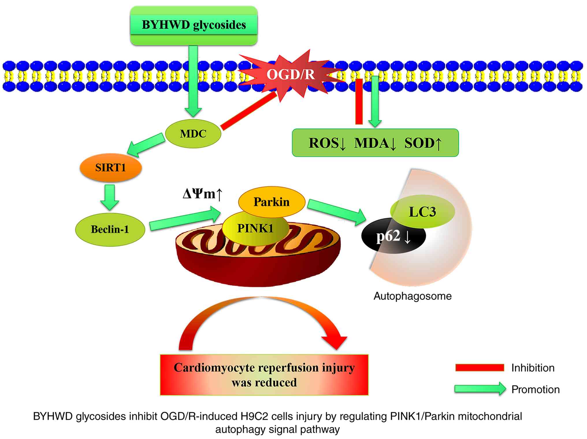
mitochondrial autophagy pathway of PINK1/Parkin (Fig. 7). The present results are therefore
important in the clinical application and validation of BYHWD
glycosides in treating MIRI.
Supplementary Material
Standard curve of astragaloside IV
measured by vanillin-sulfuric acid colorimetric method at 560 nm.
The regression equation is y=0.0551x+0.0774 (R2=0.985).
The content of astragaloside IV in the samples was calculated using
the following formula: Content of total astragaloside In
samples=[(y-yblank-0.0774)/0.0551]/concentration of
compound total glycosides (x100%).
Preparation of the astragaloside IV
standard curve for the vanillin-sulfuric acid colorimetric
method.
Determination of astragaloside IV in
BYHWD by vanillin-sulfuric acid colorimetric method.
Acknowledgements
Not applicable.
Funding
Funding: The present study was supported by the National Key
R&D Program of China under ‘Biological basic study of
supplementing qi and activating blood circulation based on vascular
lesions and its components in the treatment of cerebral
infarction/cardiac infarction’ and ‘Innovative study on the
etiology, pathogenesis, diagnosis and treatment of cerebral
infarction/myocardial infarction treated with Yiqi Huoxue recipe
based on the concept of simultaneous treatment of brain and heart’
(grant nos. 2019YFC1708604 and 2019YFC1708600).
Availability of data and materials
The data generated in the present study may be
requested from the corresponding author.
Authors' contributions
XJH was responsible for study conceptualization,
project administration, providing supervision and writing,
reviewing and editing the manuscript. JJF was responsible for study
conceptualization, data visualization and figure preparation,
methodology and protocol design, conducting the investigation,
curating data, performing formal analysis and writing the original
manuscript draft. TS was responsible for methodology development,
conducting the investigation, performing formal analysis,
validation of experimental reproducibility, and writing the
original manuscript draft. JHF was responsible for methodology
design and conducting the investigation. FMZ contributed to study
conceptualization and data interpretation, provided supervision and
wrote, reviewed and edited the manuscript. MYZ conducted the
investigation by performing molecular and cellular assays,
contributed to data interpretation, provided supervision, and
wrote, reviewed and edited the manuscript. ZSD was responsible for
study conceptualization and the provision of essential resources,
acquired the funding and wrote, reviewed and edited the manuscript.
JJF, TS, JHF, MYZ and XJH confirm the authenticity of all the raw
data. All authors have read and approved the final version of the
manuscript.
Ethics approval and consent to
participate
Not applicable.
Patient consent for publication
Not applicable.
Competing interests
The authors declare that they have no competing
interests.
References
|
1
|
Heusch G: Myocardial ischaemia-reperfusion
injury and cardioprotection in perspective. Nat Rev Cardiol.
17:773–789. 2020.PubMed/NCBI View Article : Google Scholar
|
|
2
|
Min F, Jia XJ, Gao Q, Niu F, Hu ZY, Han
YL, Shi HJ and Yu Y: Remote ischemic post-conditioning protects
against myocardial ischemia/reperfusion injury by inhibiting the
Rho-kinase signaling pathway. Exp Ther Med. 19:99–106.
2020.PubMed/NCBI View Article : Google Scholar
|
|
3
|
Moens AL, Claeys MJ, Timmermans JP and
Vrints CJ: Myocardial ischemia/reperfusion-injury, a clinical view
on a complex pathophysiological process. Int J Cardiol.
100:179–190. 2005.PubMed/NCBI View Article : Google Scholar
|
|
4
|
Zhang WW, Xu F, Wang D, Ye J and Cai SQ:
Buyang huanwu decoction ameliorates ischemic stroke by modulating
multiple targets with multiple components: In vitro evidences. Chin
J Nat Med. 16:194–202. 2018.PubMed/NCBI View Article : Google Scholar
|
|
5
|
Han X, Zhang G, Chen G, Wu Y, Xu T, Xu H,
Liu B and Zhou Y: Buyang huanwu decoction promotes angiogenesis in
myocardial infarction through suppression of PTEN and activation of
the PI3K/Akt signalling pathway. J Ethnopharmacol.
287(114929)2022.PubMed/NCBI View Article : Google Scholar
|
|
6
|
Liu Q, Li J, Wang J, Li J, Janicki JS and
Fan D: Effects and mechanisms of Chinese herbal medicine in
ameliorating myocardial ischemia-reperfusion injury. Evid Based
Complement Alternat Med. 2013(925625)2013.PubMed/NCBI View Article : Google Scholar
|
|
7
|
Wang R, Wang M, Zhou J, Wu D, Ye J, Sun G
and Sun X: Saponins in Chinese herbal medicine exerts protection in
myocardial ischemia-reperfusion injury: Possible mechanism and
target analysis. Front Pharmacol. 11(570867)2021.PubMed/NCBI View Article : Google Scholar
|
|
8
|
Fu X, Sun Z, Long Q, Tan W, Ding H, Liu X,
Wu L, Wang Y and Zhang W: Glycosides from buyang huanwu decoction
inhibit atherosclerotic inflammation via JAK/STAT signaling
pathway. Phytomedicine. 105(154385)2022.PubMed/NCBI View Article : Google Scholar
|
|
9
|
Wang QR: Yilin Gaicuo (Correcting the
Errors of Medical Works). People's Med Publ House, Beijing,
2005.
|
|
10
|
Xia JY, Chen C, Lin Q, Cui J, Wan J, Li Y
and Li D: Role of mitophagy in myocardial ischemia/reperfusion
injury and Chinese medicine treatment. Chin J Integr Med. 29:81–88.
2023.PubMed/NCBI View Article : Google Scholar
|
|
11
|
Guan Z, Chen J, Wang L, Hao M, Dong X, Luo
T, Jiang J, Lin Z, Li X, Chen P, et al: Nuanxinkang prevents the
development of myocardial infarction-induced chronic heart failure
by promoting PINK1/Parkin-mediated mitophagy. Phytomedicine.
108(154494)2022.PubMed/NCBI View Article : Google Scholar
|
|
12
|
Sun M, Zhang W, Bi Y, Xu H, Abudureyimu M,
Peng H, Zhang Y and Ren J: NDP52 protects against myocardial
infarction-provoked cardiac anomalies through promoting
autophagosome-lysosome fusion via recruiting TBK1 and RAB7.
Antioxid Redox Signal. 36:1119–1135. 2022.PubMed/NCBI View Article : Google Scholar
|
|
13
|
Bugger H and Pfeil K: Mitochondrial ROS in
myocardial ischemia reperfusion and remodeling. Biochim Biophys
Acta Mol Basis Dis. 1866(165768)2020.PubMed/NCBI View Article : Google Scholar
|
|
14
|
Matsuda N and Yamano K: Two sides of a
coin: Physiological significance and molecular mechanisms for
damage-induced mitochondrial localization of PINK1 and parkin.
Neurosci Res. 159:16–24. 2020.PubMed/NCBI View Article : Google Scholar
|
|
15
|
Horan DP, O'Malley TJ, Weber MP, Maynes
EJ, Choi JH, Patel S, Challapalli J, Luc JGY, Entwistle JW, Todd
Massey H, et al: Repair of ischemic ventricular septal defect with
and without coronary artery bypass grafting. J Card Surg.
35:1062–1071. 2020.PubMed/NCBI View Article : Google Scholar
|
|
16
|
Livak KJ and Schmittgen TD: Analysis of
relative gene expression data using real-time quantitative PCR and
the 2(-delta delta C(T)) method. Methods. 25:402–408.
2001.PubMed/NCBI View Article : Google Scholar
|
|
17
|
Le AV, Parks SE, Nguyen MH and Roach PD:
Improving the vanillin-sulphuric acid method for quantifying total
saponins. Technologies. 6(84)2018.
|
|
18
|
Li S, Zhang J, Liu C, Wang Q, Yan J, Hui
L, Jia Q, Shan H, Tao L and Zhang M: The role of mitophagy in
regulating cell death. Oxid Med Cell Longev.
2021(6617256)2021.PubMed/NCBI View Article : Google Scholar
|
|
19
|
Smiley ST, Reers M, Mottola-Hartshorn C,
Lin M, Chen A, Smith TW, Steele GD Jr and Chen LB: Intracellular
heterogeneity in mitochondrial membrane potentials revealed by a
J-aggregate-forming lipophilic cation JC-1. Proc Natl Acad Sci USA.
88:3671–3675. 1991.PubMed/NCBI View Article : Google Scholar
|
|
20
|
Perelman A, Wachtel C, Cohen M, Haupt S,
Shapiro H and Tzur A: JC-1: Alternative excitation wavelengths
facilitate mitochondrial membrane potential cytometry. Cell Death
Dis. 3(e430)2012.PubMed/NCBI View Article : Google Scholar
|
|
21
|
Vallabhajosyula S, Vallabhajosyula S,
Burstein B, Ternus BW, Sundaragiri PR, White RD, Barsness GW and
Jentzer JC: Epidemiology of in-hospital cardiac arrest complicating
non-ST-segment elevation myocardial infarction receiving early
coronary angiography. Am Heart J. 223:59–64. 2020.PubMed/NCBI View Article : Google Scholar
|
|
22
|
Hausenloy DJ and Yellon DM: Myocardial
ischemia-reperfusion injury: A neglected therapeutic target. J Clin
Invest. 123:92–100. 2013.PubMed/NCBI View
Article : Google Scholar
|
|
23
|
Frank A, Bonney M, Bonney S, Weitzel L,
Koeppen M and Eckle T: Myocardial ischemia reperfusion injury: From
basic science to clinical bedside. Semin Cardiothorac Vasc Anesth.
16:123–132. 2012.PubMed/NCBI View Article : Google Scholar
|
|
24
|
Wei L, Wang J, Chen A, Liu J, Feng X and
Shao L: Involvement of PINK1/parkin-mediated mitophagy in ZnO
nanoparticle-induced toxicity in BV-2 cells. Int J Nanomedicine.
12:1891–1903. 2017.PubMed/NCBI View Article : Google Scholar
|
|
25
|
Hao P, Jiang F, Cheng J, Ma L, Zhang Y and
Zhao Y: Traditional Chinese medicine for cardiovascular disease:
Evidence and potential mechanisms. J Am Coll Cardiol. 69:2952–2966.
2017.PubMed/NCBI View Article : Google Scholar
|
|
26
|
Zhang J, Hu Y, Wang H, Hou J, Xiao W, Wen
X, Wang T, Long P, Jiang H, Wang Z, et al: Advances in research on
the protective mechanisms of traditional Chinese medicine (TCM) in
myocardial ischaemia-reperfusion injury. Pharm Biol. 60:931–948.
2022.PubMed/NCBI View Article : Google Scholar
|
|
27
|
Wang K, Lei L, Cao J, Liang R, Duan J,
Feng Z, Ding Y, Ma Y, Yang Z and Zhang E: Network
pharmacology-based prediction of the active compounds and mechanism
of buyang huanwu decoction for ischemic stroke. Exp Ther Med.
22(1050)2021.PubMed/NCBI View Article : Google Scholar
|
|
28
|
Chen Y, Hu Z, Li Y, Lan X, Bai Y, Long H,
Deng C and Zou L: Pharmacologically active constituents of buyang
huanwu decoction against cerebral ischemia-reperfusion injury and
verification of its effects on oxidative stress, energy metabolism,
and inflammation. J Ethnopharmacol. 353(120405)2025.PubMed/NCBI View Article : Google Scholar
|
|
29
|
She Y, Shao L, Zhang Y, Hao Y, Cai Y,
Cheng Z, Deng C and Liu X: Neuroprotective effect of glycosides in
buyang huanwu Decoction on pyroptosis following cerebral
ischemia-reperfusion injury in rats. J Ethnopharmacol.
242(112051)2019.PubMed/NCBI View Article : Google Scholar
|
|
30
|
González-Montero J, Brito R, Gajardo AI
and Rodrigo R: Myocardial reperfusion injury and oxidative stress:
Therapeutic opportunities. World J Cardiol. 10:74–86.
2018.PubMed/NCBI View Article : Google Scholar
|
|
31
|
Lesnefsky EJ, Chen Q, Tandler B and Hoppel
CL: Mitochondrial dysfunction and myocardial ischemia-reperfusion:
Implications for novel therapies. Annu Rev Pharmacol Toxicol.
57:535–565. 2017.PubMed/NCBI View Article : Google Scholar
|
|
32
|
Li XM, Luo XG, Li K, Wang N, Hua EB, Zhang
Y and Zhang TC: Difference in protective effects of three
structurally similar flavonoid glycosides from hypericum ascyron
against H2O2-induced injury in H9c2
cardiomyoblasts. Mol Med Rep. 12:5423–5428. 2015.PubMed/NCBI View Article : Google Scholar
|
|
33
|
Xu H, Deng R, Li ETS, Shen J and Wang M:
Pinosylvin provides neuroprotection against cerebral ischemia and
reperfusion injury through enhancing PINK1/Parkin mediated
mitophagy and Nrf2 pathway. J Funct Foods. 71(84)2020.
|
|
34
|
Jiang M, Ni J, Cao Y, Xing X, Wu Q and Fan
G: Astragaloside IV attenuates myocardial ischemia-reperfusion
injury from oxidative stress by regulating succinate,
lysophospholipid metabolism, and ROS scavenging system. Oxid Med
Cell Longev. 2019(9137654)2019.PubMed/NCBI View Article : Google Scholar
|
|
35
|
Jones E, Gaytan N, Garcia I, Herrera A,
Ramos M, Agarwala D, Rana M, Innis-Whitehouse W, Schuenzel E and
Gilkerson R: A threshold of transmembrane potential is required for
mitochondrial dynamic balance mediated by DRP1 and OMA1. Cell Mol
Life Sci. 74:1347–1363. 2017.PubMed/NCBI View Article : Google Scholar
|
|
36
|
Zhang H, Zhang RH, Liao XM, Yang D, Wang
YC, Zhao YL, Xu GB, Liu CH, Li YJ, Liao SG and Zhou M: Discovery of
β-carboline derivatives as a highly potent cardioprotectant against
myocardial ischemia-reperfusion injury. J Med Chem. 64:9166–9181.
2021.PubMed/NCBI View Article : Google Scholar
|
|
37
|
Zeng RY, Qiu ZC, Zhao ST, Lai SQ, Qiu RB,
Wu ZM, Xu ZQ, Li JN, Luo XD, Wang BL and Wan L: Astragaloside IV
mitigates myocardial ischemia/reperfusion injury by modulating the
14-3-3η/GPX4 axis, reducing mitochondrial dysfunction, ferroptosis,
and apoptosis. Phytomedicine. 155(158082)2026.PubMed/NCBI View Article : Google Scholar
|
|
38
|
Ma S, Wang Y, Chen Y and Cao F: The role
of the autophagy in myocardial ischemia/reperfusion injury. Biochim
Biophys Acta. 1852:271–276. 2015.PubMed/NCBI View Article : Google Scholar
|
|
39
|
Saito T and Sadoshima J: Molecular
mechanisms of mitochondrial autophagy/mitophagy in the heart. Circ
Res. 116:1477–1490. 2015.PubMed/NCBI View Article : Google Scholar
|
|
40
|
Borutaite V, Jekabsone A, Morkuniene R and
Brown GC: Inhibition of mitochondrial permeability transition
prevents mitochondrial dysfunction, cytochrome c release and
apoptosis induced by heart ischemia. J Mol Cell Cardiol.
35:357–366. 2003.PubMed/NCBI View Article : Google Scholar
|
|
41
|
Burman JL, Pickles S, Wang C, Sekine S,
Vargas JNS, Zhang Z, Youle AM, Nezich CL, Wu X, Hammer JA and Youle
RJ: Mitochondrial fission facilitates the selective mitophagy of
protein aggregates. J Cell Biol. 216:3231–3247. 2017.PubMed/NCBI View Article : Google Scholar
|
|
42
|
Chen TS, Chuang SY, Shen CY, Ho TJ, Chang
RL, Yeh YL, Kuo CH, Mahalakshmi B, Kuo WW and Huang CY: Antioxidant
Sirt1/Akt axis expression in resveratrol pretreated adipose-derived
stem cells increases regenerative capability in a rat model with
cardiomyopathy induced by diabetes mellitus. J Cell Physiol.
236:4290–4302. 2021.PubMed/NCBI View Article : Google Scholar
|
|
43
|
Wei X, Xiong X, Wang P, Zhang S and Peng
D: SIRT1-mediated deacetylation of FOXO3 enhances mitophagy and
drives hormone resistance in endometrial cancer. Mol Med.
30(147)2024.PubMed/NCBI View Article : Google Scholar
|
|
44
|
Liao Y, Ke B, Long X, Xu J and Wu Y:
Abnormalities in the SIRT1-SIRT3 axis promote myocardial
ischemia-reperfusion injury through ferroptosis caused by silencing
the PINK1/parkin signaling pathway. BMC Cardiovasc Disord.
23(582)2023.PubMed/NCBI View Article : Google Scholar
|
|
45
|
Wang L, Xu C, Johansen T, Berger SL and
Dou Z: SIRT1-a new mammalian substrate of nuclear autophagy.
Autophagy. 17:593–595. 2021.PubMed/NCBI View Article : Google Scholar
|
|
46
|
Sun Y, Yao X, Zhang QJ, Zhu M, Liu ZP, Ci
B, Xie Y, Carlson D, Rothermel BA, Sun Y, et al: Beclin-1-dependent
autophagy protects the heart during sepsis. Circulation.
138:2247–2262. 2018.PubMed/NCBI View Article : Google Scholar
|
|
47
|
Zhong Y, Wang QJ, Li X, Yan Y, Backer JM,
Chait BT, Heintz N and Yue Z: Distinct regulation of autophagic
activity by Atg14L and rubicon associated with Beclin
1-phosphatidylinositol-3-kinase complex. Nat Cell Biol. 11:468–476.
2009.PubMed/NCBI View Article : Google Scholar
|
|
48
|
Deng Z, Sun M, Wu J, Fang H, Cai S, An S,
Huang Q, Chen Z, Wu C, Zhou Z, et al: SIRT1 attenuates
sepsis-induced acute kidney injury via Beclin1
deacetylation-mediated autophagy activation. Cell Death Dis.
12(217)2021.PubMed/NCBI View Article : Google Scholar
|
|
49
|
Li W, Du M, Wang Q, Ma X, Wu L, Guo F, Ji
H, Huang F and Qin G: FoxO1 promotes mitophagy in the podocytes of
diabetic male mice via the PINK1/parkin pathway. Endocrinology.
158:2155–2167. 2017.PubMed/NCBI View Article : Google Scholar
|
|
50
|
Chai D, Shan H, Wang G, Li H, Fang L, Song
J, Zhang Q, Bai J and Zheng J: AIM2 is a potential therapeutic
target in human renal carcinoma and suppresses its invasion and
metastasis via enhancing autophagy induction. Exp Cell Res.
370:561–570. 2018.PubMed/NCBI View Article : Google Scholar
|
|
51
|
Chen B, Xu Y, Tian F, Liu Y, Ouyang Y,
Zeng F, Peng Y and Liu B: Buyang huanwu decoction promotes
angiogenesis after cerebral ischemia through modulating
caveolin-1-mediated exosome MALAT1/YAP1/HIF-1α axis. Phytomedicine.
129(155609)2024.PubMed/NCBI View Article : Google Scholar
|
|
52
|
Zordoky BN and El-Kadi AO: H9c2 cell line
is a valuable in vitro model to study the drug metabolizing enzymes
in the heart. J Pharmacol Toxicol Methods. 56:317–322.
2007.PubMed/NCBI View Article : Google Scholar
|