Introduction
Meningiomas are the most common primary intracranial
tumors in adults, accounting for 40% of all intracranial tumors. It
is estimated that the incidence rate is 951/100,000 individuals,
most of whom are women, and the ratio of men to women is ~1:2.3
(1,2). Angiomatous meningioma (AM) is a
relatively rare type of meningioma, accounting for ~2.1% of the 15
subtypes of meningioma. Meningiomas are further stratified into
three grades based on their biological behavior: i) WHO Grade I
(benign), consisting of meningothelial (syncytial) meningioma,
fibrous (fibroblastic) meningioma, transitional (mixed) meningioma,
psammomatous meningioma, angiomatous meningioma, microcystic
meningioma, secretory meningioma, lymphoplasmacyte-rich meningioma
and metaplastic meningioma; ii) WHO Grade II (atypical), consisting
of clear cell meningioma, chordoid meningioma and atypical
meningioma; and iii) WHO Grade III (anaplastic), consisting of
papillary meningioma, rhabdoid meningioma and anaplastic
meningioma. AM is notably vascular, soft in texture and tends to
develop numerous small perforating arteries that integrate with
adjacent brain tissues, increasing their susceptibility to
bleeding. The most prevalent sites for AMs include the cerebral
convexity, as indicated by Hasselblatt et al (3) (42%), Ben Nsir et al (4) (58.6%) and Liu et al (5) (66.7%). AM with cystic degeneration is
even less common (6,7). Most reports are confined to
individual case studies. A total of 8 cases have been reported in
the published literature to date. Several potential mechanisms
account for cystic alterations in AM (8). Intratumoral apoplexy may cause
necrosis and further form cystic cavities inside the tumor. Glial
cells surrounding the tumor proliferate and secrete fluid
persistently, mostly generating cysts in peritumoral brain tissues.
Peritumoral brain tissue with edema or demyelination can develop
cystic lesions, and isolated fluid spaces in edematous areas may
combine into large peritumoral cysts. Trapped subarachnoid spaces
between tumor and brain tissue hinder fluid outflow, contributing
to progressive peritumoral cyst formation. These cysts lie in the
interval between the tumor and adjacent brain tissue (8). In the present study, a case of AM was
treated at The Affiliated Hospital of Hebei University (Baoding,
China) in August 2023. This case offers new diagnostic references
for similar imaging presentations and practical experience for
managing cystic-degenerated AM.
Case report
A 58-year-old man was admitted to The Affiliated
Hospital of Hebei University in August 2023 with a bilateral
frontal distending headache that had appeared 18 days before
without obvious triggering factors and was gradually worsening.
There were no other accompanying symptoms such as nausea, vomiting,
visual disturbance, limb weakness or seizures. At 15 days after the
onset of the headache, the patient visited a local hospital (Xushui
District Hospital of Traditional Chinese Medicine, Baoding, China),
where a computed tomography (CT) scan of the head revealed a space
occupying lesion in the left parietal lobe. After 3 days, the
patient was admitted to The Affiliated Hospital of Hebei University
for further diagnosis and treatment. During the outpatient visit to
the hospital, a physical examination revealed no obvious positive
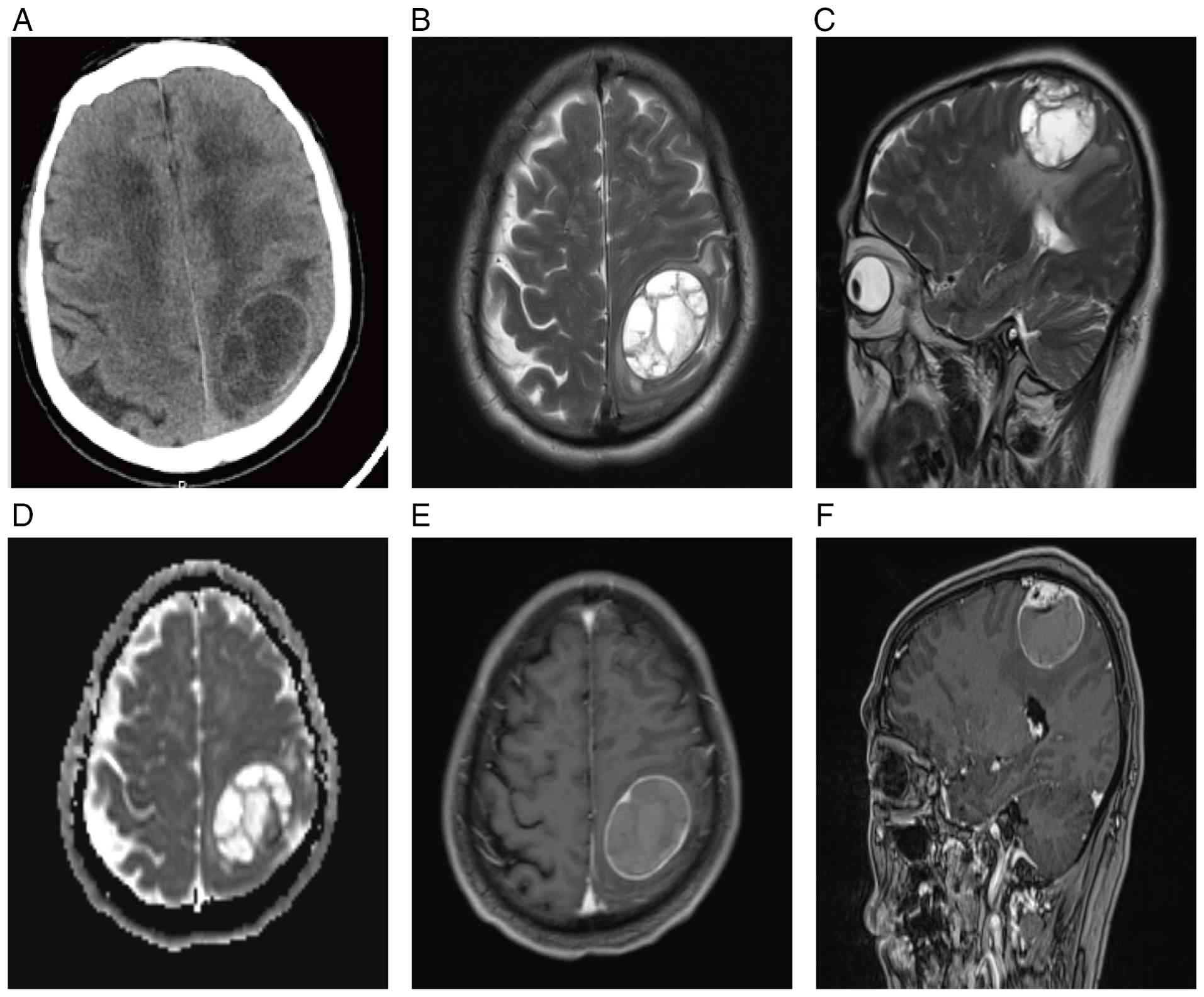
signs of nervous system involvement. A head CT scan showed a mass
lesion in the left parietal region (Fig. 1A), and a magnetic resonance imaging
(MRI) plain scan with enhancement was recommended. This scan showed
a cystic mass at the left parietal region, which was considered a
teratoid tumor (Fig. 1B-F).
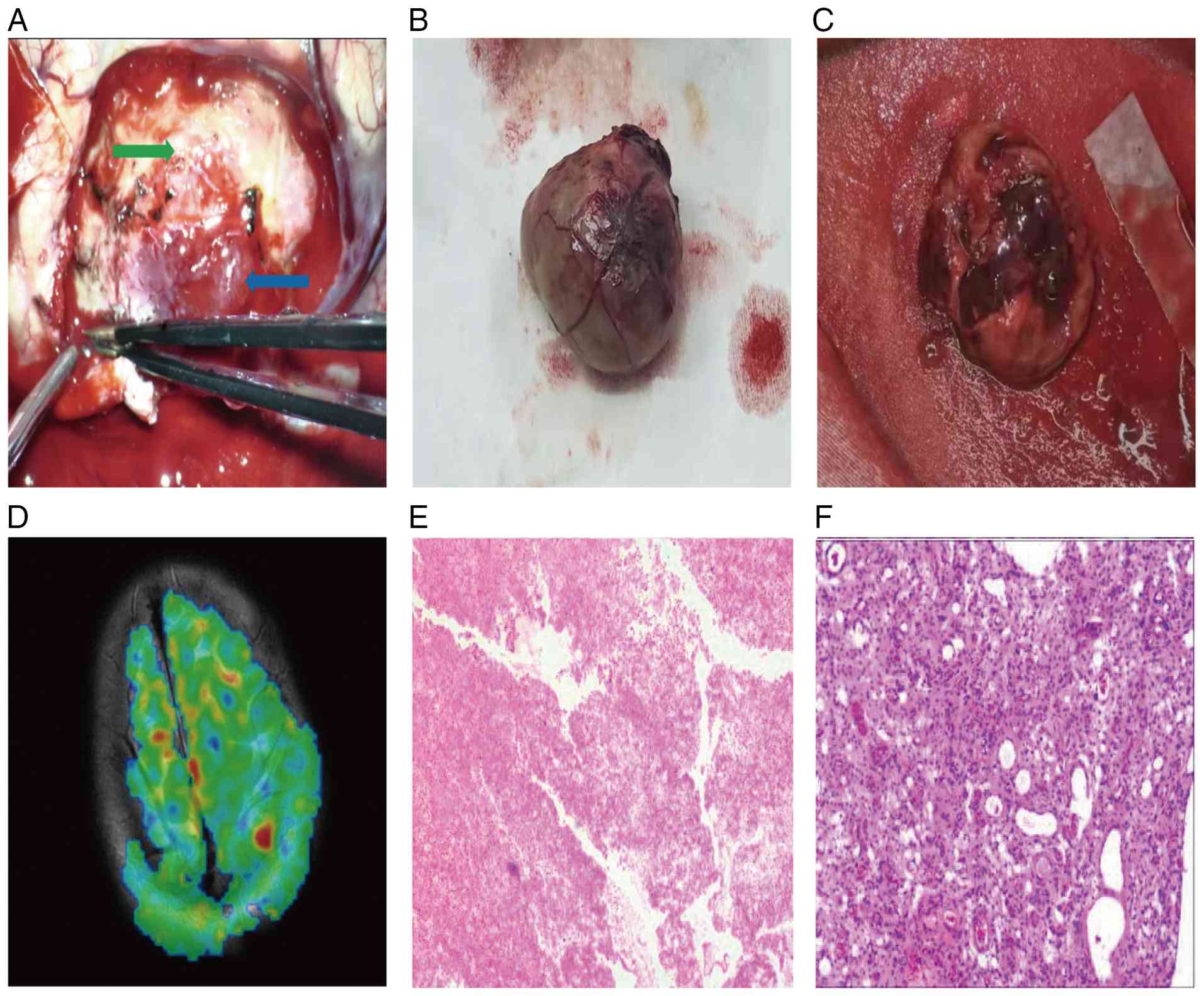
Three-dimensional arterial spin labeling (3D-ASL) showed local high
perfusion in the left parietal mass lesion (Fig. 2D). The preoperative diagnosis was
of a potential left parietal lobe teratoma. During surgery, a
horseshoe-shaped incision was made in the parietal region on the
left side, and after the milling cutter freed the bone flap,
extensive bleeding was observed in the dura mater. Following
hemostasis, the dura mater was suspended. The dura mater was cut in
an arc with the sagittal sinus as the base. In the center of the
surgical area, there was severe adhesion between the dura mater and
tumor tissue. The surgeon used electrocoagulation to cut off
adhesions. After removing the dura mater, the solid part of the
tumor appeared to be purple-red in color, with a soft texture and
abundant blood supply. The cyst wall of the cystic part of the
tumor was grayish-white, tough and had an arachnoid space with
surrounding brain tissue (Fig.
2A). Blunt separation was performed along the gap between the
tumor and brain tissue. During the separation process, the
junctional area of the tumor was separated, and some dark red
bloody fluid leaked out. Next, a puncture needle was inserted along
the fistula, with its tip kept centered in the cyst (to avoid
puncturing solid-component blood vessels), and 7 ml of dark red
bloody fluid was slowly aspirated (to prevent a sudden drop in
intracranial pressure), leading to a notable decrease in tumor
tension. After electrocoagulation was used to seal the fistula, the
separation continued, and the tumor and capsule were completely
removed (Simpson type 1 resection; a standard classification for
meningioma resection degree, where grade I indicates complete
resection of the tumor, the attached dura and abnormal bone, with
the lowest recurrence risk) (9).
After the tumor was detached, its gross appearance was spherical,
with tortuous blood vessels on the surface. Upon incision, the cyst
wall was not thick and there were partitions inside, containing a
large number of blood clots (Fig.
2B and C). The preoperative
bilateral limb muscle strength was recorded as grade 5.
Postoperatively, bilateral limb muscle strength was preserved at
Medical Research Council (MRC) grade 5, as evaluated using the MRC
muscle strength grading system (10), the gold standard 6-point ordinal
scale for quantitative neurological assessment of skeletal muscle
function. A CT scan showed a complete resection of the tumor. The
postoperative pathological report revealed a large amount of
coagulation and necrotic tissue, with meningeal tissue. For
pathological processing, the surgical specimen was fixed in 10%
neutral buffered formalin at room temperature for 24 h, then
embedded in paraffin and sectioned into 4-µm thick slices.
Histochemical staining with hematoxylin and eosin was performed at
room temperature for 10 min, and the sections were observed under a
light microscope, with a magnification of x200 for the vascular
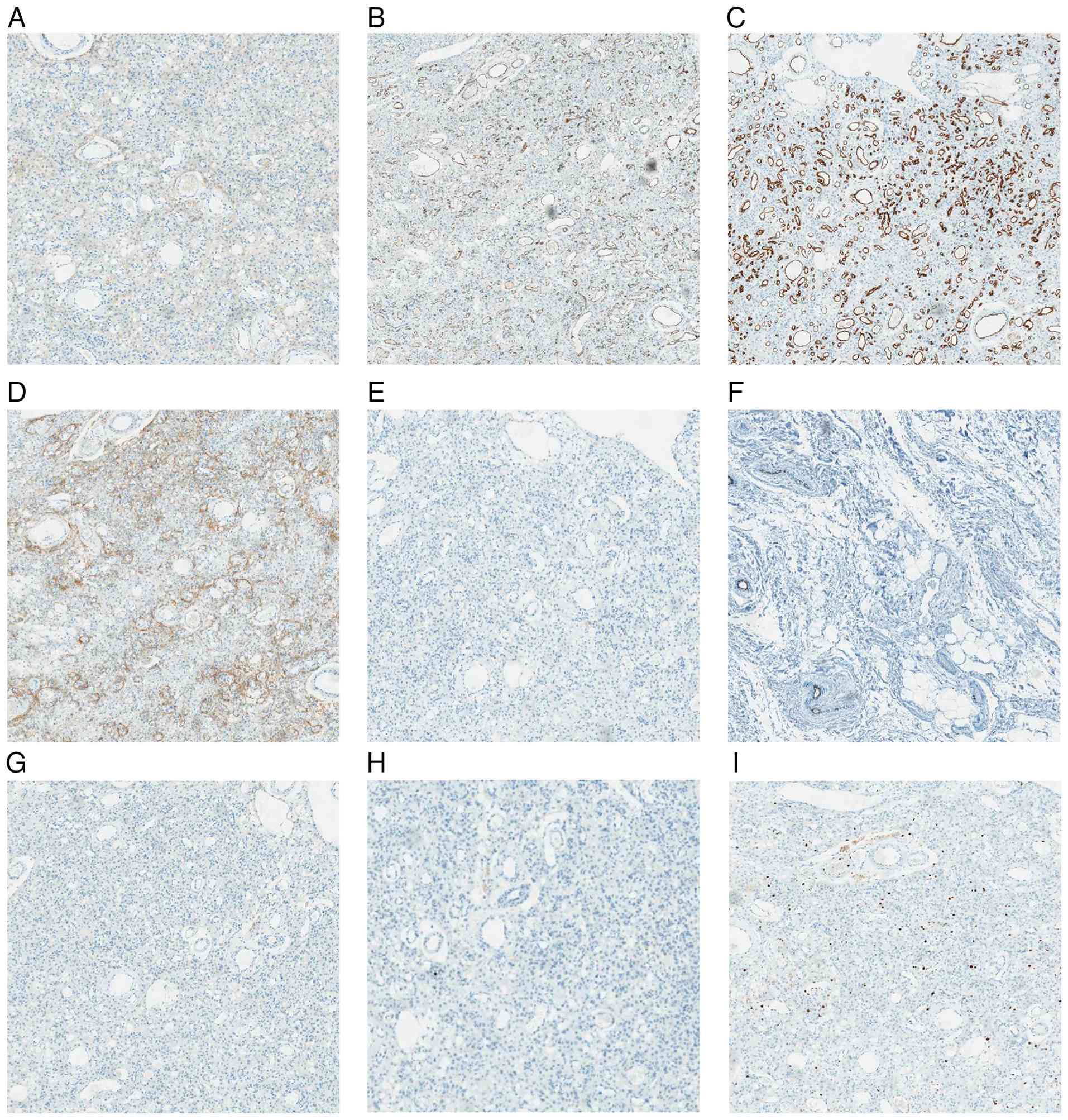
structure observation. Histochemical staining showed AM (Fig. 2E and F). Immunohistochemistry also indicated
positive results for epithelial membrane antigen, CD31 and CD34,
and a lack of staining for desmin, glial fibrillary acidic protein,
progesterone receptor (PR), S-100 and neuron-specific enolase. The
positive rate of Ki-67 was 2% (Fig.
3A-I). Endogenous peroxidase blocking reagent (cat. no.
PV-9000; Beijing Zhongshan Golden Bridge Biotechnology Co., Ltd.)
was incubated with the tissues at 37˚C for 10 min. The following
ready-to-use primary antibodies (all Beijing Zhongshan Jinqiao
Biotechnology Co., Ltd.) were applied and incubated overnight at
4˚C: Epithelial membrane antigen (cat. no. ZM-0095UM), CD31 (cat.
no. ZA-0568UM), CD34 (cat. no. ZM-0046UM), desmin (cat. no.
ZA-0610UM), glial fibrillary acidic protein (cat. no. ZA-0529UM),
PR (cat. no. ZA-0255UM), S-100 protein (cat. no. ZA-0225UM),
neuron-specific enolase (cat. no. ZM-0203UM) and Ki-67 (cat. no.
ZM-0167UM). Secondary antibody incubation was performed with
horseradish peroxidase-conjugated enhanced enzyme-labeled goat
anti-mouse/rabbit IgG polymer (cat. no. PV-9000; ready-to-use;
Beijing Zhongshan Jinqiao Biotechnology Co., Ltd.) and incubated at
37˚C for 30 min. This system was a biotin-free polymer detection
system. The sections were observed and images were captured under a
light microscope at a magnification of x200. These results were
consistent with the histopathological characteristics of AM. The
patient received no further treatment. At the 6-month follow-up
after surgery, no tumor recurrence was observed.
Discussion
AM has the histological and clinical characteristics
of a benign meningioma, and according to the 2021 WHO Central
Nervous System Tumor Classification, it belongs to the WHO Grade I
tumor group (6). The difference
between AM and other subtypes of meningioma is that AM is a highly
vascular tumor tissue, rich in abnormal vascular components in the
tumor body, with an extremely rich blood supply (11,12).
The study by Hasselblatt et al (3) suggested that meningiomas with
vascular components covering >50% of the tumor area can be
diagnosed as AM. AM is most common on the convex surface of the
brain and generally progresses slowly. Most patients have no
symptoms at the beginning, while some patients may exhibit symptoms
of chronic intracranial hypertension, such as seizures, focal
neurological disorders, sudden headaches or cranial nerve paralysis
(12). The patient in the current
case presented with a persistent headache, which may be related to
the continuous increase in tumor volume leading to dysfunction of
intracranial pressure compensation and the appearance of
intracranial hypertension.
In clinical practice, meningiomas are generally
diagnosed through CT combined with imaging examinations such as
MRI, with the characteristic features of MRI being especially
important. Meningiomas are isointense on T1-weighted imaging (T1WI)
and hyperintense on T2WI and fluid-attenuated inversion recovery,
with uniform and consistent enhancement and an obvious meningeal
tail sign on contrast-enhanced scans (13,14).
The imaging manifestations of AM are notably different from those
of classical meningiomas. Usually, AM are mainly characterized by
low signal intensity on T1WI and high signal intensity on T2WI. The
incidence of peritumoral brain edema (PTBE) is high, with moderate
or severe PTBE being the main manifestation in AM. Enhanced MRI
shows uniform enhancement (15).
Severe PTBE has always been regarded as the imaging diagnostic
basis for AM (16), and studies
have shown that this may be related to abnormal activation of the
vascular endothelial growth factor A pathway and changes in tumor
capillary length in AM (17).
However, in the present case, only mild PTBE was observed on MRI
T2WI, and the meningeal tail sign and vascular flow void shadows on
MRI enhanced scan were also atypical, which may be related to
cystic degeneration of the tumor. Based on the postoperative
pathological findings of hemorrhagic necrotic tissue and meningeal
tissue in this case, its cystic degeneration may be associated with
the rupture and hemorrhage of abnormal intratumoral blood vessels,
as well as the necrosis and liquefaction of local tissues, a
mechanism also mentioned in previous cases of cystic
angioblastoma-type meningioma (AM) (7,8).
Regarding the mild peritumoral edema in the present case, unlike
the severe peritumoral edema caused by overactivation of the VEGF-A
pathway in typical AM, although VEGF was not detected by
immunohistochemistry in this case, the ‘buffering effect’ of the
cystic structure may have alleviated the mechanical compression of
the tumor on the surrounding brain tissue (17). Meanwhile, the reduced stimulation
of necrotic tissue on vascular endothelium further diminished the
edema severity. The absence of typical vascular flow voids is
attributed to the high proportion of cystic components in the
tumor, coupled with the low vascular density and small caliber of
the intratumoral blood vessels in the solid component (as evidenced
by the slender vascular structures in pathological examination),
which failed to reach the threshold for MRI detection of flow voids
(18). The appearance of vascular
flow void shadows in AM on enhanced MRI is generally ‘root like’,
and some studies have described it as a ‘sunrise sign’ and used it
as a basis for differentiation from other blood-rich tumors
(19,20), but it is not applicable to this
patient. In addition, AM often presents a high signal on the
apparent diffusion coefficient (ADC), which is related to the tumor
tissue containing a large number of blood vessels and having low
resistance to water diffusion. This is consistent with the findings
of the patient in the present case.
In summary, the imaging findings of this patient are
not consistent with the typical presentation of AM: The solid
component of the tumor accounted for a smaller proportion of the
total lesion volume, while the cystic part was large, the PTBE was
not severe, the meningeal tail sign was not obvious on enhanced
scanning, there were few vascular flow void shadows and the
morphology was atypical. Overall, the imaging findings of this case
differ from those of typical AM. Compared with prior reported
cystic AM, it has a higher cystic proportion (far exceeding 50%)
and an absence of mild vascular flow voids, which are the main
cause of the preoperative misdiagnosis as teratoma (7). For a differential diagnosis, using
core evidence of a high ADC signal (excluding highly proliferative
tumors) + focal hyperperfusion in the solid part on 3D-ASL
(supporting hypervascular tumors), it is readily distinguishable
from cystic gliomas (low ADC signal) and intracranial metastases
(often with multiple nodules) (21). This approach offers a reference for
cases with atypical imaging. The key features are a low solid
proportion, high cystic proportion, mild peritumoral edema,
indistinct dural tail sign on enhancement, few vascular flow voids
and atypical morphology. The study by Galldiks et al
(14) also demonstrated that due
to the limitations of CT and MRI, introducing positron emission
tomography (PET) into preoperative examinations of meningiomas can
help in forming more accurate imaging diagnoses for cases with
atypical routine imaging examinations. In addition, the development
of radiomics, texture analysis based radiomics methods, ADC whole
tumor histogram analysis, pseudo-continuous arterial spin labeling
perfusion imaging and other methods have also provided new ideas
for the preoperative diagnosis of AM (21-23).
In addition, although the application of digital subtraction
angiography (DSA) in the diagnosis of meningiomas is constantly
declining, DSA can still provide important assistance for
preoperative diagnosis and surgical planning when important blood
vessels such as the sagittal sinus are infiltrated by tumors
(13). This also suggests that PET
or DSA examination may provide ideas for the preoperative diagnosis
in cases of blood-rich tumors with atypical imaging findings.
At present, the main treatment method for AM is
still surgery, and the degree of resection is an important factor
reflecting the prognosis. The Simpson classification method is
commonly used to distinguish the degree of resection (9). However, AM is highly vascular and may
even be confused with vascular malformations on histological
analysis. AM is one of the most blood-rich intracranial tumors. In
a previous study, the mean estimated amount of intraoperative
bleeding of the AM was 1,350 ml, and 13.8% of patients required a
blood transfusion to safely conclude the surgery (4). In the present case, during the
surgical procedure, the surgeon first disconnected the solid part
of the tumor from the dura mater, severed the blood supply artery
of the tumor and was able to extract a portion of the cystic fluid
for decompression during the resection process, thereby removing
all of the tumor and its attached dura mater, achieving a Simpson
type 1 resection. However, this surgical approach is only
applicable to AM with minimal solid, predominant cystic components
and 3D-ASL showing focal hyperperfusion confined to the solid
component (not global tumor hyperperfusion). The approach is
contraindicated in hypervascular AM with a solid proportion of
>50% and a vascularity score of ≥4(4); blind application to these cases may
cause a sudden intraoperative blood loss surge from tumor vessel
rupture during puncture decompression, far exceeding the reported
mean of 1,350 ml, and even induce cerebral herniation. In the
present study, the postoperative pathological report revealed
coagulation and necrotic tissue under the microscope, with
meningeal tissue also observed. The patient was followed up for 6
months after surgery. Postoperative 3-month and 6-month head MRI
reexaminations showed no tumor recurrence, and the surgical area
had no residual bleeding or edema. Neurological function evaluation
showed that the patient's bilateral limb muscle strength remained
at grade 5, with no symptoms such as headache, dizziness or limb
sensory disturbance, and the patient had returned to normal daily
life. Study limitations include a relatively short follow-up
duration (6 months); thus, the long-term recurrence risk and
stability of neurological function in cystic AM require further
validation through prolonged follow-up.
In conclusion, the imaging findings of the patient
in the present case are not typical, which poses certain
difficulties for the preoperative diagnosis. This case report
describes the rare imaging manifestations of cystic AM, providing
new diagnostic references for patients with similar imaging
manifestations. When encountering blood-rich tumors with atypical
imaging manifestations, such as AM, and when traditional imaging
examinations cannot meet diagnostic requirements, the introduction
of new radiomics methods is a wise choice. PET or DSA examination
may also provide evidence for the preoperative diagnosis of AM.
With the continuous intersection of imaging technology and other
disciplines, a pathological analysis based on MRI will also greatly
help diagnose different subtypes of meningiomas, and provide
assistance in formulating surgical planning and determining
clinical prognosis. At the same time, in order to completely remove
the tumor, first disconnecting the tumor supply artery and then
removing the cystic fluid for decompression is a safe and effective
surgical technique in the treatment process of such patients, and
blood transfusion should be performed when necessary to safely
conclude the surgical process.
Acknowledgements
Not applicable.
Funding
Funding: The study was supported by the Medicine and Health
Science and Technology Plan of Zhejiang Provincial Health
Commission (grant no. 2023RC306) and the Youth Research Fund
Project of Affiliated Hospital of Hebei University (grant no.
2023QB02).
Availability of data and materials
The data generated in the present study may be
requested from the corresponding author.
Authors' contributions
ZTB, YJS and HGL collected the clinical case data
and were major contributors to the manuscript. YW collected and
analyzed the imaging data of the patient. HPL performed the
postoperative follow-up of the patient. HPX participated in the
pathological section evaluation and analysis. SAY and JYC guided
the surgical management of patients, advised on perioperative
treatment plans and reviewed the manuscript. XCW and ZMZ supervised
the research, designed the study and critically revised the
manuscript for important intellectual content. ZTB and ZMZ confirm
the authenticity of all the raw data. All authors have read and
approved the final version of the manuscript.
Ethics approval and consent to
participate
Not applicable.
Patient consent for publication
The patient provided written informed consent for
publication of the case report and any accompanying images.
Competing interests
The authors declare that they have no competing
interests.
References
|
1
|
Nassiri F, Liu J, Patil V, Mamatjan Y,
Wang JZ, Hugh-White R, Macklin AM, Khan S, Singh O, Karimi S, et
al: A clinically applicable integrative molecular classification of
meningiomas. Nature. 597:119–125. 2021.PubMed/NCBI View Article : Google Scholar
|
|
2
|
Ostrom QT, Price M, Neff C, Cioffi G,
Waite KA, Kruchko C and Barnholtz-Sloan JS: CBTRUS statistical
report: Primary brain and other central nervous system tumors
diagnosed in the united states in 2015-2019. Neuro Oncol. 24 (Suppl
5):v1–v95. 2022.PubMed/NCBI View Article : Google Scholar
|
|
3
|
Hasselblatt M, Nolte KW and Paulus W:
Angiomatous meningioma: A clinicopathologic study of 38 cases. Am J
Surg Pathol. 28:390–393. 2004.PubMed/NCBI View Article : Google Scholar
|
|
4
|
Ben Nsir A, Chabaane M, Krifa H, Jeme H
and Hattab N: Intracranial angiomatous meningiomas: A 15-year,
multicenter study. Clin Neurol Neurosurg. 149:111–117.
2016.PubMed/NCBI View Article : Google Scholar
|
|
5
|
Liu Z, Wang C, Wang H, Wang Y, Li JY and
Liu Y: Clinical characteristics and treatment of angiomatous
meningiomas: A report of 27 cases. Int J Clin Exp Pathol.
6:695–702. 2013.PubMed/NCBI
|
|
6
|
Wen PY and Packer RJ: The 2021 WHO
Classification of tumors of the central nervous system: Clinical
implications. Neuro Oncol. 23:1215–1217. 2021.PubMed/NCBI View Article : Google Scholar
|
|
7
|
Xia J, Shan M, Sun J and Hou M: A rare
case of angiomatous meningioma with cystic degeneration. Asian J
Surg. 46:5612–5613. 2023.PubMed/NCBI View Article : Google Scholar
|
|
8
|
Sun Y, Yang S, Chang S and Wang H:
Angiomatous meningioma with cystic change: 4 cases report and
literature review. Egyp J Neur. 40(68)2025.
|
|
9
|
Simpson D: The recurrence of intracranial
meningiomas after surgical treatment. J Neurol Neurosurg
Psychiatry. 20:22–39. 1957.PubMed/NCBI View Article : Google Scholar
|
|
10
|
James MA: Use of the medical research
council muscle strength grading system in the upper extremity. J
Hand Surg Am. 32:154–156. 2006.PubMed/NCBI View Article : Google Scholar
|
|
11
|
Whittle IR, Smith C, Navoo P and Collie D:
Meningiomas. Lancet. 363:1535–1543. 2004.PubMed/NCBI View Article : Google Scholar
|
|
12
|
Hua L, Luan S, Li H, Zhu H, Tang H, Liu H,
Chen X, Bozinov O, Xie Q and Gong Y: Angiomatous meningiomas have a
very benign outcome despite frequent peritumoral edema at onset.
World Neurosurg. 108:465–473. 2017.PubMed/NCBI View Article : Google Scholar
|
|
13
|
Goldbrunner R, Stavrinou P, Jenkinson MD,
Sahm F, Mawrin C, Weber DC, Preusser M, Minniti G, Lund-Johansen M,
Lefranc F, et al: EANO guideline on the diagnosis and management of
meningiomas. Neuro Oncol. 23:1821–1834. 2021.PubMed/NCBI View Article : Google Scholar
|
|
14
|
Galldiks N, Albert NL, Sommerauer M, Grosu
AL, Ganswindt U, Law I, Preusser M, Le Rhun E, Vogelbaum MA, Zadeh
G, et al: PET imaging in patients with meningioma-report of the
RANO/PET Group. Neuro Oncol. 19:1576–1587. 2017.PubMed/NCBI View Article : Google Scholar
|
|
15
|
Zhang T, Yu JM, Wang YQ, Yin DD and Fang
LJ: WHO grade I meningioma subtypes: MRI features and pathological
analysis. Life Sci. 213:50–56. 2018.PubMed/NCBI View Article : Google Scholar
|
|
16
|
Yang L, Ren G and Tang J: Intracranial
angiomatous meningioma: A clinicopathological study of 23 cases.
Int J Gen Med. 13:1653–1659. 2020.PubMed/NCBI View Article : Google Scholar
|
|
17
|
Nassehi D, Sørensen LP, Dyrbye H, Thomsen
C, Juhler M, Laursen H and Broholm H: Peritumoral brain edema in
angiomatous supratentorial meningiomas: an investigation of the
vascular endothelial growth factor A pathway. APMIS. 121:1025–1036.
2013.PubMed/NCBI View Article : Google Scholar
|
|
18
|
Chen X, Li F, Xu G, Su J, Shi Q and Dai H:
Cerebellar metastasis manifesting as a cyst with mural nodule(s):
Differentiating it from hemangioblastoma on MRI. World Neurosurg.
175:e994–e1004. 2023.PubMed/NCBI View Article : Google Scholar
|
|
19
|
Wang C, Xu Y, Xiao X, Zhang J, Zhou F and
Zhao X: Role of intratumoral flow void signs in the differential
diagnosis of intracranial solitary fibrous tumors and meningiomas.
J Neuroradiol. 43:325–330. 2016.PubMed/NCBI View Article : Google Scholar
|
|
20
|
Li X, Lu Y, Xiong J, Wang D, She D, Kuai
X, Geng D and Yin B: Presurgical differentiation between malignant
haemangiopericytoma and angiomatous meningioma by a radiomics
approach based on texture analysis. J Neuroradiol. 46:281–287.
2019.PubMed/NCBI View Article : Google Scholar
|
|
21
|
He W, Xiao X, Li X, Guo Y, Guo L, Liu X,
Xu Y, Zhou J and Wu Y: Whole-tumor histogram analysis of apparent
diffusion coefficient in differentiating intracranial solitary
fibrous tumor/hemangiopericytoma from angiomatous meningioma. Eur J
Radiol. 112:186–191. 2019.PubMed/NCBI View Article : Google Scholar
|
|
22
|
Ashizawa K, Ogura K, Nagase S, Sakaguchi
A, Tokugawa J, Hishii M, Fukunaga M, Hirose T and Matsumoto T: A
collision tumor of solitary fibrous tumor/hemangiopericytoma and
meningioma: A case report with literature review. Pathol Int.
71(697e706)2021.PubMed/NCBI View Article : Google Scholar
|
|
23
|
Koizumi S, Sakai N, Kawaji H, Takehara Y,
Yamashita S, Sakahara H, Baba S, Hiramatsu H, Sameshima T and Namba
H: Pseudo-continuous arterial spin labeling reflects vascular
density and differentiates angiomatous meningiomas from
non-angiomatous meningiomas. J Neurooncol. 121:549–556.
2015.PubMed/NCBI View Article : Google Scholar
|