Introduction
Complete cytoreductive surgery with no residual
disease remains the gold standard for managing patients with
ovarian cancer, as it is associated with improved prognosis
(1-4).
To achieve complete cytoreduction, diaphragmatic resection is
occasionally recommended (5-9).
However, this procedure carries a relatively rare risk of
developing diaphragmatic hernia (10), with left-sided hernias being more
frequent than the right-sided ones (11). The incidence of left diaphragmatic
herniation in the literature following left diaphragm peritonectomy
or resection is 2.2% (12).
Although uncommon, diaphragmatic hernia can lead to severe clinical
manifestations and requires adequate management. The present case
report details a delayed left diaphragmatic hernia, 4 months after
interval cytoreductive surgery in a patient with advanced ovarian
cancer.
Case report
A 57-year-old woman had a history of mediastinal
large B-cell non-Hodgkin lymphoma treated with
Adriamycin-cyclophosphamide-vindesine-bleomycin and prednisone
(ACVBP) chemotherapy alone. The patient was diagnosed with
high-grade papillary serous ovarian adenocarcinoma, with extensive
peritoneal carcinomatosis involving the peritoneum and mesentery
and associated with ascites (PCI 34/39). The patient was managed at
the Gustave Roussy Institute (Paris, France) in August 2009. At
diagnosis, the CA125 level of the patient was 12,000 and the CA19.9
level was 157. Preliminary tests also included gastroscopy and
colonoscopy, both of which were normal.
Neoadjuvant chemotherapy was initiated with a
carboplatin area under the free carboplatin plasma concentration
vs. time curve value of 5 and weekly paclitaxel (80 mg/m²).
Paclitaxel was further adjusted during the third cycle of
chemotherapy to 60 mg/m² due to grade 3 hepatic toxicity. After
three cycles, the CA125 level of the patients decreased to 162 and
laparoscopic re-evaluation showed a reduction in tumor burden. The
patient then underwent an interval cytoreduction that included an
en-bloc peritoneal stripping of both diaphragmatic domes and
Morrison's pouch, without diaphragmatic or pleural injury, along
with omento-splenectomy, cholecystectomy, transverse and left
colectomy, posterior pelvic exenteration, fulguration of mesenteric
lesions and bilateral pelvic and para-aortic lymphadenectomy. A
complete resection of the peritoneum of the left diaphragmatic dome
was performed due to its extensive involvement. Pathological
examination confirmed the persistence of extensive residual
disease. The patient made a full recovery ≤1 month and resumed
chemotherapy for five additional cycles.
The patient developed an asymptomatic lymphocele
that did not require intervention. Computed tomography (CT) imaging
at 2 months postoperatively still showed moderate lymphocele, and a
CT scan 4 days prior to hernia surgery also demonstrated persistent
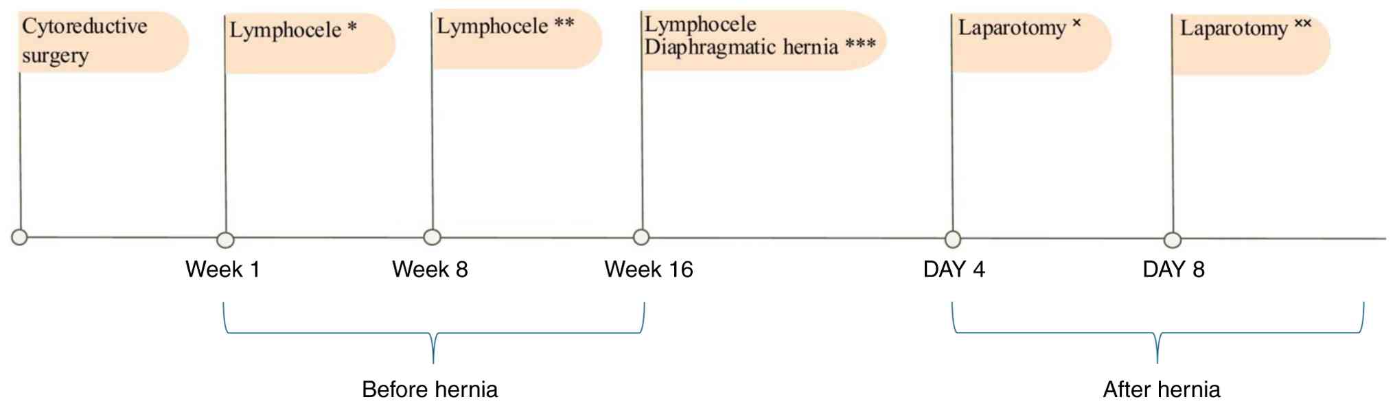
lymphatic effusion (Fig. 1). This
patient presented with malnutrition and prior to cytoreductive
surgery, the patient reported marked anorexia. The patients also
lost >10% of their body weight ≤3 months postoperatively and
their serum albumin level (measured 2 days after cytoreduction) was
1.3 g/dl. A few days before hernia surgery, serum albumin level of
the patient had increased to 3.1 g/dl.
At 4 months after cytoreductive surgery, the patient
presented with subocclusive symptoms. CT scan did not reveal a
digestive occlusion, but showed instead gastric distension with
horizontal displacement of the stomach, occupying the lower half of
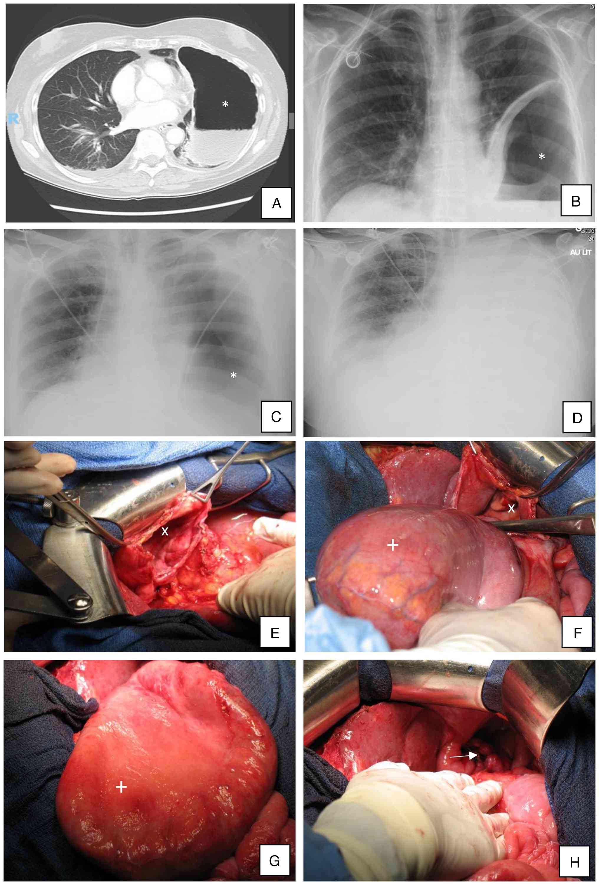
the left hemithorax, with ascites (Fig. 2A). Chest X-ray revealed elevation
of the left hemidiaphragm, marked gastric distension and partial
atelectasis of the left lower lobe (Fig. 2B). The clinical evolution was
favorable with medical treatment, allowing the discharge of the
patient. However, 3 days later, the patient was readmitted to the
intensive care unit for complete bowel obstruction and multiorgan
failure, as well as shock. Chest imaging suggested a left
pneumothorax (Fig. 2C and D). The patient was intubated for
hypercapnic respiratory failure, followed by post-intubation
collapse requiring resuscitation. Infectious tests were
negative.
Following the diagnosis of a left diaphragmatic
hernia with intrathoracic gastric migration, surgical exploration
confirmed a 4 cm defect in the left diaphragm through which the
entire stomach had herniated. The gastric hernia was reduced
without signs of ischemia. Gastric serosal sutures were further
added for reinforcement. Diaphragmatic defect was closed using
U-shaped stitches and non-absorbable running sutures (Fig. 2E-H).
The immediate postoperative period was complicated
by multiple reinterventions (Fig.
1). At 4 days after surgery, the patient developed severe
nausea and vomiting. An upper gastrointestinal endoscopy revealed
extensive necrosis of the gastric mucosa. Exploratory laparotomy
demonstrated a markedly distended stomach without evidence of
ischemia or perforation. At 4 days after this reoperation, a
deglobulization decrease in hemoglobin levels prompted a
thoraco-abdominopelvic CT scan, which revealed hemoperitoneum. An
emergency laparotomy was therefore indicated. No active bleeding
source was identified. Careful exploration of the entire digestive
tract revealed a pre-perforative area in the ileum. This lesion
required resection of a 10-cm ileal segment, followed by the
creation of a double-barrel ileostomy in the left iliac fossa. The
patient did not have a recurrence of their diaphragmatic hernia,
but experienced a relapse of their underlying disease 1 year later,
which ultimately caused mortality.
Discussion
Delayed diaphragmatic hernia is a rare, but serious
postoperative complication that can occur weeks to a range of 3 to
18 months after major cytoreductive surgery. In studies involving
patients who underwent hyperthermic intraperitoneal chemotherapy
(HIPEC), the interval is much shorter, 2 to 4 weeks (13,14).
Among patients treated with bevacizumab, the delay ranges between 6
and 12 weeks (13,14). Similar to the case described in the
present case report, the majority of cases reported in literature
report signs of intestinal obstruction as initial symptoms
(14-18).
The respiratory failure experienced by this patient might be the
result of a delayed diagnosis, leading to a more complex clinical
presentation. Notably, to the best of our knowledge, only one
similar case has been described in the literature, involving a
delayed diagnosis and postoperative complications after hernia
repair (15). This warrants
particular attention because of its diagnostic complexity and
potential therapeutic consequences (15). Clinical presentation may be
non-specific at first: Vomiting and non-specific abdominal
discomfort, before progressing to more pronounced gastrointestinal
or respiratory symptoms, including hemodynamic instability. Several
differential diagnoses should be considered in such cases: Pleural
effusion, pneumonia or pneumothorax. In the present case, chest
imaging initially suggested a left pneumothorax (Fig. 2C and D), the patient presented with hypercapnic
respiratory failure.
The initial CT scan demonstrated gastric migration
into the thoracic cavity; however, this finding was interpreted as
a subocclusive syndrome with marked gastric distension rather than
a diaphragmatic hernia. Because the patient showed clinical
improvement under conservative medical management, the index of
suspicion for diaphragmatic hernia was low, and the possibility of
a hernia was not reconsidered during follow-up. This diagnostic
oversight illustrates the intrinsic difficulty in recognizing this
rare complication, which can closely mimic more common
postoperative conditions, such as a bowel obstruction, as
aforementioned in the present case report. The case therefore
highlights the importance of maintaining a high index of suspicion
and contributes considerably to the existing literature by
reinforcing the need for heightened diagnostic vigilance.
Awareness of this condition is essential for early
recognition and intervention, before the onset of severe
complications (19). CT is
considered the gold standard for diagnosis, as it allows precise
evaluation of the size, location and contents of the diaphragmatic
defect (20). Despite its
limitations, standard chest X-ray remains a reasonable screening
tool. The most common radiographic finding is an opacity of the
hemithorax, often associated with dilated bowel loops and the
presence of intrathoracic air-fluid levels (21).
Surgery is the treatment of choice in symptomatic
patients. For small diaphragmatic defects, primary closure with
non-absorbable sutures is the fundamental principle. It is
essential to ensure a tension-free closure with good
vascularization of the margins. A synthetic or biological xenogenic
mesh may be used depending on the size of the hernia orifice
(20). Since the defect measured
~4 cm in diameter, tension free closure was possible without the
use of synthetic mesh. Hernia defect closure was performed using a
series of simple Vicryl 1 ‘U-shaped’ stitch sutures.
The most common cause of delayed diaphragmatic
hernia is partial or complete diaphragmatic resection during
cytoreduction, particularly when combined with HIPEC. Mechanical
trauma from surgery, along with the thermal effects of HIPEC, can
compromise the structural integrity of the diaphragm (13-17).
Other contributing factors may include pharmacologic agents such as
bevacizumab, an anti-angiogenic drug used in different types of
gynecological cancer. By inhibiting neovascularization, bevacizumab
may interfere with tissue regeneration and increase tissue
fragility, thereby increasing the risk of diaphragmatic rupture
(22-24).
In the present case, the patient developed a delayed
diaphragmatic hernia without undergoing HIPEC or receiving
bevacizumab, reported in only two previous cases in the literature
(15,16). Adjuvant chemotherapy alone has
rarely been associated with delayed hernia formation, although some
reports suggest it could impair healing (10,15).
Risk factors for diaphragmatic hernia include
advanced age, obesity, smoking, malnutrition and emergency surgery.
It is also important to consider the prevalence of comorbidities
that could lead to increased intra-abdominal pressure such as
constipation and ascites (24).
The present case study observed persistent lymphocele and, most
importantly, malnutrition, which seemed to be the main risk factor.
To the best of our knowledge, this has not yet been researched in
the literature on post-cytoreductive diaphragmatic hernias.
To the best of our knowledge, this is the first
reported case of delayed diaphragmatic hernia after cytoreductive
surgery in a patient with a history of mediastinal large B-cell
non-Hodgkin's lymphoma, treated with ACVBP. Although this case
occurred in 2009, subsequent changes in surgical practice and
perioperative management do not affect the interpretation of the
present case report.
High-risk patients should undergo systematic
nutritional assessment, optimization when needed and closer
post-operative monitoring. In patients with atypical
gastrointestinal or respiratory symptoms, maintaining a high index
of suspicion and considering prompt imaging may facilitate early
diagnosis and prevent severe complications.
Acknowledgements
Not applicable.
Funding
Funding: No funding was received.
Availability of data and materials
The data generated in the present study may be
requested from the corresponding author.
Authors' contributions
SG, HEH and PM contributed to the conception and
design of the present case report. MS, SG and PP contributed to the
acquisition of the data. MS, PP, SG, PM and HEH contributed to
analysis and interpretation of data. MS, PP, SG, PM and HEH
contributed to manuscript revision. All authors have read and
approved the final manuscript. MS and HEH confirm the authenticity
of all the raw data.
Ethics approval and consent to
participate
The study conformed to the French ethical standards,
and the 2008 Helsinki Declaration. According to our institutional
policy, ethics committee approval is not required for case
reports.
Patient consent for publication
The patient signed a consent form during their first
consultation, agreeing to receive care and authorizing the use of
their anonymized clinical data, including images and videos for
medical, academic and research purposes.
Competing interests
The authors declare that they have no competing
interests.
References
|
1
|
Elattar A, Bryant A, Winter-Roach BA,
Hatem M and Naik R: Optimal primary surgical treatment for advanced
epithelial ovarian cancer. Cochrane Database Syst Rev.
2011(CD007565)2011.PubMed/NCBI View Article : Google Scholar
|
|
2
|
Heitz F, Harter P, Alesina PF, Walz MK,
Lorenz D, Groeben H, Heikaus S, Fisseler-Eckhoff A, Schneider S,
Ataseven B, et al: Pattern of and reason for postoperative residual
disease in patients with advanced ovarian cancer following upfront
radical debulking surgery. Gynecol Oncol. 141:264–270.
2016.PubMed/NCBI View Article : Google Scholar
|
|
3
|
du Bois A, Reuss A, Pujade-Lauraine E,
Harter P, Ray-Coquard I and Pfisterer J: Role of surgical outcome
as prognostic factor in advanced epithelial ovarian cancer: A
combined exploratory analysis of 3 prospectively randomized phase 3
multicenter trials: by the arbeitsgemeinschaft gynaekologische
onkologie studiengruppe ovarialkarzinom (AGO-OVAR) and the groupe
d'Investigateurs nationaux pour les etudes des cancers de l'Ovaire
(GINECO). Cancer. 115:1234–1244. 2009.PubMed/NCBI View Article : Google Scholar
|
|
4
|
Bristow RE, Tomacruz RS, Armstrong DK,
Trimble EL and Montz FJ: Survival effect of maximal cytoreductive
surgery for advanced ovarian carcinoma during the platinum era: A
meta-analysis. J Clin Oncol. 20:1248–1259. 2002.PubMed/NCBI View Article : Google Scholar
|
|
5
|
Papadia A and Morotti M: Diaphragmatic
surgery during cytoreduction for primary or recurrent epithelial
ovarian cancer: A review of the literature. Arch Gynecol Obstet.
287:733–741. 2013.PubMed/NCBI View Article : Google Scholar
|
|
6
|
Benedetti Panici P, Di Donato V, Fischetti
M, Casorelli A, Perniola G, Musella A, Marchetti C, Palaia I,
Berloco P and Muzii L: Predictors of postoperative morbidity after
cytoreduction for advanced ovarian cancer: Analysis and management
of complications in upper abdominal surgery. Gynecol Oncol.
137:406–411. 2015.PubMed/NCBI View Article : Google Scholar
|
|
7
|
Tate S, Kato K, Nishikimi K, Matsuoka A
and Shozu M: Survival and safety associated with aggressive surgery
for stage III/IV epithelial ovarian cancer: A single institution
observation study. Gynecol Oncol. 147:73–80. 2017.PubMed/NCBI View Article : Google Scholar
|
|
8
|
Colombo PE and Rouanet P: Chirurgie
optimale des stades avancés: Aspects oncologiques. In: Les Cancers
Ovariens. Springer, New York, NY, pp217-238, 2006.
|
|
9
|
Einenkel J, Ott R, Handzel R, Braumann UD
and Horn LC: Characteristics and management of diaphragm
involvement in patients with primary advanced-stage ovarian,
fallopian tube, or peritoneal cancer. Int J Gynecol Cancer.
19:1288–1297. 2009.PubMed/NCBI View Article : Google Scholar
|
|
10
|
Nishikimi K, Tate S, Matsuoka A, Otsuka S,
Shozu M and Koga K: Intra- and postoperative complications
associated with diaphragmatic surgery for advanced ovarian cancer.
J Gynecol Oncol. 36(e66)2025.PubMed/NCBI View Article : Google Scholar
|
|
11
|
Leray H, Brouchet L, Tanguy Le Gac Y,
Bouharaoua S, Otal P, Ferron G, Gabiache E, Angeles MA,
Martínez-Gómez C and Martinez A: Postoperative chest liver
herniation after cardiophrenic lymph node resection by a
transdiaphragmatic approach following primary cytoreductive surgery
for advanced endometrioid ovarian cancer: A case report. Gynecol
Oncol Rep. 36(100727)2021.PubMed/NCBI View Article : Google Scholar
|
|
12
|
Kahn RM, Anderson S, Yeoshoua E, Harlev C,
Ehmann S, Zhou Q, Iasonos A, Abu-Rustum NR, Al-Niaimi A, Broach V,
et al: Evaluating the risk of diaphragmatic hernia following left
diaphragm resections during cytoreductive surgery for ovarian
cancer: A memorial sloan kettering cancer center team ovary study.
Int J Gynecol Cancer. 35(102673)2025.PubMed/NCBI View Article : Google Scholar
|
|
13
|
Lampl B, Leebmann H, Mayr M and Piso P:
Rare diaphragmatic complications following cytoreductive surgery
and HIPEC: Report of two cases. Surg Today. 44:383–386.
2014.PubMed/NCBI View Article : Google Scholar
|
|
14
|
Caronna R, Sammartino P, Framarino ML,
Sollazzo BM, Meniconi RL and Chirletti P: Intrathoracic gastric
volvulus as a severe, delayed surgical complication after left
subphrenic peritonectomy and hyperthermic intraperitoneal
chemotherapy (HIPEC) for advanced sovarian cancer. World J Surg
Oncol. 11(239)2013.PubMed/NCBI View Article : Google Scholar
|
|
15
|
Mestre A, Ferreira Simões A, Marino F and
Gonçalves Pereira J: Passing through a hole: Delayed diaphragmatic
hernia after cytoreductive surgery. Cureus.
13(e20314)2021.PubMed/NCBI View Article : Google Scholar
|
|
16
|
Ehmann S, Aviki EM, Sonoda Y, Boerner T,
Sassine D, Jones DR, Park B, Cohen M, Rosenblum NG and Chi DS:
Diaphragm hernia after debulking surgery in patients with ovarian
cancer. Gynecol Oncol Rep. 36(100759)2021.PubMed/NCBI View Article : Google Scholar
|
|
17
|
Ghattas S, Maalouf H, Hadeer RA, Elias F,
Abou Zeid HK, Kahy A and Wakim R: Gastric volvulus: A delayed
surgical complication after debulking and hyperthermic
intraperitoneal chemotherapy for advanced ovarian cancer. ACG Case
Rep J. 10(e00989)2023.PubMed/NCBI View Article : Google Scholar
|
|
18
|
Sakaguchi H, Masuda K, Kobayashi M, Oi Y,
Fujishiro A, Tanaka A, Kozasa K, Naoi H, Otsuka H and Yokoi T: Left
diaphragmatic hernia after diaphragmatic peritonectomy for
peritoneal cancer. J Clin Gynecol Obstet. 9:108–111. 2020.
|
|
19
|
Axon PR, Whatling PJ, Dwerryhouse S and
Forrester-Wood CP: Strangulated iatrogenic diaphragmatic hernia: A
late diagnosed complication. Eur J Cardiothorac Surg. 9:664–666.
1995.PubMed/NCBI View Article : Google Scholar
|
|
20
|
Testini M, Girardi A, Isernia RM, De Palma
A, Catalano G, Pezzolla A and Gurrado A: Emergency surgery due to
diaphragmatic hernia: Case series and review. World J Emerg Surg.
12(23)2017.PubMed/NCBI View Article : Google Scholar
|
|
21
|
Eren S and Ciriş F: Diaphragmatic hernia:
Diagnostic approaches with review of the literature. Eur J Radiol.
54:448–459. 2005.PubMed/NCBI View Article : Google Scholar
|
|
22
|
Al-Masari H, Nofal H, Majdalawi R, Ainawi
R, Alwahedi A and Mahdi T: Diaphragmatic hernia with gastric
volvulus and complete gastric outlet obstruction. J Minim Invasive
Surg. 23:197–200. 2020.PubMed/NCBI View Article : Google Scholar
|
|
23
|
Lacaze L and Scotté M: Diaphragmatic
rupture, a new complication of bevacizumab. Eur J Surg Oncol.
38:1079–1081. 2012.PubMed/NCBI View Article : Google Scholar
|
|
24
|
Caglià P, Tracia A, Borzì L, Amodeo L,
Tracia L, Veroux M and Amodeo C: Incisional hernia in the elderly:
Risk factors and clinical considerations. Int J Surg. 12 (Suppl
2):S164–S169. 2014.PubMed/NCBI View Article : Google Scholar
|