1. Introduction
Lung cancer is a leading global health burden,
accounting for a marked proportion of cancer-associated deaths.
Histologically, this malignancy is classified into two distinct
subtypes: Non-small cell lung carcinoma (NSCLC) and small cell lung
carcinoma (SCLC). NSCLC accounts for ~85% of all cases, whereas
SCLC constitutes ~15% of pulmonary malignancies. Lung cancer is
characterized by its aggressive biological behavior, rapid
proliferation kinetics and early systemic dissemination (1). Smoking, occupational exposure,
environmental pollution and family history are all risk factors for
lung cancer. Although numerous treatments for lung cancer are
available, including radiotherapy, surgery and chemotherapy, the
prognosis for the disease remains poor (2). The NSCLC and SCLC types of metastatic
lung cancer are both associated with poor survival, with a 5-year
survival rate of ~4% (3).
With the discovery of immunotherapy, however, the
treatment landscape for advanced lung cancer has fundamentally
changed. Immunotherapy for lung cancer primarily involves immune
checkpoint blockade. Tumor cells are able to evade the surveillance
and attack of the immune system using immune checkpoint-associated
mechanisms. Immune checkpoint inhibitors (ICIs) serve to restore
the antitumor activity of T cells by blocking these inhibitory
signals, and compared with traditional chemotherapy drugs, ICIs
have been shown to improve overall survival rates and cause fewer
adverse reactions (4).
Immune checkpoints comprise a network of inhibitory
receptors and ligands that physiologically maintain self-tolerance
and prevent excessive immune damage. Key members include cytotoxic
T-lymphocyte-associated protein 4 (CTLA-4) (5), programmed cell death protein 1 (PD-1)
(6), programmed death-ligand 1
(PD-L1) (7), lymphocyte-activating
gene 3 (LAG-3) (8), B7 homolog 4
(B7-H4) (9) and emerging targets
such as T-cell immunoglobulin and mucin-domain containing-3 (TIM-3)
(10) and T-cell immunoreceptor
with Ig and ITIM domains (TIGIT) (11). Under physiological conditions,
engagement of these molecules suppresses T-cell activation and
preserves immune homeostasis. By contrast, tumors exploit these
pathways to establish an immunosuppressive microenvironment. For
example, in tumor cells, PD-L1 expression is frequently upregulated
in response to interferon-gamma (IFN-γ) stimulation within the
tumor microenvironment (12). This
leads to T-cell dysfunction, encompassing diminished activation,
impaired proliferation, reduced cytokine secretion (including
interleukin-2 and interferon-γ) and attenuated cytotoxicity
(13), subsequently allowing
sustained tumor proliferation. Consequently, the advent of ICIs
that disrupt these interactions has transformed lung cancer
therapy. However, one disadvantage is that the clinical efficacy of
ICIs is limited by primary and acquired resistance (14). Emerging evidence indicates that
post-translational modifications (PTMs), especially ubiquitination,
notably govern the stability and function of immune checkpoint
proteins (15). Although TIM-3 and
TIGIT hold considerable clinical promise as next-generation immune
checkpoint targets, with multiple inhibitors (such as sabatolimab,
tiragolumab and ociperlimab) currently undergoing phase I-III
clinical trials for NSCLC and other solid tumors (16), their direct regulation by
ubiquitin-modifying enzymes in lung cancer remains to be fully
elucidated. While the exact E3 ligase responsible for TIM-3
proteasomal degradation has not yet been identified, the SCF
ubiquitin ligase complex is known to regulate the intracellular
transport of phosphatidylserine, a key TIM-3 ligand (17). By contrast, TIGIT has been
demonstrated to undergo K48-linked polyubiquitination mediated by
the E3 ligase STUB1, particularly under hypoxic conditions, leading
to its proteasomal degradation (15).
The ubiquitin-proteasome system (UPS) is a complex
protein degradation system composed of the ubiquitin system and
proteasomes. The proteasome is mainly involved with the degradation
mechanism, comprising the 19S regulatory particle, the 20S core
particle and the 26S proteasome (18). It recognizes and degrades proteins
with ubiquitin chains. The UPS also includes components such as
ubiquitin, E1 ubiquitin-activating enzyme, E2 ubiquitin-conjugating
enzyme and E3 ubiquitin-ligase. These components serve to label
target proteins for degradation, and through a succession of
enzymatic cascades involving the E1, E2 and E3 enzymes, ubiquitin
is transferred to the lysine residue of the substrate via its
C-terminal glycine and ε-amino groups (19). By contrast, deubiquitinating
enzymes (DUBs) catalyze the removal of ubiquitin from the
substrate. In terms of the underlying mechanism, ubiquitin is first
conjugated to E1 ubiquitin-activating enzymes. It is subsequently
moved to E2 ubiquitin-conjugating enzymes in an ATP-dependent
manner, and then ubiquitin is translocated from the ubiquitin-E2
complex to the lysine residues on the substrate. This process can
occur directly via E2 enzymes [aided by RING-type E3 ligases
(E3s)], or indirectly through homologous to the E6-AP carboxyl
terminus (HECT)- or RING-between-RING (RBR)-type E3s, which accept
ubiquitin before transferring it to the substrate (20). The process of ubiquitination is
characterized by the action of an E3 ligase, whether RING-type,
HECT-type or RBR-type, which determines its specificity (21). The E3 enzyme recognizes and
selectively binds specific substrate proteins. Ubiquitins contain
seven lysine residues (K6, K11, K27, K29, K33, K48 and K63) that
enable the formation of diverse polyubiquitin chains, and these
dictate the functional outcomes of target proteins (22). For example, K63-linked chains are
essential for signal transduction and DNA damage repair, whereas
K48- and K11-linked chains predominantly target proteasomal
degradation substrates (23).
Ubiquitin ligases (E3s), which mediate these modifications, are
classified into five major families: RING E3s, HECT E3s, RBR E3s,
cullin-RING ligases and U-box E3s (21). On the other hand, DUBs reverse
ubiquitination by cleaving the ubiquitin chains. DUBs are
categorized into six distinct subfamilies based on their catalytic
domain structure and mechanistic features: Ubiquitin-specific
proteases, ubiquitin C-terminal hydrolases, Machado-Joseph disease
proteases, ovarian tumor proteases (OTUs), MINDYs and JAMMs
(24) (Fig. 1).
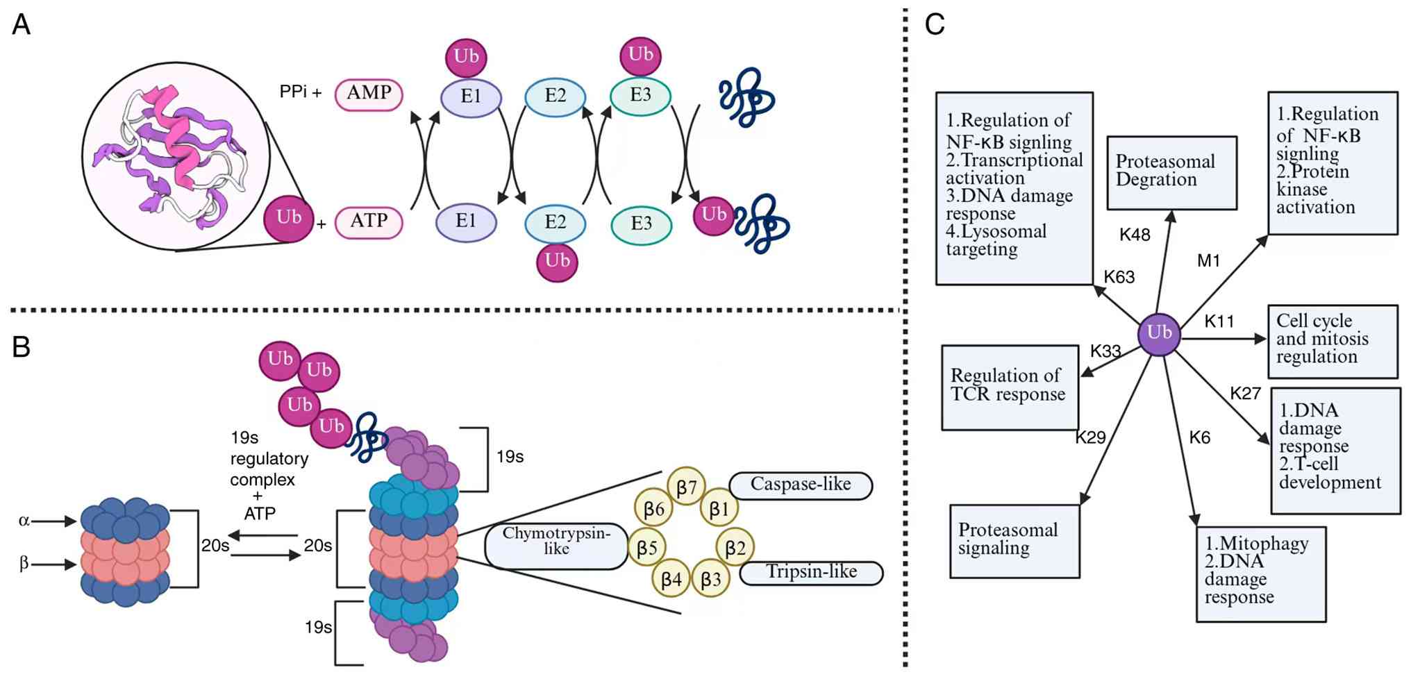 | Figure 1Ubiquitination modification process,
the ubiquitin-proteasome pathway and the types of ubiquitination
modifications. (A) The ubiquitination process is depicted. First,
with ATP providing energy, the ubiquitin-activating enzyme E1
activates the ubiquitin molecule. Secondly, the
ubiquitin-activating enzyme E1 transfers the activated ubiquitin
molecule to the ubiquitin-binding enzyme E2. Finally, the ubiquitin
ligase E3 attaches the bound ubiquitin to the target protein. (B)
The ubiquitin-proteasome pathway is shown. Ubiquitinated proteins
bind to the 19S complex and are degraded at the proteolytic β
subunit. The 19S subunit binds to multiple ubiquitin chains, and
ATP unfolds the protein substrate and transfers it to the 20S core
particle. The protein is subsequently degraded into small
oligopeptides <25 amino acids in length through the 20S core.
This mediates ubiquitin-independent protein degradation. (C) The
types of ubiquitination modifications. In the ubiquitin chain, the
ubiquitin portion can bind to its lysine residues (K11, K27, K6,
K29, K33, K63 and K48) or the N-terminal methionine residue (M1).
Each chain is recognized by different ubiquitin binding domains,
thereby targeting proteins in specific signaling pathways. Ub,
ubiquitin. |
2. Roles of ubiquitination and
deubiquitination in immune checkpoints in lung cancer
As notable regulatory mechanisms, ubiquitination and
deubiquitination perform pivotal roles in lung cancer through
modulating tumor progression and therapeutic efficacy (25). As aforementioned, the enzymes
governing these processes have emerged as promising therapeutic
targets, especially through their ability to regulate the stability
and function of key immune checkpoints, including PD-1/PD-L1, LAG-3
and CTLA-4. The present review summarizes the functions of
ubiquitinating enzymes and DUBs in lung cancer and delves into the
underlying mechanisms via which they control immune checkpoint
expression and activity (Table
I).
 | Table IE3 ligases and DUBs associated with
immune checkpoints in lung cancer. |
Table I
E3 ligases and DUBs associated with
immune checkpoints in lung cancer.
| Type | Name | Relevant immune
checkpoints | Sites of
ubiquitination or ubiquitin-linked chain | The role of immune
therapy | (Refs.) |
|---|
| E3 ligase | TRIM28 | PD-1 | Lysine(s) within
the RIPK1 kinase domain/K63-linked polyubiquitin chain | Inhibition | (50) |
| E3 ligase | FBW7 | PD-1 | Lys233 of
PD-1/K48-linked polyubiquitin chain | Promotion | (52) |
| E3 ligase | TRIM35 | PD-1 | Lys422 of
LSD1/K63-linked polyubiquitin chain | Promotion | (51) |
| E3 ligase | TRIP12 | PD-1 | Lysine 1136 of
TRIP12/K48-linked polyubiquitin chain | Promotion | (53) |
| E3 ligase | SPOP | PD-L1 | Lysine(s) within
the cytoplasmic tail of PD-L1/K48-linked polyubiquitin chain | Promotion | (37) |
| E3 ligase | Cbl-b, c-Cbl | PD-L1 | Lysines within
STAT3, AKT, and/or ERK components/- | Promotion | (63) |
| E3 ligase | AIP4 | PD-L1 | Lys263 of
PD-L1/mono-ubiquitination | Promotion | (39) |
| E3 ligase | FBXO22 | PD-L1 | Lysine(s) within
the intracellular tail of PD-L1/lysine(s) within the intracellular
tail of PD-L1 | Promotion | (38) |
| E3 ligase | DCUN1D1 | PD-L1 | Indirect (no direct
ubiquitination) | Inhibition | (41) |
| E3 ligase | RNF182 | PD-L1 | Lysine(s) within
the RNF182 substrate p65/K48-linked polyubiquitin chain | Promotion | (64) |
| DUB | USP7 | PD-1 | Unidentified
lysine(s) on p38 MAPK pathway component(s) or PD-L1
regulator(s)/- | Inhibition | (54) |
| DUB | USP12 | PD-1 | Unidentified
lysine(s) within PPM1B/- | Promotion | (55) |
| DUB | USP51 | PD-L1 | Lys280 and Lys281
of PD-L1/removal of K48-linked polyubiquitin chains | Inhibition | (42) |
| DUB | USP22 | PD-L1 | -/removal of
multiple non-K48 linkages-K6-, K11-, K27-, K29-, K33- and
K63-linked ubiquitin chains | Inhibition | (43) |
| DUB | USP5 | PD-L1 | Indirect (no direct
ubiquitination) | Inhibition | (48) |
| DUB | ATXN3 | PD-L1 | Lysine residues
within IRF1, STAT3 and HIF-2α/removal of K48-linked polyubiquitin
chains from IRF1, STAT3 and HIF-2α | Inhibition | (45) |
| DUB | UCHL1 | PD-L1 | Indirect (no direct
ubiquitination) | Inhibition | (47) |
| DUB | OTUB2 | PD-L1 | -/removal of
K48-linked polyubiquitin chains from PD-L1 | Inhibition | (45) |
| E3 ligase | WWP2 | PD-1 | Lysine(s) within
the PTEN phosphatase/K48-linked polyubiquitin chain | Inhibition | (56) |
| E3 ligase | CBL | PD-1 | Lysine residues
within EGFR, MAPKKK, MAPKK and MAPK isoforms/K48-linked
polyubiquitin chain (proteasomal degradation) and K63-linked chain
(non-proteolytic signaling) | Promotion | (59) |
| DUB | USP2 | PD-L1 | Lysine(s) within
the CD47 cytoplasmic tail/removal of K48-linked polyubiquitin
chains from CD47 | Promotion | (61) |
| DUB | USP8 | PD-L1 | Lysine(s) within
the intracellular tail of PD-L1/removal of K48-linked polyubiquitin
chains from PD-L1 | Promotion | (64) |
| E3 ligase | Cbl-b, c-Cbl | PD-1/LAG-3 | Basic-residue-rich
juxtamembrane region within the LAG3 cytoplasmic tail/K63-linked
polyubiquitin chain | Promotion | (84) |
| E3 ligase | FBXO38 | LAG-3 | Lysine(s) within
the FGL1 sequence/K48-linked polyubiquitin chain | Promotion | (85) |
| DUB | USP2a | B7-H4 | Lysine(s) within
the B7-H4 cytoplasmic tail/removal of both K48- and K63-linked
polyubiquitin chains from B7-H4 | Inhibition | (91) |
PD-1/PD-L1: A notable immune
checkpoint in lung cancer
PD-1 (or CD279) is a 55-kDa transmembrane protein
composed of 288 amino acids. Its structure includes an
extracellular N-terminal IgV-like domain, a transmembrane domain
and a cytoplasmic tail (26). The
cytoplasmic tail contains two tyrosine-based motifs located at the
N- and C-terminal regions (27).
Its ligand, PD-L1 (also known as CD274 or B7-H1), is a member of
the B7 family, which functions as a 33-kDa type I transmembrane
glycoprotein. The extracellular region of PD-L1 consists of 290
amino acids, featuring both IgV and IgC structural domains
(28). Under normal physiological
conditions, PD-L1 is expressed by macrophages, various activated T-
and B-cells, dendritic cells and certain epithelial cells,
especially in response to inflammatory conditions (29).
The PD-1 pathway functions as a key negative
feedback mechanism in normal immunity. T-cell activation, triggered
by the engagement of T-cell receptors (TCRs) with antigen-major
histocompatibility complex (MHC) and costimulatory signals, leads
to the upregulation of PD-1 on the T-cell surface. Binding of PD-1
by PD-L1 from antigen-presenting cells subsequently transmits
inhibitory signals that suppress T-cell activation, curb cell
proliferation and diminish cytokine release, thereby contributing
to the maintenance of immune tolerance (30). In the context of tumor immune
escape, cancer cells exploit this regulatory mechanism through
upregulating PD-L1 expression by means of various mechanisms, such
as in response to IFN-γ within the tumor microenvironment (31). Tumor-surface PD-L1 engages PD-1 on
tumor-infiltrating T cells, delivering inhibitory signals that lead
to both T-cell dysfunction and an impairment of the immune-mediated
clearance of tumor cells (32).
Consequently, the suppression of T-cell activity both enables
sustained tumor proliferation and promotes tumor disease
progression.
The activity of the PD-1/PD-L1 pathway is regulated
through multiple layers of control, including gene transcription,
post-transcriptional modifications, PTMs and exosomal transport
(33). Among these, PTMs, such as
glycosylation, phosphorylation, ubiquitination, palmitoylation,
SUMOylation and acetylation, fulfill particularly notable roles in
modulating the stability and protein-protein interactions of PD-1
and PD-L1(34). Notably,
substantial evidence over the past decade has established that the
expression levels of PD-1 and PD-L1 are markedly influenced by
ubiquitin-mediated proteasomal degradation. As a key regulatory
mechanism, ubiquitination is essential for controlling diverse
cellular processes, including protein stability, receptor
internalization and immune responses (35).
The subsequent part of the present review delineates
the sophisticated roles of E3s and DUBs in dynamically shaping the
tumor immune microenvironment (TIME). It details a molecular
‘tug-of-war’, wherein specific E3s [for example, speckle-type POZ
protein (SPOP), F-box protein (FBXO)22 and F-box and WD repeat
domain containing 7 (FBW7)] target PD-L1 and PD-1 for degradation
to potentiate anti-tumor immunity. By contrast, counteracting DUBs
[for example, ubiquitin-specific peptidase 22 (USP22),
ubiquitin-specific peptidase 51 (USP51) and the DUB OTU and
ubiquitin aldehyde binding 2 (OTUB2)] stabilize these checkpoints
to foster immune evasion; however, these functions are
context-dependent. The present review explores how this
ubiquitin-mediated regulation extends beyond the checkpoints
themselves to orchestrate key oncogenic signaling pathways [for
example, the phosphoinositide 3-kinase (PI3K/AKT) and
mitogen-activated protein kinase (MAPK) signaling pathways] and
modulate broader immune responses, including phagocytic clearance
mediated via cluster of differentiation (CD)47. Through
highlighting context-dependent regulators such as
ubiquitin-specific peptidase 7 (USP7) and ubiquitin-specific
peptidase 12 (USP12), the discussion underscores the therapeutic
potential of strategically manipulating this equilibrium, which can
be achieved either through augmenting E3 activity or by inhibiting
specific DUBs, thereby recalibrating the TIME and overcoming
resistance to immunotherapy in lung cancer (Fig. 2). Notably, although TIM-3 and TIGIT
represent clinically relevant immune checkpoints in lung cancer,
their direct regulation by ubiquitin-modifying enzymes remains
largely unexplored.
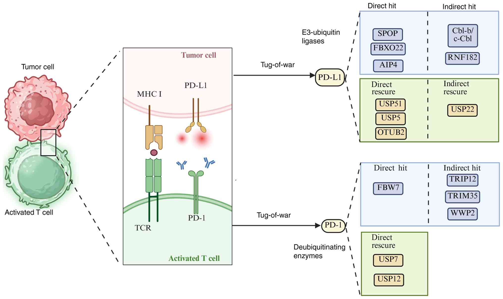 | Figure 2Role of Ubiquitinating and
Deubiquitinating enzymes in PD-1/PD-L1 immune checkpoints. A
schematic diagram of the ubiquitination regulatory network of the T
cell immune checkpoint protein PD-1 and its ligand PD-L1 in the
tumor microenvironment, showing the regulatory effects of
deubiquitinating enzymes and E3 ligases on the stability and
function of PD-1, and elucidating the molecular mechanism via which
ubiquitination modification mediates T cell exhaustion and immune
escape. PD-1, programmed cell death protein 1; PD-L1, programmed
death-ligand 1; MHC, major histocompatibility complex; TCR, T-cell
receptor; SPOP, speckle-type POZ protein; FBXO22, F-box protein 22;
FBW7, F-box and WD repeat domain-containing 7; AIP4, atrophin-1
interacting protein 4; RNF182, ring finger protein 182; Cbl,
Casitas B-lineage lymphoma; USP, ubiquitin-specific peptidase;
OTUB2, OTU and ubiquitin aldehyde-binding 2; TRIM35, tripartite
motif-containing 35; TRIP12, thyroid hormone receptor interactor
12; WWP2, WW domain-containing E3 ubiquitin-protein ligase 2. |
E3s and DUBs in direct PD-L1
modulation
Beyond simple protein degradation, the ubiquitin
system has emerged as a sophisticated signaling network that
dynamically shapes the lung cancer immune landscape. E3s function
as molecular switches, the activity of which directly determines
tumor immune recognition and therapeutic susceptibility (36); however, their effects are not
uniformly pro-immunity. SPOP exemplifies how existing drugs can be
repurposed to manipulate this system. The ability of canagliflozin
to disrupt the sodium-glucose cotransporter-2 (SGLT2)-PD-L1 axis
redirects the activity of SPOP toward PD-L1 degradation, thereby
creating a window for enhanced T-cell attack (37). Similarly, FBXO22 operates at the
intersection of DNA damage response and immunotherapy. The
inhibition of cyclin-dependent kinase 5 was unexpectedly shown to
strengthen antitumor immunity through FBXO22-mediated PD-L1 control
(38). This system demonstrates
marked versatility in terms of its regulatory strategies. Although
atrophin-1 interacting protein 4 (AIP4) has been shown to
collaborate with metastasis suppressor 1 (MTSS1) to channel PD-L1
toward lysosomal degradation (39), ring finger protein 182 (RNF182) and
casitas B-lineage lymphoma (Cbl) proteins employ indirect
mechanisms. RNF182 dismantles the transcriptional activator p65,
whereas Cbl proteins serve to constrain the upstream signal
transducer and activator of transcription 3
(STAT3)/AKT/extracellular signal-regulated kinase (ERK) signaling
pathway. These diverse approaches converge on the same goal, namely
modulating the presence of PD-L1 at the immune synapse.
Not all E3s, however, are involved in pushing toward
immune activation. DCUN1D1 (defective in cullin neddylation 1
domain containing 1), also known as SCCRO, functions as an
essential co-E3 component of the neddylation E3 complex that
promotes cullin neddylation and subsequent CRL activation (40). This E3 enhances PD-L1 expression
through activation of the focal adhesion kinase (FAK) signaling
pathway, thereby highlighting the delicate balance within this
network (41). The collective
activity of these competing forces ultimately determines whether
the TIME permits or prevents effective immune surveillance, thereby
positioning the ubiquitin system as a central arbitrator of
immunotherapeutic outcomes in lung cancer.
Beyond transcriptional control, PD-L1 expression is
dynamically shaped by DUBs that stabilize PD-L1 through distinct
PTMs, thereby creating a notable barrier to effective
immunotherapy. Multiple DUBs converge on PD-L1 protection through
specialized mechanisms. USP51 directly stabilizes PD-L1 by removing
ubiquitin chains at K280/K281, which has the effect of driving dual
resistance to both chemotherapy and immunotherapy (42). By contrast, USP22 employs a
dual-layer stabilization mechanism: Not only does it deubiquitinate
PD-L1 at multiple lysine residues, but it also stabilizes
constitutive photomorphogenesis 9 signalosome subunit 5 (CSN5).
CSN5 itself functions as a JAMM-family metalloprotease
deubiquitinase that directly binds the C-terminal region of PD-L1
and cleaves its ubiquitin moieties via MPN-domain enzymatic
activity, thereby preventing proteasomal degradation. Consequently,
USP22 fortifies PD-L1 stability through two convergent routes,
direct deubiquitination of PD-L1 and indirect stabilization of its
upstream guardian CSN5, creating a reinforced USP22-CSN5-PD-L1
signaling circuit that amplifies immune evasion (43,44).
OTUB2 operates as an early checkpoint protein that intercepts PD-L1
in the endoplasmic reticulum (ER) to subvert ER-associated
degradation, which thereby ensures that it is trafficked to the
cell surface (45).
The regulatory landscape has been shown to extend
beyond direct protein stabilization. Ataxin-3, a protein encoded by
the human ATXN3 gene that primarily functions as DUB,
determines PD-L1 upregulation through transcriptional control. It
promotes PD-L1 upregulation through two distinct microenvironmental
cues. Upon IFN-γ stimulation, it deubiquitinates and stabilizes
both interferon regulatory factor 1 (IRF1) and signal transducer
and activator of transcription 3 (STAT3), thereby preserving their
ability to bind the PD-L1 promoter and enhance its transcription.
Under hypoxic conditions, ATXN3 functions as a deubiquitinase for
hypoxia-inducible factor-2α (HIF-2α), preventing its proteasomal
degradation; the accumulated HIF-2α subsequently translocates to
the nucleus and drives PD-L1 transcription through hypoxia-response
elements in the PD-L1 promoter. These convergent transcriptional
programs ultimately increase PD-L1 abundance and foster immune
evasion (46). Similarly,
ubiquitin C-terminal hydrolase L1 was shown to enhance PD-L1
expression through AKT-P65 pathway activation, which creates an
immunosuppressive microenvironment that dampens T-cell efficacy
(47). Notably, USP5 exemplifies
how DUB function is integrated with broader cellular systems.
Classically known as isopeptidase T (IsoT), USP5 specifically
disassembles unanchored polyubiquitin chains, generated during
proteasomal processing or aberrant E2/E3 activity, into
monoubiquitin units for recycling. When these free chains
accumulate, they competitively bind proteasomal ubiquitin receptors
(such as S5a/Rpn10), obstructing the recognition and degradation of
authentic ubiquitinated substrates and thereby disrupting global
proteostasis. By clearing this inhibitory pool, USP5 preserves
proteasome capacity and maintains the cellular monoubiquitin
reservoir, which indirectly sustains PD-L1 stability and links the
general ubiquitin-proteasome system to specific immune checkpoint
control. Beyond proteostasis, USP5 also participates in DNA
double-strand break repair, Wnt/β-catenin signaling, and
NF-κB-mediated inflammatory responses, further coupling immune
checkpoint regulation to genome integrity and oncogenic signaling
networks (48,49).
This intricate network reveals how DUBs function as
sophisticated modulators of tumor immunity. The diverse range of
mechanisms that they employ, ranging from direct deubiquitination
to transcriptional reprogramming and pathway activation,
demonstrate their potential as therapeutic targets. Nevertheless,
the impact of DUB inhibition needs to be evaluated in a
context-specific manner, as the net effects elicited depend on the
particular substrate and cellular environment.
E3s and DUBs in PD-1 regulation and
contextual immune modulation
Multiple E3s form a complex regulatory network that
both modulates the TIME and responses to anti-PD-1/PD-1
immunotherapy in lung cancer. However, these enzymes exert
divergent effects on immune surveillance. Tripartite motif
containing (TRIM)28 promotes the recruitment of myeloid-derived
suppressor cells (MDSCs) and confers anti-PD-1 resistance. It
catalyzes K63-linked ubiquitination of receptor-interacting
serine/threonine kinase 1, thereby activating nuclear factor-κB
(NF-κB) signaling (50). This
process is dependent on its E3 activity. Additionally, the
DNA-binding protein TRIM35 enhances immune surveillance in NSCLC.
It mediates the K63-linked ubiquitination of lysine specific
demethylase 1, which suppresses its demethylase activity. This
inhibition subsequently leads to an upregulation of the
transcription of the protein ER-Golgi intermediate compartment 1,
stabilizes IFN-γ receptor 1 and activates IFN-γ signaling,
ultimately leading to an increased expression of MHC-I (51). Similarly, FBW7 ubiquitinates PD-1
via K48-linked polyubiquitination, thereby targeting it for
degradation and enhancing anti-PD-1 efficacy (52). This interaction is facilitated by
cyclin-dependent kinase 1-mediated phosphorylation of PD-1 at the
residue S261. Furthermore, tryptophan metabolism also engages in
this cross-talk: Tryptophan or indoleamine 2,3-dioxygenase
inhibitors have been shown to promote the tryptophanylation and
activation of the E3, thyroid hormone receptor interactor 12
(TRIP12) at K1136. TRIP12 subsequently degrades nuclear factor of
activated T cells 1 (NFATc1), which functions as a key
transcription activator of PD-1(53). This pathway enhances
CD8+ T-cell-mediated cytotoxicity and inhibits tumor
growth.
The roles of DUBs in lung cancer immunity are
context-dependent, as exemplified by the contrasting, yet
complementary, functions of USP7 and USP12. Although both are
cysteine proteases within the USP family, their inhibition leads to
divergent outcomes in the tumor microenvironment, thereby
highlighting the nuanced regulation of immunotherapy efficacy. The
targeted inhibition of USP7 presents a dual therapeutic advantage;
on one hand, it directly suppresses tumor growth through the
activation of the p38 MAPK pathway, whereas, on the other hand, it
concurrently effects an upregulation of PD-L1 expression (54). This seemingly paradoxical increase
in PD-L1 serves to sensitize the tumor to anti-PD-1 antibodies,
which thereby transforms the microenvironment into a more
responsive state for combination immunotherapy.
Alternatively, USP12 acts as a guardian of antitumor
immunity. Its knockdown exacerbates tumor progression through
fostering a powerfully immunosuppressive niche. This occurs through
enhanced macrophage recruitment, hypervascularization and T-cell
inactivation. Mechanistically, a depletion of USP12 disrupts the
stability of the protein phosphatase
Mg2+/Mn2+ dependent 1B, which serves as a
negative regulator of NF-κB signaling (55). The subsequent hyperactivation of
NF-κB then drives the overexpression of pro-tumor chemokines,
including CXCL1, CXCL8, CCL2 and CCL5, which promote the
recruitment of tumor-associated macrophages and myeloid-derived
suppressor cells (55). These
chemokines, in turn, orchestrate the immunosuppressive landscape
and drive resistance to immune checkpoint blockade (55). Therefore, a loss of USP12 function
has the effect of crippling the responsiveness of tumors to
anti-PD-1 treatment.
In summary, USP7 inhibition creates a favorable
condition that may be combined with existing immunotherapies,
whereas USP12 activity is essential for maintaining the capacity to
respond to such treatments. This delineation underscores that a
strategic targeting of the ubiquitin system requires a precise
understanding of each component's unique role in balancing tumor
suppression and immune activation.
Ubiquitin-mediated modulation of
oncogenic signaling and complementary checkpoints
Ubiquitin-dependent fine-tuning of the PI3K/AKT and
MAPK cascades orchestrates the amplitude and duration of signaling
that ultimately dictate the PD-1/PD-L1 checkpoint threshold in lung
cancer. Within the PI3K-AKT axis, E3s such as WW domain-containing
E3 ubiquitin protein ligase 2 promote Akt activation via mediating
phosphatase and tensin homolog degradation. This consequently leads
to an upregulation of PD-1 expression (56). On the other hand, PD-1 engagement
itself attenuates Akt phosphorylation (57), thereby inducing metabolic
quiescence in T cells (58).
Similarly, ubiquitination has been shown to regulate multiple
levels of the MAPK cascade. For example, Cbl-mediated
ubiquitination of the epidermal growth factor receptor (EGFR)
restricts upstream signaling, whereas the ubiquitin-dependent
turnover of MAPK kinase/MAPK shapes downstream output (59). Collectively, these events lead to
reduced ERK activity and lower PD-L1 expression (60). Therefore, dynamic ubiquitination
and deubiquitination cycles not only modulate oncogenic signaling,
but they also serve to calibrate the balance between T-cell
exhaustion and reinvigoration, providing a mechanistic rationale
for targeting ligases and DUBs to enhance immune checkpoint
blockade.
DUBs markedly shape the immunosuppressive lung
cancer microenvironment through controlling the stability of
pivotal immune checkpoint molecules. Ubiquitin-specific peptidase 2
(USP2) directly deubiquitinates and stabilizes CD47, thereby
stabilizing the protein and preserving its anti-phagocytic signal.
Either genetic or pharmacological inhibition of USP2 has been shown
to promote CD47 degradation, which thereby shifts macrophages
toward a pro-inflammatory M1 phenotype, also enhancing
CD8+ T-cell infiltration (61). In parallel, ubiquitin-specific
peptidase 8 (USP8) circumvents the TNF receptor-associated factor
6-mediated K63-linked ubiquitination of PD-L1. Inhibiting USP8
directs PD-L1 toward K48-linked polyubiquitination and degradation,
while simultaneously enhancing the NF-κB-dependent expression of
MHC-I. Elevated MHC-I surface density increases the presentation of
tumor-derived antigenic peptides to CD8+ T cells,
thereby augmenting antigen presentation and priming cytotoxic
T-cell responses (62).
Consequently, the dual targeting of USP2 and USP8 disrupts two
complementary ubiquitin-dependent immune barriers, namely
phagocytic checkpoint and T-cell suppression, thereby effectively
converting an immune-excluded tumor niche into an inflamed
microenvironment that is susceptible to PD-1 checkpoint
blockade.
Dynamic equilibrium of the ubiquitin
network in PD-1/PD-L1 immunotherapy
Notably, the fate of the immune checkpoint PD-L1 is
not sealed at its production; rather, its fate is perpetually
decided by a PTM ‘tug-of-war’ that is orchestrated within lung
cancer cells. One the one hand, the E3s strive to pull PD-L1 toward
its degradation, whereas, on the other hand, the DUBs counteract
the activity of the E3s, and their function is to ensure the
survival and continued activity of PD-L1. This dynamic equilibrium
acts as a ‘master regulator’ of tumor immune evasion.
E3s modulate PD-L1 in various ways to cause its
degradation, thereby exposing the tumor to immune attack. Ligases
such as SPOP (activated by canagliflozin), FBXO22 and AIP4 in
combination with MTSS1 directly bind to PD-L1. They decorate PD-L1
with ubiquitin chains, thereby marking it for proteasomal or
lysosomal disposal. Other players, such as Cbl-b/c-Cbl and RNF182,
however, take a more indirect approach. They do not target PD-L1
itself, but interrupt its production; Cbl-b/c-Cbl dampens the
signaling pathways (such as the STAT3/AKT/ERK pathways) that drive
PD-L1 transcription (63), whereas
RNF182 downregulates a key transcription factor, p65, that is
required for the activation of the PD-L1 gene (64).
Conversely, DUBs antagonize E3-mediated
ubiquitination by removing ubiquitin chains to rescue PD-L1 from
degradation. DUBs such as USP51, USP5 and OTUB2 act as notable
defenders. They directly intercept PD-L1, and strip off its
ubiquitin chains. A key tactical maneuver is employed by OTUB2,
which interacts with PD-L1 during its processing in the endoplasmic
reticulum (ER). This prevents its degradation before it even
reaches the cell surface. Some DUBs, such as USP22, have been shown
to provide a layered defense. USP22 not only directly
deubiquitinates and stabilizes PD-L1, but also enhances the
stability of CSN5, which itself functions as a PD-L1 stabilizer.
This establishes a cooperative positive feedback loop that
amplifies PD-L1 abundance (43).
The ultimate level of PD-L1 to be found on the tumor
cell surface (and therefore, its ability to suppress T-cells) is
the net result of this continuous ‘tug-of-war’. When E3s gain the
upper hand, immunity is enhanced (51,52),
but when DUBs dominate, the tumor persists. This refined
understanding of the crosstalk moves us beyond a static list and
reveals a dynamic signaling network. The most promising therapeutic
strategies that are coming into prominence now aim to disrupt this
balance. In terms of influencing this balance, clinicians can
either inhibit the stabilizing DUBs (for example, with USP51 or
OTUB2 inhibitors) or activate specific E3s. Through strategically
intervening in this tug-of-war, it is possible to tip the scales in
favor of the immune system and thereby overcome resistance to
immunotherapy.
The success of anti-PD-1 therapy in lung cancer is
therefore governed by a dynamic equilibrium in the ubiquitin
system, where E3s and DUBs engage in a molecular tug-of-war to
control immune recognition. E3s predominantly propel the system
toward immune activation (65)
through dismantling inhibitory checkpoints. FBW7 and TRIP12 have
been shown to degrade both PD-1 and its transcriptional regulator,
NFATc1. By contrast, TRIM35 potentiates T-cell surveillance by
enhancing IFN-γ signaling. On the other hand, DUBs provide nuanced,
context-dependent counterbalances to the action of the E3s. The
contrasting roles of USP7 and USP12 exemplify this complexity.
Inhibiting USP7 creates a synthetically vulnerable state wherein
tumor cells become sensitized to immune checkpoint blockade: USP7
inhibition concurrently suppresses tumor growth through p38 MAPK
activation and upregulates PD-L1 expression, thereby priming the
tumor microenvironment for enhanced anti-PD-1 efficacy (54). By contrast, preserving USP12 is
essential to prevent the collapse of the immune microenvironment
into a state of profound immunosuppression (55).
Therefore, the ubiquitin system acts not merely as
an on/off switch, but as a sophisticated signaling hub. Therapeutic
success hinges on being able to strategically manipulate this hub.
This strategic manipulation may be achieved either through
reinforcing the ‘pro-immunity’ force of key E3s or by precisely
inhibiting the ‘pro-resistance’ function of specific DUBs. Such
interventions tip the scales in favor of sustained antitumor
immunity (65).
3. LAG-3: An immune-checkpoint hub bridging
T-cell exhaustion and tumor immune escape
The gene known as LAG-3, which was discovered
in 1990 and was found to be a structural homologue of CD4, has been
revealed to be expressed by various types of lymphocytes and
non-lymphocyte lineage cells (66). LAG-3, an inhibitory receptor that
is notably expressed in exhausted T cells, has been identified as a
potential target for immunotherapy. LAG-3 has been shown to exert a
regulatory function characterized by the suppression of cell
proliferation, activation, effector function and homeostasis for
both CD8 and CD4 T cells. Furthermore, this process disrupts the
shared pathway of CD4 and CD8 activation, while concurrently
regulating the activation and proliferation of T memory cells
(67). As with PD-1, the
constitutive expression of LAG-3 is often observed under conditions
of T-cell exhaustion, and its expression is generally accepted as a
hallmark of CD4 and CD8 T-cell exhaustion in response to repetitive
antigenic stimulation in the context of cancer and chronic viral
infections (68). In terms of the
underlying mechanism, the expression of LAG-3 is stimulated either
by TCRs or by cytokine stimulation (69). LAG-3 subsequently binds to TCR-CD3
complexes on the T-cell membrane, thereby exerting a negative
regulatory effect on TCR signaling (53). This process ultimately leads to an
arrest of cell proliferation and of cytokine secretion for the
cells; in fact, LAG-3 has been demonstrated to physically interact
with TCRs in CD8 and CD4 T cells following TCR engagement (69). This interaction results in
downregulation of the TCR-dependent signaling cascade, with the
subsequent suppression of T-cell responses (70).
MHC-II is widely regarded as the archetypal ligand
of LAG-3, which interacts stably with the D1 domain. Following the
binding of LAG-3 to MHC-II, CD4 T-cell activation becomes inhibited
(71). Furthermore, it has been
demonstrated that the binding of LAG-3 to MHC-II facilitates tumor
evasion from apoptosis (72) and
enhances the recruitment of tumor-specific CD4 T cells, while
concomitantly reducing CD8 T-cell responses (73). Notably, the binding of galectin-3
(Gal-3) to LAG-3 is a prerequisite for the inhibition of CD8 T-cell
cytotoxic function (74). The role
of Gal-3 in anti-tumor immune responses is two-fold: First, it has
been demonstrated that Gal-3 inhibits the activation of
antigen-committed CD8 T cells; secondly, Gal-3 inhibits the
expansion of plasmacytoid dendritic cells through LAG-3 expression
in the tumor microenvironment (75). These effects, when considered
collectively, form an antitumor immune response (75).
Fibrinogen-like protein 1 (FGL1), also referred to
hepassocin, liver fibrinogen-related gene-1 or hepatocyte-derived
fibrinogen-related protein, is a component of the
fibrinogen-related protein family. It comprises two 34 kDa
homodimers that are linked by disulfide bonds to form a 68-kDa
protein (76), and contains two
distinct domains: The N-terminal signal recognition peptide
(helical domain, coiled-coil domain) and the C-terminal
fibrinogen-like domain, which binds LAG-3 in the absence of an
involvement of the membrane cross-over region (77,78).
FGL1 is a novel ligand for LAG-3, in addition to its classical
ligand, MHC-II. It forms a novel immune checkpoint pathway that is
independent of PD-1/PD-L1, which results in T-cell exhaustion and
subsequent dysfunction, as well as evasion of immune surveillance
by tumor cells (79).
The expression of LAG-3 in T cells is widely
regarded as a hallmark of aggressive tumor progression across a
wide range of human tumors, especially in SCLC (79). Previous studies have demonstrated
that tumors exhibiting immune evasion, primarily mediated by LAG-3,
have a reduced sensitivity to PD-1 blockade (80,81).
This provides a foundation for the potential future utilization of
LAG-3 as a stratified biomarker in immunotherapy (69).
Emerging evidence has suggested that
ubiquitin-mediated post-translational regulation notably governs
both LAG-3 stability and function in lung cancer. The Cbl family,
comprising c-Cbl, Cbl-b and Cbl-3, represents a class of
RING-finger E3s that are characterized by a tyrosine kinase-binding
‘antenna’, an E2-docking RING ‘catalytic core’ and a C-terminal
‘signal relay’ domain composed of proline-rich and
ubiquitin-associated/leucine zipper motifs (82,83).
In NSCLC, these ligases function as gatekeepers of immune evasion.
c-Cbl and Cbl-b mediate the redundant ubiquitination of LAG-3,
which has the effect of trapping the receptor in a
membrane-dissociated, signaling-silent state. This mechanism
positions the LAG3-to-Cbl expression ratio as a readily applicable
biomarker for predicting the response to anti-LAG3 therapy
(84). Complementing this axis,
FBXO38, an F-box component of the SKP1-cullin-F-box E3 ligase
complex, acts as a metabolic rheostat in immune evasion. Through
conjugating K48-linked ubiquitin chains to the immunosuppressive
ligand FGL1, FBXO38 targets it for proteasomal degradation. This
process not only suppresses interleukin (IL)-6-driven pro-tumor
inflammation, but it also facilitates the infiltration of
CD8+ T-cells into tumors. Clinically, a
FBXO38-high/FGL1-low/IL-6-low expression signature identifies
early-stage, immune-infiltrated NSCLC lesions, and this signature
serves as an independent predictor of favorable outcomes following
immune checkpoint blockade (85).
Considered together, Cbl-mediated checkpoint desensitization and
FBXO38-directed ligand clearance underscore a unifying biological
principle: Specific E3s can be harnessed as ‘immune-tuning
amplifiers’ that shift the ubiquitin equilibrium away from
tumor-permissive states and toward tumor-restrictive immunity.
These mechanisms offer not only quantifiable biomarkers for
clinical analysis, but also rational combination strategies that
may be employed to enhance the efficacy of PD-1/LAG3-targeted
therapies.
4. B7-H4: An emerging immune checkpoint and
the USP2a-driven axis
The co-inhibitory molecule B7-H4 is a notable
constituent of the B7 family. It is a glycosylated molecule with a
molecular mass of 50-80 kDa (86),
and is a type-I transmembrane protein composed of 282 amino acids.
As demonstrated in several studies, human B7-H4 mRNA is broadly
expressed in numerous tissue types, including the placenta, liver,
skeletal muscle, kidney, pancreas, prostate, testis, small
intestine, stomach, spleen, lung, thymus, uterus, skin, lung, heart
and brain (87). In NSCLC,
elevated expression levels of B7-H4 are notably associated with
lymph node metastasis and tumor-infiltrating lymphocytes (TILs)
(88). In terms of the underlying
mechanism, B7-H4 exerts its immunosuppressive effects through
suppressing the activation, proliferation and clonal expansion of
both CD4+ and CD8+ T lymphocytes. It
simultaneously downregulates the secretion of key cytokines,
including IL-2 and IFN-γ (89).
This immunosuppressive activity is primarily mediated by the
inhibition of TCR-mediated signaling cascades, ultimately resulting
in T-cell dysfunction. Furthermore, B7-H4 facilitates tumor
progression through activating diverse intracellular signaling
pathways that enhance cancer cell proliferation, metastatic
potential and resistance to programmed cell death (90).
In EGFR-mutant lung cancer, mutant EGFR signaling
leads to an upregulation of the DUB USP2a. USP2a, in turn,
stabilizes the immune checkpoint protein B7-H4 through cleaving its
K48- and K63-linked ubiquitin chains, thereby shielding it from
proteasomal degradation (91).
This USP2a-mediated stabilization enhances the surface expression
of B7-H4, thereby leading to suppressed T-cell activation and
facilitated tumor immune evasion (91). The positive association observed
between the protein levels of USP2a and B7-H4 in patient-derived
specimens further validates this axis, and positions USP2a as a
promising druggable target for restoring anti-tumor immunity.
5. Treatment strategies
ICIs of lung cancer
ICIs perform a pivotal role in the management of
different types of lung cancer, especially NSCLC. PD-1 inhibitors,
such as nivolumab and pembrolizumab, are currently employed in
clinical practice based on phase III randomized controlled trials
(92,93), while PD-L1 inhibitors, such
atezolizumab, have also demonstrated efficacy (94). Furthermore, for the first-line
treatment of NSCLC, tremelimumab (Imjudo), a CTLA-4 inhibitor, has
been used in combination with duvalumab (95). Drug resistance has gradually
emerged with the widespread application of immunotherapy, although
only a small proportion of patients with lung cancer respond to
immunotherapy, and this in itself poses a new challenge in the
treatment of lung cancer (14,96).
Potential therapeutic approaches
targeting ubiquitinases and DUBs
Berberine (BBR) is a naturally occurring
isoquinoline quaternary alkaloid that is isolated from Coptis
chinensis, a species of goldthread flowering plant native to
China. It exhibits broad pharmacological properties, including
anti-cancer, anti-microbial, anti-diabetic and anti-inflammatory
activities (97). In terms of the
underlying mechanism, BBR has been shown to enhance T-cell-mediated
tumor cell death through downregulating the expression of PD-L1 in
cancer cells. Specifically, BBR interacts with the glutamate
residue E76 within CSN5, thereby inhibiting its DUB activity. This
interaction promotes PD-L1 ubiquitination, and subsequent
proteasomal degradation, which effectively disrupts the PD-1/PD-L1
immune checkpoint axis (97).
Furthermore, BBR has been shown to impair the MPN domain of CSN5,
which is critical for CSN5-mediated stabilization and
deubiquitination of PD-L1(98). In
addition to its direct effects on PD-L1, BBR also enhances
anti-tumor immunity through promoting the infiltration and
activation of tumor-specific T cells. It has also been shown to
suppress the expansion and function of immunosuppressive cell
populations, such as MDSCs and regulatory T cells (97).
P5091 is a selective USP7 inhibitor, which
demonstrates potent immunomodulatory activity via reprogramming
tumor-associated macrophages through activation of the p38 MAPK
signaling pathway, which has the effect of promoting their
polarization toward the M1 phenotype. This molecular mechanism
enhances cytotoxic T lymphocyte-mediated tumor immune surveillance
and ultimately suppresses tumor progression (54). Notably, USP7 inhibition causes an
upregulation of PD-L1 expression within the tumor microenvironment,
and this creates a synergistic therapeutic window when combined
with PD-1 blockade immunotherapy (54).
Cullin 4A (Cul4A) is an 87-kDa protein, that belongs
to a family of evolutionarily conserved cullin proteins and has
been shown to be associated with the ubiquitin proteasome pathway
(99). The virus protease
inhibitor PIK-93 functions as a potent PD-L1 destabilizing agent
through facilitating the formation of the CUL4A-E3 complex with
PD-L1. This molecular interaction promotes the K48-linked
polyubiquitination of PD-L1, thereby leading to its subsequent
proteasomal degradation through the UPS (100). Administering PIK-93 has been
shown to cause a notable downregulation of PD-L1 expression in
M1-polarized macrophages, thereby augmenting their tumoricidal
activity. In both syngeneic and human peripheral blood mononuclear
cell-derived xenograft models, the combined administration of
PIK-93 and PD-L1 blockade therapy has been demonstrated to
potentiate T-cell-mediated antitumor immunity. This was evidenced
both by an enhanced infiltration of TILs and a suppression of tumor
progression (100). This
combinatorial approach served to promote the establishment of an
immunologically active tumor microenvironment, which led to a
synergistic improvement of the therapeutic efficacy of immune
checkpoint inhibition (100).
Metformin is a widely used anti-diabetic agent.
Emerging research has demonstrated that it exhibits notable
anti-tumor activity across multiple types of cancer, including
lung, prostate and colorectal malignancies (101). Moreover, mechanistic studies have
revealed that metformin-mediated stabilization of the adaptor
protein, stimulator of interferon genes (STING), and the
enhancement of T-cell cytotoxicity are both axis inhibition protein
1 (AXIN-1)-dependent. The genetic ablation of this scaffold protein
led to an abrogation of these effects. In serine/threonine kinase
11-mutant lung cancer models, metformin was shown to synergize with
the PD-1 blockade via suppressing RNF5-dependent K48-linked
polyubiquitination, and the subsequent degradation of STING. This
process was shown to require AXIN-1(102). Furthermore, metformin was found
to potentiate tumor-specific T-cell responses in vitro, and
to augment the therapeutic efficacy of PD-1 inhibitors in both
preclinical models and experimental settings.
The antidiabetic drug canagliflozin has been shown
to suppress tumor growth by reducing the expression of PD-L1 in an
‘on-target’ manner. In terms of the underlying mechanism, SGLT2
maintains PD-L1 stability during endocytic recycling through its
direct interaction with it; however, canagliflozin was shown to
have a two-fold effect, namely to disrupt the interaction between
SGLT2 and PD-L1 and to promote the interaction between PD-L1 and
the E3, SPOP (37).
PD-L1 also forms a complex with glycogen synthase
kinase-3β (GSK-3β) and the E3 β-transducin repeat-containing
protein (TrCP), leading to phosphorylation-dependent degradation of
the UPS (103). The annual herb
Centipeda minima (CM) and its active component
6-O-angeloylplenolin (6-OAP) were found to upregulate the
expression of PD-L1 via inhibiting GSK-3β/β-TRCP-mediated
ubiquitination and degradation, which led to further increases in
CD8 T-cell infiltration (104).
Therefore, mechanistically speaking, the induced expression of
PD-L1 and the enhanced cytotoxicity of CD8 T cells underlie the
beneficial effects of 6-OAP-rich CM in NSCLC. Preclinical studies
have demonstrated that CM and 6-OAP significantly enhance
ICI-induced tumor burden reduction and prolong overall survival in
NSCLC-bearing mice when administered in combination with anti-PD-L1
antibody (104). These findings
position CM as a promising candidate for combination with ICIs, and
a phase I clinical trial (NCT05735028) is currently underway to
evaluate its safety and efficacy in patients with lung cancer. As
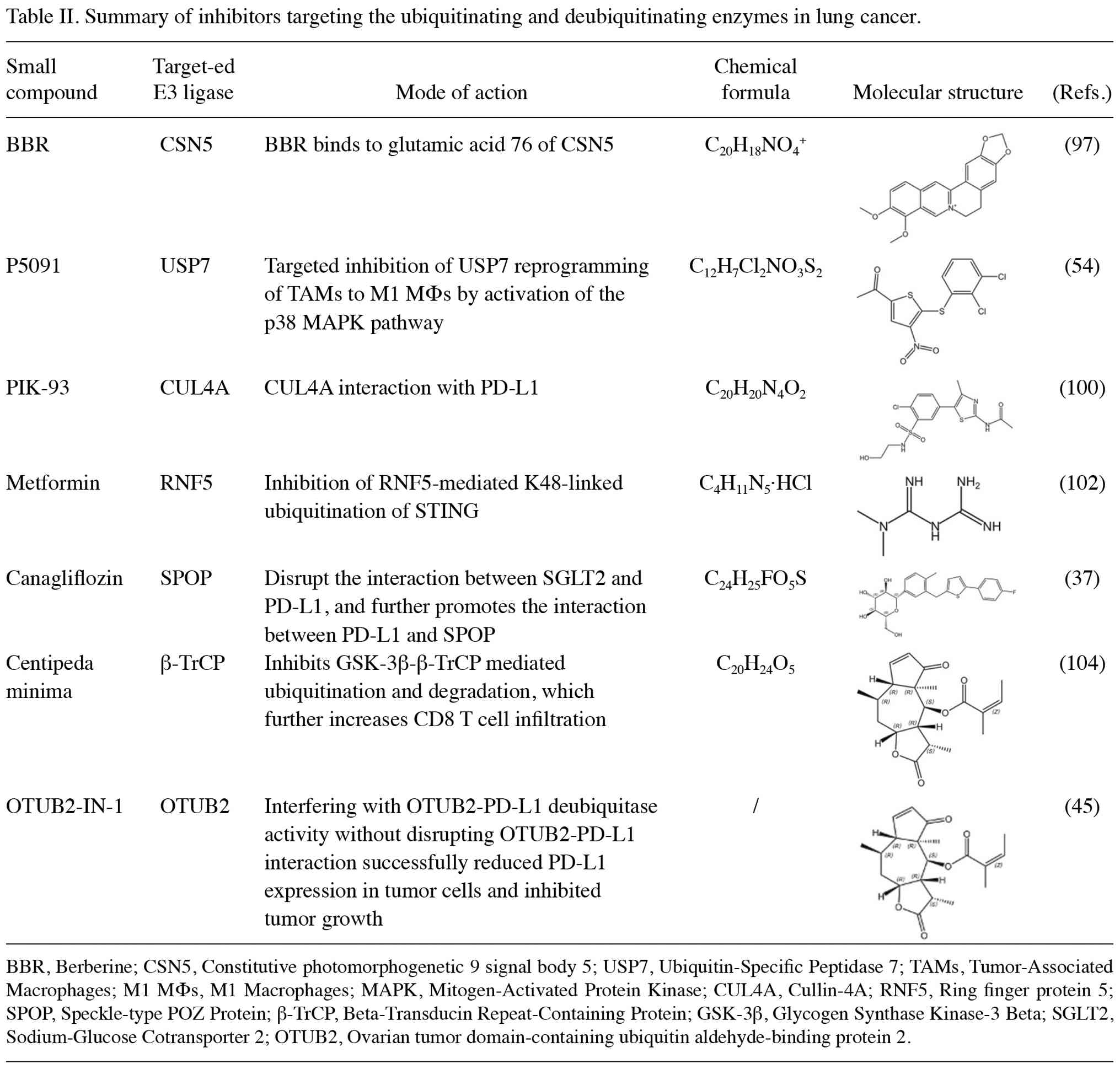
shown in Fig. 3, these diverse
strategies converge on a unified therapeutic principle:
Strategically tipping the ubiquitin equilibrium toward checkpoint
degradation or microenvironment remodeling can overcome
immunotherapy resistance in lung cancer (Table II and Fig. 3).
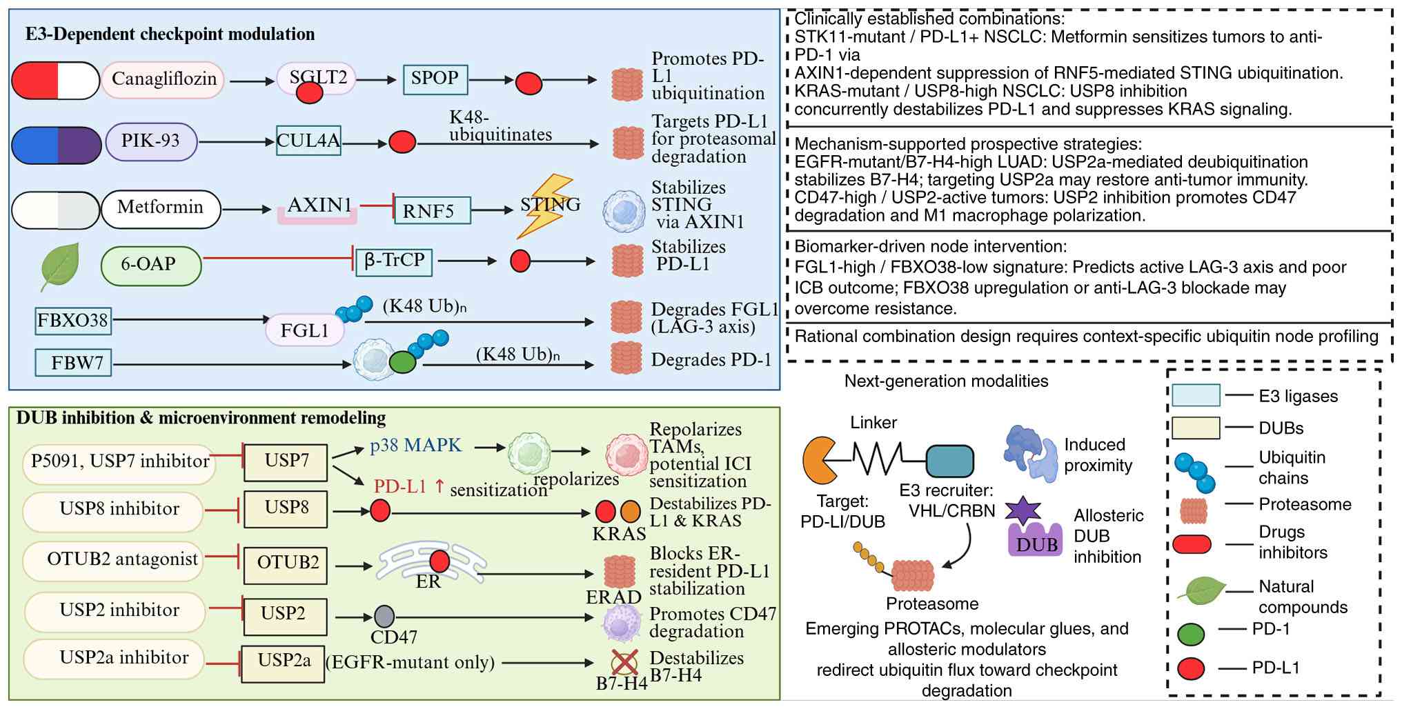 | Figure 3Therapeutic targeting of
ubiquitin-modifying enzymes to enhance immune checkpoint blockade
in lung cancer. The left panel illustrates E3 ligase activation
strategies (canagliflozin/SPOP, PIK-93/CUL4A, metformin/AXIN1,
6-OAP/β-TrCP, FBXO38, FBW7) and DUB inhibition approaches
(P5091/USP7, USP8, OTUB2, USP2, USP2a) that modulate immune
checkpoint stability and tumor microenvironment remodeling. The
right upper panel summarizes clinically established and prospective
biomarker-driven combination strategies. The lower right panel
depicts next-generation modalities, including PROTACs, molecular
glues and allosteric modulators. PD-1, programmed cell death
protein 1; PD-L1, programmed death-ligand 1; DUBs, deubiquitinating
enzymes; SPOP, speckle-type POZ protein; FBXO22, F-box protein 22;
FBW7, F-box and WD repeat domain-containing 7; RNF5, ring finger
protein 5; USP, ubiquitin-specific peptidase; OTUB2, OTU and
ubiquitin aldehyde-binding 2; ER, endoplasmic reticulum; SGLT2,
sodium-glucose cotransporter-2; CUL4A, cullin 4A; AXIN1, axis
inhibition protein 1; FGL1, fibrinogen-like protein 1; β-TrCP,
β-transducin repeat-containing protein; STING, stimulator of
interferon genes; LAG-3, lymphocyte-activating gene 3; TAMs,
tumor-associated macrophages; ICI, immune checkpoint inhibitor;
ERAD, ER-associated protein degradation; B7-H4, B7 homolog 4;
STK11, serine/threonine kinase 11; NSCLC, non-small cell lung
cancer; PROTAC, proteolysis-targeting chimera; VHL, Von
Hippel-Lindau; CRBN, cereblon; EGFR, epidermal growth factor
receptor; KRAS, Kirsten rat sarcoma viral oncogene homolog; ICB,
immune checkpoint blockade. |
6. Discussion
E3s and DUBs perform a notable role in lung cancer
pathogenesis through regulating the expression and signaling
functionality of key immune checkpoint proteins, including PD-1,
PD-L1, LAG-3 and B7-H4. Beyond directly modulating checkpoint
activity through proteasomal degradation, these enzymes are also
involved in the regulation of signaling pathways, including the
EGFR, Wnt/β-catenin and NF-κB pathways (65), thereby indirectly shaping the
immune landscape of the tumor microenvironment by influencing
immune cell infiltration and the transcription of checkpoint
molecules.
Notably, the functional outcomes of
ubiquitin-modifying enzymes in lung cancer immunotherapy are not
uniformly beneficial or detrimental. Rather, they are markedly
context-dependent. For example, E3s such as SPOP, FBXO22 and FBW7
generally promote PD-L1 or PD-1 degradation to enhance anti-tumor
immunity, DCUN1D1 paradoxically increases the expression of PD-L1
through FAK activation, thereby fostering immune evasion.
Similarly, DUBs are not exclusively pro-tumorigenic. USP7
inhibition upregulates the expression of PD-L1, and yet it
sensitizes tumors to anti-PD-1 therapy through remodeling
macrophage polarization. By contrast, USP12 activity is essential
for preserving an immunocompetent microenvironment. These
observations both caution against simplistic categorization, and
underscore the necessity of nuanced, context-specific therapeutic
strategies.
In addition, ubiquitination and deubiquitination
processes notably contribute to the efficacy of lung cancer
treatments. They exhibit synergistic interactions with conventional
therapies, such as chemotherapy and radiotherapy, while also
enhancing the response to immunotherapy (105). For example, in chemotherapy,
cisplatin has been shown to potentiate the antitumor effect of
PD-L1 blockade through upregulating the expression of Ariadne RBR
E3 ubiquitin protein ligase 1 (ARIH1) (106). ARIH1-mediated ubiquitination
leads to the degradation of DNA-dependent protein kinase catalytic
subunit. This, in turn, activates the STING pathway in tumor cells,
thereby sensitizing them to immune checkpoint inhibition (106). In radiotherapy, ubiquitination
and deubiquitination also modulate treatment outcomes.
Radiation-induced DNA damage activates innate immune pathways such
as the cyclic GMP-AMP synthase (cGAS)-STING pathway, thereby
initiating a systemic T-cell response and abscopal effects.
Notably, the activity of the cGAS-STING pathway is itself regulated
by ubiquitination and deubiquitination (107). In the context of targeted
therapy, these processes offer dual benefits, where they enhance
immunotherapy efficacy, but also directly regulate oncogenic
signaling. For example, the DUB USP8 promotes immune evasion and
tumor progression via stabilizing both PD-L1 and Kirsten rat
sarcoma viral oncogene homolog (KRAS) proteins. The inhibition of
USP8 simultaneously suppresses KRAS signaling, and augments
anti-PD-L1 efficacy, suggesting a promising strategy for treating
KRAS-mutated lung cancer (62).
From a translational perspective, the most promising
targets include SPOP activators (for example, canagliflozin)
(37), USP7 inhibitors (for
example, P5091) (40), USP8
inhibitors (62) and OTUB2
antagonists (106). These agents
have demonstrated preclinical synergy with ICIs. Additionally,
proteolysis-targeting chimeras (PROTACs) and allosteric modulators
that selectively redirect ubiquitin flux toward checkpoint proteins
have been shown to represent next-generation therapeutic modalities
(108). Nevertheless, challenges
impede clinical translation. First, the ubiquitin network exhibits
substantial heterogeneity across tumor subtypes and individuals,
and this is driven by tissue-specific enzyme expression and
compensatory pathway redundancy. In addition, pharmacokinetic
optimization, tumor-selective delivery and the management of
on-target toxicities remain hurdles. Moreover, although several
PROTACs and DUB inhibitors have entered phase I/II trials, the vast
majority of ubiquitin-targeting drug candidates remain in the
preclinical or early clinical stages (109-111),
with limited clinical validation and efficacy data. This aspect
highlights the urgent need to identify predictive biomarkers and to
rationally design combination regimens.
7. Limitations
Several limitations should be acknowledged when
interpreting the evidence presented in the current review. First,
the majority of mechanistic studies to date have been conducted in
specific cell lines or preclinical models (112), and these findings may not fully
recapitulate the heterogeneity of human lung cancer in vivo
(113). Secondly, the functional
outcomes of ubiquitin modification are context-dependent; it has
been demonstrated that the same E3 or DUB may exert divergent
effects across different genetic backgrounds, disease stages or
tumor microenvironments (114).
For example, USP7 inhibition was shown to upregulate PD-L1
expression in certain contexts, and yet it suppresses tumor growth
in others (115). This
illustrates the complexity of predicting clinical outcomes.
Thirdly, the depth of research varies across different checkpoints.
PD-1/PD-L1 ubiquitin regulation is relatively well-characterized,
whereas ubiquitin-associated studies for LAG-3, B7-H4, TIM-3 and
TIGIT in lung cancer remain scarce. In addition, the majority of
published translational studies have lacked validation in large
clinical cohorts. The pharmacokinetic profiles, tumor selectivity
and long-term toxicities of ubiquitin-targeting agents remain
incompletely defined (108).
Finally, compensatory mechanisms and pathway redundancy within the
ubiquitin network may diminish the efficacy of single-target
interventions (116). Considered
collectively, these factors constrain the generalizability of
current conclusions and underscore the need for cautious
interpretation.
8. Conclusion
The present review systematically delineated the
ubiquitin-driven regulation of immune checkpoints in lung cancer.
It emphasized that the stability and function of PD-1, PD-L1, LAG-3
and B7-H4 are governed by a dynamic interplay between E3s and DUBs.
Unlike prior reviews that have broadly surveyed ubiquitin
mechanisms in cancer immunity (36), the present work has specifically
integrated lung cancer pathobiology with checkpoint ubiquitination,
highlighting context-dependent enzyme functions and bridging
molecular mechanisms with emerging therapeutic strategies, such as
PROTACs, DUB inhibitors and drug repurposing (108). It has been underscored that E3s
and DUBs cannot be simplistically categorized as uniformly
beneficial or detrimental to immunotherapy. Rather, their effects
are dictated by substrate specificity, the cellular context and
tumor microenvironmental cues. Looking forward, precision targeting
of the ubiquitin system, guided by multi-omics profiling (117) and rational combination regimens,
holds substantial promise for overcoming immunotherapy resistance.
Nevertheless, challenges persist, including tumor heterogeneity,
network redundancy and the need to identify clinically validated
biomarkers. Future investigations should prioritize the mechanistic
dissection of understudied checkpoints, including TIM-3 and
TIGITS15). They should also develop selective enzymatic inhibitors
and establish predictive frameworks to match specific ubiquitin
targets with appropriate patient subpopulations. Over the course of
the next 5 years, it is anticipated that first-in-class USP8 or
OTUB2 inhibitors may enter early-phase clinical trials for patients
with NSCLC who are resistant to PD-1 blockade.
Acknowledgements
Not applicable.
Funding
Funding: The present review was supported by Zhejiang
Traditional Chinese Medicine Science and Technology Plan (grant no.
2024ZL152), Zhejiang Medicine and Health Plan (grant no.
2025KY1472), 2023 Ningbo Health Science and Technology Plan (grant
no. 2023Y55), Ningbo Youth Science and Technology Innovation
Leading Talents Project (grant no. 2023QL052), National Natural
Science Foundation of China (grant nos. 32270821 and 32570835),
Zhejiang Natural Science Foundation (grant no. LY24C050001) and
Zhejiang Provincial Science and Technology Plan Project [grant no.
2025C02249(SD2)].
Availability of data and materials
Not applicable.
Authors' contributions
XFJ conceived the study. LC and LRP drafted the
manuscript. ZGY and JC made substantial contributions to the
interpretations offered in the present review, both drafting the
study and revising it for important intellectual content. YTL and
JHW performed the literature search, extracted and organized
published data, contributed to the drafting and revision of the
manuscript and assisted in figure preparation. All authors have
read and approved the final version of the manuscript. Data
authentication not applicable.
Ethics approval and consent to
participate
Not applicable.
Patient consent for publication
Not applicable.
Competing interests
The authors declare that they have no competing
interests.
References
|
1
|
Hazim AZ, Leventakos K and Ernani V:
Evolving field of immunotherapy: Pioneering new paths in Small-cell
lung cancer. JCO Oncol Pract. 21:942–949. 2025.PubMed/NCBI View Article : Google Scholar
|
|
2
|
Yau STY, Hung CT, Leung EYM, Lee A and
Yeoh EK: The interactions among factors associated with the risk of
lung cancer among diabetes patients: A survival tree analysis. NPJ
Prim Care Respir Med. 35(20)2025.PubMed/NCBI View Article : Google Scholar
|
|
3
|
Boloker G, Wang C and Zhang J: Updated
statistics of lung and bronchus cancer in United States (2018). J
Thorac Dis. 10:1158–1161. 2018.PubMed/NCBI View Article : Google Scholar
|
|
4
|
Sutanto H, Safira A and Fetarayani D: From
tumor to tolerance: A comprehensive review of immune checkpoint
inhibitors and immune-related adverse events. Asia Pac Allergy.
14:124–138. 2024.PubMed/NCBI View Article : Google Scholar
|
|
5
|
Leach DR, Krummel MF and Allison JP:
Enhancement of antitumor immunity by CTLA-4 blockade. Science.
271:1734–1736. 1996.PubMed/NCBI View Article : Google Scholar
|
|
6
|
Ishida Y, Agata Y, Shibahara K and Honjo
T: Induced expression of PD-1, a novel member of the immunoglobulin
gene superfamily, upon programmed cell death. EMBO J. 11:3887–3895.
1992.PubMed/NCBI View Article : Google Scholar
|
|
7
|
Dong H, Zhu G, Tamada K and Chen L: B7-H1,
a third member of the B7 family, co-stimulates T-cell proliferation
and interleukin-10 secretion. Nat Med. 5:1365–1369. 1999.PubMed/NCBI View
Article : Google Scholar
|
|
8
|
Triebel F, Jitsukawa S, Baixeras E,
Roman-Roman S, Genevee C, Viegas-Pequignot E and Hercend T: LAG-3,
a novel lymphocyte activation gene closely related to CD4. J Exp
Med. 171:1393–1405. 1990.PubMed/NCBI View Article : Google Scholar
|
|
9
|
Prasad DV, Richards S, Mai XM and Dong C:
B7S1, a novel B7 family member that negatively regulates T cell
activation. Immunity. 18:863–873. 2003.PubMed/NCBI View Article : Google Scholar
|
|
10
|
Monney L, Sabatos CA, Gaglia JL, Ryu A,
Waldner H, Chernova T, Manning S, Greenfield EA, Coyle AJ, Sobel
RA, et al: Th1-specific cell surface protein Tim-3 regulates
macrophage activation and severity of an autoimmune disease.
Nature. 415:536–541. 2002.PubMed/NCBI View
Article : Google Scholar
|
|
11
|
Yu X, Harden K, Gonzalez LC, Francesco M,
Chiang E, Irving B, Tom I, Ivelja S, Refino CJ, Clark H, et al: The
surface protein TIGIT suppresses T cell activation by promoting the
generation of mature immunoregulatory dendritic cells. Nat Immunol.
10:48–57. 2009.PubMed/NCBI View Article : Google Scholar
|
|
12
|
Tumeh PC, Harview CL, Yearley JH, Shintaku
IP, Taylor EJ, Robert L, Chmielowski B, Spasic M, Henry G, Ciobanu
V, et al: PD-1 blockade induces responses by inhibiting adaptive
immune resistance. Nature. 515:568–571. 2014.PubMed/NCBI View Article : Google Scholar
|
|
13
|
Wherry EJ: T cell exhaustion. Nat Immunol.
12:492–499. 2011.PubMed/NCBI View Article : Google Scholar
|
|
14
|
Sharma P, Hu-Lieskovan S, Wargo JA and
Ribas A: Primary, adaptive, and acquired resistance to cancer
immunotherapy. Cell. 168:707–723. 2017.PubMed/NCBI View Article : Google Scholar
|
|
15
|
Hsieh HC, Ling LL and Wang YC:
Post-translational modifications of immune checkpoints: Molecular
mechanisms, tumor microenvironment remodeling, and therapeutic
implications. J Biomed Sci. 33(3)2026.PubMed/NCBI View Article : Google Scholar
|
|
16
|
Lu C and Tan Y: Promising immunotherapy
targets: TIM3, LAG3, and TIGIT joined the party. Mol Ther Oncol.
32(200773)2024.PubMed/NCBI View Article : Google Scholar
|
|
17
|
Ye P, Chi X, Cha JH, Luo S, Yang G, Yan X
and Yang WH: Potential of E3 ubiquitin ligases in cancer immunity:
Opportunities and challenges. Cells. 10(3309)2021.PubMed/NCBI View Article : Google Scholar
|
|
18
|
Arkinson C, Dong KC, Gee CL and Martin A:
Mechanisms and regulation of substrate degradation by the 26S
proteasome. Nat Rev Mol Cell Biol. 26:104–122. 2025.PubMed/NCBI View Article : Google Scholar
|
|
19
|
Zhang Y, Yang J, Min J, Huang S, Li Y and
Liu S: The emerging role of E3 ubiquitin ligases and
deubiquitinases in metabolic dysfunction-associated steatotic liver
disease. J Transl Med. 23(368)2025.PubMed/NCBI View Article : Google Scholar
|
|
20
|
Damgaard RB: The ubiquitin system: From
cell signalling to disease biology and new therapeutic
opportunities. Cell Death Differ. 28:423–426. 2021.PubMed/NCBI View Article : Google Scholar
|
|
21
|
Buetow L and Huang DT: Structural insights
into the catalysis and regulation of E3 ubiquitin ligases. Nat Rev
Mol Cell Biol. 17:626–642. 2016.PubMed/NCBI View Article : Google Scholar
|
|
22
|
Chen X, Zhou J, Dong X, Xu Y and Chen B:
Regulatory role of E3 ubiquitin ligases in multiple myeloma: From
molecular mechanisms to therapeutic strategies. Front Cell Dev
Biol. 13(1620097)2025.PubMed/NCBI View Article : Google Scholar
|
|
23
|
Agrata R and Komander D: Ubiquitin-A
structural perspective. Mol Cell. 85:323–346. 2025.PubMed/NCBI View Article : Google Scholar
|
|
24
|
Liu F, Chen J, Li K, Li H, Zhu Y, Zhai Y,
Lu B, Fan Y, Liu Z, Chen X, et al: Ubiquitination and
deubiquitination in cancer: From mechanisms to novel therapeutic
approaches. Mol Cancer. 23(148)2024.PubMed/NCBI View Article : Google Scholar
|
|
25
|
Dewson G, Eichhorn PJA and Komander D:
Deubiquitinases in cancer. Nat Rev Cancer. 23:842–862.
2023.PubMed/NCBI View Article : Google Scholar
|
|
26
|
Lin X, Kang K, Chen P, Zeng Z, Li G, Xiong
W, Yi M and Xiang B: Regulatory mechanisms of PD-1/PD-L1 in
cancers. Mol Cancer. 23(108)2024.PubMed/NCBI View Article : Google Scholar
|
|
27
|
Neel BG, Gu H and Pao L: The 'Shp'ing
news: SH2 domain-containing tyrosine phosphatases in cell
signaling. Trends Biochem Sci. 28:284–293. 2003.PubMed/NCBI View Article : Google Scholar
|
|
28
|
Sanmamed MF and Chen L: Inducible
expression of B7-H1 (PD-L1) and its selective role in tumor site
immune modulation. Cancer J. 20:256–261. 2014.PubMed/NCBI View Article : Google Scholar
|
|
29
|
Peng Q, Qiu X, Zhang Z, Zhang S, Zhang Y,
Liang Y, Guo J, Peng H, Chen M, Fu YX and Tang H: PD-L1 on
dendritic cells attenuates T cell activation and regulates response
to immune checkpoint blockade. Nat Commun. 11(4835)2020.PubMed/NCBI View Article : Google Scholar
|
|
30
|
Zhang P, Wang Y, Miao Q and Chen Y: The
therapeutic potential of PD-1/PD-L1 pathway on immune-related
diseases: Based on the innate and adaptive immune components.
Biomed Pharmacother. 167(115569)2023.PubMed/NCBI View Article : Google Scholar
|
|
31
|
Knopf P, Stowbur D, Hoffmann SHL, Hermann
N, Maurer A, Bucher V, Poxleitner M, Tako B, Sonanini D,
Krishnamachary B, et al: Acidosis-mediated increase in
IFN-γ-induced PD-L1 expression on cancer cells as an immune escape
mechanism in solid tumors. Mol Cancer. 22(207)2023.PubMed/NCBI View Article : Google Scholar
|
|
32
|
Zhao F, Zhang X, Tang Y, Yang H, Pan H, Li
B, An R, Geyemuri W, Yang C, Wan F and Wu J: Engineered PD-L1
co-expression in PD-1 knockout and MAGE-C2-targeting TCR-T cells
augments the cytotoxic efficacy toward target cancer cells. Sci
Rep. 15(11894)2025.PubMed/NCBI View Article : Google Scholar
|
|
33
|
Strati A, Adamopoulos C, Kotsantis I,
Psyrri A, Lianidou E and Papavassiliou AG: Targeting the PD-1/PD-L1
signaling pathway for cancer therapy: Focus on biomarkers. Int J
Mol Sci. 26(1235)2025.PubMed/NCBI View Article : Google Scholar
|
|
34
|
Sun C, Mezzadra R and Schumacher TN:
Regulation and function of the PD-L1 checkpoint. Immunity.
48:434–452. 2018.PubMed/NCBI View Article : Google Scholar
|
|
35
|
Gao Y, Nihira NT, Bu X, Chu C, Zhang J,
Kolodziejczyk A, Fan Y, Chan NT, Ma L, Liu J, et al:
Acetylation-dependent regulation of PD-L1 nuclear translocation
dictates the efficacy of anti-PD-1 immunotherapy. Nat Cell Biol.
22:1064–1075. 2020.PubMed/NCBI View Article : Google Scholar
|
|
36
|
Liu J, Cheng Y, Zheng M, Yuan B, Wang Z,
Li X, Yin J, Ye M and Song Y: Targeting the
ubiquitination/deubiquitination process to regulate immune
checkpoint pathways. Signal Transduct Target Ther.
6(28)2021.PubMed/NCBI View Article : Google Scholar
|
|
37
|
Ding L, Chen X, Zhang W, Dai X, Guo H, Pan
X, Xu Y, Feng J, Yuan M, Gao X, et al: Canagliflozin primes
antitumor immunity by triggering PD-L1 degradation in endocytic
recycling. J Clin Invest. 133(e154754)2023.PubMed/NCBI View Article : Google Scholar
|
|
38
|
De S, Holvey-Bates EG, Mahen K, Willard B
and Stark GR: The ubiquitin E3 ligase FBXO22 degrades PD-L1 and
sensitizes cancer cells to DNA damage. Proc Natl Acad Sci USA.
118:2021.PubMed/NCBI View Article : Google Scholar
|
|
39
|
Wang Y, Jia Z, Liang C, He Y, Cong M, Wu
Q, Tian P, He D, Miao X, Sun B, et al: MTSS1 curtails lung
adenocarcinoma immune evasion by promoting AIP4-mediated PD-L1
monoubiquitination and lysosomal degradation. Cell Discov.
9(20)2023.PubMed/NCBI View Article : Google Scholar
|
|
40
|
Huang G, Kaufman AJ, Ramanathan Y and
Singh B: SCCRO (DCUN1D1) promotes nuclear translocation and
assembly of the neddylation E3 complex. J Biol Chem.
286:10297–10304. 2011.PubMed/NCBI View Article : Google Scholar
|
|
41
|
Li J, Yu T, Yan M, Zhang X, Liao L, Zhu M,
Lin H, Pan H and Yao M: DCUN1D1 facilitates tumor metastasis by
activating FAK signaling and up-regulates PD-L1 in non-small-cell
lung cancer. Exp Cell Res. 374:304–314. 2019.PubMed/NCBI View Article : Google Scholar
|
|
42
|
Li J, Xiao X, Ou Y, Cao L, Guo M, Qi C,
Wang Z, Liu Y, Shuai Q, Wang H, et al: USP51/PD-L1/ITGB1-deployed
juxtacrine interaction plays a cell-intrinsic role in promoting
chemoresistant phenotypes in non-small cell lung cancer. Cancer
Commun (Lond). 43:765–787. 2023.PubMed/NCBI View Article : Google Scholar
|
|
43
|
Guo J, Zhao J, Fu W, Xu Q and Huang D:
Immune evasion and drug resistance mediated by USP22 in cancer:
Novel targets and mechanisms. Front Immunol.
13(918314)2022.PubMed/NCBI View Article : Google Scholar
|
|
44
|
Lim SO, Li CW, Xia W, Cha JH, Chan LC, Wu
Y, Chang SS, Lin WC, Hsu JM, Hsu YH, et al: Deubiquitination and
stabilization of PD-L1 by CSN5. Cancer Cell. 30:925–939.
2016.PubMed/NCBI View Article : Google Scholar
|
|
45
|
Ren W, Xu Z, Chang Y, Ju F, Wu H, Liang Z,
Zhao M, Wang N, Lin Y, Xu C, et al: Pharmaceutical targeting of
OTUB2 sensitizes tumors to cytotoxic T cells via degradation of
PD-L1. Nat Commun. 15(9)2024.PubMed/NCBI View Article : Google Scholar
|
|
46
|
Wang S, Iyer R, Han X, Wei J, Li N, Cheng
Y, Zhou Y, Gao Q, Zhang L, Yan M, et al: CRISPR screening
identifies the deubiquitylase ATXN3 as a PD-L1-positive regulator
for tumor immune evasion. J Clin Invest.
133(e167728)2023.PubMed/NCBI View Article : Google Scholar
|
|
47
|
Mao R, Tan X, Xiao Y, Wang X, Wei Z, Wang
J, Wang X, Zhou H, Zhang L and Shi Y: Ubiquitin C-terminal
hydrolase L1 promotes expression of programmed cell death-ligand 1
in non-small-cell lung cancer cells. Cancer Sci. 111:3174–3183.
2020.PubMed/NCBI View Article : Google Scholar
|
|
48
|
Pan J, Qiao Y, Chen C, Zang H, Zhang X, Qi
F, Chang C, Yang F, Sun M, Lin S, et al: USP5 facilitates non-small
cell lung cancer progression through stabilization of PD-L1. Cell
Death Dis. 12(1051)2021.PubMed/NCBI View Article : Google Scholar
|
|
49
|
Wang J, Fang S, Jiang Y and Hua Q:
Unraveling the mechanism of action of Ubiquitin-specific Protease 5
and its inhibitors in tumors. Clin Med Insights Oncol.
18(11795549241281932)2024.PubMed/NCBI View Article : Google Scholar
|
|
50
|
Liang M, Sun Z, Chen X, Wang L, Wang H,
Qin L, Zhao W and Geng B: E3 ligase TRIM28 promotes anti-PD-1
resistance in non-small cell lung cancer by enhancing the
recruitment of myeloid-derived suppressor cells. J Exp Clin Cancer
Res. 42(275)2023.PubMed/NCBI View Article : Google Scholar
|
|
51
|
Tang F, Lu C, He X, Lin W, Xie B, Gao X,
Peng Y, Yang D, Sun L and Weng L: E3 ligase Trim35 inhibits LSD1
demethylase activity through K63-linked ubiquitination and enhances
anti-tumor immunity in NSCLC. Cell Rep. 42(113477)2023.PubMed/NCBI View Article : Google Scholar
|
|
52
|
Liu J, Wei L, Hu N, Wang D, Ni J, Zhang S,
Liu H, Lv T, Yin J, Ye M and Song Y: FBW7-mediated ubiquitination
and destruction of PD-1 protein primes sensitivity to anti-PD-1
immunotherapy in non-small cell lung cancer. J Immunother Cancer.
10(e005116)2022.PubMed/NCBI View Article : Google Scholar
|
|
53
|
Qin R, Zhao C, Wang CJ, Xu W, Zhao JY, Lin
Y, Yuan YY, Lin PC, Li Y, Zhao S and Huang Y: Tryptophan
potentiates CD8(+) T cells against cancer cells by TRIP12
tryptophanylation and surface PD-1 downregulation. J Immunother
Cancer. 9(e002840)2021.PubMed/NCBI View Article : Google Scholar
|
|
54
|
Dai X, Lu L, Deng S, Meng J, Wan C, Huang
J, Sun Y, Hu Y, Wu B, Wu G, et al: USP7 targeting modulates
anti-tumor immune response by reprogramming Tumor-associated
macrophages in lung cancer. Theranostics. 10:9332–9347.
2020.PubMed/NCBI View Article : Google Scholar
|
|
55
|
Yang Z, Xu G, Wang B, Liu Y, Zhang L, Jing
T, Tang M, Xu X, Jiao K, Xiang L, et al: USP12 downregulation
orchestrates a protumourigenic microenvironment and enhances lung
tumour resistance to PD-1 blockade. Nat Commun.
12(4852)2021.PubMed/NCBI View Article : Google Scholar
|
|
56
|
Jin JO, Puranik N, Bui QT, Yadav D and Lee
PC: The ubiquitin system: An emerging therapeutic target for lung
cancer. Int J Mol Sci. 22(9629)2021.PubMed/NCBI View Article : Google Scholar
|
|
57
|
Parry RV, Chemnitz JM, Frauwirth KA,
Lanfranco AR, Braunstein I, Kobayashi SV, Linsley PS, Thompson CB
and Riley JL: CTLA-4 and PD-1 receptors inhibit T-cell activation
by distinct mechanisms. Mol Cell Biol. 25:9543–9553.
2005.PubMed/NCBI View Article : Google Scholar
|
|
58
|
Patsoukis N, Bardhan K, Chatterjee P, Sari
D, Liu B, Bell LN, Karoly ED, Freeman GJ, Petkova V, Seth P, et al:
PD-1 alters T-cell metabolic reprogramming by inhibiting glycolysis
and promoting lipolysis and fatty acid oxidation. Nat Commun.
6(6692)2015.PubMed/NCBI View Article : Google Scholar
|
|
59
|
Ren J, Lv L, Tao X, Zhai X, Chen X, Yu H,
Zhao X, Kong X, Yu Z, Dong D and Liu J: The role of CBL family
ubiquitin ligases in cancer progression and therapeutic strategies.
Front Pharmacol. 15(1432545)2024.PubMed/NCBI View Article : Google Scholar
|
|
60
|
Quan Z, Yang Y, Zheng H, Zhan Y, Luo J,
Ning Y and Fan S: Clinical implications of the interaction between
PD-1/PD-L1 and PI3K/AKT/mTOR pathway in progression and treatment
of non-small cell lung cancer. J Cancer. 13:3434–3443.
2022.PubMed/NCBI View Article : Google Scholar
|
|
61
|
Dai P, Sun Y, Huang Z, Liu YT, Gao M, Liu
HM, Shi J, He C, Xiang B, Yao Y, et al: USP2 inhibition unleashes
CD47-restrained phagocytosis and enhances anti-tumor immunity. Nat
Commun. 16(4564)2025.PubMed/NCBI View Article : Google Scholar
|
|
62
|
Xiong W, Gao X, Zhang T, Jiang B, Hu MM,
Bu X, Gao Y, Zhang LZ, Xiao BL, He C, et al: USP8 inhibition
reshapes an inflamed tumor microenvironment that potentiates the
immunotherapy. Nat Commun. 13(1700)2022.PubMed/NCBI View Article : Google Scholar
|
|
63
|
Wang S, Xu L, Che X, Li C, Xu L, Hou K,
Fan Y, Wen T, Qu X and Liu Y: E3 ubiquitin ligases Cbl-b and c-Cbl
downregulate PD-L1 in EGFR wild-type non-small cell lung cancer.
FEBS Lett. 592:621–630. 2018.PubMed/NCBI View Article : Google Scholar
|
|
64
|
Zeng X, Tang X, Chen X and Wen H: RNF182
induces p65 ubiquitination to affect PDL1 transcription and
suppress immune evasion in lung adenocarcinoma. Immun Inflamm Dis.
11(e864)2023.PubMed/NCBI View Article : Google Scholar
|
|
65
|
Sun D, Duan X, Li N, Qiao O, Hou Y, Ma Z,
Liu S, Gong Y and Liu Z: Construction of ubiquitination-related
risk model for predicting prognosis in lung adenocarcinoma. Sci
Rep. 15(11787)2025.PubMed/NCBI View Article : Google Scholar
|
|
66
|
Aggarwal V, Workman CJ and Vignali DAA:
LAG-3 as the third checkpoint inhibitor. Nat Immunol. 24:1415–1422.
2023.PubMed/NCBI View Article : Google Scholar
|
|
67
|
Maçon-Lemaître L and Triebel F: The
negative regulatory function of the lymphocyte-activation gene-3
co-receptor (CD223) on human T cells. Immunology. 115:170–178.
2005.PubMed/NCBI View Article : Google Scholar
|
|
68
|
Blackburn SD, Shin H, Haining WN, Zou T,
Workman CJ, Polley A, Betts MR, Freeman GJ, Vignali DA and Wherry
EJ: Coregulation of CD8+ T cell exhaustion by multiple inhibitory
receptors during chronic viral infection. Nat Immunol. 10:29–37.
2009.PubMed/NCBI View Article : Google Scholar
|
|
69
|
Chocarro L, Blanco E, Zuazo M, Arasanz H,
Bocanegra A, Fernández-Rubio L, Morente P, Fernández-Hinojal G,
Echaide M, Garnica M, et al: Understanding LAG-3 Signaling. Int J
Mol Sci. 22(5282)2021.PubMed/NCBI View Article : Google Scholar
|
|
70
|
Hannier S, Tournier M, Bismuth G and
Triebel F: CD3/TCR complex-associated lymphocyte activation gene-3
molecules inhibit CD3/TCR signaling. J Immunol. 161:4058–4065.
1998.PubMed/NCBI
|
|
71
|
Ming Q, Antfolk D, Price DA, Manturova A,
Medina E, Singh S, Mason C, Tran TH, Smalley KSM, Leung DW and Luca
VC: Structural basis for mouse LAG3 interactions with the MHC class
II molecule I-Ab. Nat Commun. 15(7513)2024.PubMed/NCBI View Article : Google Scholar
|
|
72
|
Hemon P, Jean-Louis F, Ramgolam K,
Brignone C, Viguier M, Bachelez H, Triebel F, Charron D, Aoudjit F,
Al-Daccak R and Michel L: MHC class II engagement by its ligand
LAG-3 (CD223) contributes to melanoma resistance to apoptosis. J
Immunol. 186:5173–5183. 2011.PubMed/NCBI View Article : Google Scholar
|
|
73
|
Donia M, Andersen R, Kjeldsen JW, Fagone
P, Munir S, Nicoletti F, Andersen MH, Thor Straten P and Svane IM:
Aberrant expression of MHC class II in melanoma attracts
inflammatory Tumor-specific CD4+ T-cells, which dampen CD8+ T-cell
antitumor reactivity. Cancer Res. 75:3747–3759. 2015.PubMed/NCBI View Article : Google Scholar
|
|
74
|
Merenstein A, Obeidat L, Zaravinos A and
Bonavida B: The role of YY1 in the regulation of LAG-3 expression
in CD8 T cells and immune evasion in cancer: Therapeutic
implications. Cancers (Basel). 17(19)2024.PubMed/NCBI View Article : Google Scholar
|
|
75
|
Kouo T, Huang L, Pucsek AB, Cao M, Solt S,
Armstrong T and Jaffee E: Galectin-3 shapes antitumor immune
responses by suppressing CD8+ T cells via LAG-3 and inhibiting
expansion of plasmacytoid dendritic cells. Cancer Immunol Res.
3:412–423. 2015.PubMed/NCBI View Article : Google Scholar
|
|
76
|
Personnaz J, Guillou H and Kautz L:
Fibrinogen-like 1: A hepatokine linking liver physiology to
hematology. Hemasphere. 8(e115)2024.PubMed/NCBI View Article : Google Scholar
|
|
77
|
Yu J, Li J, Shen J, Du F, Wu X, Li M, Chen
Y, Cho CH, Li X, Xiao Z and Zhao Y: The role of Fibrinogen-like
proteins in cancer. Int J Biol Sci. 17:1079–1087. 2021.PubMed/NCBI View Article : Google Scholar
|
|
78
|
Nayeb-Hashemi H, Desai A, Demchev V,
Bronson RT, Hornick JL, Cohen DE and Ukomadu C: Targeted disruption
of fibrinogen like protein-1 accelerates hepatocellular carcinoma
development. Biochem Biophys Res Commun. 465:167–173.
2015.PubMed/NCBI View Article : Google Scholar
|
|
79
|
Liu TY, Yan JS, Li X, Xu L, Hao JL, Zhao
SY, Hu QL, Na FJ, Li HM, Zhao Y and Zhao MF: FGL1: A novel
biomarker and target for non-small cell lung cancer, promoting
tumor progression and metastasis through KDM4A/STAT3 transcription
mechanism. J Exp Clin Cancer Res. 43(213)2024.PubMed/NCBI View Article : Google Scholar
|
|
80
|
Andrews LP, Somasundaram A, Moskovitz JM,
Szymczak-Workman AL, Liu C, Cillo AR, Lin H, Normolle DP, Moynihan
KD, Taniuchi I, et al: Resistance to PD1 blockade in the absence of
metalloprotease-mediated LAG3 shedding. Sci Immunol.
5(eabc2728)2020.PubMed/NCBI View Article : Google Scholar
|
|
81
|
Kim JY, Kim J, Cho EY, Park YH, Ahn JS,
Kim KM and Im YH: Lymphocyte-activating gene 3 expression in tumor
cells predicts immune checkpoint inhibitor response in triple
negative breast cancer. Front Oncol. 13(1146934)2023.PubMed/NCBI View Article : Google Scholar
|
|
82
|
Tsygankov AY, Teckchandani AM, Feshchenko
EA and Swaminathan G: Beyond the RING: CBL proteins as multivalent
adapters. Oncogene. 20:6382–6402. 2001.PubMed/NCBI View Article : Google Scholar
|
|
83
|
Meng W, Sawasdikosol S, Burakoff SJ and
Eck MJ: Structure of the amino-terminal domain of Cbl complexed to
its binding site on ZAP-70 kinase. Nature. 398:84–90.
1999.PubMed/NCBI View
Article : Google Scholar
|
|
84
|
Jiang Y, Dai A, Huang Y, Li H, Cui J, Yang
H, Si L, Jiao T, Ren Z, Zhang Z, et al: Ligand-induced
ubiquitination unleashes LAG3 immune checkpoint function by
hindering membrane sequestration of signaling motifs. Cell.
188:2354–2371.e18. 2025.PubMed/NCBI View Article : Google Scholar
|
|
85
|
Tian T, Xie X, Yi W, Zhou Y, Xu Y, Wang Z,
Zhang J, Lin M, Zhang R, Lv Z, et al: FBXO38 mediates FGL1
ubiquitination and degradation to enhance cancer immunity and
suppress inflammation. Cell Rep. 42(113362)2023.PubMed/NCBI View Article : Google Scholar
|
|
86
|
Wescott EC, Sun X, Gonzalez-Ericsson P,
Hanna A, Taylor BC, Sanchez V, Bronzini J, Opalenik SR, Sanders ME,
Wulfkuhle J, et al: Epithelial expressed B7-H4 drives differential
immunotherapy response in murine and human breast cancer. Cancer
Res Commun. 4:1120–1134. 2024.PubMed/NCBI View Article : Google Scholar
|
|
87
|
Zang X, Loke P, Kim J, Murphy K, Waitz R
and Allison JP: B7x: A widely expressed B7 family member that
inhibits T cell activation. Proc Natl Acad Sci USA.
100:10388–10392. 2003.PubMed/NCBI View Article : Google Scholar
|
|
88
|
Sun Y, Wang Y, Zhao J, Gu M, Giscombe R,
Lefvert AK and Wang X: B7-H3 and B7-H4 expression in non-small-cell
lung cancer. Lung Cancer. 53:143–151. 2006.PubMed/NCBI View Article : Google Scholar
|
|
89
|
Qi Y, Hu L, Ji C, Yang X, Yao J, Chen D
and Yao Y: B7-H4 reduces the infiltration of CD8+T cells and
induces their anti-tumor dysfunction in gliomas. Neoplasia.
54(101007)2024.PubMed/NCBI View Article : Google Scholar
|
|
90
|
Wang JY and Wang WP: B7-H4, a promising
target for immunotherapy. Cell Immunol. 347(104008)2020.PubMed/NCBI View Article : Google Scholar
|
|
91
|
Lu Y, Sun Y, Zhang J, Kong M, Zhao Z, Sun
B, Wang Y, Jiang Y, Chen S, Wang C, et al: The deubiquitinase USP2a
promotes tumor immunosuppression by stabilizing immune checkpoint
B7-H4 in lung adenocarcinoma harboring EGFR-activating mutants.
Cancer Lett. 596(217020)2024.PubMed/NCBI View Article : Google Scholar
|
|
92
|
Brahmer J, Reckamp KL, Baas P, Crinò L,
Eberhardt WE, Poddubskaya E, Antonia S, Pluzanski A, Vokes EE,
Holgado E, et al: Nivolumab versus docetaxel in advanced
Squamous-cell Non-Small-cell lung cancer. N Engl J Med.
373:123–135. 2015.PubMed/NCBI View Article : Google Scholar
|
|
93
|
Reck M, Rodríguez-Abreu D, Robinson AG,
Hui R, Csőszi T, Fülöp A, Gottfried M, Peled N, Tafreshi A, Cuffe
S, et al: Pembrolizumab versus chemotherapy for PD-L1-Positive
Non-small-cell lung cancer. N Engl J Med. 375:1823–1833.
2016.PubMed/NCBI View Article : Google Scholar
|
|
94
|
Rittmeyer A, Barlesi F, Waterkamp D, Park
K, Ciardiello F, von Pawel J, Gadgeel SM, Hida T, Kowalski DM, Dols
MC, et al: Atezolizumab versus docetaxel in patients with
previously treated non-small-cell lung cancer (OAK): A phase 3,
open-label, multicentre randomised controlled trial. Lancet.
389:255–265. 2017.PubMed/NCBI View Article : Google Scholar
|
|
95
|
Johnson ML, Cho BC, Luft A,
Alatorre-Alexander J, Geater SL, Laktionov K, Kim SW, Ursol G,
Hussein M, Lim FL, et al: Durvalumab with or without tremelimumab
in combination with chemotherapy as First-Line therapy for
metastatic Non-Small-Cell lung cancer: The phase III POSEIDON
study. J Clin Oncol. 41:1213–1227. 2023.PubMed/NCBI View Article : Google Scholar
|
|
96
|
Haslam A and Prasad V: Estimation of the
percentage of US patients with cancer who are eligible for and
respond to checkpoint inhibitor immunotherapy drugs. JAMA Netw
Open. 2(e192535)2019.PubMed/NCBI View Article : Google Scholar
|
|
97
|
Liu Y, Liu X, Zhang N, Yin M, Dong J, Zeng
Q, Mao G, Song D, Liu L and Deng H: Berberine diminishes cancer
cell PD-L1 expression and facilitates antitumor immunity via
inhibiting the deubiquitination activity of CSN5. Acta Pharm Sin B.
10:2299–2312. 2020.PubMed/NCBI View Article : Google Scholar
|
|
98
|
Lingaraju GM, Bunker RD, Cavadini S, Hess
D, Hassiepen U, Renatus M, Fischer ES and Thomä NH: Crystal
structure of the human COP9 signalosome. Nature. 512:161–165.
2014.PubMed/NCBI View Article : Google Scholar
|
|
99
|
Hung MS, Chen IC, You L, Jablons DM, Li
YC, Mao JH, Xu Z, Lung JH, Yang CT and Liu ST: Knockdown of cullin
4A inhibits growth and increases chemosensitivity in lung cancer
cells. J Cell Mol Med. 20:1295–1306. 2016.PubMed/NCBI View Article : Google Scholar
|
|
100
|
Lin CY, Huang KY, Kao SH, Lin MS, Lin CC,
Yang SC, Chung WC, Chang YH, Chein RJ and Yang PC: Small-molecule
PIK-93 modulates the tumor microenvironment to improve immune
checkpoint blockade response. Sci Adv. 9(eade9944)2023.PubMed/NCBI View Article : Google Scholar
|
|
101
|
Kirtonia A, Gala K, Fernandes SG, Pandya
G, Pandey AK, Sethi G, Khattar E and Garg M: Repurposing of drugs:
An attractive pharmacological strategy for cancer therapeutics.
Semin Cancer Biol. 68:258–278. 2021.PubMed/NCBI View Article : Google Scholar
|
|
102
|
Wang Z, Lu C, Zhang K, Lin C, Wu F, Tang
X, Wu D, Dou Y, Han R, Wang Y, et al: Metformin Combining PD-1
inhibitor enhanced Anti-tumor efficacy in STK11 mutant lung cancer
through AXIN-1-dependent inhibition of STING ubiquitination. Front
Mol Biosci. 9(780200)2022.PubMed/NCBI View Article : Google Scholar
|
|
103
|
Li CW, Lim SO, Xia W, Lee HH, Chan LC, Kuo
CW, Khoo KH, Chang SS, Cha JH, Kim T, et al: Glycosylation and
stabilization of programmed death ligand-1 suppresses T-cell
activity. Nat Commun. 7(12632)2016.PubMed/NCBI View Article : Google Scholar
|
|
104
|
Wang M, Guo H, Sun BB, Jie XL, Shi XY, Liu
YQ, Shi XL, Ding LQ, Xue PH, Qiu F, et al: Centipeda minima and
6-O-angeloylplenolin enhance the efficacy of immune checkpoint
inhibitors in non-small cell lung cancer. Phytomedicine.
132(155825)2024.PubMed/NCBI View Article : Google Scholar
|
|
105
|
Hong Z, Liu F and Zhang Z: Ubiquitin
modification in the regulation of tumor immunotherapy resistance
mechanisms and potential therapeutic targets. Exp Hematol Oncol.
13(91)2024.PubMed/NCBI View Article : Google Scholar
|
|
106
|
Liu X, Cen X, Wu R, Chen Z, Xie Y, Wang F,
Shan B, Zeng L, Zhou J, Xie B, et al: ARIH1 activates
STING-mediated T-cell activation and sensitizes tumors to immune
checkpoint blockade. Nat Commun. 14(4066)2023.PubMed/NCBI View Article : Google Scholar
|
|
107
|
Lippert TP and Greenberg RA: The abscopal
effect: A sense of DNA damage is in the air. J Clin Invest.
131(e148274)2021.PubMed/NCBI View Article : Google Scholar
|
|
108
|
Chirnomas D, Hornberger KR and Crews CM:
Protein degraders enter the clinic-a new approach to cancer
therapy. Nat Rev Clin Oncol. 20:265–278. 2023.PubMed/NCBI View Article : Google Scholar
|
|
109
|
Antao AM, Tyagi A, Kim KS and Ramakrishna
S: Advances in deubiquitinating enzyme inhibition and applications
in cancer therapeutics. Cancers (Basel). 12(1579)2020.PubMed/NCBI View Article : Google Scholar
|
|
110
|
Liu Z, Hu M, Yang Y, Du C, Zhou H, Liu C,
Chen Y, Fan L, Ma H, Gong Y and Xie Y: An overview of PROTACs: A
promising drug discovery paradigm. Mol Biomed. 3(46)2022.PubMed/NCBI View Article : Google Scholar
|
|
111
|
Hsu SK, Chou CK, Lin IL, Chang WT, Kuo IY
and Chiu CC: Deubiquitinating enzymes: Potential regulators of the
tumor microenvironment and implications for immune evasion. Cell
Commun Signal. 22(259)2024.PubMed/NCBI View Article : Google Scholar
|
|
112
|
Sajjad H, Imtiaz S, Noor T, Siddiqui YH,
Sajjad A and Zia M: Cancer models in preclinical research: A
chronicle review of advancement in effective cancer research.
Animal Model Exp Med. 4:87–103. 2021.PubMed/NCBI View Article : Google Scholar
|
|
113
|
Proietto M, Crippa M, Damiani C, Pasquale
V, Sacco E, Vanoni M and Gilardi M: Tumor heterogeneity:
Preclinical models, emerging technologies, and future applications.
Front Oncol. 13(1164535)2023.PubMed/NCBI View Article : Google Scholar
|
|
114
|
Dar AA: Editorial: Ubiquitination in tumor
pathogenesis and progression and its therapeutic potential. Front
Immunol. 17(1786943)2026.PubMed/NCBI View Article : Google Scholar
|
|
115
|
Ding P, Ma Z, Fan Y, Feng Y, Shao C, Pan
M, Zhang Y, Huang D, Han J, Hu Y and Yan X: Emerging role of
ubiquitination/deubiquitination modification of PD-1/PD-L1 in
cancer immunotherapy. Genes Dis. 10:848–863. 2023.PubMed/NCBI View Article : Google Scholar
|
|
116
|
Zhang H, Yan H, Liu Y, Zeng A and Song L:
The ubiquitination-autophagy axis in cancer therapy resistance:
Mechanistic insights and therapeutic opportunities. Front
Pharmacol. 16(1722559)2025.PubMed/NCBI View Article : Google Scholar
|
|
117
|
Su Y, Wang Y, Liu F and Chen Q: Pan-cancer
multi-omics profiling reveals ubiquitin D as a novel biomarker for
diagnosis, immune microenvironment remodeling and prognostic
prediction. Discov Oncol. 16(1707)2025.PubMed/NCBI View Article : Google Scholar
|