Introduction
In vitro fertilization (IVF) has proved
beneficial for assisted reproductive technologies by opening new
possibilities for couples experiencing infertility. An important
step in this process is the selection of embryos with the highest
survival rate and the lowest risk of genetic abnormalities
(1). Preimplantation genetic
testing (PGT) has serves an important role in this selection,
allowing the detection of chromosomal aneuploidies and single-gene
disorders before embryo transfer (2). However, conventional PGT methods,
such as blastomere, polar body and trophectoderm biopsy, rely on
invasive biopsy procedures, which not only raise ethical concerns
but may also create a small risk to embryo viability and
implantation potential. In previous years, the identification of
cell-free DNA (cfDNA) in embryo spent culture media has proven a
novel perspective in non-invasive PGT (niPGT) (3). During early embryonic development,
fragments of nuclear and mitochondrial DNA are released into the
culture medium of the embryo, providing a key source of genetic
information without exposing the embryo to invasive procedures.
This discovery represents a promising method for both research and
clinical applications, as it enables genetic screening while
preserving the structural integrity of the embryo. By using
advanced molecular techniques such as digital PCR and
next-generation sequencing, niPGT aims to provide a comprehensive
and reliable assessment of the genetic profile of the embryo,
offering the potential to improve implantation outcomes, reduce the
risk of heritable diseases and optimize reproductive success in IVF
programs (4).
Rhesus D (RhD) incompatibility between the mother
and the fetus occurs when an Rh-negative mother carries an
Rh-positive fetus. In this context, maternal antibodies cross the
placenta and destroy fetal RhD antigens (5). Without any treatment, this condition
can result in fetal anemia, hydrops fetalis, neonatal jaundice
(hyperbilirubinemia) and even fetal mortality. Rh-induced hemolytic
disease of the fetus and newborn (HDFN) is a blood disorder that is
triggered by the incompatibility between a mother and fetus; this
disease remains an important clinical problem, with the worldwide
prevalence decreasing from ~99 to 44 cases per 100,000 live births
after the introduction of routine anti-D immunoprophylaxis, which
is applied to prevent RhD incompatibility (6,7). In
this approach, Rh-negative pregnant women receive anti-D
immunoglobulin at ~28 weeks of gestation to reduce the risk of
fetomaternal hemorrhage. The RhD antigen is considered one of the
most important immunogenic antigens in transfusion medicine
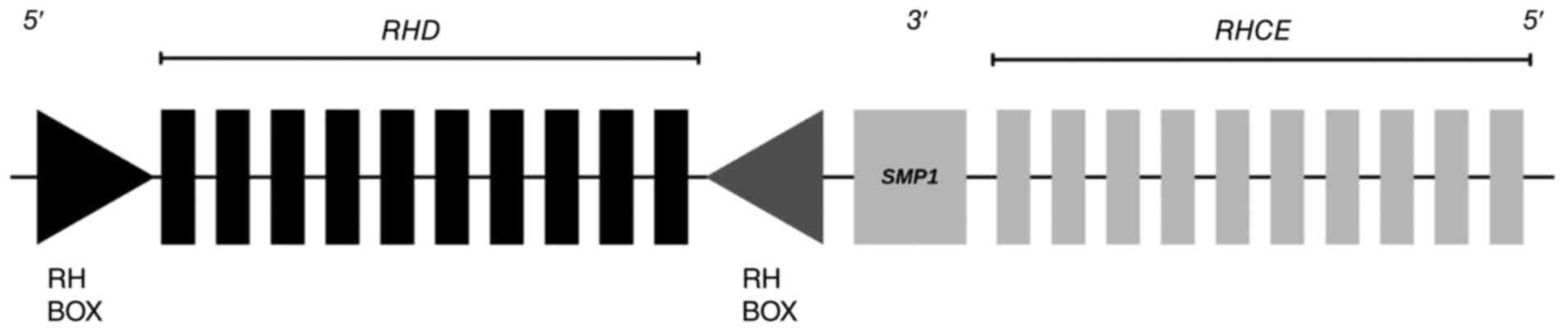
(8). RhD antigens are encoded by
the RHD gene, which is located on chromosome 1, close to the
Rh blood group CcEe antigens (RHCE) gene (Fig. 1). RHD and RHCE genes
exhibit 92-97% sequence similarity and both genes contain 10 exons.
This high similarity can make genetic testing more difficult due to
the risk of cross-reaction (9).
In non-invasive RHD genotyping, exon 7 and
exon 10 are the main targets. RHD exon 7 is important as it
includes variations specific to RHD, while exon 10 is used
as an additional control to improve accuracy and reliability. Using
both exons together helps to separate RHD from RHCE,
resulting in an advantageous method for non-invasive prenatal
RHD genotyping. Non-invasive methods for fetal RhD
determination also exhibit the potential to influence infertility
treatments. In particular, cell-free DNA (cfDNA), found in embryo
culture media during IVF, has emerged as a new biological source
for PGT (4). These analyses are
performed without harming the embryo and non-invasive
preimplantation genetic testing has attracted great attention in
recent years (4,10). Specifically, for RhD genotyping,
this method provides the possibility to detect Rh incompatibility
without physical intervention on the embryo (9).
An important application of IVF is the screening and
prevention of sex-linked genetic disorders, which are inherited
conditions caused by mutations on the X or Y chromosomes and often
exhibit distinct patterns of inheritance and clinical manifestation
between men and women (11). These
disorders are influenced not only by the genome but also by
lifestyle, nutrition, environment and sex hormones such as
estrogen, progesterone and testosterone, which affect immune
responses and gene expressions (12). The X chromosome carries ~867 genes
that are not only involved in sex determination but also serve
notable roles in the development and function of major organs such
as the brain, heart, liver, kidneys and skin, as a number of these
genes encode structural proteins, enzymes and regulatory molecules
necessary for normal cellular and physiological processes. X-linked
disorders are usually more severe in male patients as men carry
only one X chromosome, while females may exhibit milder symptoms
due to a second X chromosome being present. However, abnormal X
chromosome inactivation or homozygosity can still cause serious
disease in females, including >500 X-linked disorders, including
Duchenne muscular dystrophy, hemophilia A and fragile X syndrome,
which appear to be more severe in males (12).
Since the Y chromosome is present only in males,
Y-linked disorders are inherited exclusively through the paternal
line and their phenotypic expression is restricted to male
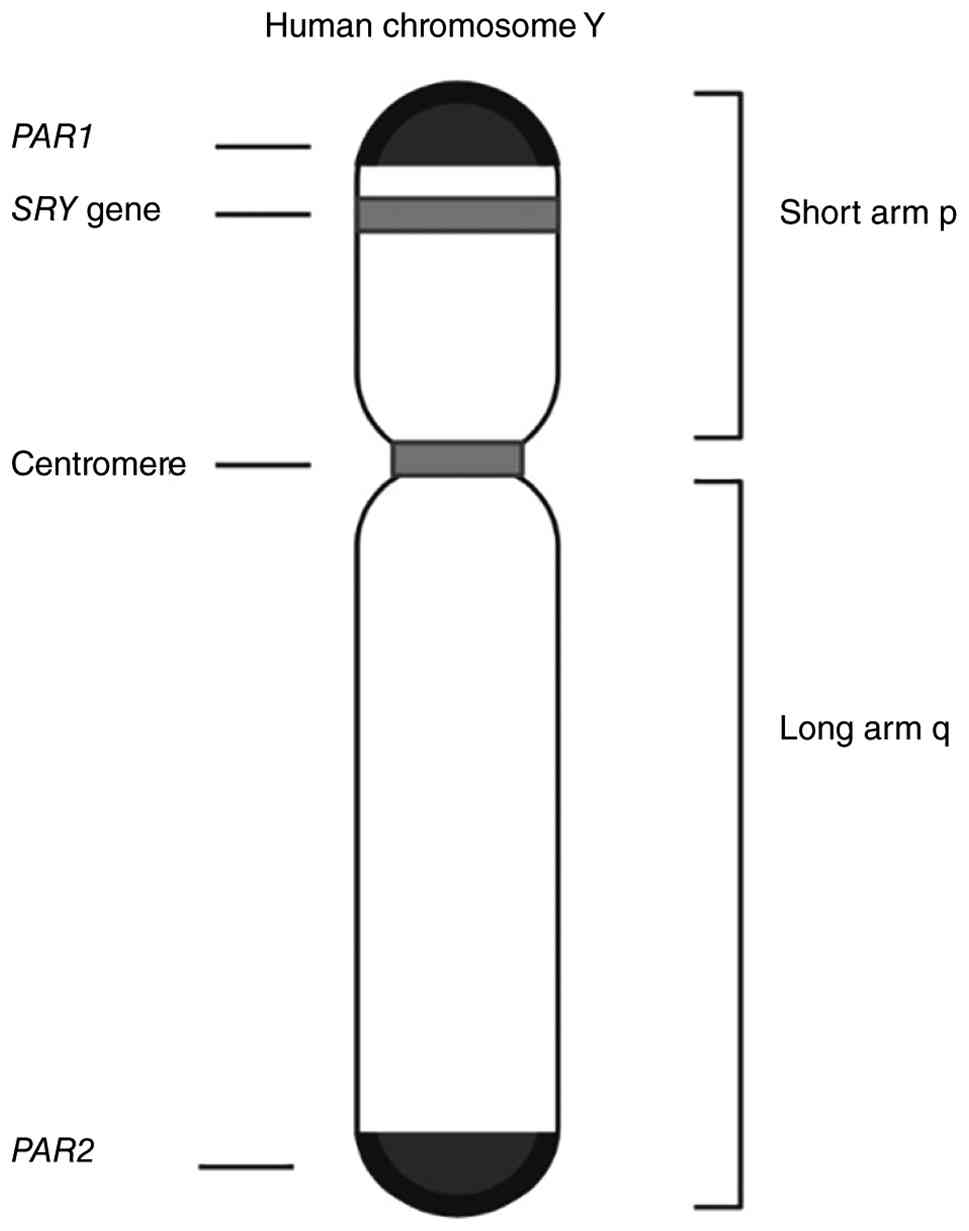
patients, as females lack the Y chromosome. An important gene on
the Y chromosome is sex-determining region Y (SRY), located
at p11.2 (Fig. 2) (13). This gene produces the testis
determining factor, which initiates testis development. During
early development, SRY activates other genes such as Sox9,
leading to Sertoli cell formation and anti-Müllerian hormone
production, which directs male reproductive system formation.
Mutations or deletions of the SRY gene can cause conditions
such as gonadal dysgenesis or hypospadias. Therefore, SRY is
a key biomarker for sex differentiation and genetic diagnosis
(13).
As the mitochondrial function of reproductive cells
deteriorates, the quality of the germ cells declines and in the
majority of cases, it prevents healthy embryo development (14). Mitochondrially encoded cytochrome b
(MT-CYB) makes it possible to check for the quality of the
reproductive cells beforehand as a part of niPGT. MT-CYB
encodes a key protein component of mitochondrial complex III of the
respiratory chain (the cytochrome bc1 complex), which
serves a central role in cellular energy production through
oxidative phosphorylation. Mitochondrial complex III is located in
the mitochondrial inner membrane (15) and is responsible for catalyzing the
electron transfer from ubiquinol to cytochrome c (16). Thus, it represents an important
step in establishing the proton gradient across the inner
mitochondrial membrane (17). This
complex also has 11 subunits, of which only one subunit is encoded
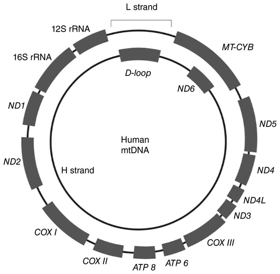
by mitochondrial DNA and is named cytochrome b. MT-CYB gene
is located between the positions 14747 and 15887 of mitochondrial
DNA (Fig. 3) and the cytochrome b
protein, which is the only protein subunit that is encoded by
mitochondrial DNA, is 380 amino acids long (17). In addition, the relatively high
abundance of this gene in mitochondrial genomes, as well as its key
function in oxidative phosphorylation, make MT-CYB an ideal
target for quantification studies such as droplet digital PCR
(ddPCR). Notably, pathogenic variants in the gene have been
associated with mitochondrial complex III deficiency, impaired
cellular energy production, and a broad spectrum of clinical
disorders affecting tissue viability and organismal health
(18).
The sensitivity of MT-CYB expression and
mutations to cellular stress and metabolic changes makes it a
potential biomarker of mitochondrial dysfunction, especially in IVF
(19). In previous years, it has
become common to analyze cfDNA samples in niPGT. Recent
advancements in niPGT have allowed the use of cfDNA and
mitochondrial cfDNA (cf-mtDNA) samples to identify the qualities of
examined germ cells. Subsequently, this increases the chance of
obtaining pregnancy with advanced maternal age by allowing the
monitoring of reproductive cell quality (20).
Therefore, the aim of the present study was to
identify Rh incompatibility, which can be lethal for the embryo, by
analyzing the Rh factor in cfDNA obtained from spent culture medium
(SCM) through niPGT and ensuring correct embryo selection before
implantation. In addition, it was intended to use the identified
genes associated with sex-linked diseases for screening, thereby
providing an effective method for selecting the healthiest
blastocyst. Furthermore, the present study sought to investigate
whether there is an association between the amount of cfDNA in SCM
and maternal age, with the aim of providing insights for future
research as a part of promising biomarkers in niPGT.
Materials and methods
Study design
Embryos from routine clinical procedures of IVF
treatment were used to conduct the present study. During the IVF
procedure, fertilized embryos were cultured in individual droplets
for incubation, observation and morphological assessment. For the
present study, the SCM from each embryo was collected. After sample
collection, cfDNA was isolated from SCM samples using a magnetic
bead-based extraction method to improve cfDNA recovery. Primers
were designed and validated by quantitative PCR (qPCR) for target
genes. ddPCR was then performed to amplify the target sequences and
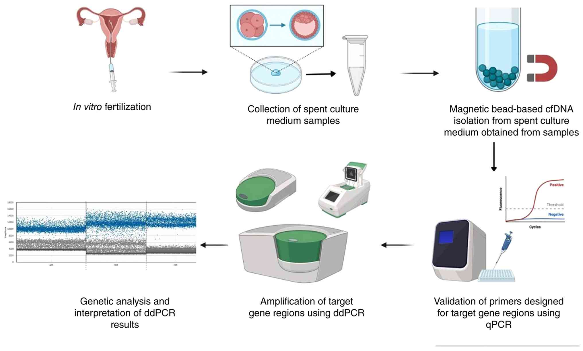
the resulting data were analyzed accordingly. The overall workflow
of the present study is presented in Fig. 4.
Study criteria
SCM samples from embryos were provided by the IVF
Center of Acıbadem Fulya Hospital (Istanbul, Turkey). All samples
were transferred to the Department of Molecular Biology and
Genetics, Istanbul University (Istanbul, Turkey). All patients were
requested to sign informed consent. The necessary documents were
obtained from patients according to the ethical regulations
approved by the Ethics Committee of Istanbul University Faculty of
Medicine (Istanbul, Turkey; approval no. 2023/1488 in 2023). The
present study was conducted without the use of embryos,
human/animal tissues or any other biological material derived from
living organisms; only cfDNA from the SCM of embryos was
analyzed.
Couples who participated in the present study: i)
Had a family history of genetic diseases or an infertility
background; ii) were 29-45 years old; and iii) presented with
infertility indications, as shown in Table I. Patients with systemic diseases,
active infections or any condition that contraindicated IVF
treatment, such as diabetes, active malignancies and uncontrolled
thyroid disorders, were excluded from the present study. The
minimum sample size for the present study was calculated using
G*Power version 3.1(21) analysis
using t-test for means, two independent means (two group) with a
power of 0.85 (Fig. S1).
 | Table IDemographic characteristics and
clinical indications of the study population. |
Table I
Demographic characteristics and
clinical indications of the study population.
| Indication | Total number of
patients | Female
patients | Male patients | Mean age ± SD,
years |
|---|
| Advanced maternal
age | 12 | 12 | - | 41.6±2.8 |
| Uterine-related
miscarriage | 8 | 8 | - | 38.3±4.8 |
| Diminished ovarian
reserve | 5 | 5 | - | 38.8±3.5 |
| Male related
factors | 5 | - | 5 | 38.4±3.5 |
| Unexplained
ınfertility | 1 | 1 | 1 | N/A |
| Recurrent pregnancy
loss | 1 | 1 | 1 | N/A |
SCM collection
All samples were collected from the IVF Center of
Acıbadem Fulya Hospital between January 2024 and December 2025. IVF
procedures in the medical center were carried out as routine
procedures; oocytes were inseminated by intracytoplasmic sperm
injection (ICSI) and used for embryo culturing. Prior to ICSI,
cumulus-corona cells were removed through standard denudation
procedures to minimize residual maternal cell carryover. All
embryology procedures were performed under validated IVF laboratory
protocols in controlled clean-room conditions using certified
sterile consumables and aseptic handling practices (22,23).
Fresh embryos were placed in media containing human serum albumin
(cat no: H5GT-010; LifeGlobal™). Each embryo was placed
in a different culture dish to prevent samples from getting
contaminated. Individual culture handling, dedicated pipettes/tips
and single-embryo culture drops were used to reduce the risk of
cross-contamination and environmental DNA exposure. Procedures,
including sample transfer, were conducted in biosafety cabinets
under sterile conditions. During the incubation process, embryo
growth and division were carefully monitored. On day 5-6, the
majority of embryos were expected to be in the blastocyst stage.
Embryos were graded according to the Gardner Grading Scale based on
their morphology (24).
A total of ~30 µl spent media was collected from
each embryo culture after trophoectoderm biopsy on day 5-6. Samples
were transferred into RNase-DNase-free PCR tubes containing 5 µl
lysis buffer (cat no: 19075; Qiagen GmbH). Collection tubes were
prepared in advance and samples were processed individually to
further minimize exogenous DNA contamination. Negative control
media droplets processed in parallel were included, where
applicable, as contamination monitoring controls. All samples were
stored at -20˚C for a day in the clinic at Acıbadem Fulya Hospital
before being transported to Istanbul University, maintaining cold
chain conditions. In the laboratory, all samples were frozen at
-80˚C until experiments were initiated.
DNA purification
cfDNA from SCM samples was isolated using a QIAamp
MinElute ccfDNA Kit (cat no: 55284; Qiagen GmbH), which utilizes
magnetic bead technology for extra yield. Extracted cfDNA was
eluted in 50 µl elution buffer (Buffer EB; cat. no. 19086; Qiagen
GmbH) for subsequent steps according to the manufacturer's
instructions. Immediately after purification, samples were
quantified using a NanoDrop™ 2000 spectrophotometer
(Thermo Fisher Scientific, Inc.). Processed samples were frozen at
-80˚C until amplification.
Primer design
For the detection of selected gene regions,
including GAPDH and β-globin as housekeeping genes, 6
primer sets were designed using the PrimerQuest™ PCR
tool (Integrated DNA Technologies, Inc.), targeting RHD exon
5, RHD exon 10, SRY, MT-CYB, GAPDH and
β-globin (Table II).
Primer specificity was validated using an in silico PCR tool
(University of California Santa Cruz In-Silico PCR)
(25), before primers determined
to be specific were then outsourced for synthesis. The primer
sequences for RHD genotyping were selected to ensure maximal
specificity. The primers for RHD exon 10 were modified from
research conducted by Hromadnikova et al (26), which underwent clinical validation
for the detection of fetal RHD and was specifically tested on
Rh-negative (dd) individuals to ensure the elimination of
false-positive signals. For exon 7, the primers were carefully
designed in silico to only target RHD-specific sequences
that don't have any similarities with the RHCE gene. To
avoid cross-reactivity, particular attention was paid to the 3' end
of the primers. This made sure that the amplification was specific
to the RHD gene, even when RHCE sequences were highly
homologous. For further validation, whole-blood analysis was
performed on Rh (-) and Rh (+) individuals. Genomic DNA isolated
from whole-blood samples using the QIAamp MinElute Media Kit
(Qiagen GmbH) was amplified with ddPCR for RHD exon 7, exon
10 and GAPDH genes.
| Table IIDesigned primers and their
sequences. |
Table II
Designed primers and their
sequences.
| Gene | Direction | Sequence |
|---|
| RHD exon
7 | Forward |
5'-GGTGTTGTAACCGAGTGCT-3' |
| | Reverse |
5'-CACCAGCAGCACAATGTAGA-3' |
| RHD exon
10 | Forward |
5'-CCTCTCACTGTTGCCTGCATT-3' |
| | Reverse |
5'-AGTGCCTGCGCGAACATT-3' |
| MT-CYB | Forward |
5'-TGAAACTTCGGCTCACTCCT-3' |
| | Reverse |
5'-AATGTATGGGATGGCGGATA-3' |
| SRY | Forward |
5'-TCCTCAAAAGAAACCGTGCAT-3' |
| | Reverse |
5'-AGATTAATGGTTGCTAAGGACTGGAT-3' |
|
β-globin | Forward |
5'-GCAGGTTGGTATCAAGGTTACA-3' |
| | Reverse |
5'-GTGCCTATCAGAAACCCAAGAG-3' |
| GAPDH | Forward |
5'-GGAGTGAGTGGAAGACAGAATG-3' |
| | Reverse |
5'-GAGGGACACAAGGTTACCATATAC-3' |
ddPCR
A ddPCR protocol was prepared and validated for the
present study using a QX Droplet Digital™ PCR system
(Bio-Rad Laboratories, Inc.; Table
III). Each sample and reaction mix was loaded into a cartridge
well along with a droplet generator oil (Bio-Rad Laboratories,
Inc.) for droplet generation, a process which separates DNA samples
into ~20,000 droplets. Generated droplets were gently transferred
to a 96-well plate for ddPCR before the plate was sealed.
Amplification conditions were as follows: 5 min at 95˚C for enzyme
activation, 30 sec at 95˚C for initial denaturation, 1 min at 58˚C
(40 cycles of annealing and extension), 5 min at 4˚C and 5 min at
90˚C for signal stabilization. After thermal cycling was conducted,
the plate was placed into a QX200 Droplet Reader (Bio-Rad
Laboratories, Inc.) for signal detection.
| Table IIIReaction mix preparation for
ddPCR. |
Table III
Reaction mix preparation for
ddPCR.
| Component | Volume per
reaction, µl | Final
concentration |
|---|
| 2x
QX200™ ddPCR™ EvaGreen®
Supermix | 10 | 1X |
| Forward primer | 1 | 100 nM |
| Reverse primer | 1 | 100 nM |
| DNA template and
RNase-/DNase-free water | 8 | Up to 100 ng |
| Total | 20 | |
Statistical analysis
The built-in droplet reader algorithm (QX Manager;
version 2.2; Bio-Rad Laboratories, Inc.) was used for droplet
analysis as it provides the positive and negative droplet counts
for each well corresponding to the target gene region. Based on the
number of positive droplets, the software applies the Poisson
statistical model to automatically provide the absolute copy number
of the target DNA (copies/µl). The threshold for the outlined
result was determined as 3 positive droplets per sample. McNemar's
tests were applied for RHD gene outcomes. For calculating
the correlation between maternal age and mitochondrial DNA levels,
Spearman's rank correlation tests were used. Statistical analyses
were performed using R software (version 4.5.3; R Foundation for
Statistical Computing) and two-sided P<0.05 was considered to
indicate a statistically significant difference.
Results
Patient characteristics and the
interpretation of the study results
A total of 33 SCM samples were collected from IVF
patients between July 2024 and July 2025. Among these samples, 18
samples were used for RHD status prediction (targeting exons
7 and 10), 31 samples were analyzed for SRY gene detection
and the same 33 samples were also examined for MT-CYB gene
expression to assess embryo quality in association with maternal
age. The total droplet counts exceeded 10,000 in all cases,
ensuring the reliability of ddPCR data. No positive droplets were
observed in negative controls. A sample was considered positive
when ≥3 positive droplets were detected. This threshold was
determined empirically based on repeated no-template control
reactions, which consistently yielded 0-1 positive droplets only.
Samples with detectable droplets above background were considered
positive. Lower droplet counts (<50) were interpreted as
low-abundance signals, whereas higher counts reflected robust
target detection.
Determination of RH status from
SCM
RHD assay results for ddPCR in Table IV demonstrated positive
amplification in 8 samples for exon 7 (47.1%) and 10 samples for
exon 10 (58.8%) out of 17 samples. The McNemar test was used to
verify consistency between exon 7 and exon 10 detection. The result
was a 52.94% (9/17) conformity rate. Statistical analysis indicated
no significant discordance between the detection rates of the two
exons (P=0.7266; 95% CI for the difference in proportions: -44.1%
to +20.6%), implying that both genetic regions were identified with
similar efficiency in cfDNA obtained from SCM.
| Table IVRHD exon 7 and exon 10 digital
droplet PCR positive droplet counts and predicted Rh status. |
Table IV
RHD exon 7 and exon 10 digital
droplet PCR positive droplet counts and predicted Rh status.
| Sample no. | RHD exon 7
positive droplet count | Total droplet
count | RHD exon 10
positive droplet count | Total droplet
count | RHD exon
7 | RHD exon
10 |
|---|
| 1 | 11,785 | 17,097 | 0 | 14,358 | Positive | Negative |
| 2 | 11,762 | 15,434 | 0 | 16,063 | Positive | Negative |
| 3 | 0 | 13,165 | 0 | 15,170 | Negative | Negative |
| 4 | 58 | 13,621 | 0 | 16,269 | Borderline
positive | Negative |
| 5 | 0 | 13,574 | 0 | 15,843 | Negative | Negative |
| 6 | 42 | 12,527 | 91 | 17,445 | Borderline
positive | Borderline
positive |
| 7 | 0 | 12,563 | 35 | 17,719 | Negative | Borderline
positive |
| 8 | 0 | 12,626 | 13,912 | 16,815 | Negative | Positive |
| 9 | 164 | 11,514 | 12,774 | 17,507 | Positive | Positive |
| 10 | 0 | 12,791 | 8,857 | 13,715 | Negative | Positive |
| 11 | 0 | 11,142 | 7,008 | 10,863 | Negative | Positive |
| 12 | 10 | 17,707 | 7,780 | 11,224 | Borderline
positive | Positive |
| 13 | 0 | 16,333 | 11,762 | 17,897 | Negative | Positive |
| 14 | 12,030 | 17,411 | 9,516 | 15,487 | Positive | Positive |
| 15 | 10,778 | 16,338 | 13,420 | 17,340 | Positive | Positive |
| 16 | 12 | 4,226 | | | Borderline
positive | |
| 17 | 0 | 13,605 | 0 | 14,785 | Negative | Negative |
| 18 | 0 | 9,347 | 0 | 12,635 | Negative | Negative |
| NTC | 0 | 10,079 | 0 | 10,355 | | |
Available clinical follow-up data for the five
embryos that resulted in live births are shown in Table V. Among these, one newborn was
RhD-negative and four were RhD-positive. ddPCR correctly predicted
the Rh status of the RhD-negative newborn, while false-negative
results were obtained in two cases (ddPCR negative and clinically
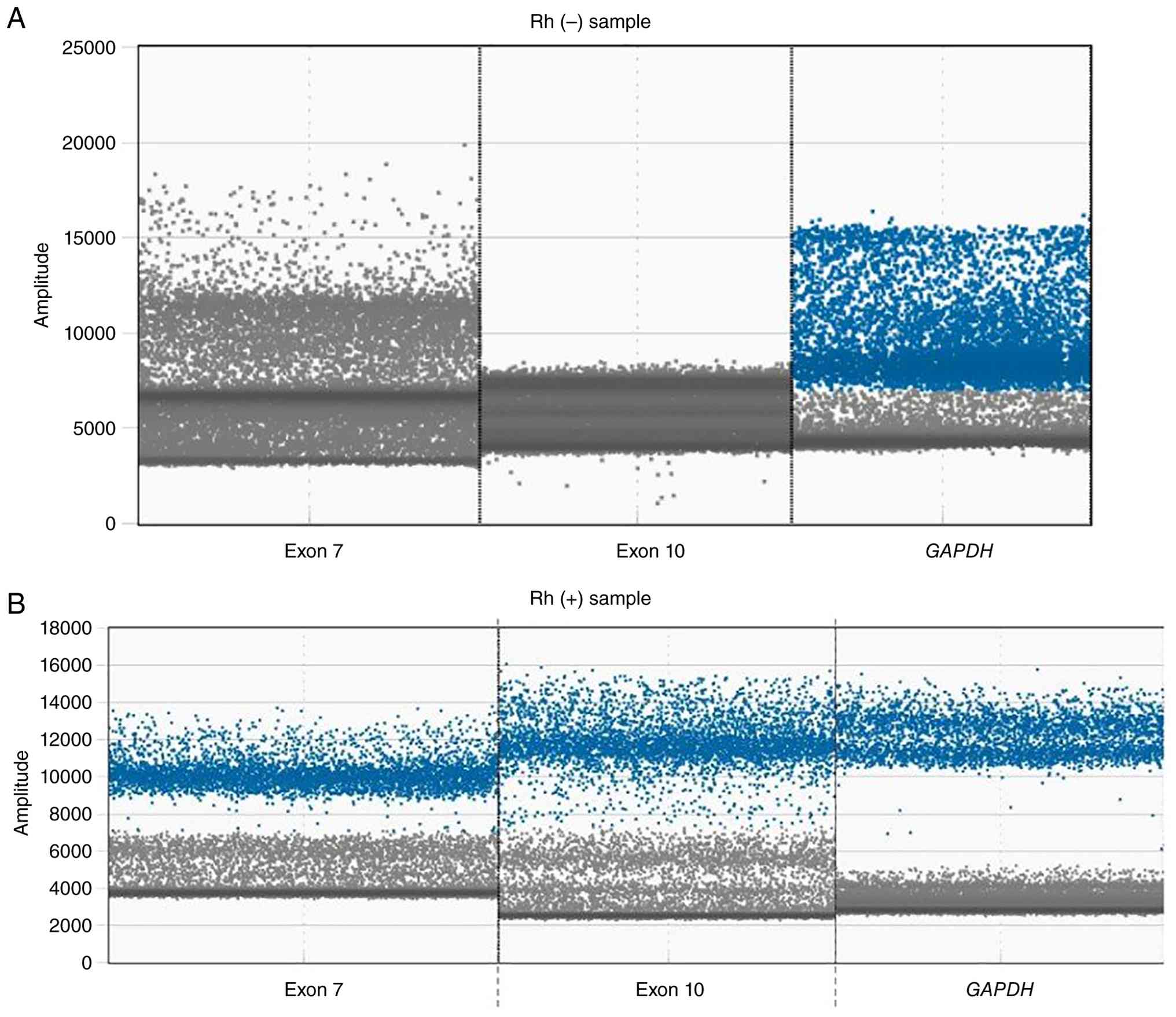
RhD-positive). The validation data of RHD exon 7 and 10 in whole
blood presented in Fig. 5
demonstrated that the primer sets were suitable for distinguishing
between RHCE and RHD.
| Table VCorrelation between RHD exon 7
and RHD exon 10 ddPCR results and clinical data. |
Table V
Correlation between RHD exon 7
and RHD exon 10 ddPCR results and clinical data.
| | ddPCR Rh status
prediction results | Clinical
records |
|---|
| Sample no. | RHD exon
7 | RHD exon
10 | Pregnancy
confirmation | Compatibility |
|---|
| 3 | Negative | Negative | Rh positive | Not compatible |
| 15 | Negative | Negative | Rh negative | Compatible |
| 16 | Negative | Negative | Rh positive | Not compatible |
| 17 | Positive | Positive | Rh Positive | Compatible |
| 18 | Positive | | Rh Positive | Compatible |
Determination of embryonic sex region
from SCM
A total of 31 samples were analyzed by ddPCR for the
detection of the SRY gene. The presence of the SRY
gene, indicated as either positive or negative are demonstrated in
Table VI. According to this
analysis, 16 samples (51.6%) were classified as SRY
positive, whereas 15 samples (48.4%) were SRY negative.
Among the SRY-positive samples, the proportion of positive
droplets was consistently clustered around 0.38-0.44, indicating a
reproducible detection threshold for true positivity. By contrast,
SRY-negative samples exhibited ratios close to 0
(0.00-0.02), consistent with the absence of the target gene.
Notably, two cases (sample 1: ratio 0.020 and sample 3: ratio
0.005) exhibited very low levels of positive droplets, which fell
between the negative and clearly positive ranges (Fig. S2). These borderline results may
have represented either low-level true positives or technical
difficulties such as non-specific amplification or contamination.
Overall, the ddPCR assay demonstrated a clear distinction between
SRY-positive and SRY-negative samples, with the
majority of results exhibiting conclusive classification.
| Table VIDigital droplet PCR results of the
SRY gene. |
Table VI
Digital droplet PCR results of the
SRY gene.
| Sample no. | Positive droplet
count | Negative droplet
count | Total droplet
count | SRY gene
presence |
|---|
| 1 | 30,000 | 16,905 | 16,935 | Borderline
positive |
| 2 | 0 | 15,149 | 15,149 | Negative |
| 3 | 63.000 | 11,727 | 11,790 | Positive |
| 4 | 0 | 16,720 | 16,720 | Negative |
| 5 | 12,238 | 19,188 | 31,426 | Positive |
| 6 | 9,525.000 | 15,856 | 25,381 | Positive |
| 7 | 0 | 16,669 | 16,669 | Negative |
| 8 | 0 | 15,062 | 15,062 | Negative |
| 9 | 0 | 16,929 | 16,929 | Negative |
| 10 | 0 | 16,011 | 16,011 | Negative |
| 11 | 0 | 18,734 | 18,734 | Negative |
| 12 | 14.086 | 17,902 | 31,988 | Positive |
| 13 | 71.000 | 16,743 | 16,814 | Positive |
| 14 | 11.794 | 17,066 | 28,860 | Positive |
| 15 | 0 | 14,589 | 14,589 | Negative |
| 16 | 12,214 | 16,416 | 28,630 | Positive |
| 17 | 0 | 16,397 | 16,397 | Negative |
| 18 | 13,741 | 18,758 | 32,499 | Positive |
| 19 | 10,435 | 15,054 | 25,489 | Positive |
| 20 | 0 | 19,163 | 19,163 | Negative |
| 21 | 9,995.000 | 14,910 | 24,905 | Positive |
| 22 | 0 | 12,151 | 12,151 | Negative |
| 23 | 9,707 | 15,905 | 25,612 | Positive |
| 24 | 12,840 | 17,687 | 30,527 | Positive |
| 25 | 0 | 19,238 | 19,238 | Negative |
| 26 | 9,511.000 | 13,542 | 23,507 | Positive |
| 27 | 9,631.000 | 13,996 | 23,627 | Positive |
| 28 | 11,170 | 15,511 | 26,681 | Positive |
| 29 | 0 | 13,438 | 13,438 | Negative |
| 30 | 0 | 12,032 | 12,032 | Negative |
| 31 | 0 | 8,529 | 8,529.000 | Negative |
Detection of the SRY gene by ddPCR analysis
demonstrated complete concordance with the pregnancy determination
results in Table VII.
Specifically, samples 3, 14 and 18 were identified as
SRY-positive, whereas samples 29 and 30 were
SRY-negative. In all five cases, the molecular findings were
fully consistent with the clinical confirmation of pregnancy
outcomes, corresponding to an accuracy rate of 100%. Although the
sample size was limited (n=5), these results provided strong
preliminary evidence supporting the reliability and specificity of
ddPCR for the detection of the SRY gene in this experimental
setting.
| Table VIIddPCR SRY gene results and
clinical data. |
Table VII
ddPCR SRY gene results and
clinical data.
| Sample no. | ddPCR SRY
prediction results | Clinical
records |
|---|
| 3 | Positive | Compatible |
| 14 | Positive | Compatible |
| 18 | Positive | Compatible |
| 29 | Negative | Compatible |
| 30 | Negative | Compatible |
Determination of the correlation
between maternal age and cf-mtDNA amount from SCM
In the present study, cfDNA samples derived from 33
different embryo SCMs were examined. The maternal age for the
examined embryos ranged between 31 and 48 years, allowing for the
evaluation of age-related changes in embryo quality across a broad
spectrum. The MT-CYB gene was successfully amplified in 19
samples and the β-globin gene was successfully amplified in
12 samples, as shown in Table
VIII. Furthermore, in 6 of the samples, both MT-CYB and
β-globin genes were detected during ddPCR analysis. Of the 6
results obtained, one extreme result was excluded from the analysis
as the positive droplets of the reference gene were considered low,
indicating insufficient template DNA for reliable quantitative
analysis. The cf-mtDNA/cf-DNA ratio was subsequently evaluated
using a threshold maternal age of 40 years, as it was stated that
advanced maternal age is considered to be >35 years in previous
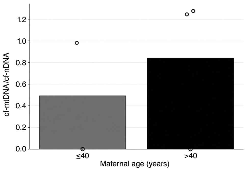
studies (27,28). In Fig.
6, the cf-mtDNA/cf-nDNA ratio was found to be moderately higher
in the ≥40 years group. In SCM samples obtained from women
undergoing IVF, women aged ≥40 years exhibited a moderately higher
cf-mtDNA/cf-nDNA ratio compared with those aged <40 years,
although these results did not reach significance.
| Table VIIIDigital droplet PCR results of the
MT-CYB gene. |
Table VIII
Digital droplet PCR results of the
MT-CYB gene.
| Sample no. | Maternal age,
years | MT-CYB
positive droplet count | β-globin
positive droplet count |
|---|
| 1 | 38 | 0 | 0 |
| 2 | 31 | 0 | 0 |
| 3 | 45 | 0 | 0 |
| 4 | 38 | 0 | 0 |
| 5 | 31 | 8 | 0 |
| 6 | 34 | 0 | 0 |
| 7 | 48 | 13 | 0 |
| 8 | 41 | 11,764 | 9,463 |
| 9 | 42 | 76 | 0 |
| 10 | 37 | 11,887 | 0 |
| 11 | 41 | 35 | 0 |
| 12 | 40 | 7 | 0 |
| 13 | 44 | 23 | 0 |
| 14 | 37 | 5 | 0 |
| 15 | 44 | 12 | 12,679 |
| 16 | 37 | 0 | 0 |
| 17 | 37 | 0 | 0 |
| 18 | 45 | 0 | 10,630 |
| 19 | 31 | 99 | 0 |
| 20 | 37 | 25 | 0 |
| 21 | 38 | 0 | 46 |
| 22 | 41 | 0 | 12,860 |
| 23 | 36 | 0 | 0 |
| 24 | 38 | 0 | 12,814 |
| 25 | 37 | 0 | 10,649 |
| 26 | 41 | 10,373 | 8,137 |
| 27 | 39 | 28 | 11,100 |
| 28 | 40 | 9,389 | 9,575 |
| 29 | 44 | 6,962 | 0 |
| 30 | 40 | 11,563 | 0 |
| 31 | 38 | 0 | 49 |
| 32 | 33 | 9,312 | 10 |
| 33 | 41 | 10,556 | 0 |
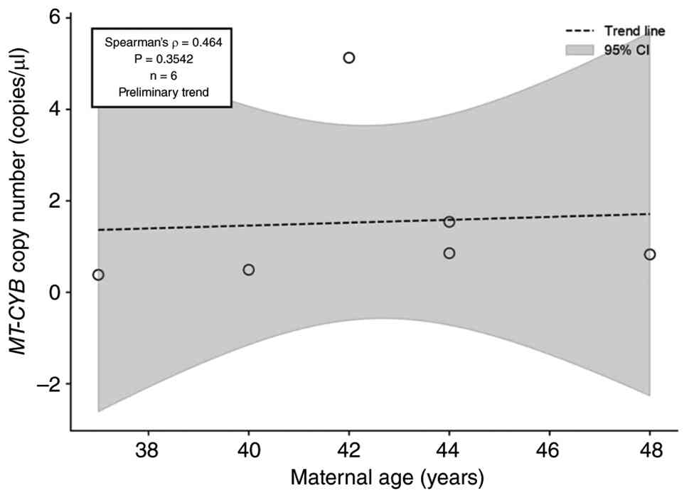
In Fig. 7, the
association between maternal age and the ratio of MT-CYB
copy number per µl was examined, which was obtained from 6 out of
33 samples collected from SCM. A Spearman's rank correlation test
was conducted to evaluate the results, revealing a coefficient of
r=0.464, indicating a moderate positive trend. However, since
P=0.354 and P>0.05, the difference was not statistically
significant. Although the data suggested that the copy number ratio
may have increased with advancing age, the limited sample size
prevented this association from reaching statistical significance.
Under these conditions, this ratio only served as a
hypothesis-generating preliminary evidence for it to be considered
as a biomarker for mitochondrial dysfunction as the maternal age
increases, especially in the experiment groups with >30
samples.
Discussion
niPGT has emerged as a new approach in reproductive
medicine that avoids invasive techniques and reduces risks, such as
structural damage to the embryo or mosaicism associated with
biopsy. Since the first discovery of cell-free fetal DNA in
maternal plasma in 1997(29),
researchers have started to move towards less invasive methods for
genetic analysis in assisted reproductive technologies. The
identification of cfDNA and cf-mtDNA in SCM represented new
possibilities for analyzing embryonic genetic status without
compromising embryo viability. With the help of technological
advancements such as ddPCR, these techniques established the way
for highly sensitive and precise quantification of genetic material
from minimal and fragmented DNA sources, offering marked clinical
potential for embryo selection in IVF.
In the present study, cfDNA and cf-mtDNA isolated
from SCM were analyzed using ddPCR to understand their potential in
niPGT. The present findings showed that ddPCR offered a highly
sensitive detection of low-input genetic targets, an important
advantage in niPGT applications whereby cfDNA quantities are often
limited and highly fragmented.
Despite ddPCR having demonstrated high analytical
sensitivity for RHD detection in low-input samples, the
clinical concordance rate remained moderate. The clinical data
concordance rate was 60% for five samples, considering the
challenging nature of SCM-derived cfDNA. This discrepancy may have
partly stemmed from the intrinsic characteristics of SCM-derived
cfDNA, which contain a variable mixture of embryonic and maternal
DNA fragments. The present findings are clinically relevant given
that accurate RhD genotyping is important in preventing HDFN.
Previous studies have shown that maternal DNA contamination and
allelic drop-out are major limiting factors in RhD genotyping when
fetal or embryonic DNA input is low (29,30).
In addition, the RHD gene exhibits structural complexity,
including pseudogenes and hybrid alleles, which may lead to
false-positive or false-negative results depending on the region
being targeted (31). These
biological and technical constraints may explain why exon 7 and
exon 10 amplification results were not always consistent across
samples. To reduce these diagnostic risks in future clinical
applications, the present study recommends employing a more
effective multiplexing approach. Targeting at least three exons of
the RHD gene at the same time in a single ddPCR reaction
(such as exons 5, 7 and 10) could make detection notably more
precise, compared with just relying on one or two exons.
Despite these limitations, the fact that ddPCR was
able to detect RHD in fragmented cfDNA samples highlights
its robustness and supports its potential for future niPGT
applications, particularly when combined with
contamination-reduction strategies or embryo-specific molecular
markers. Previous studies using qPCR have reported reduced
sensitivity at low cfDNA concentrations and in early gestation
stages (32). By contrast, ddPCR
partitions the DNA template into thousands of nanoliter-sized
droplets, allowing absolute quantification of target sequences
without the need for standard curves, thus providing a higher
tolerance to PCR inhibitors (33).
This may explain why, in the present study, ddPCR could reliably
detect RHD sequences even in samples with low cfDNA input,
supporting its advantages compared with conventional PCR methods
for niPGT.
SRY gene amplification was successful in
65.3% of samples, with 100% concordance in the five cases with
available pregnancy outcome data. SRY detection exhibited a
higher clinical concordance compared with RHD, which may be
attributed to its male-specific nature and the absence of maternal
background interference. However, the overall amplification rate
may indicate that SRY detection is still challenged by the
low abundance of Y-chromosome fragments in SCM. Studies have
reported that Y-derived cfDNA is often less abundant due to both
biological factors (for example, fewer copies of Y-chromosomal
sequences) and technical factors such as preferential degradation
of longer Y-chromosome segments (34,35).
The 100% concordance in the clinically validated samples
nevertheless suggested that when detectable, ddPCR provided highly
reliable sex determination for niPGT. It also supports that ddPCR
may improve the precision of fetal sex determination, particularly
in pregnancies at risk of X-linked disorders, by detecting
male-specific DNA at earlier gestational ages and with greater
sensitivity compared with qPCR.
It has previously been determined that the condition
of mitochondrial DNA serves a key role in understanding the quality
of the embryo and that this quality decreases as the maternal age
advances (20). In the present
study, the quality of cf-mtDNA samples derived from SCM was
identified and evaluated with the MT-CYB gene as a part of a
potential biomarker in niPGT. Mitochondrial cfDNA analysis with
MT-CYB gene droplet count using ddPCR revealed a positive
correlation between maternal age and cf-mtDNA levels, suggesting
that mitochondrial stress and dysfunction increase with advancing
maternal age, ultimately impacting embryo quality. In a study by
May-Panloup et al (36), it
was stated that as the age of the mother progresses, accumulation
of the mtDNA mutations in oocytes increases and not only
deteriorates the quality of the oocyte but also increases the risk
of allowing mitochondrial abnormalities inherited by the offspring
(36).
Given the central role of mitochondria in embryo
quality and development, this finding may indicate an
age-associated increase in mitochondrial dysfunction in oocytes,
which in turn could contribute to reduced fertility. An elevated
cf-mtDNA/cf-nDNA ratio may reflect enhanced release of damaged or
dysfunctional mitochondria into the extracellular space,
potentially impairing the overall health and developmental
competence of the oocyte, unless there is embryo fragmentation or
degeneration. In the case of embryo fragmentation and degeneration,
release of mt-DNA into the SCM may increase regardless of embryo
quality and maternal age. Under optimal conditions, which state
physiologically intact and healthy embryo samples, the
cf-mtDNA/cf-nDNA ratio effect, as presented in Fig. 6, was consistent with the findings
of Liu et al (37), further
supporting the evidence that cf-mtDNA levels increase with maternal
age. The cf-mtDNA/cf-nDNA ratio graph shown in Fig. 6 has also been reported in a study
by Tsirka et al (38),
where the ratio was investigated in the context of IVF treatment.
In this study, follicular fluid samples were analyzed using qPCR
with the ribulose-5-phosphate-3-epimerase gene for cf-nDNA
detection. The study found a direct association between increasing
maternal age and elevated cf-mtDNA/cf-nDNA ratios. Although the
amplification methods and primers used in the present study
differed from those employed by previous studies (37,38),
the present results corroborate their findings. Nevertheless, this
observation should be considered an exploratory finding due to the
limited sample size (n=6) and thus requires validation in larger
cohorts.
In recent studies, >100 different samples have
been analyzed (37,38). Compared with these studies, the
reliability of the present findings was statistically less powerful
in terms of detecting significant correlations due to the smaller
dataset. Thus, obtained results of the present hypothesis should be
valued as preliminary and require larger sample data set analysis
to eliminate the main limitation in the present research for
further studies. The relatively low detection rate of β-globin
(12/33 samples) suggested that nuclear cfDNA may be present at
lower abundance or may be more susceptible to degradation compared
with mitochondrial DNA in SCM. Therefore, apparent increases in
mtDNA relative to β-globin should be interpreted cautiously, as
they may reflect reduced recovery or detectability of nuclear cfDNA
rather than true mitochondrial enrichment alone. Despite these
limitations, the present research holds marked value as it was
conducted within a Turkish patient group and provides a basis for
future studies with larger datasets. Expanding this research within
the Turkish patient population has the potential to introduce a
notable advancement in reproductive medicine. Anticipating embryo
quality prior to implantation may increase the likelihood of
achieving successful pregnancies in couples undergoing IVF for
certain indications. Consequently, IVF success rates and embryo
selection strategies may be improved. Particularly in cases where
age-associated declines in oocyte quality are anticipated, cf-mtDNA
level measurements could guide the selection of the most viable
embryos for implantation, thereby enabling successful pregnancies,
particularly in women of advanced maternal age. Overall, the
application of niPGT utilizing cf-mtDNA demonstrates potential
clinical utility with minimal invasiveness.
The sample size in the present research was
concordant with the G*Power analysis, however was below the
threshold for the specificity and sensitivity. Due to the
fragmented structure of the cfDNA samples, the amplification rate,
despite being higher compared with qPCR, was not satisfactory in
all targets. The reliability of ddPCR results when cfDNA is
fragmented beyond amplification was evaluated by Liu (39), who demonstrated how some genes,
although present, can be silent during the early embryonic stages.
In the early stages of embryonic development, even housekeeping
genes may preserve a heterochromatin structure, therefore are not
part of transcription.
Furthermore, early-stage embryos may release a
limited quantity of nuclear DNA and the presence of mitochondrial
DNA-rich but nuclear DNA-poor droplets could lead to stochastic
loss of the target genes during partitioning, such as housekeeping
genes. This limitation may also explain the inconsistent
amplification observed between different targets, particularly
between RHD exon 7 and exon 10, whereby unequal representation of
fragmented genomic regions may result in exon-specific drop-out.
Therefore, negative amplification results should be interpreted
cautiously, as they may reflect technical limitations associated
with low-input fragmented cfDNA rather than the true absence of the
target sequence. Genes with high embryo-specificity, such as
SRY, demonstrate higher clinical concordance, whereas
targets susceptible to maternal background noise or genomic
structural variation, such as RHD, require additional
optimization. Inclusion of additional RHD targets, such as
multiplexing methods of at least three exons, may improve detection
reliability and concordance in future studies. In addition, the
incorporation of internal embryonic DNA markers or polymorphic
markers may function as a quality control mechanism to determine
the presence of adequate embryonic DNA, thereby differentiating a
genuine Rh-negative result from a technical amplification failure.
This indicates that although niPGT exhibits good potential for
diagnosis based on biomarkers, in the present context, it may be
insufficient to determine a precise conclusion and should instead
be used as a predictive model before further analysis.
An additional limitation of the present study was
the absence of systematic validation using paired conventional
embryo biopsy-based PGT results. Since embryo sex determination is
not permitted to be reported under ethical regulations in Turkey
and RHD status is not routinely evaluated within standard PGT
protocols (40), clinical data
were not available for all embryos included in the present study.
Therefore, outcome information could only be obtained from a
limited number of transferred embryos that resulted in live birth,
where postnatal sex and/or Rh status could be determined. Although
these limited follow-up data showed general concordance with
SCM-based findings, larger prospective studies incorporating paired
biopsy-PGT validation and broader clinical follow-up are required
to determine the diagnostic accuracy and clinical applicability of
this non-invasive approach. In addition, the correlation between
the cf-mtDNA/cfDNA ratio and maternal age warrants further
investigation. The subsample used for this specific correlation
consisted of only 6 samples (n=6), which was markedly below the
minimum sample size established by the present G*Power analysis.
Therefore, the findings for this endpoint are underpowered and
should be considered strictly preliminary. Nevertheless,
cf-mtDNA-based parameters may still serve as promising non-invasive
biomarkers in future studies.
Supplementary Material
G*Power analysis for the
present study. Associated power.sample sizes are marked red.
1D amplitude plots of samples 1 and
3.
Acknowledgements
Not applicable.
Funding
Funding: The present study was funded by the Istanbul University
Scientific Research Projects Department (grant nos. 41128 and
41083) and TUBITAK 2209-A (grant nos. 1919B012433914 and
1919B012447586).
Availability of data and materials
The data generated in the present study may be
requested from the corresponding author.
Authors' contributions
TG, EGA, SS and EB conceptualized and designed the
study. SS, MC, ZS, EK, BKB, HGC, EB and FB prepared materials and
performed the study. SS, EGA and MC performed data collection and
analysis. TG, SS, MC, ZS and EK prepared the original draft. EGA,
BKB, HGC, EB, FB and TG reviewed and edited the manuscript. TG
supervised the study. EGA and TG confirm the authenticity of all
the raw data. All authors read and approved the final
manuscript.
Ethics approval and consent to
participate
The present study was conducted in accordance with
institutional and national guidelines for research involving human
biological materials. Ethical approval was obtained from the Ethics
Committee of Istanbul University Faculty of Medicine (Istanbul,
Turkey; approval no. 2023/1488). Written informed consent to
participate and consent for the use of anonymized biological
samples were obtained in accordance with institutional
regulations.
Patient consent for publication
Not applicable.
Competing interests
The authors declare that they have no competing
interests.
References
|
1
|
Leaver M and Wells D: Non-invasive
preimplantation genetic testing (niPGT): The next revolution in
reproductive genetics? Hum Reprod Update. 26:16–42. 2020.PubMed/NCBI View Article : Google Scholar
|
|
2
|
Kuznyetsov V, Madjunkova S, Abramov R,
Antes R, Ibarrientos Z, Motamedi G, Zaman A, Kuznyetsova I and
Librach CL: Minimally invasive cell-free human embryo aneuploidy
testing (miPGT-A) utilizing combined spent embryo culture medium
and blastocoel fluid-towards development of a clinical assay. Sci
Rep. 10(7244)2020.PubMed/NCBI View Article : Google Scholar
|
|
3
|
Vera-Rodriguez M, Diez-Juan A,
Jimenez-Almazan J, Martinez S, Navarro R, Peinado V, Mercader A,
Meseguer M, Blesa D, Moreno I, et al: Origin and composition of
cell-free DNA in spent medium from human embryo culture during
preimplantation development. Hum Reprod. 33:745–756.
2018.PubMed/NCBI View Article : Google Scholar
|
|
4
|
Chen K, Hu Z, Lian Y, Han Y, Zhou X, Li Y,
Xiang L, Jiang W, Li M, Zeng P, et al: The diagnostic accuracy of
preimplantation genetic testing (PGT) in assessing the genetic
status of embryos: A systematic review and meta-analysis. Reprod
Biol Endocrinol. 23(39)2025.PubMed/NCBI View Article : Google Scholar
|
|
5
|
Günel T, Kalelioğlu I, Ermiş H and Aydınlı
K: Detection of fetal RhD gene from maternal blood. J Turk Ger
Gynecol Assoc. 11:82–85. 2010.PubMed/NCBI View Article : Google Scholar
|
|
6
|
Hall V, Vadakekut ES, Maines J and
Avulakunta ID: Hemolytic disease of the fetus and newborn. [Updated
2025 Jan. 22]. In: StatPearls [Internet]. Treasure Island (FL):
StatPearls Publishing, 2026.
|
|
7
|
Drozdowska-Szymczak A, Łukawska S,
Mazanowska N, Ludwin A and Krajewski P: Management and treatment
outcomes of hemolytic disease of the fetus and newborn (HDFN)-a
retrospective cohort study. J Clin Med. 13(4785)2024.PubMed/NCBI View Article : Google Scholar
|
|
8
|
Alalshaikh MA, Alsughayir AH, Alsaif AS,
Ababtain SA, Aloyouni SY, Aldilaijan KE and Alsubaie SF: Molecular
background of RhD-positive and RhD-negative phenotypes in a Saudi
population. Saudi J Med Med Sci. 12:210–215. 2024.PubMed/NCBI View Article : Google Scholar
|
|
9
|
Clausen FB: Lessons learned from the
implementation of non-invasive fetal RHD screening. Expert Rev Mol
Diagn. 18:423–431. 2018.PubMed/NCBI View Article : Google Scholar
|
|
10
|
Brouillet S, Martinez G, Coutton C and
Hamamah S: Is cell-free DNA in spent embryo culture medium an
alternative to embryo biopsy for preimplantation genetic testing? A
systematic review. Reprod Biomed Online. 40:779–796.
2020.PubMed/NCBI View Article : Google Scholar
|
|
11
|
Migeon BR: X-linked diseases: Susceptible
females. Genet Med. 22:1156–1174. 2020.PubMed/NCBI View Article : Google Scholar
|
|
12
|
Sinclair AH, Berta P, Palmer MS, Hawkins
JR, Griffiths BL, Smith MJ, Foster JW, Frischauf AM, Lovell-Badge R
and Goodfellow PN: A gene from the human sex-determining region
encodes a protein with homology to a conserved DNA-binding motif.
Nature. 346:240–244. 1990.PubMed/NCBI View Article : Google Scholar
|
|
13
|
Graves JA: Sex chromosome specialization
and degeneration in mammals. Cell. 124:901–914. 2006.PubMed/NCBI View Article : Google Scholar
|
|
14
|
Emmanuele V, Sotiriou E, Rios PG, Ganesh
J, Ichord R, Foley AR, Akman HO and Dimauro S: A novel mutation in
the mitochondrial DNA cytochrome b gene (MTCYB) in a patient with
mitochondrial encephalomyopathy, lactic acidosis, and strokelike
episodes syndrome. J Child Neurol. 28:236–242. 2013.PubMed/NCBI View Article : Google Scholar
|
|
15
|
Barel O, Shorer Z, Flusser H, Ofir R,
Narkis G, Finer G, Shalev H, Nasasra A, Saada A and Birk OS:
Mitochondrial complex III deficiency associated with a homozygous
mutation in UQCRQ. Am J Hum Genet. 82:1211–1216. 2008.PubMed/NCBI View Article : Google Scholar
|
|
16
|
Saleh Jaweesh M, Hammadeh ME, Dahadhah FW,
Al Zoubi MS and Amor H: Association between the single nucleotide
variants of the mitochondrial cytochrome B gene (MT-CYB) and the
male infertility. Mol Biol Rep. 49:3609–3616. 2022.PubMed/NCBI View Article : Google Scholar
|
|
17
|
Wallace DC: Mitochondria and cancer. Nat
Rev Cancer. 12:685–698. 2012.PubMed/NCBI View Article : Google Scholar
|
|
18
|
Blakely EL, Mitchell AL, Fisher N, Meunier
B, Nijtmans LG, Schaefer AM, Jackson MJ, Turnbull DM and Taylor RW:
A mitochondrial cytochrome b mutation causing severe respiratory
chain enzyme deficiency in humans and yeast. FEBS J. 272:3583–3592.
2005.PubMed/NCBI View Article : Google Scholar
|
|
19
|
Glick I, Kadish E and Rottenstreich M:
Management of pregnancy in women of advanced maternal age:
Improving outcomes for mother and baby. Int J Womens Health.
13:751–759. 2021.PubMed/NCBI View Article : Google Scholar
|
|
20
|
Fragouli E, Spath K, Alfarawati S, Kaper
F, Craig A, Michel CE, Kokocinski F, Cohen J, Munne S and Wells D:
Altered levels of mitochondrial DNA are associated with female age,
aneuploidy, and provide an independent measure of embryonic
implantation potential. PLoS Genet. 11(e1005241)2015.PubMed/NCBI View Article : Google Scholar
|
|
21
|
Faul F, Erdfelder E, Buchner A and Lang
AG: Statistical power analyses using G*Power 3.1: Tests for
correlation and regression analyses. Behav Res Methods.
41:1149–1160. 2009.PubMed/NCBI View Article : Google Scholar
|
|
22
|
ESHRE Guideline Group on Good Practice in
IVF Labs. De los Santos MJ, Apter S, Coticchio G, Debrock S, Lundin
K, Plancha CE, Prados F, Rienzi L, Verheyen G, et al: Revised
guidelines for good practice in IVF laboratories (2015). Hum
Reprod. 31:685–686. 2016.PubMed/NCBI View Article : Google Scholar
|
|
23
|
ESHRE PGT Consortium Steering Committee.
Carvalho F, Coonen E, Goossens V, Kokkali G, Rubio C,
Meijer-Hoogeveen M, Moutou C, Vermeulen N and De Rycke M: ESHRE PGT
consortium good practice recommendations for the organisation of
PGT. Hum Reprod Open. 2020(hoaa021)2020.PubMed/NCBI View Article : Google Scholar
|
|
24
|
Gardner DK and Schoolcraft WB: In vitro
culture of human blastocyst. In: Jansen R, Mortimer D (eds),
Towards Reproductive Certainty: Infertility and Genetics Beyond
1999: The Plenary Proceedings of the 11th World Congress on In
Vitro Fertilization and Human Reproductive Genetics, Parthenon
Press, Pearl River, pp378-388, 1999.
|
|
25
|
Casper J, Speir ML, Raney BJ, Perez G,
Nassar LR, Lee CM, Hinrichs AS, Gonzalez JN, Fischer C, Diekhans M,
et al: The UCSC genome browser database: 2026 update. Nucleic Acids
Res. 54 (D1):D1331–D1335. 2026.PubMed/NCBI View Article : Google Scholar
|
|
26
|
Hromadnikova I, Vechetova L, Vesela K,
Benesova B, Doucha J, Kulovany E and Vlk R: Non-invasive fetal RHD
exon 7 and exon 10 genotyping using real-time PCR testing of fetal
DNA in maternal plasma. Fetal Diagn Ther. 20:275–280.
2005.PubMed/NCBI View Article : Google Scholar
|
|
27
|
Lo YM, Corbetta N, Chamberlain PF, Rai V,
Sargent IL, Redman CW and Wainscoat JS: Presence of fetal DNA in
maternal plasma and serum. Lancet. 350:485–487. 1997.PubMed/NCBI View Article : Google Scholar
|
|
28
|
American College of Obstetricians and
Gynecologists Committee on Gynecologic Practice and Practice
Committee: Female age-related fertility decline. Committee opinion
No. 589. Fertil Steril. 101:633–634. 2014.PubMed/NCBI View Article : Google Scholar
|
|
29
|
Ontario Health (Quality). Noninvasive
fetal RhD blood group genotyping: A health technology assessment.
Ont Health Technol Assess Ser. 20:1–160. 2020.PubMed/NCBI
|
|
30
|
Thornhill AR and Snow K: Molecular
diagnostics in preimplantation genetic diagnosis. J Mol Diagn.
4:11–29. 2002.PubMed/NCBI View Article : Google Scholar
|
|
31
|
Flegel WA: The genetics of the Rhesus
blood group system. Blood Transfus. 5:50–57. 2007.PubMed/NCBI View Article : Google Scholar
|
|
32
|
Witchel SF, Rajkovic A and Yatsenko SA:
Discrepancies between sex prediction and fetal sex after prenatal
noninvasive cell-free DNA screening. J Endocr Soc.
9(bvaf007)2025.PubMed/NCBI View Article : Google Scholar
|
|
33
|
Lo YM, Tein MS, Lau TK, Haines CJ, Leung
TN, Poon PM, Wainscoat JS, Johnson PJ, Chang AM and Hjelm NM:
Quantitative analysis of fetal DNA in maternal plasma and serum:
Implications for noninvasive prenatal diagnosis. Am J Hum Genet.
62:768–775. 1998.PubMed/NCBI View
Article : Google Scholar
|
|
34
|
Hindson BJ, Ness KD, Masquelier DA,
Belgrader P, Heredia NJ, Makarewicz AJ, Bright IJ, Lucero MY,
Hiddessen AL, Legler TC, et al: High-throughput droplet digital PCR
system for absolute quantitation of DNA copy number. Anal Chem.
83:8604–8610. 2011.PubMed/NCBI View Article : Google Scholar
|
|
35
|
Chiu RWK, Chan KCA, Gao Y, Lau VYM, Zheng
W, Leung TY, Foo CHF, Xie B, Tsui NBY, Lun FMF, et al: Noninvasive
prenatal diagnosis of fetal chromosomal aneuploidy by massively
parallel genomic sequencing of DNA in maternal plasma. Proc Natl
Acad Sci USA. 105:20458–20463. 2008.PubMed/NCBI View Article : Google Scholar
|
|
36
|
May-Panloup P, Boucret L, Chao de la Barca
JM, Desquiret-Dumas V, Ferré-L'Hotellier V, Morinière C, Descamps
P, Procaccio V and Reynier P: Ovarian ageing: The role of
mitochondria in oocytes and follicles. Hum Reprod Update.
22:725–743. 2016.PubMed/NCBI View Article : Google Scholar
|
|
37
|
Liu Y, Shen Q, Zhao X, Zou M, Shao S, Li
J, Ren X and Zhang L: Cell-free mitochondrial DNA in human
follicular fluid: A promising bio-marker of blastocyst
developmental potential in women undergoing assisted reproductive
technology. Reprod Biol Endocrinol. 17(54)2019.PubMed/NCBI View Article : Google Scholar
|
|
38
|
Tsirka G, Zikopoulos A, Papageorgiou K,
Kostoulas C, Tsigkas I, Moustakli E, Kaltsas A, Sarafi E,
Michaelidis TM and Georgiou I: The ratio of cf-mtDNA vs cf-nDNA in
the follicular fluid of women undergoing IVF is positively
correlated with age. Genes (Basel). 14(1504)2023.PubMed/NCBI View Article : Google Scholar
|
|
39
|
Liu Y: At the dawn: Cell-free DNA
fragmentomics and gene regulation. Br J Cancer. 126:379–390.
2022.PubMed/NCBI View Article : Google Scholar
|
|
40
|
Republic of Turkey Ministry of Health:
Regulation on assisted reproductive technology applications and
assisted reproductive technology centers. Official Gazette of the
Republic of Turkey. Legislation no. 29135, Turkey, 2014. Available
from: https://www.mevzuat.gov.tr/mevzuat?MevzuatNo=20085&MevzuatTur=7&MevzuatTertip=5
(In Turkish). Accessed 23/06/2026.
|