1. Introduction
Osteoporosis is a systemic skeletal disorder
characterized by low bone mass and deterioration of the bone
microarchitecture, resulting in increased bone fragility and
fracture risk (1). It represents a
major global public health concern, affecting hundreds of millions
of individuals, particularly postmenopausal women and older adults,
and is associated with substantial morbidity, mortality and
economic burden (2,3). The pathophysiology of osteoporosis
primarily involves an imbalance in bone remodeling, in which
osteoclast-mediated bone resorption exceeds osteoblast
(OB)-mediated bone formation (4).
The mainstay of clinical management includes antiresorptive agents,
such as bisphosphonates and denosumab, and anabolic agents, such as
teriparatide and romosozumab (5).
Although effective, these pharmacological interventions have
several limitations. Long-term bisphosphonate use has been
associated with rare but serious adverse events, including
osteonecrosis of the jaw and atypical femoral fractures (6). Furthermore, concerns regarding
patient adherence, treatment costs and contraindications in certain
populations underscore the need for alternative or adjunctive
therapeutic strategies (7).
Mechanical loading is a fundamental regulator of skeletal
homeostasis. According to Wolff's law, bone adapts its mass and
architecture in response to the mechanical demands imposed upon it
(8). Importantly, the osteogenic
response is not determined solely by high-magnitude strain.
Pioneering studies have demonstrated that mechanical signals of low
magnitude-several orders of magnitude lower than those generated
during strenuous exercise-can exert potent anabolic effects on bone
when delivered at high frequencies (9,10).
This phenomenon provides the basis for low-magnitude mechanical
stress (LMMS), also referred to as low-intensity vibration. The
therapeutic appeal of LMMS lies in its non-invasive nature, the
localized concentration of mechanical strain at structurally
vulnerable sites, and its potentially self-optimizing effect,
whereby newly formed bone reduces local strain and consequently
attenuates the mechanical stimulus (11,12).
The signals generated are physiologically relevant and resemble
those produced by postural muscle contractions (10). For many patients with osteoporosis,
particularly frail older adults, conventional high-impact exercise
may be impractical or contraindicated because of the elevated risk
of fracture (13). LMMS-based
interventions, including whole-body vibration platforms, may
therefore provide a passive, low-risk and easily administered means
of stimulating bone formation and limiting bone loss (14,15).
Over the past two decades, extensive preclinical and clinical
research has helped elucidate the complex biological mechanisms
through which LMMS influences bone metabolism.
This review aims to synthesize current knowledge of
the biological mechanisms linking LMMS to the prevention and
treatment of osteoporosis. First, the fundamental principles of
mechanotransduction in bone cells are outlined. The review then
examines how LMMS regulates the fate and function of bone marrow
mesenchymal stem cells (BMSCs), OBs and osteoclasts, with
particular attention to major signaling pathways, including
Wnt/β-catenin, MAPK and PI3K/AKT. The central role of osteocytes as
mechanosensory cells is also highlighted. Finally, translational
applications, the current clinical evidence and future directions
for the development of LMMS as a mechanotherapeutic approach to
osteoporosis are discussed.
To perform this narrative review, PubMed (https://pubmed.ncbi.nlm.nih.gov/) and Crossref
(https://search.crossref.org/) were
searched until 11 July 2026 using combinations of the following
terms: ‘low-magnitude mechanical stress’, ‘low-intensity
vibration’, ‘whole-body vibration’, ‘osteoporosis’, ‘bone
mechanotransduction’, ‘osteoblast’, ‘osteoclast’, ‘RANKL’, ‘OPG’
and ‘bone marrow mesenchymal stem cell’. Priority was given to
recent systematic reviews, randomized controlled trials and
mechanistic studies, while classic studies were retained when they
established foundational mechanisms. The evidence was synthesized
narratively because the intervention protocols, experimental models
and reported outcomes were too heterogeneous to permit quantitative
pooling within the scope of this review.
2. Biological mechanisms of LMMS
The anabolic effects of LMMS on bone are mediated by
a sophisticated mechanotransduction cascade through which physical
forces are converted into biochemical signals that ultimately
regulate gene expression and cellular behavior. This process
depends on the coordinated actions of cellular mechanosensors, the
cytoskeleton and nuclear structures.
Mechanosensing and initial signal
perception
The perception of LMMS begins at the plasma membrane
and the cell-extracellular matrix (ECM) interface. Integrins,
particularly αVβ3 and α5β1 in osteocytes and OBs, function as major
mechanoreceptors (16,17). These transmembrane receptors bind
to specific ECM proteins, including fibronectin and vitronectin,
and form focal adhesions that physically connect the extracellular
environment to the intracellular actin cytoskeleton. Mechanical
stimulation induces integrin clustering and conformational changes,
thereby activating associated kinases, such as focal adhesion
kinase and Src-family kinases, and initiating downstream signaling
cascades (18). The distinctive
dendritic morphology of osteocytes within the lacunar-canalicular
network acts as a biological amplifier of low-level mechanical
strain. The ‘tethering elements’ connecting osteocyte processes to
the canalicular walls concentrate mechanical signals, enabling
osteocytes to detect subtle stimuli that might otherwise fall below
the mechanosensory threshold (19,20).
Cytoskeletal remodeling and force
transmission
Following mechanosensing, mechanical forces are
rapidly transmitted through the actin cytoskeleton, which undergoes
dynamic reorganization. LMMS has been shown to upregulate the
expression and modify the spatial organization of key cytoskeletal
and adaptor proteins, including α-actinin, vinculin (VCL) and
cadherin-11 (5,21). This remodeling increases
intracellular tension and cytoskeletal stiffness, thereby enhancing
the capacity of cells to withstand and transmit mechanical loads.
Rather than functioning merely as a passive structural scaffold,
the cytoskeleton serves as an active signaling platform that
regulates mechanosensitive proteins and transcription factors.
Nuclear mechanotransduction and gene
regulation
Mechanical signals are ultimately transmitted to the
nucleus, where they influence chromatin organization and gene
transcription. This critical step is mediated by the linker of
nucleoskeleton and cytoskeleton (LINC) complex, which comprises SUN
and nesprin proteins spanning the nuclear envelope (4,22).
By physically coupling the cytoskeleton to the nuclear lamina, the
LINC complex enables the direct transmission of mechanical forces
to the nucleus. It has been demonstrated that the structural
integrity of the LINC complex is essential for the osteogenic
response to LMMS, as disruption of this complex abolishes the
mechanically induced expression of osteogenic genes such as RUNX
family transcription factor 2 (Runx2) and Osterix (23). Force-induced alterations in nuclear
morphology and chromatin accessibility further modulate
transcriptional programs, thereby promoting a pro-osteogenic
cellular state (24).
Intercellular communication via gap
junctions
Mechanical signals are propagated throughout the
bone multicellular unit through gap junctional intercellular
communication. Connexin 43 (Cx43) is the predominant gap junction
protein expressed in bone cells. LMMS upregulates Cx43 expression
and promotes the formation of functional gap junctions,
facilitating the direct transfer of ions, small metabolites and
second messengers, including Ca2+ and inositol
trisphosphate, among neighboring osteocytes, OBs and bone-lining
cells (6,25). This communication network is
essential for coordinating cellular responses and distributing
anabolic and catabolic signals across extensive regions of bone
tissue. In addition, unopposed Cx43 hemichannels located on the
cell surface may open in response to mechanical stimulation,
allowing the release of paracrine factors such as prostaglandin
E2 and ATP, both of which are potent mediators of bone
formation (9,26). The opening of these hemichannels
involves an integrin αVβ3-PI3K-AKT signaling axis, linking primary
mechanosensing to the amplification of paracrine signaling
(10).
Interface between mechanosensing and
cell-specific responses
The membrane, cytoskeletal and nuclear
mechanosensors described above converge on several major signaling
pathways, including PI3K/AKT, MAPK/ERK, Wnt/β-catenin and ras
homolog family member A/Rho associated coiled-coil containing
protein kinase 1-Yes1-associated transcriptional
regulator/transcriptional co-activator with PDZ-binding motif
signaling. To avoid repetition of cell-specific evidence, these
downstream pathways are examined in section 3 in relation to BMSC
lineage commitment, osteoclastogenesis and OB function (27-32).
Restoration of mechanosensitivity in
osteoporotic bone
A defining feature of osteoporosis is the diminished
skeletal response to mechanical loading. This mechanosensory
impairment is associated with dysfunction in the pathways described
above, including defective integrin signaling, altered cytoskeletal
organization and increased sclerostin expression (33,34).
LMMS interventions may help counteract these abnormalities.
Preclinical studies indicate that LMMS can accelerate the recovery
of bone mechanical properties, including ultimate load and
stiffness, in osteoporotic models. These effects may be mediated in
part by the restoration of angiogenic-osteogenic coupling and
improvement of the bone vascular supply, which is frequently
compromised in osteoporosis (11,12,35).
In summary, LMMS coordinates a multistage mechanotransduction
program extending from integrin-mediated sensing at the cell
periphery to force-dependent transcriptional regulation within the
nucleus. This process is integrated through cytoskeletal remodeling
and intercellular communication. Together, these mechanisms enable
bone tissue to adapt its structure and mass efficiently in response
to subtle, high-frequency mechanical signals and provide the
mechanistic basis for the potential therapeutic application of LMMS
in osteoporosis. A schematic overview of these principal mechanisms
is presented in Fig. 1.
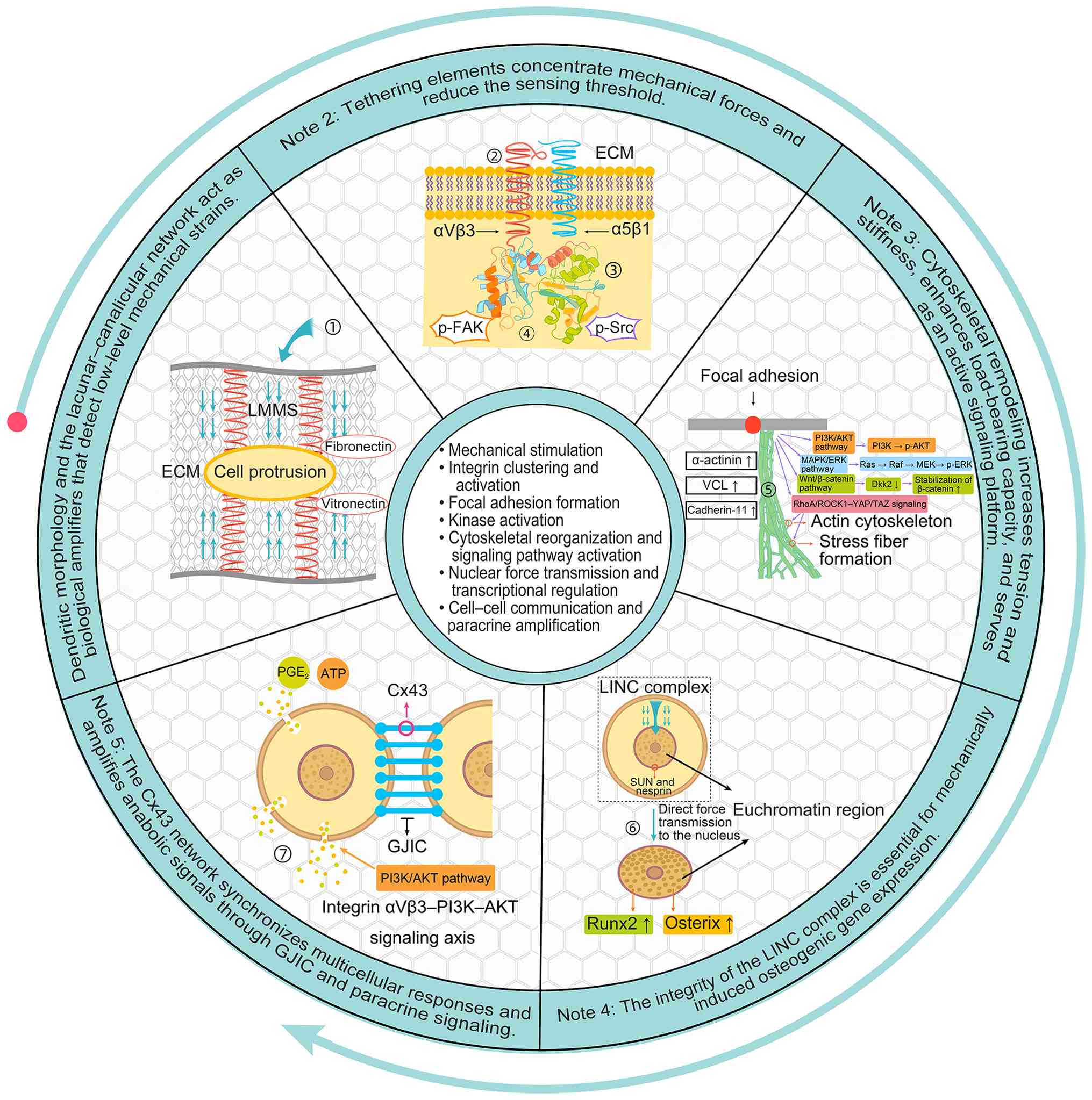 | Figure 1Biological mechanisms of
low-magnitude mechanical stress stimulation. LMMS activates
integrin/focal-adhesion kinases at the cell-ECM interface, inducing
cytoskeletal remodeling and LINC-mediated nuclear force
transmission. Cx43 channels coordinate intercellular and paracrine
signals. AKT, protein kinase B; ATP, adenosine triphosphate; Cx43,
connexin 43; Dkk2, Dickkopf-related protein 2; ECM, extracellular
matrix; ERK, extracellular signal-regulated kinase; FAK, focal
adhesion kinase; GJIC, gap junctional intercellular communication;
LINC, linker of nucleoskeleton and cytoskeleton; LMMS,
low-magnitude mechanical stress; MAPK, mitogen-activated protein
kinase; PGE2, prostaglandin E2; PI3K,
phosphoinositide 3-kinase; MEK, MAPK/ERK kinase; nesprin, nuclear
envelope spectrin repeat protein; Raf, rapidly accelerated
fibrosarcoma kinase; Ras, rat sarcoma virus protein; RhoA, Ras
homolog family member A; ROCK1, Rho-associated
coiled-coil-containing protein kinase 1; Runx2, RUNX family
transcription factor 2; Src, Src family kinase; SUN, Sad1/UNC84
domain protein; TAZ, transcriptional co-activator with PDZ-binding
motif; VCL, vinculin; Wnt, wingless-related integration site; YAP,
Yes-associated protein; αVβ3, integrin αVβ3; α5β1, integrin α5β1;
p-, phosphorylated. |
3. Regulatory mechanisms linking low
mechanical stress to osteoporosis
Effects of low mechanical stress on
BMSCs
BMSCs can differentiate into osteogenic and
adipogenic lineages. During aging and osteoporosis, the balance
between RUNX2-driven osteoblastogenesis and peroxisome proliferator
activated receptor (PPAR)γ-driven adipogenesis shifts toward
increased marrow adiposity (36,37).
Promotion of osteogenic differentiation.
Low-magnitude vibration may promote osteogenic commitment by
increasing RUNX2 expression and suppressing PPARγ2 through
ERK-dependent signaling (38).
Cellular senescence is increasingly recognized as a contributor to
age-related osteoporosis (39,40).
In naturally aged rats, low-magnitude vibration partially restored
osteogenic-cell function and was associated with increased sirtuin
1 (SIRT1) expression and reduced p53/p21 signaling. Importantly,
the source study compared 22-month-old and 3-month-old Sprague
Dawley rats rather than rats and mice (41). A separate study compared
20-month-old rats with 6-month-old rat controls and demonstrated
that vibration-induced microRNA (miR)-378a-3p suppressed growth
factor receptor bound protein 2 expression and increased
osteogenic-marker expression in BMSCs derived from aged rats
(42). Although these findings
provide useful mechanistic insights, they remain limited to
preclinical models.
Suppression of adipogenic differentiation.
Mechanical loading reduces PPARγ signaling and may redirect
mesenchymal lineage commitment away from adipogenesis (38). The p38 MAPK pathway has also been
implicated in osteoporosis-related lineage regulation (43). Before vibration treatment, BMSCs
from aged rats exhibited fewer mineralized nodules and greater
lipid-droplet accumulation. The intervention partially corrected
this imbalance through changes associated with the SIRT1/p53/p21
pathway (41). In ovariectomized
models, estrogen receptor (ER)α/Wnt signaling has also been
implicated in the suppression of adipogenic commitment (44). However, because these findings were
derived predominantly from two-dimensional cell cultures and rodent
models, the magnitude and persistence of these effects within the
three-dimensional human bone marrow niche remain uncertain.
Effects of low mechanical stress on
osteoclasts
Low mechanical stress may regulate
osteoclastogenesis through both cell-autonomous focal-adhesion
signaling and coupling factors released by osteocytes. VCL connects
integrins to the actin cytoskeleton and transmits mechanical force
within focal adhesions (45,46).
In vibration-based models, changes in VCL tension and
focal-adhesion dynamics have been associated with reductions in the
number of multinucleated tartrate-resistant acid
phosphatase-positive osteoclasts. However, these findings are
model-dependent and do not independently establish a clinically
meaningful antiresorptive effect (47).
The TNF superfamily member 11
(RANKL)/osteoprotegerin (OPG) axis represents a second and
biologically central mechanism. Low-intensity whole-body vibration
has been reviewed as a potential intervention in postmenopausal
osteoporosis (48). RANKL binds to
RANK on osteoclast precursors and promotes their differentiation,
whereas OPG functions as a soluble decoy receptor. Consequently, a
reduced RANKL/OPG ratio would be expected to inhibit
osteoclastogenesis. Osteocytes are a major source of RANKL in
remodeling bone (49). In MLO-Y4
osteocyte-like cells exposed to high-glucose conditions, mechanical
vibration increased OPG expression and secretion while reducing
RANKL expression and secretion, consistent with attenuation of
pro-osteoclastogenic signaling (50). These findings support an
osteocyte-mediated mechanism but should be interpreted cautiously
because they were obtained using a two-dimensional cellular model
under diabetic conditions. Whether a comparable shift in the
RANKL/OPG balance occurs across different loading protocols or in
human osteoporotic bone remains unresolved.
Effects of low mechanical stress on
OBs
Low mechanical stress plays an important role in
promoting osteogenesis, as illustrated in Fig. 1. Exposure to low-magnitude
mechanical vibration at 35 Hz and 0.25 g has been shown to
stimulate OB proliferation by upregulating miR-150-5p and
downregulating miR-182-5p and miR-125b-2-3p, thereby activating the
MAPK and actin-regulatory signaling pathways (51). Low mechanical stress also activates
Wnt/β-catenin signaling. Following mechanical loading, the
expression of pathway-related genes, including β-catenin, COL1,
osteocalcin and Runx2, is increased. Immunofluorescence analysis
has further demonstrated that low mechanical stress promotes the
nuclear translocation of β-catenin in mouse MC3T3-E1 cells,
indicating activation of the Wnt/β-catenin pathway. This pathway
also contributes positively to the mechanically induced expression
of Runx2(52). In diabetes-induced
secondary osteoporosis, low mechanical stress has similarly been
shown to promote OB proliferation and differentiation through
Wnt/β-catenin signaling. Under high-glucose conditions, the
expression of Dickkopf-related protein 2 (Dkk2), an inhibitor of
Wnt signaling, is increased, while β-catenin levels decline because
of phosphorylation-dependent degradation. These changes reduce the
expression of Cyclin D1 and Osterix, thereby suppressing OB
proliferation and differentiation. Dkk2 overexpression inhibits
MC3T3-E1 cell proliferation, as reflected by a marked reduction in
the proportion of cells in the S and G2/M phases and a
corresponding increase in the G0/G1 phase. It also impairs
osteogenic differentiation, resulting in reduced alkaline
phosphatase activity and fewer mineralized nodules. Low mechanical
stress attenuates the high-glucose-induced increase in Dkk2 and
restores the expression of β-catenin, cyclin D1 and Osterix. These
effects promote OB proliferation and differentiation and partially
alleviate the impairment of bone formation caused by a high-glucose
environment (53). Low mechanical
stress may also exert protective effects in diabetes-induced
secondary osteoporosis through the PI3K/AKT pathway. In MC3T3-E1
cells exposed to high-glucose conditions, activation of PI3K/AKT
signaling promotes OB proliferation and reduces matrix
metalloproteinase 9 (MMP9) expression. Low mechanical stress also
modulates the upstream TNF receptor associated factor 6-ERK/p38
MAPK pathway involved in MMP9 regulation, thereby potentially
suppressing osteoclast differentiation and maturation, bone
resorption and the migration of osteoclast precursor cells
(54). The ER is considered a
susceptibility-related factor in osteoporosis, and both OBs and
osteoclasts express ERs. Estrogen binding to receptors in OBs
regulates the production of collagenase, cytokines and growth
factors and thereby influences osteogenic differentiation. For
example, low-intensity vibration at 45 Hz and 0.9 g significantly
enhanced OB activity in ovariectomized osteoporotic rats by
increasing ERα expression and altering its subcellular
localization, ultimately promoting bone formation (55). In estrogen deficiency-induced
secondary osteoporosis, low-frequency mechanical vibration has also
been reported to increase bone morphogenetic protein 2 and Runx2
expression, followed by the upregulation of osteocalcin,
osteopontin, bone sialoprotein and COL1. These responses are
accompanied by activation of the ERK1/2 signaling pathway and
enhanced bone anabolism (56).
Overall, low mechanical stress promotes osteogenesis by activating
major signaling pathways and osteogenic regulatory factors. It may
also counteract adverse cellular conditions associated with
different forms of secondary osteoporosis. However, much of the
available evidence is derived from cell and animal models, and the
clinical relevance of these mechanisms requires further
validation.
4. Biophysical and mechanical interventions
relevant to osteoporosis
The translation of preclinical findings on LMMS into
clinical practice has prompted the development and investigation of
several non-invasive, physiotherapy-based interventions for
osteoporosis prevention and management. These approaches aim to
deliver controlled, safe and measurable stimuli to the skeleton
through whole-body or targeted regional application. However, not
all of these interventions constitute LMMS and their mechanisms and
clinical evidence should therefore be considered separately.
Whole-body vibration (WBV)
therapy
WBV is the closest clinical analogue of LMMS.
Low-magnitude protocols generally use accelerations below 1 g,
although the frequency, posture, platform type, cumulative exposure
and transmission of vibration to individual skeletal sites vary
considerably among studies.
Clinical findings remain heterogeneous. The earlier
meta-analysis by Slatkovska et al (14) and a 2023 meta-analysis of
randomized trials (57) reported
small improvements in bone mineral density (BMD) that depended on
the skeletal site and intervention protocol. Device trials in older
adults have also highlighted challenges related to recruitment,
adherence and delivery of low-magnitude mechanical stimulation
(58). By contrast, a 12-month
randomized trial using vibration at 0.3 g and either 30 or 90 Hz
found no improvement in BMD or bone microarchitecture (59). An overview of systematic reviews
also concluded that most available reviews were of critically low
methodological quality (60). WBV
may therefore be considered an adjunctive intervention for selected
patients, but current evidence does not support its use as an
established replacement for pharmacotherapy or exercise programs
designed to prevent fractures.
Pulsed electromagnetic fields
(PEMF)
PEMF therapy is an electromagnetic rather than a
mechanical intervention. It is included here only as a related
biophysical modality and should not be classified as LMMS. Its
proposed effects involve the induction of electrical currents and
subsequent modulation of intracellular signaling. Any resulting
micromechanical effects are indirect and PEMF has not been
demonstrated to be equivalent to mechanical vibration (61). Evidence supporting its use in
osteoporosis remains limited and highly dependent on the treatment
protocol (62,63). PEMF should therefore be evaluated
separately from interventions involving direct mechanical
loading.
Therapeutic exercise and physical activity.
Therapeutic exercise represents a mixed-magnitude mechanical
intervention rather than a pure LMMS exposure. Muscle-bone
interactions mediated by insulin-like growth factor I provide an
additional biological basis for the skeletal effects of exercise
(64). Resistance, weight-bearing,
balance and jumping exercises generate heterogeneous skeletal
strains that vary according to body mass, movement pattern, loading
rate and anatomical site (13,65).
Their potential benefits include modest, site-specific improvements
in BMD, enhanced muscle strength and physical function, and a
reduced risk of falls. These benefits should not, however, be
attributed exclusively to low-magnitude mechanical strain.
Similarly, Tai Chi is more appropriately characterized as a
low-impact, multicomponent form of exercise that improves balance
and functional capacity rather than as a form of LMMS (66).
Low-intensity pulsed ultrasound
(LIPUS)
LIPUS delivers acoustic pressure waves that can
generate localized micromechanical strain and fluid-streaming
potentials. It is therefore mechanistically closer to a targeted
mechanical stimulus than PEMF. Nevertheless, evidence for its use
in osteoporosis is derived predominantly from preclinical studies
or research on fracture healing (67,68).
Direct clinical evidence demonstrating that LIPUS improves BMD or
prevents fragility fractures remains insufficient.
Clinical considerations and future
directions
Clinical interpretation should distinguish evidence
of efficacy from uncertainty regarding implementation. Outcomes
vary according to vibration frequency, acceleration, platform type,
posture, cumulative exposure, adherence, baseline skeletal status
and anatomical site (57,59,60).
Evidence for fracture-risk reduction remains limited and vulnerable
patients may require supervision because balance impairment, recent
fractures, acute pain, cardiovascular instability, implanted
medical devices and other comorbidities may affect the suitability
and safety of treatment. These considerations currently preclude
the development of a universally applicable LMMS prescription.
Key priorities include adequately powered,
preregistered trials that report delivered rather than nominal
acceleration, together with adherence, adverse events and
site-specific changes in BMD and bone microarchitecture. Falls and
fractures should also be included as endpoints whenever feasible.
Head-to-head dose-response studies and subgroup analyses based on
age, sex, baseline osteoporosis severity, medication use and
mechanical transmissibility are required before personalized
treatment protocols can be recommended.
Critical appraisal of the evidence. Positive
findings in cellular models do not necessarily translate to intact
bone. Two-dimensional cultures expose isolated cells to simplified
substrates and do not reproduce the three-dimensional confinement,
viscoelastic ECM, fluid flow, cell-cell interactions, vascular
supply or remodeling history of living bone. Consequently,
mechanotransduction in three-dimensional environments may differ
qualitatively from that observed on two-dimensional elastic
surfaces (69). The anisotropic
and multiscale architecture of the bone matrix further influences
the transmission and distribution of local strains (70). At the clinical level, null trials
and inconsistent site-specific findings from meta-analyses indicate
that non-response is common (57,59,60).
The field should therefore distinguish mechanistic plausibility
from demonstrated antifracture efficacy and avoid extrapolating the
effects of a single frequency or acceleration across cellular,
animal and human systems.
5. Conclusion and future perspectives
LMMS engages a distributed mechanobiological network
involving membrane receptors, cytoskeletal remodeling, nuclear
coupling and osteocyte-mediated intercellular communication.
Available evidence supports biologically plausible effects on
mesenchymal lineage commitment, OB activity and
osteoclast-regulatory signaling. However, much of this evidence has
been derived from specific two-dimensional cell cultures or rodent
models and should not be generalized across different doses,
diseases or species.
Clinical translation remains less certain than the
preclinical literature might suggest. The effects of WBV on BMD
appear to be modest and dependent on the skeletal site and
intervention protocol. Evidence based on fracture endpoints remains
sparse, while null trials demonstrate considerable variability and
non-response. PEMF and therapeutic exercise should therefore be
evaluated as distinct interventions rather than being
mechanistically grouped with LMMS.
Immediate priorities include standardizing the
reporting of delivered acceleration, frequency, posture, cumulative
dose, adherence, adverse events and the skeletal sites assessed.
Dose-response relationships should be investigated in adequately
powered randomized controlled trials, and clinically meaningful
outcomes-including falls, fractures and measures of bone
quality-should be evaluated alongside BMD. Mechanistic studies
should incorporate three-dimensional matrices, osteocyte networks,
vascular and immune components, and aged or osteoporotic human
cells to more accurately reproduce the in vivo bone
microenvironment.
Until these evidence gaps are addressed, LMMS should
be regarded as an investigational or adjunctive intervention rather
than a standalone treatment for osteoporosis. Future studies should
identify biological and biomechanical predictors of treatment
response and determine whether LMMS provides additional benefits
when combined with guideline-recommended exercise and
pharmacological therapy.
In conclusion, LMMS provides a valuable framework
for investigating how weak mechanical signals influence bone
remodeling. However, its mechanistic promise currently exceeds the
certainty of the clinical evidence. Further progress will depend on
experimental models that more accurately reproduce the
three-dimensional bone niche and clinical trials that apply
standardized exposure reporting and assess clinically meaningful
outcomes.
Acknowledgements
Not applicable.
Funding
Funding: This work was supported by the Gansu Provincial Joint
Scientific Research Fund (grant no. 24JRRA878), the Gansu
Provincial Talent Project (grant no. 2025RCXM006), the Gansu
Provincial Natural Science Foundation (grant no. 24JRRA1020), the
Gansu Provincial Key Research and Development Program for
International Scientific and Technological Cooperation (grant no.
25YFWA024) and the Graduate Innovation and Entrepreneurship Fund of
Gansu University of Chinese Medicine (grant no. 2025CXCY-025).
Availability of data and materials
Not applicable.
Authors' contributions
ZL and ZS conceived and designed the review. ZL
wrote the original draft. ZL, DH and XP conducted the literature
search and analyzed the literature. YW contributed to the
interpretation of the literature findings. HL and ZS critically
revised the manuscript. Data authentication is not applicable. All
authors read and approved the final manuscript.
Ethics approval and consent to
participate
Not applicable.
Patient consent for publication
Not applicable.
Competing interests
The authors declare that they have no competing
interests.
References
|
1
|
No authors listed. Consensus development
conference: Diagnosis, prophylaxis, and treatment of osteoporosis.
Am J Med. 94:646–650. 1993.PubMed/NCBI View Article : Google Scholar
|
|
2
|
Johnell O and Kanis JA: An estimate of the
worldwide prevalence and disability associated with osteoporotic
fractures. Osteoporos Int. 17:1726–1733. 2006.PubMed/NCBI View Article : Google Scholar
|
|
3
|
Si L, Winzenberg TM, Jiang Q, Chen M and
Palmer AJ: Projection of osteoporosis-related fractures and costs
in China: 2010-2050. Osteoporos Int. 26:1929–1937. 2015.PubMed/NCBI View Article : Google Scholar
|
|
4
|
Rachner TD, Khosla S and Hofbauer LC:
Osteoporosis: Now and the future. Lancet. 377:1276–1287.
2011.PubMed/NCBI View Article : Google Scholar
|
|
5
|
Eastell R, Rosen CJ, Black DM, Cheung AM,
Murad MH and Shoback D: Pharmacological management of osteoporosis
in postmenopausal women: An endocrine society* clinical
practice guideline. J Clin Endocrinol Metab. 104:1595–1622.
2019.PubMed/NCBI View Article : Google Scholar
|
|
6
|
Shane E, Burr D, Abrahamsen B, Adler RA,
Brown TD, Cheung AM, Cosman F, Curtis JR, Dell R, Dempster DW, et
al: Atypical subtrochanteric and diaphyseal femoral fractures:
Second report of a task force of the American society for bone and
mineral research. J Bone Miner Res. 29:1–23. 2014.PubMed/NCBI View Article : Google Scholar
|
|
7
|
Compston J, Cooper A, Cooper C, Gittoes N,
Gregson C, Harvey N, Hope S, Kanis JA, McCloskey EV, Poole KES, et
al: UK clinical guideline for the prevention and treatment of
osteoporosis. Arch Osteoporos. 12(43)2017.PubMed/NCBI View Article : Google Scholar
|
|
8
|
Frost HM: Wolff's Law and bone's
structural adaptations to mechanical usage: An overview for
clinicians. Angle Orthod. 64:175–188. 1994.PubMed/NCBI View Article : Google Scholar
|
|
9
|
Rubin C, Turner AS, Bain S, Mallinckrodt C
and McLeod K: Anabolism. Low mechanical signals strengthen long
bones. Nature. 412:603–604. 2001.PubMed/NCBI View
Article : Google Scholar
|
|
10
|
Rubin CT, Sommerfeldt DW, Judex S and Qin
Y: Inhibition of osteopenia by low magnitude, high-frequency
mechanical stimuli. Drug Discov Today. 6:848–858. 2001.PubMed/NCBI View Article : Google Scholar
|
|
11
|
Ozcivici E, Luu YK, Adler B, Qin YX, Rubin
J, Judex S and Rubin CT: Mechanical signals as anabolic agents in
bone. Nat Rev Rheumatol. 6:50–59. 2010.PubMed/NCBI View Article : Google Scholar
|
|
12
|
Qin YX, Lam H, Ferreri S and Rubin C:
Dynamic skeletal muscle stimulation and its potential in bone
adaptation. J Musculoskelet Neuronal Interact. 10:12–24.
2010.PubMed/NCBI
|
|
13
|
Beck BR, Daly RM, Singh MA and Taaffe DR:
Exercise and sports science Australia (ESSA) position statement on
exercise prescription for the prevention and management of
osteoporosis. J Sci Med Sport. 20:438–445. 2017.PubMed/NCBI View Article : Google Scholar
|
|
14
|
Slatkovska L, Alibhai SMH, Beyene J and
Cheung AM: Effect of whole-body vibration on BMD: A systematic
review and meta-analysis. Osteoporos Int. 21:1969–1980.
2010.PubMed/NCBI View Article : Google Scholar
|
|
15
|
Lau RWK, Liao LR, Yu F, Teo T, Chung RC
and Pang MYC: The effects of whole body vibration therapy on bone
mineral density and leg muscle strength in older adults: A
systematic review and meta-analysis. Clin Rehabil. 25:975–988.
2011.PubMed/NCBI View Article : Google Scholar
|
|
16
|
Wang Y, McNamara LM, Schaffler MB and
Weinbaum S: A model for the role of integrins in flow induced
mechanotransduction in osteocytes. Proc Natl Acad Sci USA.
104:15941–15946. 2007.PubMed/NCBI View Article : Google Scholar
|
|
17
|
Litzenberger JB, Kim JB, Tummala P and
Jacobs CR: Beta1 integrins mediate mechanosensitive signaling
pathways in osteocytes. Calcif Tissue Int. 86:325–332.
2010.PubMed/NCBI View Article : Google Scholar
|
|
18
|
Salasznyk RM, Klees RF, Williams WA,
Boskey A and Plopper GE: Focal adhesion kinase signaling pathways
regulate the osteogenic differentiation of human mesenchymal stem
cells. Exp Cell Re. 313:22–37. 2007.PubMed/NCBI View Article : Google Scholar
|
|
19
|
Wang L, Ciani C, Doty SB and Fritton SP:
Delineating bone's interstitial fluid pathway in vivo. Bone.
34:499–509. 2004.PubMed/NCBI View Article : Google Scholar
|
|
20
|
You L, Cowin SC, Schaffler MB and Weinbaum
S: A model for strain amplification in the actin cytoskeleton of
osteocytes due to fluid drag on pericellular matrix. J Biomech.
34:1375–1386. 2001.PubMed/NCBI View Article : Google Scholar
|
|
21
|
Arnsdorf EJ, Tummala P, Kwon RY and Jacobs
CR: Mechanically induced osteogenic differentiation-the role of
RhoA, ROCKII and cytoskeletal dynamics. J Cell Sci. 122:546–553.
2009.PubMed/NCBI View Article : Google Scholar
|
|
22
|
Lombardi ML, Jaalouk DE, Shanahan CM,
Burke B, Roux KJ and Lammerding J: The interaction between nesprins
and sun proteins at the nuclear envelope is critical for force
transmission between the nucleus and cytoskeleton. J Biol Chem.
286:26743–26753. 2011.PubMed/NCBI View Article : Google Scholar
|
|
23
|
Uzer G, Thompson WR, Sen B, Xie Z, Yen SS,
Miller S, Bas G, Styner M, Rubin CT, Judex S, et al: Cell
mechanosensitivity to extremely low-magnitude signals is enabled by
a LINCed nucleus. Stem Cells. 33:2063–2076. 2015.PubMed/NCBI View Article : Google Scholar
|
|
24
|
Alam SG, Lovett D, Kim DI, Roux KJ,
Dickinson RB and Lele TP: The nucleus is an intracellular
propagator of tensile forces in NIH 3T3 fibroblasts. J Cell Sci.
128:1901–1911. 2015.PubMed/NCBI View Article : Google Scholar
|
|
25
|
Lloyd SA, Lewis GS, Zhang Y, Paul EM and
Donahue HJ: Connexin 43 deficiency attenuates loss of trabecular
bone and prevents suppression of cortical bone formation during
unloading. J Bone Miner Res. 27:2359–2372. 2012.PubMed/NCBI View Article : Google Scholar
|
|
26
|
Cherian PP, Siller-Jackson AJ, Gu S, Wang
X, Bonewald LF, Sprague E and Jiang JX: Mechanical strain opens
connexin 43 hemichannels in osteocytes: A novel mechanism for the
release of prostaglandin. Mol Biol Cell. 16:3100–3106.
2005.PubMed/NCBI View Article : Google Scholar
|
|
27
|
Case N, Thomas J, Sen B, Styner M, Xie Z,
Galior K and Rubin J: Mechanical regulation of glycogen synthase
kinase 3β (GSK3β) in mesenchymal stem cells is dependent on Akt
protein serine 473 phosphorylation via mTORC2 protein. J Biol Chem.
286:39450–39456. 2011.PubMed/NCBI View Article : Google Scholar
|
|
28
|
Zayzafoon M, Gathings WE and McDonald JM:
New Collective Author. Modeled microgravity inhibits osteogenic
differentiation of human mesenchymal stem cells and increases
adipogenesis. Endocrinology. 145:2421–2432. 2004.PubMed/NCBI View Article : Google Scholar
|
|
29
|
Robinson JA, Chatterjee-Kishore M,
Yaworsky PJ, Cullen DM, Zhao W, Li C, Kharode Y, Sauter L, Babij P,
Brown EL, et al: Wnt/beta-catenin signaling is a normal
physiological response to mechanical loading in bone. J Biol Chem.
281:31720–31728. 2006.PubMed/NCBI View Article : Google Scholar
|
|
30
|
Tu X, Rhee Y, Condon KW, Bivi N, Allen MR,
Dwyer D, Stolina M, Turner CH, Robling AG, Plotkin LI and Bellido
T: Sost downregulation and local Wnt signaling are required for the
osteogenic response to mechanical loading. Bone. 50:209–217.
2012.PubMed/NCBI View Article : Google Scholar
|
|
31
|
Dupont S, Morsut L, Aragona M, Enzo E,
Giulitti S, Cordenonsi M, Zanconato F, Le Digabel J, Forcato M,
Bicciato S, et al: Role of YAP/TAZ in mechanotransduction. Nature.
474:179–183. 2011.PubMed/NCBI View Article : Google Scholar
|
|
32
|
Aragona M, Panciera T, Manfrin A, Giulitti
S, Michielin F, Elvassore N, Dupont S and Piccolo S: A mechanical
checkpoint controls multicellular growth through YAP/TAZ regulation
by actin-processing factors. Cell. 154:1047–1059. 2013.PubMed/NCBI View Article : Google Scholar
|
|
33
|
Tu X, Delgado-Calle J, Condon KW, Maycas
M, Zhang H, Carlesso N, Taketo MM, Burr DB, Plotkin LI and Bellido
T: Osteocytes mediate the anabolic actions of canonical
Wnt/β-catenin signaling in bone. Proc Natl Acad Sci USA.
112:E478–E486. 2015.PubMed/NCBI View Article : Google Scholar
|
|
34
|
Cabahug-Zuckerman P, Frikha-Benayed D,
Majeska RJ, Tuthill A, Yakar S, Judex S and Schaffler MB: Osteocyte
apoptosis caused by hindlimb unloading is required to trigger
osteocyte RANKL production and subsequent resorption of cortical
and trabecular bone in mice femurs. J Bone Miner Res. 31:1356–1365.
2016.PubMed/NCBI View Article : Google Scholar
|
|
35
|
Prisby RD, Ramsey MW, Behnke BJ, Dominguez
JM II, Donato AJ, Allen MR and Delp MD: Aging reduces skeletal
blood flow, endothelium-dependent vasodilation, and NO
bioavailability in rats. J Bone Miner Res. 22:1280–1288.
2007.PubMed/NCBI View Article : Google Scholar
|
|
36
|
Park SR, Oreffo RO and Triffitt JT:
Interconversion potential of cloned human marrow adipocytes in
vitro. Bone. 24:549–554. 1999.PubMed/NCBI View Article : Google Scholar
|
|
37
|
Vernochet C, Milstone DS, Iehlé C,
Belmonte N, Phillips B, Wdziekonski B, Villageois P, Amri EZ,
O'Donnell PE, Mortensen RM, et al: PPARgamma-dependent and
PPARgamma-independent effects on the development of adipose cells
from embryonic stem cells. FEBS Lett. 510:94–98. 2002.PubMed/NCBI View Article : Google Scholar
|
|
38
|
David V, Martin A, Lafage-Proust MH,
Malaval L, Peyroche S, Jones DB, Vico L and Guignandon A:
Mechanical loading down-regulates peroxisome proliferator-activated
receptor gamma in bone marrow stromal cells and favors
osteoblastogenesis at the expense of adipogenesis. Endocrinology.
148:2553–2562. 2007.PubMed/NCBI View Article : Google Scholar
|
|
39
|
Khosla S, Farr JN and Kirkland JL:
Inhibiting cellular senescence: A new therapeutic paradigm for
age-related osteoporosis. J Clin Endocrinol Metab. 103:1282–1290.
2018.PubMed/NCBI View Article : Google Scholar
|
|
40
|
Marie PJ: Bone cell senescence: Mechanisms
and perspectives. J Bone Miner Res. 29:1311–1321. 2014.PubMed/NCBI View Article : Google Scholar
|
|
41
|
Wen J, Bao M, Tang M, He X, Yao X and Li
L: Low magnitude vibration alleviates age-related bone loss by
inhibiting cell senescence of osteogenic cells in naturally
senescent rats. Aging (Albany NY). 13:12031–12045. 2021.PubMed/NCBI View Article : Google Scholar
|
|
42
|
Yu X, Zeng Y, Bao M, Wen J, Zhu G, Cao C,
He X and Li L: Low-magnitude vibration induces osteogenic
differentiation of bone marrow mesenchymal stem cells via
miR-378a-3p/Grb2 pathway to promote bone formation in a rat model
of age-related bone loss. FASEB J. 34:11754–11771. 2020.PubMed/NCBI View Article : Google Scholar
|
|
43
|
Qin H, Kang T and Liu G: Long non-coding
RNA directly or indirectly affects osteoporosis through the p38MAPK
signaling pathway. Chin J Tissue Eng Res. 29:175–184. 2025.
|
|
44
|
Li H, Wu W, He X, Cao C, Yu X, Zeng Y and
Li L: Applying vibration in early postmenopausal osteoporosis
promotes osteogenic differentiation of bone marrow-derived
mesenchymal stem cells and suppresses postmenopausal osteoporosis
progression. Biosci Rep. 39(BSR20191011)2019.PubMed/NCBI View Article : Google Scholar
|
|
45
|
Ji L, Lim J and Danuser G: Fluctuations of
intracellular forces during cell protrusion. Nat Cell Biol.
10:1393–1400. 2008.PubMed/NCBI View Article : Google Scholar
|
|
46
|
Balaban NQ, Schwarz US, Riveline D,
Goichberg P, Tzur G, Sabanay I, Mahalu D, Safran S, Bershadsky A,
Addadi L and Geiger B: Force and focal adhesion assembly: A close
relationship studied using elastic micropatterned substrates. Nat
Cell Biol. 3:466–472. 2001.PubMed/NCBI View Article : Google Scholar
|
|
47
|
Xiong X, Huo Q, Li K, Cui C, Chang C, Park
C, Ku B, Hong CS, Lim H, Pandya PH, et al: Enhancing anti-tumor
potential: Low-intensity vibration suppresses osteosarcoma
progression and augments MSCs' tumor-suppressive abilities.
Theranostics. 14:1430–1449. 2024.PubMed/NCBI View Article : Google Scholar
|
|
48
|
Li HM and Li L: Whole body vibration with
low intensity and postmenopausal osteoporosis. J Biomed Eng.
35:301–306, 313. 2018.
|
|
49
|
Nakashima T, Hayashi M, Fukunaga T, Kurata
K, Oh-Hora M, Feng JQ, Bonewald LF, Kodama T, Wutz A, Wagner EF, et
al: Evidence for osteocyte regulation of bone homeostasis through
RANKL expression. Nat Med. 17:1231–1234. 2011.PubMed/NCBI View Article : Google Scholar
|
|
50
|
Sun T, Yan Z, Cai J, Shao X, Wang D, Ding
Y, Feng Y, Yang J, Luo E, Feng X and Jing D: Effects of mechanical
vibration on cell morphology, proliferation, apoptosis, and
cytokine expression/secretion in osteocyte-like MLO-Y4 cells
exposed to high glucose. Cell Biol Int. 44:216–228. 2020.PubMed/NCBI View Article : Google Scholar
|
|
51
|
Liu YW and Zhu D: Experimental study on
the effect of cytoskeleton and MAPK signaling pathway on osteoblast
proliferation under low-load mechanical vibration. J Med Biomech.
34 (S1)(S103)2019.
|
|
52
|
Hao BH: Experimental study on the effect
of continuous and intermittent low-load mechanical vibration on
fracture healing through the Wnt/β-catenin signaling pathway
(Doctoral dissertation, Jilin University), 2019.
|
|
53
|
Fu ZY: Experimental study on the
improvement of diabetic osteoporosis by low-intensity
high-frequency mechanical vibration and its mechanism (Doctoral
dissertation, Jilin University), 2022.
|
|
54
|
Wu B: Molecular mechanism of
high-frequency low-load mechanical vibration in improving diabetic
osteoporosis (Doctoral dissertation, Jilin University), 2023.
|
|
55
|
Zhu GG, Yu XQ, Wen JR and Chen W:
Low-intensity vibration promotes osteogenic bone formation through
estrogen receptor α in ovariectomized osteoporotic rats. J Biomed
Eng. 37:825–833. 2020.
|
|
56
|
Li M, Wu W, Tan L, Mu D, Zhu D, Wang J and
Zhao B: Low-magnitude mechanical vibration regulates expression of
osteogenic proteins in ovariectomized rats. Biochem Biophys Res
Commun. 465:344–348. 2015.PubMed/NCBI View Article : Google Scholar
|
|
57
|
de Oliveira RDJ, de Oliveira RG, de
Oliveira LC, Santos-Filho SD, Sá-Caputo DC and Bernardo-Filho M:
Effectiveness of whole-body vibration on bone mineral density in
postmenopausal women: A systematic review and meta-analysis of
randomized controlled trials. Osteoporos Int. 34:29–52.
2023.PubMed/NCBI View Article : Google Scholar
|
|
58
|
Kiel DP, Hannan MT, Barton BA, Bouxsein
ML, Lang TF, Brown KM, Shane E, Magaziner J, Zimmerman S and Rubin
CT: Insights from the conduct of a device trial in older persons:
Low magnitude mechanical stimulation for musculoskeletal health.
Clin Trials. 7:354–367. 2010.PubMed/NCBI View Article : Google Scholar
|
|
59
|
Slatkovska L, Alibhai SM, Beyene J, Hu H,
Demaras A and Cheung AM: Effect of 12 months of whole-body
vibration therapy on bone density and structure in postmenopausal
women: A randomized trial. Ann Intern Med. 155:668–679.
2011.PubMed/NCBI View Article : Google Scholar
|
|
60
|
Yin S, Liu Y, Zhong Y and Zhu F: Effects
of whole-body vibration on bone mineral density in postmenopausal
women: An overview of systematic reviews. BMC Womens Health.
24(444)2024.PubMed/NCBI View Article : Google Scholar
|
|
61
|
Ross CL and Harrison BS: The use of
magnetic fields in bone healing: A review of the literature and
current state of the art. Electromagn Biol Med. 32:577–586.
2013.
|
|
62
|
Tabrah F, Hoffmeier M, Gilbert F Jr,
Batkin S and Bassett CA: Bone density changes in osteoporosis-prone
women exposed to pulsed electromagnetic fields (PEMFs). J Bone
Miner Res. 5:437–442. 1990.PubMed/NCBI View Article : Google Scholar
|
|
63
|
Zhou J, He H, Yang L, Chen S, Guo H, Xia L
and Yu H: Effects of pulsed electromagnetic fields on bone mineral
density in postmenopausal women: A systematic review and
meta-analysis. Bioelectromagnetics. 39:97–110. 2018.
|
|
64
|
Bikle DD, Tahimic C, Chang W, Wang Y,
Philippou A and Barton ER: Role of IGF-I signaling in muscle bone
interactions. Bone. 80:79–88. 2015.PubMed/NCBI View Article : Google Scholar
|
|
65
|
Zhao R, Zhao M and Zhang L: Efficiency of
jumping exercise in improving bone mineral density among
premenopausal women: A meta-analysis. Sports Med. 44:1393–1402.
2014.PubMed/NCBI View Article : Google Scholar
|
|
66
|
Wayne PM, Kiel DP, Krebs DE, Davis RB,
Savetsky-German J, Connelly M and Buring JE: The effects of Tai Chi
on bone mineral density in postmenopausal women: A systematic
review. Arch Phys Med Rehabil. 88:673–680. 2007.PubMed/NCBI View Article : Google Scholar
|
|
67
|
Cheung WH, Chin WC, Qin L and Leung KS:
Low intensity pulsed ultrasound enhances fracture healing in both
ovariectomy-induced osteoporotic and age-matched normal bones. J
Orthop Res. 30:129–136. 2012.PubMed/NCBI View Article : Google Scholar
|
|
68
|
Padilla F, Puts R, Vico L and Raum K:
Stimulation of bone repair with ultrasound: A review of the
possible mechanic effects. Ultrasonics. 54:1125–1145.
2014.PubMed/NCBI View Article : Google Scholar
|
|
69
|
Saraswathibhatla A, Indana D and Chaudhuri
O: Cell-extracellular matrix mechanotransduction in 3D. Nat Rev Mol
Cell Biol. 24:495–516. 2023.PubMed/NCBI View Article : Google Scholar
|
|
70
|
Ma C, Du T, Niu X and Fan Y: Biomechanics
and mechanobiology of the bone matrix. Bone Res.
10(59)2022.PubMed/NCBI View Article : Google Scholar
|

















