Introduction
Bladder cancer ranks as the ninth most common
malignancy globally, exhibiting marked epidemiological burden
particularly among men (1). A
notable challenge in the management of bladder cancer is the
accurate differentiation between muscle-invasive bladder cancer
(MIBC) and non-MIBC (NMIBC), as this distinction has implications
for treatment selection and prognosis (2). Although established approaches, such
as the vesical imaging-reporting and data system (VI-RADS) based on
multiparametric magnetic resonance imaging (mp-MRI), have
demonstrated utility in assessing muscularis propria invasion
(3), and emerging biomarkers,
including urinary lactoferrin and immune gene signatures, provide
additional prognostic information (4), limitations remain. In particular,
conventional diffusion-weighted imaging (DWI)-derived metrics, such
as the apparent diffusion coefficient (ADC), despite their clinical
value, are constrained by relatively low spatial resolution,
limited tumor specificity and challenges related to inter-scanner
and inter-protocol standardization (5). Consequently, there remains a need for
more refined, non-invasive imaging biomarkers to improve the
assessment of muscle invasiveness. Advanced DWI models, including
continuous-time random walk (CTRW) and stretched-exponential model
(SEM) that capture complex tissue microstructure beyond Gaussian
diffusion, have exhibited diagnostic potential inother tumor types,
including prostate, breast, renal and head-and-neck cancers
(6-8),
while their specific utility and diagnostic performance for
evaluating muscle layer invasion in bladder cancer, particularly
vs. VI-RADS and conventional ADC, have not yet been well assessed
(9).
Accurate discrimination between MIBC and NMIBC is
essential for guiding treatment strategies and predicting prognosis
(10). Although conventional
imaging biomarkers such as DWI-derived ADC and VIRADS provide
clinically useful information, their diagnostic utility is
constrained by suboptimal spatial resolution, limited tumor
specificity and inter-scanner/inter-protocol variability that
compromise reproducibility (11,12).
Advanced non-Gaussian diffusion models, such as the CTRW and SEM,
have exhibited promise in other tumor types, including prostate,
breast, renal and head-and-neck cancer (6-8),
while their specific utility lies in the ability to capture complex
microstructural heterogeneity of tissues via two parameters: α
reflecting temporal diffusion heterogeneity and β reflecting
spatial diffusion heterogeneity. However, their specific role in
evaluating muscularis propria invasion in bladder cancer remains
inadequately investigated (13).
Against this background, a more refined,
non-invasive quantitative imaging approach is required to improve
the assessment of muscle invasion.
MRI, particularly DWI and its quantitative
parameter, the ADC, has become an area of active investigation in
the diagnosis and staging of bladder cancer. DWI provides important
information on tumor microenvironmental alterations by
characterizing the diffusion behavior of water molecules in
tissues. Numerous studies have demonstrated that ADC values possess
notable predictive value for assessing muscularis propria invasion
in bladder cancer (14,15). DWI has exhibited notable
performance in bladder cancer staging and grading. Comparative
analyses of single-parameter, dual-parameter and extended diffusion
models have revealed marked differences in their ability to
discriminate tumor stage and grade, particularly differences in ADC
values between NMIBC and MIBC. Consequently, the combined
application of DWI and ADC measurements provides valuable
diagnostic and pathological classification information, including
the differentiation of benign lesions from malignant bladder
lesions (15). Despite these
advantages, DWI is subject to several limitations, which include
relatively low spatial resolution, limited tumor specificity and a
lack of standardization in image acquisition protocols and
post-processing methodologies, collectively restricting its
clinical applicability and compromising the reproducibility of ADC
measurements (16). The mp-MRI and
biparametric MRI approaches have demonstrated improved diagnostic
performance for predicting muscle layer invasion in bladder cancer,
particularly when integrated with the VI-RADS framework (17,18).
Beyond staging, DWI also exhibits potential in assessing tumor
biological behavior. Specifically, ADC values have been proposed as
imaging biomarkers of clinical aggressiveness, enabling
differentiation between high- and low-grade tumors, as well as
early- and advanced-stage disease (19). Furthermore, in patients with MIBC,
combined DWI and ADC analysis has exhibited to promote the
evaluation of response to neoadjuvant chemotherapy, contributing to
improved long-term clinical outcomes (20).
Compared with conventional ADC, multi-b-value DWI
models provide improved characterization of complex tissue
heterogeneity. Advanced non-Gaussian diffusion models, such as
intravoxel incoherent motion (IVIM) and diffusion kurtosis imaging
(DKI), have demonstrated notable diagnostic utility in other
malignancies, involving distinguishing histological grades in renal
cell carcinoma (6), predicting
distant metastasis in head and neck cancer (7) and characterizing orbital lesions
(21). Similarly, advanced
non-Gaussian diffusion models, including CTRW and SEM, are capable
of characterizing complex diffusion behavior that reflects
underlying tissue microstructural heterogeneity. Although advanced
non-Gaussian diffusion models have demonstrated preliminary utility
in bladder cancer grading (14),
the diagnostic performance of CTRW- and SEM-derived parameters for
assessing muscularis propria invasion, particularly their ability
to discriminate MIBC from NMIBC and their comparative effectiveness
relative to established standards, such as the VI-RADS and
conventional ADC measurements, remains insufficiently investigated
and has not been clearly established.
To address this knowledge gap, the present study
systematically evaluated CTRW- and SEM-derived parameters for the
assessment of MIBC and compared their diagnostic performance with
that of conventional ADC and VI-RADS scores. Furthermore, by
integrating multiple imaging-derived metrics and analytical
approaches, the present study aimed to improve the accuracy of
muscle invasion assessment and to provide more robust, non-invasive
evidence to support clinical decision-making.
Therefore, the present study aimed to: (i) Determine
whether CTRW and SEM-derived parameters can effectively
discriminate MIBC from NMIBC; (ii) compare their diagnostic
performance against conventional ADC and VIRADS; and (iii) assess
whether multiparametric integration enhances diagnostic accuracy,
thereby providing a robust, non-invasive imaging biomarker to
support clinical decision-making.
Materials and methods
Patient enrollment
The present study was approved by the Ethics
Committee of The Second Affiliated Hospital of Kunming Medical
University (Kunming, China; approval no. 2024-217). A prospective
case-control study was conducted on 106 patients diagnosed with
bladder cancer between April 2025 and August 2025. According to the
2016 World Health Organization tumor classification system and the
National Comprehensive Cancer Network clinical practice guidelines
(22,23), patients were categorized into MIBC
(62 cases) and NMIBC (44 cases) groups. Patient clinical and
pathological data were collected, including age, sex, tumor stage
and grade. Surgical procedures included transurethral resection of
bladder tumor (TURBT) or radical cystectomy. The resected tissues
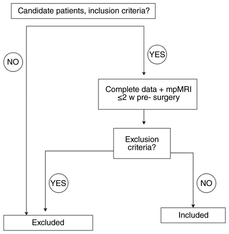
or organs were sent for pathological examination. Inclusion
criteria were as follows: i) Availability of complete clinical
data; and ii) mp-MRI performed within 2 weeks prior to surgery.
Exclusion criteria were as follows: i) Prior treatment before
undergoing mp-MRI, including TURBT within 3 months, bladder biopsy
within 2 weeks or cystoscopy within 3 days before imaging; ii)
inadequate DWI quality; iii) tumor lesions <5 mm in maximum
diameter, precluding reliable quantitative analysis; and iv)
incomplete pathological information from TURBT (Fig. 1). Although the enrollment interval
was relatively short (April-August 2025), all eligible patients
presenting during this period were consecutively included. The
Second Affiliated Hospital of Kunming Medical University maintains
a stable, high-volume bladder cancer service, and no eligible cases
were omitted, ensuring that the cohort reflects a representative
clinical population. To preserve the representative nature of the
sample and to minimize selection bias, uniform application of
inclusion and exclusion criteria were used. The pathological
reference standard was obtained from either TURBT or radical
cystectomy specimens, depending on the definitive treatment
received. For patients with NMIBC, complete TURBT specimens
containing muscularis propria were used as the staging reference,
whereas full-thickness radical cystectomy specimens served as the
gold standard for patients with MIBC. To avoid the risk of
understaging, cases with incomplete or indeterminate TURBT
pathology were excluded according to the predefined criteria.
MRI acquisition
In the present study, all enrolled subjects
underwent imaging examinations using the GE SIGNA Premier 3. 0T MRI
scanner. The entire examination process strictly followed
standardized protocols. Participants were instructed to empty their
bladder 1 h prior to the examination and to ingest 500 ml of water
in a standardized manner (a fixed volume consumed over a defined
30-min interval under consistent conditions) beginning 30 min prior
to imaging to achieve adequate bladder distension. Once sufficient
bladder distension was attained, the MRI sequences were acquired
sequentially, including T1-weighted imaging (T1WI), T2-weighted
imaging (T2WI), DWI and dynamic contrast-enhanced MRI
(DCE-MRI).
The specific MRI acquisition protocols were as
follows: Conventional T2WI was performed in the sagittal and axial
planes, and fat-suppressed T2WI sequences were acquired in the
coronal and axial planes. T1WI was obtained in the axial plane,
including both non-fat-suppressed and fat-suppressed acquisitions.
DWI was performed in the axial plane, with additional sagittal and
coronal orientations. Axial DWI employed a multi-b-value
acquisition, and ADC maps were generated through simultaneous image
reconstruction. Contrast-enhanced imaging was carried out using
gadopentetate dimeglumine administered at a dose of 0.2 ml/kg.
Multiphase dynamic imaging was acquired using the liver acquisition
with volume acceleration sequence, including axial arterial, venous
and delayed phases, as well as sagittal and coronal equilibrium
phases. In addition to multi-b-value DWI, DKI data were acquired
using the same single-shot echo-planar imaging sequence with an
extended high-b-value range to ensure stable higher-order diffusion
modeling. Specifically, DKI reconstruction utilized b values of 0,
500, 1,000, 1,500, 2,000 and 2,400 sec/mm², consistent with
established DKI acquisition practices (24). These b values were selected to
optimize sensitivity to non-Gaussian diffusion behavior and to
enable robust kurtosis estimation while maintaining acceptable
signal-to-noise ratio. Detailed acquisition parameters for all MRI
sequences are summarized in Table
I.
 | Table IDetailed list of MRI scan
parameters. |
Table I
Detailed list of MRI scan
parameters.
| Name of
sequence | B,
sec/mm2 | Repetition time,
msec | Echo time,
msec | Deflection
angle | Scan field, cm | Layer thickness,
mm | Layer spacing,
mm |
|---|
| Diffusion-weighted
imaging | 0, 50, 100, 150,
200(1), 500(2), 800(2), 1,000(4), 1,300(4), 1,800(6), 2,400(8) | 2,018 | 68.8 | 90˚ | 24x24 | 4 | 1 |
| T2-weighted
imaging | - | 6,416 | 102.4 | 111˚ | 24x24 | 4 | 1 |
| Dynamic
contrast-enhanced | - | 3.4 | 1.5 | 15˚ | 38x38 | 3 | 1.5 |
Imaging examinations
In the present study, all enrolled subjects
underwent imaging examinations using the GE SIGNA Premier 3.
Patients were instructed to ingest 500 ml of water 30 min prior to
imaging (as detailed in the MRI acquisition section) to achieve
moderate bladder distension (target ~300 ml); in selected cases, up
to 1,000 ml was permitted when adequate distension was otherwise
not achieved. The imaging field of view involved the entire urinary
bladder, proximal urethra and pelvic lymph nodes. The core MRI
acquisition parameters were as follows. T1WI was performed using a
spin-echo sequence [repetition time (TR)/echo time (TE)=600/11
msec; field of view (FOV)=240 mm; matrix=256x192; slice thickness=5
mm]. Axial T2WI was acquired with the following parameters:
TR/TE=4,050/85 msec, FOV=200 mm, matrix=320x224 and slice
thickness=6 mm. Fat-suppressed T2WI sequences were obtained in the
coronal (TR/TE=5,000/120 msec; FOV=220 mm) and sagittal
(TR/TE=4,000/85 msec; FOV=300 mm) planes. DCE-MRI was performed
using a three-dimensional gradient-echo sequence (TR/TE=3.9/1.4
msec; FOV=260 mm, matrix=192x192; slice thickness=3 mm) following
intravenous administration of a gadolinium-based contrast agent at
a dose of 0.1 mmol/kg and an injection rate of 1.5-2.0 ml/sec. A
total of five to six dynamic phases were acquired over a temporal
window of 20-131 sec. Multi-b-value DWI was performed using a
single-shot echo-planar imaging sequence. The applied b values were
0, 50, 100, 150, 200, 500, 800, 1,000, 1,300, 1,800 and 2,400
sec/mm², with corresponding numbers of signal averages of 1, 1, 1,
1, 1, 2, 2, 4, 4, 6 and 8, respectively. The total DWI acquisition
time was ~9 min. All imaging protocols were implemented in strict
accordance with the VIRADS recommendations (25,26)
to ensure reproducible assessment of muscularis propria invasion
and pathological tumor grading.
Data analysis
DKI parameter estimation was performed using a
standard diffusion kurtosis model to quantify non-Gaussian water
diffusion. For each voxel, the diffusion-weighted signal S(b) was
fitted to the second-order polynomial expression:
S(b)=So·exp[−b·Dapp +
(1/6)b²·Dapp²·Kapp], where Dapp
represents the apparent diffusion coefficient and Kapp
denotes the apparent kurtosis coefficient. Model fitting was
performed using non-linear least-squares optimization across the
dedicated DKI b values (0-2,400 sec/mm²). Kurtosis maps were
automatically generated by the iCare Spin workstation (Spin
Imaging; Finnish company iCare, version 1.2.2), and mean
Kapp in each region-of-interest (ROI) was recorded as
the representative DKI-kurtosis value.
The ADC value was calculated using the single
exponential model formula S=S0e-(DDC-b)µ,
where DDC denotes the distributed diffusion coefficient (equivalent
to ADC under the mono-exponential model) and S and S0 are the
signal strengths at b=800 and b=0 sec/mm², respectively. The CTRW
model parameters (D, α and β) were fitted to multi-b-value
diffusion data (b=0-3,000 sec/mm²) using the Mittag-Leffler
function-based signal model: S(b)=S0·E_α[−(bD)^β], where
E_α denotes the Mittag-Lefler function of order α and D represents
the anomalous diffusion coefficient, and α and β quantify the
temporal and spatial heterogeneity of diffusion, respectively.
For the SEM, the parameters µ and distributed
diffusion coefficient (DDC) were calculated using the
stretched-exponential formulation. In addition, the SEM-kurtosis
parameter was derived from a modified SEM model incorporating
diffusion kurtosis effects. Signal decay was fitted according to
the following equation:
S(b)=S0·exp[-(b·DDC)µ+(1/6)b2.DDC2·K],
where K denotes the SEM-kurtosis parameter, reflecting the degree
of non-Gaussian diffusion.
Model parameters were extracted using non-linear
least-squares fitting across multiple b values (0-2,400 sec/mm²).
The model fitting was performed using the iCare Spin workstation
via the non-linear Levenberg-Marquardt algorithm with a subsection
optimization strategy. Specifically, D was initially estimated
using low b values (≤1,000 sec/mm²), after which the fixed D values
were used for global fitting of α and β (CTRW model) or µ and DDC
(SEM model).
ROI delineation was independently performed by two
experienced radiologists with 10 and 13 years of expertise in
urological imaging. ROIs were manually drawn on the axial DWI
images at the slice showing the maximum tumor diameter (b=800
sec/mm²), with careful exclusion of necrotic or cystic areas.
Diffusion parameter values for all pixels within each ROI were
automatically extracted by the workstation, and the mean value of
each parameter was calculated as the representative metric. The
standard deviation and median were also recorded to characterize
the dispersion and skewness of the parameter distributions.
To assess the reproducibility of quantitative
diffusion parameters, interobserver agreement for ROI-based
measurements between the two radiologists was evaluated using the
intraclass correlation coefficient (ICC) based on a two-way
random-effects model. Parameters with ICC values ≥0.75 were
considered to demonstrate good reliability, and the averaged
measurements from both readers were used in subsequent
analyses.
VI-RADS scoring
A total of two experienced radiologists
independently scored the cases using multi-parametric VI-RADS
criteria, integrating comprehensive analysis of T2WI, DWI and DCE
sequences (25). Both physicians
had >1-year experience of diagnostic scoring, ensuring
consistency in scoring standards. During the procedure, the
physicians were blinded to clinical history and pathological
results, and a 5-point scale score was assigned to each the bladder
tumor from each patient. Scores ranged from 1 (definitely NMIBC) to
5 (definitely MIBC). Key observations included bladder wall layer
disruption on T2WI, high signal diffusion restriction features on
DWI and early enhancement patterns on DCE. In cases of initial
discrepancies in image interpretation, consensus was achieved
through joint image review and consultation of the VI-RADS decision
tree, in which the highest assigned VI-RADS score was recorded as
the final representative value. This process strictly adhered to
multi-parametric integration principles, emphasizing the core value
of DWI and DCE in differentiating T1/T2 staging. It was also
validated that multi-parametric methods (mp-VI-RADS) could improve
specificity and the area under the receiver operating
characteristic (ROC) curve (AUC) for muscle invasion assessment
compared with parametric approaches (for example bp-VI-RADS using
only T2WI and DWI) (27).
Interobserver agreement for VI-RADS scoring was quantified using
Cohen's κ statistic. A κ>0.80 was interpreted as excellent
agreement, 0.61-0.80 as good agreement and <0.60 as moderate or
poor agreement. The final VI-RADS score used in the analysis was
the consensus value reached after initial independent scoring.
Decision curve analysis (DCA)
DCA was performed to evaluate the clinical utility
of the predictive models by quantifying the net benefit across a
range of threshold probabilities. Net benefit was calculated as:
Net benefit=(True positives/N) - (False positives/N) x [P_t/(1 -
P_t)], where N represents the total sample size (N=106), and P_t
represents the threshold probability. The threshold probability
represents the minimum predicted probability of muscle invasion at
which a clinician would opt for radical cystectomy over
bladder-preserving therapy. A total of four strategies were
compared: i) Treat-none (net benefit=0); i) treat-all (net
benefit=prevalence - [1 - prevalence] x [P_t/(1 - P_t)]); iii)
VI-RADS alone; and iv) the combined model (SEM-ADC + CTRW-α +
VI-RADS). The clinically relevant threshold probability range was
predefined as 20-80% based on clinical guidelines suggesting that
patients with ≥20% predicted risk of muscle invasion should be
considered for radical cystectomy, while thresholds >80% may
lead to excessive overtreatment. DCA was conducted using the rmda
package (version 1.6; https://cran.r-project.org/web/packages/rmda/) in R.
The net benefit difference between the combined model and VI-RADS
alone was calculated at 5% intervals within the clinically relevant
range, and the mean net benefit difference was reported. Bootstrap
resampling (n=1,000 iterations) was used to estimate 95% confidence
intervals for the net benefit curves.
Statistical analysis
Statistical analysis was conducted using SPSS
software (version [28.0]; IBM Corp.) for basic descriptive
statistics and parametric comparisons, while R (version [4.2.1]; R
Foundation for Statistical Computing) and Python (version [3.9.13];
Python Software Foundation) were employed for advanced modeling and
machine learning tasks. Prior to parametric testing, the normality
of distribution for all continuous variables was assessed using the
Shapiro-Wilk test. The Shapiro-Wilk test revealed that age,
mono-exponential ADC, SEM-ADC and CTRW-ADC followed normal
distributions (all P>0.05), whereas CTRW-α, CTRW-β, SEM-kurtosis
and DKI-kurtosis showed non-normal distributions (all P<0.05).
Consequently, normally distributed continuous variables were
compared between groups using Student's t-test or one-way analysis
of variance, whereas non-normally distributed variables were
analyzed using the Mann-Whitney U test. For pairwise comparisons
following one-way analysis of variance, Tukey's honestly
significant difference (HSD) post-hoc test was applied. Categorical
variables were analyzed via the χ2 test. Data are
presented as mean ± standard deviation for normally distributed
variables and median (interquartile range) for non-normally
distributed variables. For non-normally distributed continuous
variables that were included in multivariate logistic regression
models, natural log transformation was applied to approximate
normal distribution prior to model entry. The normality of
transformed variables was confirmed using the Shapiro-Wilk test
(all P>0.05 after transformation). Diagnostic performance of
individual biomarkers and combined models was assessed using
receiver operating characteristic (ROC) curve analysis, with the
area under the curve (AUC) calculated using the trapezoidal rule.
Pairwise comparisons of AUCs between different models were
performed using DeLong's test for two correlated ROC curves. To
control for Type I error inflation resulting from multiple pairwise
comparisons, Bonferroni correction was applied by multiplying the
raw P-values by the number of comparisons performed (m).
Specifically, for comparisons involving k models, the total number
of pairwise comparisons was m=k(k-1)/2. A corrected P<0.05 was
considered to indicate a statistically significant difference. All
ROC and DeLong's test analyses were conducted using the pROC
package (version 1.18.0; https://cran.r-project.org/web/packages/pROC/) in
R.
To identify effective predictive factors, univariate
logistic regression was employed to screen variables with
P<0.05, followed by multivariate analysis using stepwise
regression and least absolute shrinkage and selection operator
(LASSO) regularization to address multicollinearity and select the
most significant features. For multi-parameter integration,
principal component analysis (PCA) was applied to reduce
dimensionality of multi-b-value DWI parameters, extracting
principal components with cumulative variance contribution >85%
to retain key information. The diagnostic performance of the
multi-parameter model was evaluated using ROC curve analysis, with
DeLong's test employed to compare the AUC between the
multi-parameter model and the VI-RADS score. Calibration curves and
DCA further validated the clinical utility and net benefit of the
combined model. It should be noted that multivariate logistic
regression was performed to identify independent statistical
predictors, whereas the combined diagnostic model was developed
using predictive-performance optimization. Because several
diffusion parameters exhibited substantial inter-correlation,
dimensionality-reduction (PCA) and regularization (LASSO) were
applied to prevent multicollinearity and overfitting. These
procedures consistently selected SEM-ADC and CTRW-α as the most
informative quantitative predictors, even though CTRW-β remained
statistically significant in the multivariate inferential model.
Optimal cut-off values for all quantitative imaging parameters were
determined using the Youden index (J=sensitivity + specificity-1)
on the ROC curve. The corresponding thresholds reported for
SEM-ADC, CTRW parameters, DKI kurtosis and mono-exponential ADC
therefore represent the points that maximized the Youden index. For
the VI-RADS score, the commonly used threshold of ≥4 (28,29)
was also confirmed by the Youden index as the optimal discriminator
between NMIBC and MIBC in the present cohort.
To minimize the risk of model overfitting, the
events-per-variable (EPV) ratio was calculated for all regression
models, and only variables achieving an EPV ≥10 were retained in
the final multivariate analyses. Internal validation was performed
using 1,000-bootstrap resampling with replacement to estimate
optimism-corrected regression coefficients and calibration
performance. The discriminative ability of each model was further
corrected for optimism by computing the bias-adjusted AUC using
bootstrap-based Harrell's method. For machine-learning models and
LASSO feature selection, 10-fold cross-validation was applied to
ensure model robustness and stability. All final reported AUC
values, calibration curves and decision curve analyses were based
on these internally validated, optimism-adjusted estimates.
Results
Clinical characteristics of
participants
During data validation, the initially reported
VI-RADS ≥4 distribution was found to be inconsistent with the
logistic regression results. The results exhibited a higher
proportion of VI-RADS ≥4 in the MIBC group, consistent with its
role as a notable predictor of muscle invasion. No significant
differences were identified between the two groups in terms of age
(P=0.50) or sex (P=0.66); however, the VI-RADS score, which
followed a non-normal distribution, was significantly higher in the
MIBC group [4 (3-5)]
compared with the NMIBC group [3 (2-4)]
(P=0.003). The proportion of patients with VI-RADS ≥4 was 61.3%
(38/62) in the MIBC group and 20.5% (9/44) in the NMIBC group
(P<0.001). Interobserver reproducibility analyses revealed good
to excellent reliability for ROI-based diffusion metrics
(ICC=0.78-0.89). VI-RADS scoring demonstrated excellent agreement
between the two radiologists, with a Cohen's κ value of 0.82.
Detailed results are presented in Table II.
| Table IIComparison of clinical
characteristics between the two groups of patients. |
Table II
Comparison of clinical
characteristics between the two groups of patients.
| Feature | MIBC group
(n=62) | NMIBC group
(n=44) | Z/t/χ² | P-value |
|---|
| Age, years | 65.9±10.2 | 67.4±11.8 | -0.68 | 0.500 |
| Sex
(male/female) | 54/8 | 37/7 | 0.19 | 0.660 |
| VIRADS score
(median) | 4 (3-5) | 3 (2-4) | 2.98 | 0.003 |
| Proportion of
VIRADS ≥4, % (n/total n) | 61.3 (38/62) | 20.5 (9/44) | 14.22 | <0.001 |
| Tumor stage (T
category), n (%) | | | 106.05 | <0.001 |
|
Ta | 0 (0.0%) | 26 (59.1%) | | |
|
T1 | 0 (0.0%) | 18 (40.9%) | | |
|
T2 | 34 (54.8%) | 0 (0.0%) | | |
|
T3 | 19 (30.6%) | 0 (0.0%) | | |
|
T4 | 9 (14.5%) | 0 (0.0%) | | |
| Histological grade,
n (%) | | | 23.11 | <0.001 |
|
Low grade
(G1-G2) | 9 (14.5%) | 26 (59.1%) | | |
|
High grade
(G3) | 53 (85.5%) | 18 (40.9%) | | |
Comparison of DWI, CTRW, SEM
parameters and VI-RADS between the NMIBC and MIBC groups
The DWI, CTRW and SEM parameters were compared
between the two groups. For normally distributed variables, SEM-ADC
was significantly lower in the MIBC group than in the NMIBC group
(0.93±0.20 vs. 1.05±0.17; independent t-test, P=0.002). Similarly,
CTRW-ADC (1.37±0.41 vs. 1.58±0.44; P=0.013) and DWI-ADC (1.69±0.44
vs. 1.88±0.41; P=0.026) were significantly lower in the MIBC group.
For non-normally distributed variables analyzed using the
Mann-Whitney U test, CTRW-α [median (IQR): 0.83 (0.72-0.94) vs.
0.95 (0.86-1.04); P<0.0001] and CTRW-β [0.93 (0.88-0.98) vs.
0.99 (0.96-1.02); P<0.0001] were significantly lower in the MIBC
group. No significant difference was identified in SEM-kurtosis
[0.71 (0.45-0.98) vs. 0.84 (0.62-1.06); P=0.362]. Detailed results
are summarized in Table III.
| Table IIIComparison of DWI, CTRW, SEM
parameters and VI-RADS between NMIBC and MIBC groups. |
Table III
Comparison of DWI, CTRW, SEM
parameters and VI-RADS between NMIBC and MIBC groups.
| Parameter | MIBC group
(n=62) | NMIBC group
(n=44) | Mean difference
(95% CI) | Statistical
test | Test statistic | P-value |
|---|
| Normally
distributed variables | | | | | | |
|
SEM-ADC | 0.93±0.20
(0.88-0.98) | 1.05±0.17
(1.00-1.10) | -0.12 (-0.19 -
-0.05) | Independent
t-test | t=-3.235 | 0.0020 |
|
CTRW-ADC | 1.37±0.41
(1.27-1.47) | 1.58±0.44
(1.45-1.71) | -0.21 (-0.38 -
-0.04) | Independent
t-test | t=-2.520 | 0.0130 |
|
DWI-ADC | 1.69±0.44
(1.58-1.80) | 1.88±0.41
(1.76-2.00) | -0.19 (-0.36 -
-0.02) | Independent
t-test | t=-2.252 | 0.0260 |
| Non-normally
distributed variables | | | | | | |
|
CTRW-α
[median (IQR)] | 0.83
(0.72-0.94) | 0.95
(0.86-1.04) | - | Mann-Whitney U
test | Z=-4.284 | <0.0001 |
|
CTRW-β
[median (IQR)] | 0.93
(0.88-0.98) | 0.99
(0.96-1.02) | - | Mann-Whitney U
test | Z=-4.399 | <0.0001 |
|
SEM-kurtosis
[median (IQR)] | 0.71
(0.45-0.98) | 0.84
(0.62-1.06) | - | Mann-Whitney U
test | Z=-0.915 | 0.3620 |
Typical cases
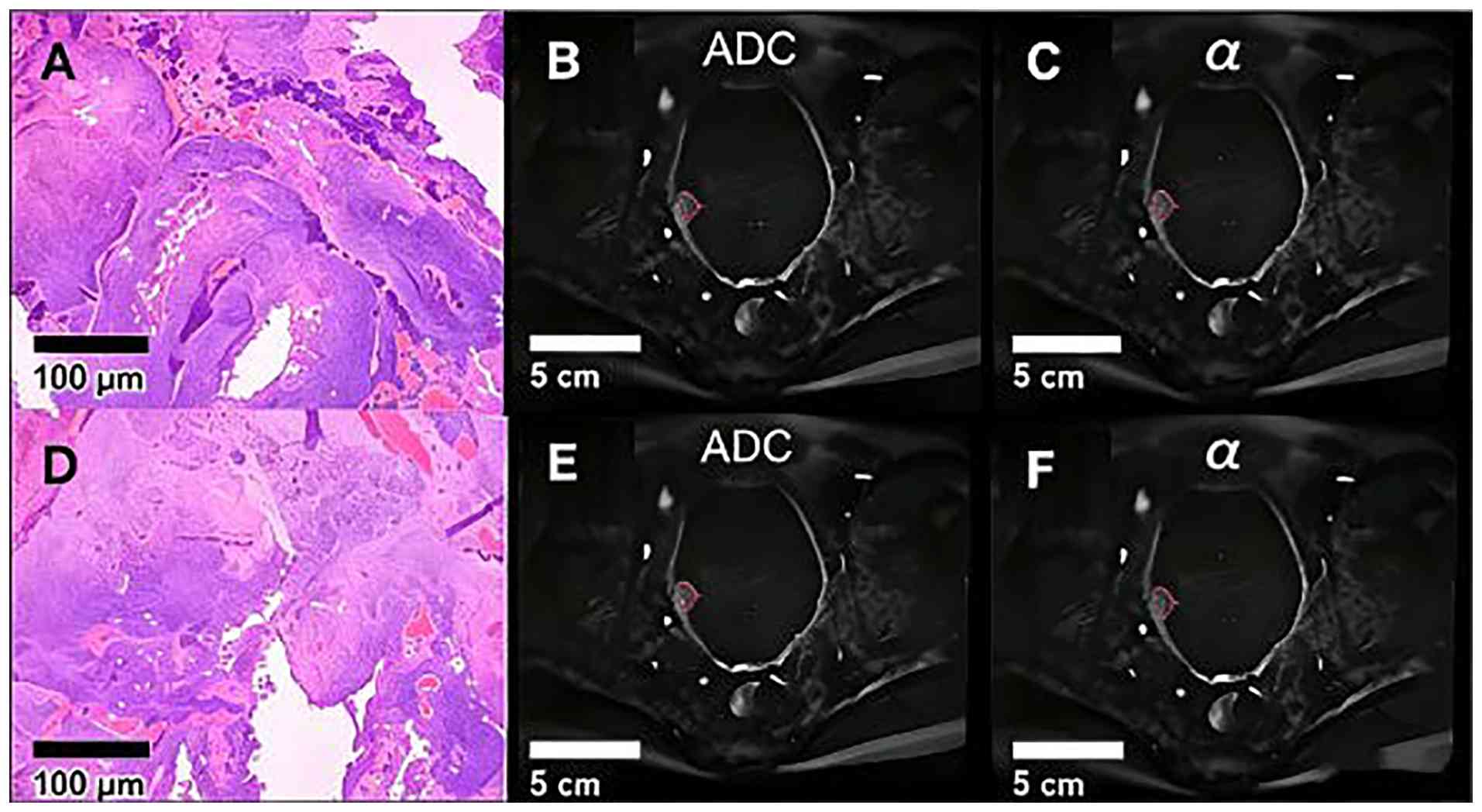
An example patient was a 70-year-old woman diagnosed
with high-grade papillary urothelial carcinoma. The tumor
infiltrated the superficial muscular layer of the bladder wall, and
no evidence of vascular or perineural invasion was identified.
Carcinoma in situ lesions were present adjacent to the
primary tumor. Another patient was a 65-year-old man with low-grade
non-invasive papillary urothelial carcinoma, characterized by
inverted growth, with no involvement of the muscularis propria.
Representative color maps of the ROI and corresponding SEM- and
CTRW-derived parameters (ADC and α) are illustrated on the original
diffusion-weighted image (b=1,000 sec/mm²). Compared with low-grade
non-invasive papillary urothelial carcinoma, high-grade papillary
urothelial carcinoma demonstrated lower mean diffusion parameter
values (Figs. 2 and 3).
Independent predictive factors of
muscle invasion
Univariate and multivariate logistic regression
analyses were performed to evaluate the predictive value of
multiparametric MRI parameters and clinical indicators for muscle
invasion in bladder cancer. In the univariate logistic regression
analysis, several variables were significantly associated with
muscle-invasive disease, including SEM-ADC [P=0.001; odds ratio
(OR)=0.05; 95% confidence interval (CI): 0.01-0.29], CTRW-α
(P=0.002; OR=0.05; 95% CI: 0.01-0.32), CTRW-β (P=0.001; OR=0.01;
95% CI: 0.00-0.15), VI-RADS score (P=0.002; OR=6.35; 95% CI:
1.99-20.28) and VI-RADS ≥4 (P=0.001; OR=11.36; 95% CI: 2.61-49.46).
Multivariate logistic regression analysis identified VI-RADS ≥4
(P=0.009; OR=6.49; 95% CI: 1.58-26.65) and CTRW-β (P=0.011;
OR=0.04, 95% CI: 0.00-0.52) as independent predictors of MIBC. All
included variables demonstrated VIF values between 1 and 5,
indicating only mild multicollinearity and acceptable model
stability. Detailed results are presented in Tables SI and SII.
Performance of multiple metrics for
assessing muscle invasion
The diagnostic performance of individual biomarkers
and combined models is summarized in Tables IV and SI, SII, SIII. Among individual indicators, the
VI-RADS score demonstrated the highest diagnostic performance, with
an AUC of 0.85 (95% CI: 0.78-0.92), yielding a sensitivity of 82%
and a specificity of 79% at a cut-off value of ≥4. CTRW-α_median
(AUC=0.83; 95% CI: 0.75-0.90) and DKI-kurtosis (AUC=0.81; 95% CI:
0.73-0.89) exhibited slightly lower discriminative ability
(Fig. 4). The mono-exponential
ADC_mean exhibited the lowest diagnostic performance (AUC=0.78; 95%
CI: 0.70-0.86). Pairwise AUC comparisons using DeLong's test with
Bonferroni correction for 15 comparisons (m=6x5/2) are presented in
Table V. The combined model
incorporating SEM-ADC, CTRW-α and VI-RADS achieved the highest
overall performance (AUC=0.91; 95% CI: 0.86-0.96). This combined
model significantly outperformed VI-RADS alone (raw P=0.002;
Bonferroni-corrected P=0.030) and mono-exponential ADC_mean (raw
P=0.001; Bonferroni-corrected P=0.015). Similarly, Combined Model 2
(DKI-kurtosis + SEM-ADC + CTRW-α, AUC=0.91; 95% CI: 0.85-0.96)
significantly exceeded VI-RADS alone (raw P<0.001;
Bonferroni-corrected P=0.006) and mono-exponential ADC_mean (raw
P=0.001; Bonferroni-corrected P=0.015). No statistically
significant difference was observed between Combined Model 1 and
Combined Model 2 after Bonferroni correction (raw P=0.89;
Bonferroni-corrected P>0.999) (Fig.
5). Comparative ROC analysis indicated that CTRW-α (AUC=0.83)
was significantly superior to mono-exponential ADC (ΔAUC=0.05; raw
P=0.03; Bonferroni-corrected P=0.450). Calibration plots
demonstrated good agreement between predicted and observed MIBC
probabilities for both combined models (Hosmer-Lemeshow test:
Combined Model 1, χ²=6.42, P=0.60; Combined Model 2, χ²=7.15,
P=0.52), indicating reliable probability estimates (Fig. S1).
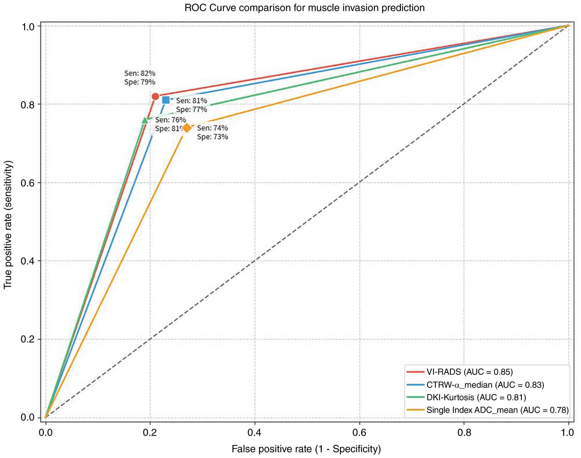 | Figure 4ROC analysis of imaging biomarkers
for the diagnosis of muscle-invasive bladder cancer. Each curve
represents the diagnostic performance of a single continuous
parameter directly used as the test variable without transformation
or model fitting: VI-RADS score (ordinal, 1-5), CTRW-α median
(continuous, non-normally distributed), DKI-kurtosis (continuous,
non-normally distributed), mono-exponential ADC mean (continuous,
normally distributed), and CTRW-α mean (continuous, normally
distributed). These curves represent the raw diagnostic information
contained in each parameter individually. The x-axis
(1-specificity) and y-axis (sensitivity) are derived by
thresholding each parameter across its observed range. No
statistical modeling, probability transformation or
cross-validation is applied, as each parameter is evaluated in
isolation. ROC, receiver operating characteristic; AUC, area under
the curve; ADC, apparent diffusion coefficient; CTRW,
continuous-time random walk; DKI, diffusion kurtosis imaging; Sen,
sensitivity; Spe, specificity. |
 | Figure 5ROC analysis of combined
multi-parametric models for the diagnosis of muscle-invasive
bladder cancer. Shown are ROC curves for the combined models:
ADCmean + VI-RADS (AUC=0.87, solid purple line), CTRW-α + CTRW-β
(AUC=0.88, dashed green line), and the tri-parameter model (ADC +
CTRW-α + DKI-kurtosis; AUC=0.91, dash-dot red line); the blue dot
marks the optimal operating point of VI-RADS alone (AUC=0.85). The
gray dashed diagonal line represents no discrimination (AUC=0.50).
The inset lists the AUC improvement (Δ) of each combined model over
VI-RADS alone, with pairwise P-values from DeLong tests under
Bonferroni correction (m=3). ROC, receiver operating
characteristic; AUC, area under the curve; ADC, apparent diffusion
coefficient; CTRW, continuous-time random walk; DKI, diffusion
kurtosis imaging; VI-RADS, vesical imaging-reporting and data
system. |
| Table IVSummary of diagnostic
performance. |
Table IV
Summary of diagnostic
performance.
|
Model/biomarker | AUC | 95% CI | Sensitivity
(%) | Specificity
(%) | Cut-off |
|---|
| Individual
biomarkers | | | | | |
|
VI-RADS
score | 0.85 | 0.78-0.92 | 82 | 79 | ≥4 |
|
CTRW-α_median | 0.83 | 0.75-0.90 | 81 | 77 | 0.89 |
|
DKI-kurtosis | 0.81 | 0.73-0.89 | 76 | 81 | 0.68 |
|
Mono-exponential
ADC_mean | 0.78 | 0.70-0.86 | 74 | 73 |
1.05x10-³ mm²/sec |
|
CTRW-α_mean | 0.83 | 0.81-0.88 | - | - | - |
| Combined
models | | | | | |
|
Combined
Model 1 (SEM-ADC + CTRW-α + VI-RADS) | 0.91 | 0.86-0.96 | 88 | 84 | - |
|
Combined
Model 2 (DKI-kurtosis + SEM-ADC + CTRW-α) | 0.91 | 0.85-0.96 | 86 | 85 | - |
| Table VDeLong test pairwise comparison
results matrix. |
Table V
DeLong test pairwise comparison
results matrix.
| Comparison | ΔAUC | Raw P-value |
Bonferroni-corrected P-value | Significant |
|---|
| Combined Model 1
vs. Mono-exp ADC | 0.13 | 0.001 | 0.015 | Yes |
| Combined Model 1
vs. VI-RADS | 0.06 | 0.002 | 0.030 | Yes |
| Combined Model 2
vs. Mono-exp ADC | 0.13 | 0.001 | 0.015 | Yes |
| Combined Model 2
vs. VI-RADS | 0.06 | <0.001 | 0.006 | Yes |
| Combined Model 1
vs. DKI-kurtosis | 0.10 | 0.003 | 0.045 | Yes |
| Combined Model 2
vs. DKI-kurtosis | 0.10 | 0.003 | 0.045 | Yes |
| Combined Model 1
vs. CTRW-α_median | 0.08 | 0.008 | 0.120 | No |
| Combined Model 2
vs. CTRW-α_median | 0.08 | 0.010 | 0.150 | No |
| VI-RADS vs.
Mono-exp ADC | 0.07 | 0.012 | 0.180 | No |
| CTRW-α_median vs.
Mono-exp ADC | 0.05 | 0.030 | 0.450 | No |
| Combined Model 1
vs. CTRW-α_mean | 0.08 | 0.025 | 0.375 | No |
| Combined Model 2
vs. CTRW-α_mean | 0.08 | 0.028 | 0.420 | No |
| VI-RADS vs.
DKI-kurtosis | 0.04 | 0.120 | 1.000 | No |
| CTRW-α_median vs.
DKI-kurtosis | 0.02 | 0.350 | 1.000 | No |
| Combined Model 1
vs. Combined Model 2 | 0.00 | 0.890 | 1.000 | No |
DCA
Across the clinically relevant threshold probability
range of 20-80%, the combined model consistently demonstrated
positive net benefit and outperformed both the treat-all and
treat-none strategies (Fig. 6A).
The combined model achieved a maximum net benefit of 0.42 (95% CI:
0.31-0.53) at a threshold probability of 35%, compared with 0.34
(95% CI: 0.24-0.44) for VI-RADS alone at the same threshold
(Table VI). Compared with VI-RADS
alone, the combined model provided a mean net benefit difference of
0.10 (95% CI: 0.06-0.14) across the 20-80% threshold range,
translating to ~10 additional correct diagnoses per 100 patients
(range: 6-14) without increasing the rate of unnecessary
interventions. The net benefit gain was most pronounced in the
25-50% threshold range, where the combined model provided a net
benefit difference of 0.12-0.15 compared with VI-RADS alone
(Fig. 6B). At the predefined
clinical threshold of 20%, the net benefit was 0.38 for the
combined model vs. 0.28 for VI-RADS alone (difference: 0.10). At
the 50% threshold, the net benefit was 0.25 vs. 0.16 (difference:
0.09). The treat-all strategy yielded negative net benefit below
62% threshold probability, while the treat-none strategy yielded
zero net benefit across all thresholds (Table VI).
 | Figure 6Decision curve analysis evaluating
the clinical utility of predictive models for muscle-invasive
bladder cancer. Net benefit curves for four clinical decision
strategies across threshold probabilities from 0 to 100%: The
combined model (SEM-ADC + CTRW-α + VI-RADS; solid blue line),
VI-RADS alone (dashed red line), treat-all (dotted gray line), and
treat-none (horizontal black line at net benefit=0). The shaded
green area indicates the predefined clinically relevant threshold
range (20-80%). The shaded orange area between the combined model
and VI-RADS alone curves represents the net benefit gain achieved
by the combined model. The combined model demonstrated superior net
benefit across the entire clinically relevant range (20-80%), with
a maximum net benefit of 0.42 at 35% threshold probability. At 20%
threshold, the combined model yielded 0.38 vs. 0.28 for VI-RADS
alone, corresponding to 10 additional correct diagnoses per 100
patients. ADC, apparent diffusion coefficient; CTRW,
continuous-time random walk; SEM, stretched-exponential model;
VI-RADS, vesical imaging-reporting and data system. |
| Table VISummary of decision curve analysis
results. |
Table VI
Summary of decision curve analysis
results.
| Threshold
probability (%) | Combined model net
benefit (95% CI) | VI-RADS alone net
benefit (95% CI) | Treat-all net
benefit | Treat-none net
benefit | Net benefit
difference (95% CI) |
|---|
| 20 | 0.38
(0.28-0.48) | 0.28
(0.18-0.38) | 0.15 | 0 | 0.10
(0.04-0.16) |
| 25 | 0.40
(0.30-0.50) | 0.27
(0.17-0.37) | 0.05 | 0 | 0.13
(0.07-0.19) |
| 30 | 0.41
(0.31-0.51) | 0.29
(0.19-0.39) | -0.05 | 0 | 0.12
(0.06-0.18) |
| 35 | 0.42
(0.31-0.53) | 0.34
(0.24-0.44) | -0.15 | 0 | 0.08
(0.02-0.14) |
| 40 | 0.40
(0.29-0.51) | 0.31
(0.21-0.41) | -0.25 | 0 | 0.09
(0.03-0.15) |
| 45 | 0.35
(0.24-0.46) | 0.26
(0.16-0.36) | -0.35 | 0 | 0.09
(0.03-0.15) |
| 50 | 0.25
(0.14-0.36) | 0.16
(0.06-0.26) | −0.45 | 0 | 0.09
(0.02-0.16) |
| 55 | 0.18
(0.07-0.29) | 0.10
(0.00-0.20) | -0.55 | 0 | 0.08
(0.01-0.15) |
| 60 | 0.12
(0.02-0.22) | 0.05
(-0.05-0.15) | -0.65 | 0 | 0.07
(0.00-0.14) |
| 65 | 0.08
(-0.02-0.18) | 0.02
(-0.08-0.12) | -0.75 | 0 | 0.06
(-0.02-0.14) |
| 70 | 0.04
(-0.06-0.14) | -0.01
(-0.11-0.09) | -0.85 | 0 | 0.05
(-0.04-0.14) |
| 75 | 0.01
(-0.09-0.11) | -0.03
(-0.13-0.07) | -0.95 | 0 | 0.04
(-0.05-0.13) |
| 80 | -0.02
(-0.12-0.08) | -0.05
(-0.15-0.05) | -1.05 | 0 | 0.03
(-0.06-0.12) |
| Mean (20-80%) | 0.22
(0.15-0.29) | 0.12
(0.05-0.19) | - | - | 0.10
(0.06-0.14) |
Discussion
The present study demonstrated that quantitative
parameters derived from the CTRW anomalous diffusion exponent (α)
and the SEM-ADC could significantly enhance the differentiation
between MIBC and NMIBC. When integrated with the VI-RADS score,
these parameters achieved a diagnostic AUC of 0.91, exceeding the
performance of VI-RADS alone, as well as that of the conventional
mono-exponential single-b-value ADC. These findings highlight the
ability of multi-b-value diffusion models to capture intratumoral
microstructural heterogeneity, supporting their potential role as
non-invasive imaging biomarkers for clinical decision-making,
including the selection between radical cystectomy and
bladder-preserving treatment strategies. A methodological
consideration in the present study is the comparison between
conventional univariate ROC curves and multivariate model-based ROC
curves. While these curves differ in their construction, raw
parameter values vs. predicted probabilities, several safeguards
were implemented to ensure statistical comparability. First, the
use of 10-fold cross-validation for multivariate models mitigates
overfitting, ensuring that Fig. 5
reflects out-of-sample performance rather than in-sample optimism.
Second, it was demonstrated that univariate logistic regression
predicted probabilities for VI-RADS yielded an identical AUC to
conventional univariate ROC (0.85 vs. 0.85), confirming that the
modeling framework per se does not inflate diagnostic performance
estimates. Third, calibration assessment validated that the
predicted probabilities represent true probabilistic predictions.
Fourth, supplementary metrics including NRI and IDI corroborated
the AUC-based conclusions, providing evidence that the observed AUC
improvement reflects genuine clinical reclassification rather than
statistical artifact.
Compared with NMIBC, MIBC is characterized by
notably lower ADC values, reflecting restricted water diffusion
associated with increased cellular density and architectural
complexity. ADC has exhibited to partially reflect tumor
invasiveness and proliferative activity in bladder cancer. Previous
research has reported notably lower ADC values in MIBC compared
with NMIBC, being consistent with the higher cellular density and
more aggressive biological behavior of muscle-invasive tumors
(28). Furthermore, ADC values
have been demonstrated to be negatively associated with the Ki-67
labeling index, supporting their association with tumor
proliferative potential (30).
However, despite its utility, the diagnostic performance of ADC
alone in differentiating MIBC from NMIBC remains limited (31), and its independent predictive value
for tumor recurrence has been reported to be suboptimal in some
studies (31). Previous evidence
demonstrated that combining ADC measurements with VI-RADS
assessments derived from mp-MRI and bp-MRI (biparametric MRI) can
improve diagnostic accuracy for muscle invasion (26). In particular, the integration of
VI-RADS scores with ADC values has exhibited to markedly enhance
diagnostic performance in the assessment of MIBC (32). Nevertheless, even with these
combined approaches, ADC-based metrics alone generally provide only
moderate discriminative ability in certain clinical settings,
highlighting the added value of advanced non-Gaussian diffusion
models, such as CTRW and SEM.
In the present study, CTRW-α and CTRW-β outperformed
ADC in the detection of MIBC. The decrease in CTRW-α values is
indicative of tissue disruption, whereas the reduction in CTRW-β
values reflects diffusion abnormalities caused by microstructural
chaos (8). The CTRW model
exhibited marked diagnostic advantages in distinguishing benign
lesions from malignant lesions. The CTRW model more effectively
characterizes tissue microstructural alterations through variations
in its α and β parameters, which is particularly relevant for the
detection of MIBC, a disease characterized by marked structural
complexity and heterogeneity (28,30).
Conventional ADC measurements are subject to notable variability
across MRI scanners, acquisition protocols and post-processing
methods, thereby limiting their robustness and reliability as
diagnostic biomarkers (33).
Although multi-b-value diffusion techniques improve the stability
of ADC estimation, they remain insufficient for fully
characterizing diffusion behavior in biologically complex tissues.
By contrast, the CTRW model explicitly incorporates both temporal
and spatial aspects of anomalous diffusion, enabling a more
comprehensive representation of non-Gaussian water diffusion in
heterogeneous tumor microenvironments. By capturing subtle
deviations from mono-exponential diffusion decay, the CTRW
framework enhances sensitivity and specificity for detecting
structurally complex lesions, such as MIBC, and provides a more
biologically informative assessment of tumor invasiveness.
The SEM-ADC possesses notable advantages in
discriminative capability, while the kurtosis parameter does not
reach statistical significance, which has been validated and
discussed in multiple studies. Previous research indicated that
SEM-ADC more accurately reflects diffusion heterogeneity in
biological tissues, whereas conventional mono-exponential ADC may
underestimate such heterogeneity (34). SEM-ADC has demonstrated higher
sensitivity and specificity across a range of tissue types,
particularly in differentiating benign lesions from malignant
lesions (35), reflecting notable
clinical potential in applications requiring high-precision tissue
characterization. Nevertheless, SEM-ADC remains limited in its
ability to detect subtle microstructural abnormalities, especially
when compared with the anomalous diffusion parameters (α and β)
derived from the CTRW model. The CTRW framework has shown to more
effectively capture microstructural abnormalities by characterizing
non-Gaussian diffusion behavior, as supported by previous research
(36). Although SEM-ADC provides a
robust summary of overall diffusion characteristics, its
sensitivity to fine-scale microstructural alterations is inferior
to that of the CTRW α and β parameters (24). In this context, VIRADS ≥4 emerged
as the strongest single predictor of MIBC (26,29),
reflecting its value in standardized MRI interpretation. However,
the reliance of VI-RADS on qualitative integration of T2WI, DWI,
and DCE-MRI may contribute to false-positive assessments,
particularly in the presence of inflammatory changes. Integrating
VI-RADS ≥4 with quantitative diffusion metrics, including CTRW-β
and tumor size, further improves diagnostic performance by reducing
subjectivity and enhancing model robustness. Overall, multi-b-value
diffusion models, such as CTRW and SEM, outperform conventional
single-b-value ADC in the detection of MIBC. By sampling diffusion
behavior across multiple b-values, these models capture
non-Gaussian diffusion characteristics that are essential for
accurately characterizing tumor complexity and heterogeneity.
The findings of the present study indicated that
multi-parametric fusion (ADC + CTRW-α + VI-RADS) is advantageous in
reducing false-negative results. By integrating multiple imaging
parameters, the present study aimed to enhance diagnostic accuracy
for bladder cancer, particularly in distinguishing NMIBC from MIBC.
A prospective study confirmed that VI-RADS, as a standardized
reporting method, achieved high clinical accuracy, with a
sensitivity of 85.7% and a specificity of 86.9%, highlighting its
notable clinical utility; however, the standalone use of VI-RADS
has limitations, notably in terms of false-negative rates. Emerging
evidence indicated that clinical factors, such as patient age and
prostate-specific antigen density, which are not incorporated into
the PI-RADSv2 algorithm, may contribute to false-negative
diagnostic outcomes (36).
Incorporation of complementary quantitative imaging parameters,
including ADC and the CTRW-derived α parameter, provides a more
comprehensive assessment of tissue microstructure and diffusion
behavior, thereby mitigating the risk of false-negative findings.
This multiparametric strategy integrates heterogeneous imaging
features to enhance lesion characterization and improve diagnostic
sensitivity for malignant tissue detection (37). The clinical utility of the combined
model was further supported by DCA, which demonstrated a mean net
benefit gain of 0.10 across the clinically relevant threshold range
(20-80%). This translates to ~10 additional correct diagnoses per
100 patients without increasing unnecessary interventions. The net
benefit was most pronounced at threshold probabilities of 25-50%,
which aligns with the clinical scenario where intermediate-risk
patients pose the greatest diagnostic uncertainty. Notably, the
treat-all strategy yielded negative net benefit <62% threshold,
highlighting the substantial harm of overtreatment if all patients
were subjected to radical cystectomy without risk stratification.
Conversely, the treat-none strategy provided zero net benefit,
underscoring the necessity of predictive modeling to identify
candidates for aggressive therapy.
The present study has certain limitations that
should be addressed. The present study was conducted at a single
center, which might introduce inherent selection bias and limit the
generalizability of the findings. The patient population, imaging
protocols and clinical practices might not fully represent those of
other institutions. While the multi-b-value protocol used in the
present study possesses advantages in certain aspects of diffusion
imaging, it requires longer acquisition time. Prolonged acquisition
time increases the likelihood of involuntary motion during
scanning, which in turn raises the risk of motion artifacts,
potentially interfering with image quality and subsequent
quantitative analysis. Therefore, future multi-center prospective
studies with larger and more diverse cohorts are required to
validate the robustness and external applicability of the present
predictive model and imaging biomarkers.
In conclusion, the present study found that the α
parameter derived from the CTRW model and the APC obtained from the
SEM-ADC model could be effective in differentiating MIBC from
NMIBC. Furthermore, integration of these quantitative diffusion
parameters with the VI-RADS score, or their evaluation in
comparison with VI-RADS alone, resulted in the improved diagnostic
accuracy. These findings demonstrate that CTRW- and SEM-based
diffusion metrics may serve as promising non-invasive imaging
biomarkers to support the pathological stratification of bladder
cancer.
Supplementary Material
Calibration plots for multivariate
predictive models. (A) Combined model 1 (SEM-ADC + CTRW-α +
VI-RADS). (B) Combined model 2 (DKI-kurtosis + SEM-ADC + CTRW-α).
The x-axis shows predicted probability deciles; the y-axis shows
observed MIBC frequency. Error bars represent 95% confidence
intervals (Wilson score method). The dashed diagonal line
represents perfect calibration. Numbers adjacent to data points
indicate sample size per decile. Hosmer-Lemeshow goodness-of-fit
test: (A) χ2=6.42, df (degrees of freedom)=8, P=0.600;
(B) χ2=7.15, df (degrees of freedom)=8, P=0.520. Both
models demonstrate good calibration (P>0.05). ADC, apparent
diffusion coefficient; CTRW, continuous-time random walk; SEM,
stretched-exponential model; VI-RADS, vesical imaging-reporting and
data system; MIBC, muscle-invasive bladder cancer.
Univariate logistic regression
analysis (dependent variable: myometrial infiltration status,
MIBC=1, non-MIBC=0).
Multivariate logistic regression
analysis.
Shapiro-Wilk normality test
results.
Acknowledgements
Not applicable.
Funding
Funding: The present study was supported by the Yunnan
Provincial Department of Education Fund Project (grant no.
2024J03480) and Yunnan Provincial Department of Science and
Technology Kunming Medical University Joint Special Fund (grant no.
202401AY070001-337).
Availability of data and materials
The data generated in the present study may be
requested from the corresponding author.
Authors' contributions
YS and XZhou were responsible for the research
concept and design; YW and XZhu collected and/or assembled of data;
CL, LG and JH performed the data analysis and interpretation; YW
and XZhu wrote the article; YS and XZhou performed critical
revision of the article. All authors have read and approved the
final version of the manuscript. YS and XZhou confirm the
authenticity of all the raw data.
Ethics approval and consent to
participate
The present study was approved by the Ethics
Committee of the Second Affiliated Hospital of Kunming Medical
University (approval no. 2024-217), and written informed consent
was obtained from all patients. The study was carried out in
accordance with the Declaration of Helsinki and the Strengthening
of the Reporting of Observational Studie in Epidemiology (STROBE)
guidelines.
Patient consent for publication
Not applicable.
Competing interests
The authors declare that they have no competing
interests.
References
|
1
|
Huang Q, Zhou Q, Zhang H, Liu Z, Zeng H,
Chen Y, Qu Y, Xiong Y, Wang J, Chang Y, et al: Identification and
validation of an excellent prognosis subtype of muscle-invasive
bladder cancer patients with intratumoral CXCR5+ CD8+ T cell
abundance. Oncoimmunology. 9(1810489)2020.PubMed/NCBI View Article : Google Scholar
|
|
2
|
Zangouei AS, Rahimi HR, Mojarrad M and
Moghbeli M: Non coding RNAs as the critical factors in chemo
resistance of bladder tumor cells. Diagn Pathol.
15(136)2020.PubMed/NCBI View Article : Google Scholar
|
|
3
|
Mir MC, Marchioni M, Zargar H,
Zargar-Shoshtari K, Fairey AS, Mertens LS, Dinney CP, Krabbe LM,
Cookson MS, Jacobsen NE, et al: Nomogram Predicting bladder
cancer-specific mortality after neoadjuvant chemotherapy and
radical cystectomy for muscle-invasive bladder cancer: Results of
an International consortium. Eur Urol Focus. 7:1347–1354.
2021.PubMed/NCBI View Article : Google Scholar
|
|
4
|
Drakaki A, Pantuck A, Mhatre SK, Dhillon
PK, Davarpanah N, Degaonkar V, Surinach A, Chamie K and Grivas P:
‘Real-world’ outcomes and prognostic indicators among patients with
high-risk muscle-invasive urothelial carcinoma. Urol Oncol.
39:76.e15–76.e22. 2021.PubMed/NCBI View Article : Google Scholar
|
|
5
|
Matsumura E, Kosuge N, Nakanishi S, Suda
T, Sugawa A, Fujimura T, Miyagi R, Yoshimi N and Saito S: Urine
lactoferrin as a potential biomarker reflecting the degree of
malignancy in urothelial carcinoma of the bladder. Tohoku J Exp
Med. 252:225–244. 2020.PubMed/NCBI View Article : Google Scholar
|
|
6
|
Cheng Q, Ren A, Xu X, Meng Z, Feng X,
Pylypenko D, Dou W and Yu D: Application of DKI and IVIM imaging in
evaluating histologic grades and clinical stages of clear cell
renal cell carcinoma. Front Oncol. 13(1203922)2023.PubMed/NCBI View Article : Google Scholar
|
|
7
|
Fujima N, Sakashita T, Homma A, Yoshida D,
Kudo K and Shirato H: Utility of a Hybrid IVIM-DKI model to predict
the development of distant metastasis in head and neck squamous
cell carcinoma patients. Magn Reson Med Sci. 17:21–27.
2018.PubMed/NCBI View Article : Google Scholar
|
|
8
|
He L, Li F, Qin Y, Li Y, Hu Q, Liu Z,
Zhang Y and Ai T: Enhanced preoperative prediction of breast lesion
pathology, prognostic biomarkers, and molecular subtypes using
multiple models diffusion-weighted MR imaging. Sci Rep.
15(4704)2025.PubMed/NCBI View Article : Google Scholar
|
|
9
|
Jin K, Qiu S, Jin D, Zhou X, Zheng X, Li
J, Liao X, Yang L and Wei Q: Development of prognostic signature
based on immune-related genes in muscle-invasive bladder
cancer:bioinformatics analysis of TCGA database. Aging (Albany NY).
13:1859–1871. 2021.PubMed/NCBI View Article : Google Scholar
|
|
10
|
Li R, Naidu S, Fan W, Rose K, Huelster H,
Grass GD, Vosoughi A, Dhillon J, Kim Y, Gupta S, et al:
Effectiveness of perioperative chemotherapy and radical cystectomy
in treating bladder cancer. Urol Oncol. 41:457.e17–457.e24.
2023.PubMed/NCBI View Article : Google Scholar
|
|
11
|
Garneau CA, Marcotte N, Lacombe L, Fradet
Y, Fradet V, Pouliot F, Toren P and Lodde M: Salvage therapy for
BCG failure with intravesical sequential gemcitabine and docetaxel
in patients with recurrent NMIBC. Can Urol Assoc J. 18:33–40.
2024.PubMed/NCBI View Article : Google Scholar
|
|
12
|
Tan X, Liu Z, Cai T, Wang Y, Wu Z, Qin Z,
Li Z, Liu Z, Yuan G, Zhou Q and Yao K: Prognostic significance of
HER2 expression in patients with bacillus calmette-guérin-exposed
non-muscle-invasive bladder cancer. Eur Urol Oncol. 7:760–769.
2024.PubMed/NCBI View Article : Google Scholar
|
|
13
|
Kim SW, Yu H, Kim Y, Nam KH, Chae HK, Nam
W, Eom DW, Park JY and Kim SJ: HER2 overexpression predicts
pathological T2 stage and improved survival in de novo
muscle-invasive bladder cancer after immediate radical cystectomy:a
retrospective cohort study. Int J Surg. 110:847–858.
2024.PubMed/NCBI View Article : Google Scholar
|
|
14
|
Wang Y, Hu D, Yu H, Shen Y, Tang H, Kamel
IR and Li Z: Comparison of the diagnostic value of monoexponential,
biexponential, and stretched exponential diffusion-weighted MRI in
differentiating tumor stage and histological grade of bladder
cancer. Acad Radiol. 26:239–246. 2019.PubMed/NCBI View Article : Google Scholar
|
|
15
|
Dağgülli M, Onur MR, Fırdolaş F, Onur R,
Kocakoç E and Orhan İ: Role of diffusion MRI and apparent diffusion
coefficient measurement in the diagnosis, staging and pathological
classification of bladder tumors. Urol Int. 87:346–352.
2011.PubMed/NCBI View Article : Google Scholar
|
|
16
|
Lin WC and Chen JH: Pitfalls and
limitations of diffusion-weighted magnetic resonance imaging in the
diagnosis of urinary bladder cancer. Transl Oncol. 8:217–230.
2015.PubMed/NCBI View Article : Google Scholar
|
|
17
|
Liu P, Cai L, Jiang L, Chen H, Cao Q, Bai
K, Bai R, Wu Q, Yang X and Lu Q: Comparative diagnostic performance
of VI-RADS based on biparametric and multiparametric MRI in
predicting muscle invasion in bladder cancer. BMC Med Imaging.
25(60)2025.PubMed/NCBI View Article : Google Scholar
|
|
18
|
Kobayashi S, Koga F, Yoshida S, Masuda H,
Ishii C, Tanaka H, Komai Y, Yokoyama M, Saito K, Fujii Y, et al:
Diagnostic performance of diffusion-weighted magnetic resonance
imaging in bladder cancer:potential utility of apparent diffusion
coefficient values as a biomarker to predict clinical
aggressiveness. Eur Radiol. 21:2178–2186. 2011.PubMed/NCBI View Article : Google Scholar
|
|
19
|
Hafeez S, Koh M, Jones K, Ghzal AE, D'Arcy
J, Kumar P, Khoo V, Lalondrelle S, McDonald F, Thompson A, et al:
Diffusion-weighted MRI to determine response and long-term clinical
outcomes in muscle-invasive bladder cancer following neoadjuvant
chemotherapy. Front Oncol. 12(961393)2022.PubMed/NCBI View Article : Google Scholar
|
|
20
|
Xu S, Yao Q, Liu G, Jin D, Chen H, Xu J,
Li Z and Wu G: Combining DWI radiomics features with transurethral
resection promotes the differentiation between muscle-invasive
bladder cancer and non-muscle-invasive bladder cancer. Eur Radiol.
30:1804–1812. 2020.PubMed/NCBI View Article : Google Scholar
|
|
21
|
Lecler A, Duron L, Zmuda M, Zuber K,
Bergès O, Putterman M, Savatovsky J and Fournier L: Intravoxel
incoherent motion (IVIM) 3 T MRI for orbital lesion
characterization. Eur Radiol. 31:14–23. 2021.PubMed/NCBI View Article : Google Scholar
|
|
22
|
Bezhanova SD: Tumors of the kidney. The
new 2016 WHO classification of tumors of the genitourinary system.
Arkh Patol. 79:48–52. 2017.PubMed/NCBI View Article : Google Scholar : (In Russian).
|
|
23
|
Ettinger DS, Agulnik M, Cates JM, Cristea
M, Denlinger CS, Eaton KD, Fidias PM, Gierada D, Gockerman JP,
Handorf CR, et al: NCCN clinical practice guidelines occult
primary. J Natl Compr Canc Netw. 9:1358–1395. 2011.PubMed/NCBI View Article : Google Scholar
|
|
24
|
Wang P, Thapa D, Wu G, Sun Q, Cai H and
Tuo F: A study on diffusion and kurtosis features of cervical
cancer based on non-Gaussian diffusion weighted model. Magn Reson
Imaging. 47:60–66. 2018.PubMed/NCBI View Article : Google Scholar
|
|
25
|
Pecoraro M, Cipollari S, Messina E,
Laschena L, Dehghanpour A, Borrelli A, Del Giudice F, Muglia VF,
Vargas HA and Panebianco V: Multiparametric MRI for bladder
cancer:A practical approach to the clinical application of VI-RADS.
Radiology. 314(e233459)2025.PubMed/NCBI View Article : Google Scholar
|
|
26
|
Wang Z, Shang Y, Luan T, Duan Y, Wang J,
Wang H and Hao J: Evaluation of the value of the VI-RADS scoring
system in assessing muscle infiltration by bladder cancer. Cancer
Imaging. 20(26)2020.PubMed/NCBI View Article : Google Scholar
|
|
27
|
Zhang X, Wang Y, Xu X, Hu M, Wang S, Chen
Y and Zhao X: Prospective comparison of 2D and 3D T2-weighted
imaging in multiparametric MRI for assessing muscle invasion
accuracy using VI-RADS in bladder cancer. Acad Radiol.
31:5034–5041. 2024.PubMed/NCBI View Article : Google Scholar
|
|
28
|
Wang F, Wu LM, Hua XL, Zhao ZZ, Chen XX
and Xu JR: Intravoxel incoherent motion diffusion-weighted imaging
in assessing bladder cancer invasiveness and cell proliferation. J
Magn Reson Imaging. 47:1054–1060. 2018.PubMed/NCBI View Article : Google Scholar
|
|
29
|
Marchioni M, Primiceri G, Delli Pizzi A,
Basilico R, Berardinelli F, Mincuzzi E, Castellucci R, Sessa B, Di
Nicola M and Schips L: Could bladder multiparametric MRI be
introduced in routine clinical practice? Role of the new VI-RADS
score: Results from a prospective study. Clin Genitourin Cancer.
18:409–415.e1. 2020.PubMed/NCBI View Article : Google Scholar
|
|
30
|
Kobayashi S, Koga F, Kajino K, Yoshita S,
Ishii C, Tanaka H, Saito K, Masuda H, Fujii Y, Yamada T and Kihara
K: Apparent diffusion coefficient value reflects invasive and
proliferative potential of bladder cancer. J Magn Reson Imaging.
39:172–178. 2014.PubMed/NCBI View Article : Google Scholar
|
|
31
|
Funatsu H, Imamura A, Takano H, Ueda T and
Uno T: Can pretreatment ADC values predict recurrence of bladder
cancer after transurethral resection? Eur J Radiol. 81:3115–3119.
2012.PubMed/NCBI View Article : Google Scholar
|
|
32
|
Zhang Z, Xiao W, Wang Y, Zhang W and Luo
M: Exploring the potential of the combined diagnostic model of ADC
value and bp-MRI VI-RADS in the evaluation of muscle invasion in
bladder cancer. Abdom Radiol (NY). 50:3100–3107. 2025.PubMed/NCBI View Article : Google Scholar
|
|
33
|
Alshuhri MS, Alhulail AA, Alqahtani AGM,
Madkhali Y, Aljuhani M, Alghuraybi RA, Alqahtani S, Alomair OI,
Alqahtani M, Qaisi AH and Almalki MS: Stability of ADC measurements
in diffusion MRI:A multicenter phantom study using multi-point
b-values. Radiography (Lond). 31:212–219. 2025.PubMed/NCBI View Article : Google Scholar
|
|
34
|
Park H, Kim SH, Lee Y and Son JH:
Comparison of diagnostic performance between diffusion kurtosis
imaging parameters and mono-exponential ADC for determination of
clinically significant cancer in patients with prostate cancer.
Abdom Radiol (NY). 45:4235–4243. 2020.PubMed/NCBI View Article : Google Scholar
|
|
35
|
Nakajo M, Fukukura Y, Hakamada H, Yoneyama
T, Kamimura K, Nagano S, Nakajo M and Yoshiura T: Whole-tumor
apparent diffusion coefficient (ADC) histogram analysis to
differentiate benign peripheral neurogenic tumors from soft tissue
sarcomas. J Magn Reson Imaging: Feb 22, 2018 (Epub ahead of
print).
|
|
36
|
Mao C, Hu L, Jiang W, Qiu Y, Yang Z, Liu
Y, Wang M, Wang D, Su Y, Lin J, et al: Discrimination between human
epidermal growth factor receptor 2 (HER2)-low-expressing and
HER2-overexpressing breast cancers:a comparative study of four MRI
diffusion models. Eur Radiol. 34:2546–2559. 2024.PubMed/NCBI View Article : Google Scholar
|
|
37
|
Salka B, Troost JP, Gaur S, Shankar PR,
Diab AR, Hakim C, Mervak BM, Khalatbari S and Davenport MS:
Clinical and imaging predictors of false-positive and
false-negative results in prostate multiparametric MRI Using
PI-RADS version 2. Radiol Imaging Cancer. 7(e240019)2025.PubMed/NCBI View Article : Google Scholar
|