Introduction
Thyroid cancer (TC) is a common tumor of the
endocrine system and its incidence rate has been rapidly increasing
in recent years (1). The global
incidence rate of TC stands at 6.7 per 100,000 individuals, with
190,000 new cases reported annually in China (2). TC primarily includes four
pathological types, including papillary thyroid carcinoma (PTC),
follicular thyroid carcinoma (FTC), undifferentiated thyroid
carcinoma and medullary thyroid carcinoma. Among these, PTC and FTC
are referred to as differentiated thyroid carcinoma (DTC) (3). PTC is the most common type,
accounting for 85-90% of the global total incidence rate (4). DTC (including PTC and FTC) generally
exhibits a favorable prognosis, whereas poorly DTC (PDTC) and
anaplastic thyroid carcinoma (ATC) are associated with
progressively worse outcomes. Notably, programmed death-ligand 1
(PD-L1) expression and immune checkpoint activation differ notably
across these subtypes, with ATC exhibiting the highest PD-L1
positivity rates (reported as 80-100% in ATC vs. 30-60% in PTC in
previous studies) (5,6). This subtype-specific difference has
notable implications for patient stratification and immunotherapy
strategies. Due to the relatively indolent biological behavior of
PTC, the 10-year survival rate for patients is >90% (7). Although the prognosis is favorable,
10-15% of patients with TC exhibit aggressive phenotypes such as
local invasion, metastasis and drug resistance, resulting in a
5-year survival rate of 35% (8).
Studies have reported that TC typically invades lymph node regions,
leading to multifocal TC and increased lymph node metastasis rates,
which markedly affect the selection of treatment options and
prognosis (9,10). Currently, the main treatment
modalities for TC are surgical resection, iodine-131 (I-131)
therapy and thyroid stimulating hormone suppression therapy. The
majority of patients experience notable improvement after
undergoing these standardized treatments. However, studies have
reported that 14.9% of patients still experience disease
persistence or recurrence and 7-23% of patients develop distant
metastasis (11-13).
Due to the high I-131 uptake in TC tumor lesions, surgical
resection and I-131 therapy are often curative. However, ~30% of
patients with TC with metastasis lose the ability to concentrate
iodine, resulting in a lower survival rate and poor prognosis
(14). Therefore, there is an
urgent need to discover and evaluate new biomarkers and therapeutic
targets for TC.
These subtype-specific differences in PD-L1
expression are thought to reflect underlying biological differences
among TC subtypes: ATC and PDTC typically carry a higher burden of
driver mutations (including tumor protein 53 and telomerase reverse
transcriptase promoter mutations) and a more desmoplastic,
inflammatory tumor microenvironment (TME) with denser immune-cell
infiltration, both of which favor the upregulation of PD-L1 and the
engagement of the programmed cell death protein-1 (PD-1)/PD-L1
checkpoint. By contrast, DTC (and in particular PTC) is generally
associated with a more immunologically ‘cold’ microenvironment
(sparse tumor-infiltrating lymphocytes and low
PD-L1/tumor-mutational-burden) and comparatively lower PD-L1
positivity (15,16). Consequently, immune checkpoint
activation is hypothesized to be most pronounced in ATC,
intermediate in PDTC and least pronounced in DTC, which has direct
implications for patient stratification and the design of
immunotherapy trials across these subtypes.
The immune system serves a key role in recognizing
and eliminating tumor cells, making immunotherapy an effective
treatment for a number of types of cancer, such as melanoma,
non-small cell lung cancer, renal cell carcinoma and Hodgkin's
lymphoma (17). Studies have shown
that the PD-1/PD-L1 pathway is key in maintaining immune tolerance
and regulating immune responses. Upregulation of PD-1/PD-L1 can
suppress immune responses, allowing tumor cells to evade immune
surveillance (18,19). PD-1, also known as CD279, is a type
I transmembrane glycoprotein encoded by the PDCD1 gene located on
chromosome 2. It is primarily expressed by T and B lymphocytes,
dendritic cells, activated natural killer cells and
tumor-infiltrating lymphocytes (20). PD-L1 (also known as CD274, the
ligand of PD-1) is a type I transmembrane glycoprotein with a
relative molecular weight of 33,000. Its extracellular region
consists of 290 amino acids and two protein domains, primarily
expressed by cancer cells (21).
Research has found that, compared with normal thyroid cells, PD-1
is more highly expressed in TC cells. Inhibiting PD-1 expression
has been shown to notably suppress the phosphorylation of MEK1/2
and the levels of MAPK, as well as inhibiting the proliferation and
migration of tumor cells (22).
Furthermore, a study revealed that microRNA (miR)-199a-5p was
downregulated in TC, while the expression of PD-L1 was positively
associated with the elevated expression of tight junction protein
1(23). Experimental
overexpression of miR-199a-5p inhibited the expression of PD-L1 and
tight junction protein 1, thereby suppressing the proliferation,
metastasis and invasion abilities of TC cells (23). However, the exact effects and
associated mechanisms of PD-1/PD-L1 on the tumorigenesis and
metastasis of TC are still unclear.
Beyond its tumor-intrinsic functions, the PD-1/PD-L1
axis serves a key immunological role within the TME. PD-1
engagement by PD-L1 on tumor cells or antigen-presenting cells
suppresses T-cell receptor signaling, leading to T-cell exhaustion,
reduced cytotoxic activity and impaired anti-tumor immunity
(24). In TC, tumor-associated
macrophages (TAMs) are abundant in the TME and contribute to PD-L1
expression, immune suppression and disease progression (15). Furthermore, inflammatory cytokines
such as IL-17 have been implicated in TC progression through the
recruitment of myeloid-derived suppressor cells and the promotion
of a pro-tumorigenic inflammatory milieu (25). Banerjee et al (24) demonstrated that cytotoxic T-cell
activity and PD-1 immune checkpoint pathway activation are closely
associated with clinical outcomes in papillary thyroid carcinoma
and further showed that IL-17A is associated with disease
progression in PTC (25). These
findings highlight the complex interplay between PD-1/PD-L1
checkpoint signaling, IL-17-driven inflammation and TGF-β pathway
activation in the thyroid tumor microenvironment. In addition to
its canonical role on immune cells, accumulating evidence has
indicated that PD-1 is expressed directly on a number of cancer
cells, including melanoma, lung cancer and breast cancer cells,
where it functions as an intrinsic oncogenic driver (26,27).
In TC, Liotti et al (22)
demonstrated that PD-1 is expressed on TC cells and promotes tumor
growth through Src homology region 2 domain-containing
phosphatase-2/Ras/MAPK signaling (20), providing a notable rationale for
investigating tumor-intrinsic PD-1 functions.
TGF-β is a cytokine that serves a central role in
regulating the cell cycle and participating in the processes of
cell-cycle control, cell proliferation, differentiation, apoptosis,
extracellular-matrix remodeling and epithelial-mesenchymal
transition (28). Studies have
shown that in TC cells, the expression of TGF-β is increased by
52-100% (29). TGF-β protein
activates both Smad and non-Smad signaling pathways. The TGF-β/Smad
signaling pathway is an important intracellular signaling pathway
implicated in the regulation of biological behaviors such as tumor
cell proliferation, differentiation, apoptosis, migration and
invasion, angiogenesis and immune responses (30,31).
Research has demonstrated that the activation of the TGF-β/Smad
signaling pathway is associated with TC cell migration, lymph node
metastasis, epithelial-mesenchymal transition and
radioiodine/therapy resistance (32). A previous study showed that Foxp3
activates the phosphorylation of Smad3 by inducing the secretion of
TGF-β1, thereby inhibiting the expression of sodium/iodine
symporter (NIS) in TC (33).
However, the role of the TGF-β/Smad signaling pathway in PD-1/PD-L1
regulation of PTC cells has not been extensively studied.
Therefore, the aim of the present study was to
investigate the function and precise mechanisms of PD-1/PD-L1 in
modulating tumorigenesis and metastasis of PTC partially through
regulating TGF-β/Smad signaling pathway, indicating that PD-1/PD-L1
might represent potential therapeutic targets for the treatment of
patients with PTC.
Materials and methods
Clinical samples
A total of 30 pairs of TC tissues and their
corresponding adjacent non-cancerous thyroid tissues were obtained
from patients who underwent surgical resection between February
2025 and December 2025 in the People's Hospital of Ningxia Hui
Autonomous Region (Yinchuan, China). The diagnosis of TC was
histologically confirmed by two independent pathologists according
to the World Health Organization classification criteria (34). All participants provided written
informed consent and the present study protocol was approved by the
ethics committee of People's Hospital of Ningxia Hui Autonomous
Region (Yinchuan, China; approval no. 2025-NZR-291). Freshly
collected tissue samples were immediately frozen in liquid nitrogen
and stored at -80˚C for subsequent protein and RNA analysis.
Detailed clinicopathological characteristics of the 30 patients,
including histological subtype, TNM stage, lymph node metastasis
status, BRAF V600E mutation status (where available from clinical
records), age and sex distribution, are presented in Table SI. The final cohort comprised 30
patients, including 11 males and 19 females (female:male, 1.7:1);
22 patients (73.3%) were aged <55 years and 8 patients (26.7%)
were ≥55 years. The mean age of patients aged <55 years was 37.2
years (range, 21-54 years) and the mean age of patients aged ≥55
years was 61.3 years (range, 55-71 years) (Table SI). Inclusion criteria were: i)
Histologically confirmed differentiated thyroid cancer after total
or partial thyroidectomy at the People's Hospital of Ningxia Hui
Autonomous Region between February and December 2025; ii)
availability of complete clinicopathological records; and iii)
written informed consent obtained from all participants. Exclusion
criteria were: i) A history of another primary malignancy; ii)
receipt of radioiodine (I-131), chemotherapy, external radiotherapy
or targeted/anti-PD-1 therapy before surgery; and iii) incomplete
clinical data.
Cell culture and transfection
Immortalized normal human thyroid follicular
epithelial cells (Nthy-ori3-1) and TC cell lines (TPC-1 and BCPAP)
were obtained from the American Type Culture Collection. Both cell
lines were authenticated by short tandem repeat profiling within
the past three years, with results matching the corresponding cell
line databases. Both cell lines were routinely tested for
Mycoplasma contamination using a PCR-based method, with
negative results determined prior to experiments. Cells were
cultured in DMEM containing 10% FBS (Gibco; Thermo Fisher
Scientific, Inc.) at 37˚C in a humidified incubator with 5%
CO2. As reported in the literature (35,36),
BCPAP, although originally derived from PTC, is now regarded as a
poorly differentiated/de-differentiated cell line. Throughout the
present study, BCPAP is therefore described as a TC cell line
rather than specifically a PTC cell line.
For short hairpin (sh)-RNA-mediated knockdown, three
independent shRNA sequences targeting human PD-1 (https://www.ncbi.nlm.nih.gov/nuccore/NM_005018.3/) and
PD-L1 (https://www.ncbi.nlm.nih.gov/nuccore/NM_014143.4)
were designed using the RNAi Consortium design algorithm
(http://portals.broadinstitute.org/gpp/public/) and
synthesized by Shanghai GenePharma Co., Ltd. The most efficient
shRNA sequence for each target was selected based on preliminary
validation experiments. PD-1 and PD-L1 are distinct genes encoded
on different chromosomes (PD-1: PDCD1 on chromosome 2q37.3; PD-L1:
CD274 on chromosome 9p24.1). We hypothesize that the observed
cross-knockdown effect, where sh-PD-L1 reduced PD-1 expression and
sh-PD-1 reduced PD-L1 expression, may reflect reciprocal regulatory
feedback between these two molecules at the transcriptional or
post-transcriptional level, rather than direct sequence homology.
shRNA-expressing lentiviral particles were prepared as
concentrated, ready-to-use stocks supplied by Shanghai GenePharma
Co., Ltd., and lentiviral particles were thus collected and
concentrated by the manufacturer (titre: 1x10⁸ TU/ml)
and stored at -80˚C until use. TPC-1 and BCPAP cells were
transfected with 2 µg shRNA-expressing lentiviruses targeting PD-1
or PD-L1 at an MOI of 10 in 6-well culture plates, in the presence
of polybrene (8 µg/ml) (MilliporeSigma). Cells were incubated at
37˚C in a 5% CO2 humidified incubator for 12-16 h, after
which the virus-containing medium was replaced with fresh complete
medium. A non-targeting shRNA (sh-NC) was used as a negative
transfection control (sequences provided in Table SII). Stable clones were selected
using puromycin (2 µg/ml) for 7 days, after which positive stable
clones were maintained in complete culture medium supplemented with
puromycin (1 µg/ml). Knockdown efficiency was determined by reverse
transcription-quantitative PCR (RT-qPCR) and western blotting
analysis 48 h post-transfection.
Cell Counting Kit-8 (CCK-8) assay
TC cell viability after transfection was assessed
using a CCK-8 assay (Beyotime Biotechnology). Cells with a density
of 1x10⁴ were cultured in 96-well plates and after
incubation for 0, 24, 48 and 72 h respectively, cells were
incubated with 10 µl CCK-8 reagent for another 4 h at 37˚C.
Subsequently, the absorbance was detected at the wavelength of 490
nm.
EdU assay
TC cell proliferation after transfection was
assessed using an EdU assay (MilliporeSigma). Transfected TC cells
(4x10⁴/well) from different groups were inoculated into
the 24-well plates for 48 h. After being fixed with 4%
paraformaldehyde at room temperature for 15 min, goat serum
(Beyotime Biotechnology) was used to block cells at room
temperature for another 1 h. Furthermore, cells were incubated with
the prepared EdU reaction mixture for 30 min at room temperature,
protected from light, then washed twice with 3% BSA in PBS to
remove unreacted dye. Cell nuclei were counterstained with DAPI (5
µg/ml) (MilliporeSigma) for 10 min at room temperature in the dark,
then washed twice more with PBS before imaging.
Colony formation assay
TC cell colony formation ability after transfection
was assessed using a colony formation assay. Transfected TC cells
(1x10³/well) from different groups were cultured in
6-well plates and the medium was replaced every 2-3 days for 2
weeks. Subsequently, cells were fixed with 4% paraformaldehyde at
room temperature for 15 min, then stained using crystal violet (1%)
at room temperature for 20 min and imaged using a light microscope.
Visible colonies with a diameter >1 mm (or containing >50
individual cells) were defined as positive scoring colonies. All
colonies were counted manually by three independent investigators
blinded to the group assignments, and the average count was
calculated for statistical analysis.
Apoptosis analysis
TC cell apoptosis ability after transfection was
assessed using a flow cytometry assay. Briefly, transfected TC
cells after transfection were cultured in a 6-well plate at a
density of 1x10⁵ cells/well and cultivated for 24 h.
Afterwards, cells were suspended in binding buffer and incubated at
room temperature in the dark for 15 min. Annexin-V-FITC and
propidium iodide were added to cell suspension for 15 min in the
dark at room temperature according to the protocol of the
manufacturer of the Annexin V-FITC/PI Apoptosis Detection Kit (BD
Biosciences). Next, stained cells were detected via the BD
FACSVerse™ flow cytometer (BD Biosciences), and data
were analyzed using FlowJo software version 10.8 (BD
Biosciences).
Wound healing assay
Cell motility of TC cells after transfection was
evaluated by a wound healing assay. Cells at a density of
1x10⁵/ml were seeded in a 6-well plate and carefully
scratched using a yellow pipette tip. Subsequently, cellular debris
was removed by washing with serum-free DMEM and cells were
subsequently cultured in serum-free DMEM to minimize the
confounding effect of proliferation on wound closure. Images of the
crosses of each well were taken under a light microscope (Olympus
IX73; Olympus Corporation) at 0 and 48 h, respectively. For
quantification, the wound width was measured at three fixed
positions along the scratch for each image. The wound closure rate
was calculated as the difference between the average wound width at
0 h and the average wound width at 48 h, divided by the average
wound width at 0 h, multiplied by 100 to get a percentage
value.
Transwell assay
TC cell migration and invasion ability after
transfection were evaluated by a Transwell assay. For the migration
assay, PTC cells after transfection were trypsinized into a single
cell suspension, washed 3 times in serum-free medium and then
adjusted to a concentration of 2x10⁵/ml. The lower
chambers of the 24-well Transwell system (Corning Costar) were
filled with DMEM containing 10% FBS as a chemoattractant, while 200
µl of the prepared cell suspension (equal to 4x10⁴ total
cells in serum-free DMEM) was added to the upper inserts without
Matrigel coating. Following a 48-h incubation period at 37˚C,
non-migratory cells remaining on the upper surface of the Transwell
membrane were wiped off with a cotton swab. The migrated cells that
attached to the lower surface of the membrane were fixed and
stained with 0.1% crystal violet at room temperature for 15 min.
The stained cells were observed and counted under a light
microscope (Olympus CX33; Olympus Corporation). For Transwell
invasion assays, Matrigel was pre-diluted with serum-free DMEM at a
ratio of 1:3, then uniformly coated onto the polycarbonate membrane
of the upper Transwell chamber and left to solidify for 1 h at
37˚C. After Matrigel polymerization, the same volume and number of
cells (200 µl; 1x10⁵ total cells in serum-free DMEM) as
the migration assay was seeded into the coated upper chamber. Lower
chamber preparation, incubation, post-incubation processing and
cell counting steps were identical to the migration assay protocol
aforementioned. Five random fields of view were selected to count
the number of migrated/invaded cells for each chamber using a light
microscope, and results were averaged across three independent
biological replicates.
RNA extraction and RT-qPCR
analysis
Total RNA was extracted from human PTC tissues and
cell lines using QIAzol Lysis Reagent (Qiagen GmbH). Subsequently,
total RNA was reverse transcribed into cDNA using an RNeasy Plus
Micro kit (Qiagen GmbH) according to the manufacturer's standard
protocol, which was used as starting material for RT-qPCR, which
was performed using SYBR Premix Ex Taq (TaKaRa Biotechnology Co.
Ltd.) on the Step One System (Thermo Fisher Scientific, Inc.).
Primer sequences used were designed as follows: PD-1 forward,
5'-CCGTGCCTGTGTTCTCTGTG-3', PD-1 reverse,
5'-GGTGGCATACTCCGTCTGCT-3', PD-L1 forward, 5'-GGCTGTTGAAGGACCAGCT
C-3', PD-L1 reverse, 5'-CTTGTAGTCGGCACCACCATAG-3', GAPDH forward,
5'-AGAAGGCTGGGGCTCATTTG-3' and GAPDH reverse,
5'-AGGGGCCATCCACAGTCTTC-3'. Conditions for RT-qPCR were as follows:
95˚C for 10 min, 40 cycles of 95˚C for 15 sec and 60˚C for 1 min.
All target gene transcripts were normalized to GAPDH using the
2-ΔΔCq method as previously described by Livak and
Schmittgen (2001) (37).
Western blotting analysis
Protein of TC tissues and cell lines was lysed in
RIPA lysis buffer (Beyotime Biotechnology). After 12,000 x g
centrifugation for 15 min at 4˚C, the total protein concentrations
were determined using a BCA protein assay kit. Equal amounts of
protein (30 µg per lane) were separated on 10% gels using SDS-PAGE
and then transferred onto a PVDF membrane for 2 h. Membranes were
blocked with 5% non-fat skimmed milk in TBST (with Tween-20) buffer
at room temperature for 1 h and incubated with the following
primary antibodies: PD-1, PD-L1, NIS, Bax, Bcl-2, caspase-3,
cleaved caspase-3, E-cadherin, N-cadherin, vimentin, TGF-β1,
phosphorylated (p)-Smad2, Smad2, p-Smad3 and Smad3 at 4˚C
overnight. After washing three times with TBST, secondary
antibodies [Goat anti-Rabbit IgG (HRP) or Goat anti-Mouse IgG
(HRP)] were added and incubated for 2 h at room temperature. An ECL
detection system was used to evaluate the protein expression
levels. GAPDH was used as the loading control. Detailed information
regarding antibody suppliers, catalog numbers and dilution ratios
is provided in Table SIII.
Immunohistochemistry (IHC)
IHC staining was performed on 4-µm-thick 10% neutral
buffered formalin-fixed, paraffin-embedded tissue sections (fixed
at room temperature for 24 h). The same 30 pairs of TC and adjacent
non-cancerous thyroid tissues used for RT-qPCR and western blotting
were employed for IHC analysis; no additional tissue collection was
required. Briefly, sections were deparaffinized in xylene and
rehydrated through graded ethanol solutions. Antigen retrieval was
performed by heating sections in citrate buffer (10 mM; pH 6.0) at
95˚C for 20 min. Endogenous peroxidase activity was blocked with 3%
hydrogen peroxide in methanol for 10 min.
Sections were then incubated with 5% normal goat
serum (Beyotime Biotechnology) for 30 min. Primary antibodies
against PD-1 (1:200; cat. no. ab137132; Abcam) and PD-L1 (1:200;
cat. no. ab205921; Abcam) were applied and incubated overnight at
4˚C. After washing with PBS, sections were incubated with an
HRP-conjugated secondary antibody (1:500; cat. no. ab205718; Abcam)
for 30 min at room temperature. Signal was developed using DAB
substrate (Dako; Agilent Technologies) for 3-5 min and
counterstained with hematoxylin at room temperature for 2 min.
Slides were dehydrated, cleared and mounted. IHC images were
acquired using an Olympus BX53 light microscope (Olympus
Corporation) at x200 and x400 magnification. PD-1 expression was
evaluated as positive when membranous and/or cytoplasmic staining
was observed in tumor-infiltrating lymphocytes (TILs) and/or tumor
cells. PD-L1 expression was evaluated based on membranous staining
in tumor cells.
Statistical analysis
All experiments were carried out in at least three
independent biological replicates, each with three technical
replicates and the data are presented as the mean ± SD analyzed by
SPSS (version 22.0; IBM Corp). Paired Student's t-test or one-way
ANOVA was used to measure statistical significance of differences
between two groups or multiple groups, respectively. P<0.05 was
considered to indicate a statistically significant difference. The
Shapiro-Wilk test was used to assess normality before applying
parametric tests; the Mann-Whitney U test or Kruskal-Wallis test
was used when the normality assumption was violated. Following
significant one-way ANOVA results, Tukey's HSD or Bonferroni
post-hoc correction was applied for multiple comparisons.
Associations between PD-1/PD-L1 expression and clinicopathological
parameters in Table SIV were
evaluated using Fisher's exact test/χ2 test as
appropriate.
Results
PD-1 and PD-L1 are upregulated in
clinical PTC tissues and cell lines
To investigate the effects and possible mechanisms
of PD-1/PD-L1 on the development and progression of PTC, firstly,
RT-qPCR and western blotting were adopted to evaluate the
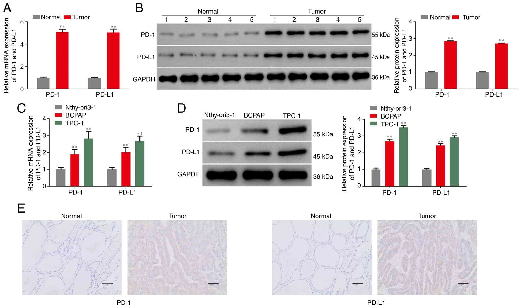
expression levels of PD-1/PD-L1 in clinical PTC tissues. As shown
in Fig. 1A and B, the expression of PD-1 and PD-L1 was
significantly upregulated in PTC tissues compared with those in
paracancerous tissues. Furthermore, the expression levels of PD-1
and PD-L1 in TPC-1 and BCPAP cells were also through with RT-qPCR
and western blotting assays. As hypothesized, PD-1 and PD-L1
expression levels were significantly increased relative to those in
immortalized normal thyroid follicular epithelial cells
(Nthy-ori3-1), as demonstrated in Fig.
1C and D. Given that these
were pure tumor cell populations cultured in vitro, the
detected PD-1 expression represents tumor-intrinsic PD-1.
To evaluate the expression profiles of PD-1 and
PD-L1 in the present patient cohort, IHC staining was performed on
tissue sections. IHC staining for PD-1 was performed on TC tissues
and matched adjacent non-tumorous tissues. In tumor tissues, PD-1
expression was predominantly detected in TILs, with strong positive
staining observed. In addition, weak to moderate PD-1 expression
was also noted in tumor cells themselves. By contrast, adjacent
normal thyroid tissues exhibited primarily negative PD-1 staining
in follicular epithelial cells, with only occasional PD-1-positive
lymphocytes present. These results demonstrate that PD-1 is
expressed in both TILs and tumor cells within the thyroid cancer
microenvironment, while its expression is minimal in adjacent
non-cancerous tissues. Correlation analyses between PD-1/PD-L1
expression levels and available clinicopathological parameters are
presented in Table SIV. High and
low expression groups for PD-1 and PD-L1 were defined based on the
median 2-ΔΔCq value of RT-qPCR from all 30 PTC tissue
samples; values above the median were classified as high
expression, while those at or below the median were classified as
low expression. Despite the data exhibiting a trend toward higher
PD-L1 expression in tumors >2 cm (94.1%) compared to those ≤2 cm
(61.5%), as well as in lymph node-positive (N1) patients (93.8%)
compared to node-negative (N0) patients (64.3%), these differences
did not reach statistical significance. Similarly, PD-1 expression
showed no significant correlation with age, gender, TNM stage or
BRAF mutation status. Overall, there were no statistically
significant associations between PD-1/PD-L1 expression and the
available clinicopathological parameters in the present study
group. These data suggested that PD-1/PD-L1 might act as oncogenes
in development and progression of PTC.
Downregulation of PD-1/PD-L1 inhibits
the proliferation and induces the apoptosis of TC cells
To investigate the functional role of PD-1/PD-L1 in
PTC development and progression, sh-NC, sh-PD-1 and sh-PD-L1 were
transfected into TC cells, with the transfection efficiency being
shown in Fig. 2A and B.
NIS exhibits an important role in TC progression
(38). Previous studies have
confirmed that loss of NIS function is a hallmark of TC
dedifferentiation, which correlates with enhanced tumor
invasiveness, distant metastasis and the development of radioactive
iodine resistance, the main cause of mortality from advanced TC.
Moreover, recent research has revealed that NIS expression is
correlated with the tumor immune microenvironment and the
expression of immune checkpoint molecules, including PD-1 and
PD-L1, suggesting it may coordinate with immune remodeling to
promote TC progression. Thus, western blotting was carried out to
evaluate the effects of PD-1/PD-L1 on the expression of NIS.
Fig. 2C shows that downregulation
of PD-1/PD-L1 significantly promoted the expression of NIS. In
addition, a CCK-8 assay was used to evaluate the effects of
PD-1/PD-L1 on the viability of TC cells. As shown in Fig. 2D, downregulation of PD-1/PD-L1
significantly suppressed the viability of TC cells. An EdU assay
was carried out to assess the effects of PD-1/PD-L1 on the
proliferation of TC cells. As demonstrated in Fig. 2E, downregulation of PD-1/PD-L1
significantly inhibited the proliferation of TC cells. A colony
formation assay was carried out to assess the effects of PD-1/PD-L1
on the clonogenic ability of TC cells and the Fig. 2F indicates that downregulation of
PD-1/PD-L1 significantly suppressed the formation of TC cell
colonies.
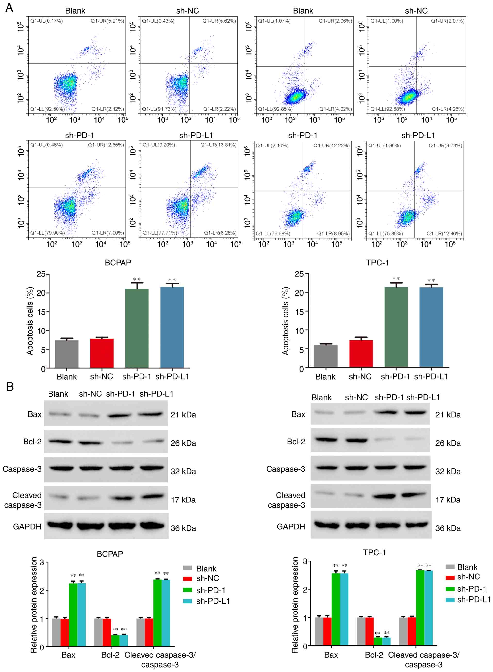
In addition, flow cytometry analysis was adopted to
evaluate the effects of PD-1/PD-L1 on the apoptosis of PTC cells.
Fig. 3A demonstrates that
downregulation of PD-1/PD-L1 significantly promoted the apoptosis
of TC cells. Furthermore, western blotting was performed to analyze
the expression levels of proteins associated with apoptosis. As
demonstrated in Fig. 3B,
downregulation of PD-1/PD-L1 significantly reduced the protein
expression level of Bcl-2, while promoting the expression of Bax
and increasing the ratio of cleaved caspase-3/caspase-3 in TC
cells. These findings indicate that downregulation of PD-1/PD-L1
inhibited proliferation and induced the apoptosis of TC cells.
Downregulation of PD-1/PD-L1 inhibits
the migration and invasion of TC cells
Wound healing and Transwell assays were performed to
analyze the effects of PD-1/PD-L1 on the migration and invasion of
TC cells. As shown in Fig. 4A-C,
downregulation of PD-1/PD-L1 significantly suppressed the migration
and invasion of TC cells. In addition, western blotting was
performed to analyze the expression levels of proteins associated
with EMT progression. As demonstrated in Fig. 4D, downregulation of PD-1/PD-L1
significantly reduced the protein expression level of N-cadherin
and Vimentin, while promoted the expression levels of E-cadherin in
TC cells. These findings indicated that downregulation of
PD-1/PD-L1 inhibited the migration and invasion of TC cells by
suppressing EMT.
 | Figure 4Downregulation of PD-1/PD-L1
inhibited the migration and invasion of TC cells. (A) Wound healing
(scale bar, 100 µm), (B) Transwell migration (scale bar, 50 µm) and
(C) invasion assays (scale bar, 50 µm) were performed to analyze
the effects of PD-1/PD-L1 on the migration and invasion of TC
cells. (D) Western blotting assay was performed to evaluate the
effects of PD-1/PD-L1 on the protein expressions associated with
EMT progress of TC cells. **P<0.01 vs. the Blank
group. RT-qPCR, reverse transcription-quantitative PCR; PD-1,
programmed death-1; PD-L1, programmed death-ligand 1; TC, thyroid
cancer; EMT, epithelial-mesenchymal transition; sh, short hairpin;
NC, negative control. |
Downregulation of PD-1/PD-L1
suppresses PTC progression partially by regulating the TGF-β/Smad
signaling pathway
TGF-β/Smad signaling pathways are key regulators of
EMT, cell proliferation, apoptosis and extracellular matrix
remodeling in cancer. During PTC progression, TGF-β/Smad exerts a
key role by: i) Directly promoting EMT through transcriptional
induction of Snail, Slug and Twist; ii) suppressing
thyroid-specific genes including NIS through Smad-mediated
transcriptional repression; iii) modulating the TME by regulating
immune cell infiltration and cytokine production and; iv)
cooperating with BRAF V600E signaling to drive TC dedifferentiation
(39-42).
Thus, western blot analysis was performed to evaluate the
expression levels of TGF-β/Smad pathway. As shown in Fig. 5A, downregulation of PD-1/PD-L1
significantly inhibited the expression levels of TGF-β/Smad
pathway. In addition, exogenous TGF-β (10 ng/ml) was used to treat
TC cells with sh-PD-1 or sh-PD-L1 and western blotting was used to
detect the TGF-β/Smad pathway. PD-L1 knockdown significantly
suppresses basal and TGF-β-induced phosphorylation of Smad2 and
Smad3 in TPC-1 cells, indicating that PD-L1 positively regulates
the canonical TGF-β/Smad signaling pathway. Moreover, exogenous
TGF-β partially rescues the suppression of p-Smad2/3 caused by
PD-L1 silencing, suggesting a functional crosstalk between PD-L1
and TGF-β signaling in TC progression (Fig. 5B). CCK-8 assays showed that sh-PD-1
or sh-PD-L1 cells exhibited significantly reduced proliferation at
48 and 72 h compared with sh-NC (Fig.
6A). NIS was significantly downregulated upon PD-1 or PD-L1
knockdown (Fig. 6B). The result of
CCK-8 assays was corroborated by EdU incorporation assays (Fig. 6C). Colony formation capacity was
similarly suppressed (Fig. 6D),
while apoptosis was increased (Fig.
6E). Importantly, all these phenotypic changes were
significantly attenuated upon TGF-β supplementation.
PD-1 or PD-L1 deficiency significantly impaired
wound closure (Fig. 7A), migration
(Fig. 7B) and invasion (Fig. 7C). This coincided with a pronounced
shift toward an epithelial state: E-cadherin was upregulated,
whereas mesenchymal markers N-cadherin and Vimentin were
downregulated (Fig. 7D). Moreover,
TGF-β treatment reversed this molecular signature in sh-PD-L1 or
sh-PD-1 cells. These findings indicated that downregulation of
PD-1/PD-L1 suppressed TC progression partially through the
TGF-β/Smad signaling pathway.
 | Figure 7Downregulation of PD-1/PD-L1
inhibited the migration and invasion of TC cells regulating
TGF-β/Smad signaling pathway. (A) Wound healing (scale bar, 100
µm), (B) Transwell migration (scale bar, 50 µm) and (C) invasion
assays (scale bar, 50 µm) were performed to analyze the migration
and invasion of TC cells. (D) Western blotting was performed to
evaluate the expression levels associated with EMT progress of TC
cells. **P<0.01 vs. Blank or sh-NC group,
##P<0.01 vs. sh-PD-1 group and
&&P<0.01 vs. PD-L1 group. PD-1, programmed
death-1; PD-L1, programmed death-ligand 1; TC, Thyroid cancer; sh,
short hairpin; NC, negative control; EMT, epithelial-mesenchymal
transition. |
Discussion
PD-1 and PD-L1 were found to be significantly
upregulated in PTC tissues and cell lines, with the present study
demonstrating their intrinsic carcinogenic effects through
functional experiments. Knockdown of PD-1 or PD-L1 effectively
inhibited the proliferation, colony formation, migration and
invasion abilities of TC cells, while promoting apoptosis and
upregulating the expression of the key differentiation/therapeutic
target NIS. The mechanisms of action revealed that PD-1/PD-L1
achieved their pro-cancer effects by regulating the TGF-β/Smad
signaling pathway. Intracellular PD-1/PD-L1 signaling activates
transcription factors (such as STAT3 and NF-κB) that bind the TGFB1
promoter, upregulating TGF-β1 transcription. PD-1/PD-L1 signaling
may also stabilize TGF-β1 mRNA or enhance its secretion. TGF-β1
then acts in an autocrine manner to activate canonical Smad2/3
signaling, as evidenced by increased Smad2/3 phosphorylation
following PD-1/PD-L1 knockdown (43,44),
specifically manifested as knockdown of PD-1/PD-L1 inhibiting
TGF-β1 expression and Smad2/3 phosphorylation. A key rescue
experiment further determined that exogenous TGF-β treatment could
partially reverse the suppression of malignant phenotype caused by
knockdown of PD-1/PD-L1, thus establishing the causal centrality of
this signaling pathway. These results collectively constructed a
clear evidence chain, indicating that the PD-1/PD-L1-TGF-β/Smad
axis was a key intrinsic mechanism driving the malignant biological
behavior of PTC.
The findings of the present study regarding the high
expression of PD-1/PD-L1 and its intrinsic tumor functions were
consistent with and complementary to a series of published
articles, furthering our understanding of the role of this pathway
in PTC. Previous studies have shown that PD-L1 expression was
particularly prominent in TC, especially in highly aggressive
subtypes such as anaplastic TC and was clearly associated with BRAF
V600E mutations, disease progression and poor prognosis (5,6,15).
With regard to PD-1 expression, different studies have reached
varying conclusions on clinical relevance. For example, one study
found that the positive rate of PD-1 in tumor cells or infiltrating
lymphocytes in thyroid follicular carcinoma samples was 46.67% and
its expression was associated with lower TNM staging and improved
progression-free survival (45).
Whilst the aforementioned study observed the basic high expression
of PD-1/PD-L1 in TC cell lines, it proposed at the molecular level
that this axis participated in the direct intrinsic signaling
mechanism of tumor cell EMT and proliferation-apoptosis through the
TGF-β/Smad pathway, which was an important supplement to its
intrinsic tumor functions (46).
This not only demonstrated its potential value as prognostic
biomarkers but also clarified the role as a driving force in tumor
progression, providing a scientific hypothesis for further
understanding the therapeutic efficacy differences among
patients.
Beyond tumor-intrinsic mechanisms, the PD-1/PD-L1
axis exerts notable effects on the immune TME. PD-L1 expression on
tumor cells engages PD-1 on tumor-infiltrating T lymphocytes,
promoting T-cell exhaustion and functional impairment (24). This immune checkpoint mechanism
likely contributes to the immunosuppressive TME observed in
aggressive thyroid cancers. TAMs, particularly the M2-polarized
subtype, are abundant in thyroid cancer stroma and have been shown
to express PD-L1, further amplifying immune evasion (15). In addition, IL-17A, a
pro-inflammatory cytokine produced by Th17 cells, has been
associated with disease progression in PTC (25). The canonical IL-17 signaling
pathway is initiated by IL-17 binding to the IL-17RA/IL-17RC
receptor complex, which recruits the adaptor protein Act1 and
activates downstream NF-κB and MAPK signaling cascades, leading to
the production of pro-inflammatory cytokines including IL-6, TNF-α
and TGF-β. IL-17 has been shown to upregulate PD-L1 expression in
non-small cell lung cancer, hepatocellular carcinoma, colorectal
cancer and ovarian cancer through NF-κB-dependent mechanisms, and
TGF-β serves as a downstream mediator of IL-17-driven inflammation.
Thus, IL-17 signaling may potentiate the PD-1/PD-L1-TGF-β/Smad axis
through both direct transcriptional regulation of PD-L1 and
indirect amplification of TGF-β signaling within the tumor
microenvironment. This crosstalk warrants further investigation in
TC.
The interaction between IL-17 signaling and the
TGF-β pathway is particularly relevant to the present findings, as
IL-17 can potentiate TGF-β-mediated EMT and promote a
pro-tumorigenic inflammatory microenvironment (25). Studies by Banerjee et al
(24,25) have highlighted the clinical
importance of these interactions in PTC, including their
association with higher tumor nodal stage, increased extrathyroidal
invasion and elevated risk of lymph node metastasis, all clinical
features linked to poor prognosis in patients with PTC. Future
investigations examining the interplay between PD-1/PD-L1
signaling, IL-17-mediated inflammation, and TGF-β activation in the
TME may provide further insights into the immune regulatory
networks governing thyroid cancer progression.
It is important to reconcile the present findings
with previous studies that have reported associations between PD-1
expression and favorable clinical outcomes in certain thyroid
cancer contexts. For example, Ning et al (45) observed that PD-1 positivity in
thyroid follicular carcinoma samples was associated with lower TNM
staging and improved progression-free survival (40). This apparent discrepancy may
reflect the context-dependent nature of PD-1 signaling. A number of
possible explanations may therefore be hypothesized: First, PD-1
expression on tumor-infiltrating lymphocytes may serve as a marker
of an active anti-tumor immune response, which would be associated
with improved prognosis, whereas tumor-intrinsic PD-1 signaling (as
investigated in the present study) may promote malignant behavior.
Second, differences in detection methods (IHC vs. RT-qPCR/western
blotting) and the specific antibodies used may capture different
aspects of PD-1 expression, with IHC typically detecting
cell-surface PD-1 protein on intact tumor tissue, while RT-qPCR
measures total transcript levels and western blotting quantifies
total cellular protein (including both intracellular and
membrane-localized pools). Third, the clinical importance of PD-1
may differ across thyroid cancer subtypes and disease stages. The
coexistence of these context-dependent roles underscores the
complexity of PD-1/PD-L1 biology and highlights the need for
integrated analyses that simultaneously evaluate both
tumor-intrinsic and immune microenvironment-associated
mechanisms.
The clinical importance of the present findings is
further underscored by the observed upregulation of NIS expression
following PD-1 or PD-L1 knockdown. NIS is the primary mediator of
radioiodine uptake in thyroid cells and its downregulation is a
hallmark of radioiodine-refractory thyroid cancer. TGF-β has been
shown to repress NIS expression through Smad-dependent signaling
(40-42)
and the present data suggest that PD-1/PD-L1-mediated activation of
the TGF-β/Smad pathway may contribute to NIS silencing and
subsequent radioiodine resistance. These findings suggest that
targeting the PD-1/PD-L1/TGF-β/Smad axis could restore radioiodine
sensitivity in refractory patients, although this hypothesis
requires rigorous preclinical and clinical evaluation.
At a mechanistic level, the present study was able
to associate PD-1/PD-L1 with the TGF-β/Smad signaling pathway,
providing a new molecular explanation framework that enhances the
existing literature. Previous studies have primarily focused on the
‘interaction’ between PD-1/PD-L1 and immune function within the
tumor microenvironment, revealing crosstalk with other
cancer-promoting pathways such as MAPK and PI3K/Akt (47,48).
The TGF-β/Smad pathway was selected based on the following
rationale: i) TGF-β is the most consistently upregulated cytokine
in TC and is associated with EMT and NIS repression; ii) BRAF V600E
induces TGF-β secretion which suppresses NIS expression through
Smad signaling; iii) the present preliminary data showed that
PD-1/PD-L1 knockdown predominantly affected TGF-β/Smad signaling
with minimal changes in MAPK or PI3K/Akt pathways and; iv) the role
of PD-1/PD-L1 in TGF-β-driven tumor-intrinsic signaling was
previously unknown in TC (47,48).
In addition, the TGF-β/Smad signaling pathway is an
important intracellular signaling pathway involved in the malignant
biological processes of tumor cells. Studies have shown that the
activation of the TGF-β/Smad signaling pathway was associated with
the migration of TC cells and lymph node metastasis. For example,
REGγ mediated the upregulation of TGF-β by degrading Smad7,
enhancing the dedifferentiation of TC cells. The absence of REGγ
restored the expression of thyroid-specific genes, especially the
expression of the NIS in TC cells, thereby enhancing the uptake of
radioactive iodine (40). In TC,
the BRAF V600E gene could induce the secretion of TGF-β, which in
turn inhibited NIS expression (34). Furthermore, reduced NADPH oxidase 4
was found to be a key mediator of NIS downregulation in BRAF
V600E-induced TC and this process was associated with the
TGF-β/Smad signaling pathway (40). Therefore, inhibiting this signaling
pathway may exert an anti-tumor effect on TC.
The novelty of the present study lies in: i)
Demonstrating that PD-1 and PD-L1 function through the TGF-β/Smad
pathway specifically in thyroid cancer cells, establishing a
tumor-intrinsic mechanism beyond immune checkpoint function; ii)
revealing that this signaling axis regulates NIS expression, which
has direct clinical implications for radioiodine therapy and; iii)
providing comprehensive functional evidence to associate intrinsic
PD-1/PD-L1 signaling with the full spectrum of TC malignant
behaviors (proliferation, apoptosis, migration, invasion, and EMT)
through the TGF-β/Smad pathway.
Regarding the exclusion of other signaling pathways,
the following additional considerations apply. For the MAPK
pathway, although BRAF V600E is the most frequent oncogenic driver
in PTC and signals principally through the MAPK/ERK cascade, the
present preliminary screening showed that PD-1/PD-L1 knockdown
produced negligible changes in ERK1/2 phosphorylation, indicating
that the tumor-intrinsic effects observed in the present study were
not primarily mediated by MAPK signaling. For the PI3K/Akt pathway:
similarly, no consistent change in Akt phosphorylation was detected
following PD-1/PD-L1 knockdown, and the PI3K/Akt axis is more
commonly implicated in FTC and radioiodine-refractory disease
rather than in the PTC-derived models used here. Collectively, the
combination of the present preliminary pathway-screening data, the
well-established association between TGF-β/Smad signaling and NIS
repression/EMT in thyroid cancer (49,50)
and the absence of changes in MAPK or PI3K/Akt activity supports
the prioritization of the TGF-β/Smad pathway, although it should be
acknowledged that parallel or compensatory signalling cannot be
fully excluded on the basis of the present experiments (47,48).
Based on the present study and existing literature,
it could be considered that strategies targeting the PD-1/PD-L1
pathway, especially combination therapies, hold potential in the
treatment of TC. When exploring combination therapy strategies,
additional studies have also provided support. For instance, basic
experiments have found that simultaneous inhibition of IRE1α and
PD-L1 in the endoplasmic reticulum stress pathway yields notable
synergistic effects in inhibiting TC in both in vitro and
in vivo models (51).
Similarly, a further study has observed that in a mouse-derived TC
model, the expression of TGF-βR2 promoted immune-associated adverse
events in the liver induced by anti-PD-1/PD-L1 treatment (52). Analysis of the experiments carried
out in the present study revealed that exogenous TGF-β treatment
could partially reverse the malignant phenotype suppression caused
by knockdown of PD-1/PD-L1. These findings collectively suggest
that simultaneously targeting immune checkpoints and related
signaling networks may be an effective approach to overcome
monotherapy resistance and enhance therapeutic efficacy and that
targeting TGF-β signaling may represent a potential combination for
achieving synergistic effects. However, it must be emphasized that
these combination strategies remain highly speculative at present
and extensive preclinical validation in appropriate in vivo
models, followed by carefully designed clinical trials, is needed
before any clinical translation can be considered.
The present study exhibits certain limitations. The
present research was primarily based on in vitro cell
experiments and future verification in xenograft animal models
(subcutaneous and orthotopic mouse models) is currently being
planned, along with further exploration of whether there were
differences in this mechanism among different TC subtypes. No dose
response analysis or alternative pathway inhibitors were used to
rule off target effects or other parallel pathways. In addition,
PD-1/PD-L1 may also regulate other pathways besides TGF-β/Smad,
which remain to be revealed using more comprehensive omics
technologies. The lack of corresponding rescue experiments
involving PD-1/PD-L1 overexpression limits the ability to
definitively rule out potential off-target effects, although the
consistency of the present findings support the proposed mechanism.
Finally, reviewing published literature revealed that the efficacy
prediction of PD-1/PD-L1 not only depended on expression levels,
but was also influenced by factors such as the inflammatory
background of patients, epigenetic regulation and complex
interactions with other signaling pathways. Future preclinical and
clinical studies should aim to take these complex regulatory
networks into account.
In summary, the present study provided experimental
evidence for the direct carcinogenic role of the PD-1/PD-L1 axis in
the progression of PTC and clarified the core molecular mechanisms
through which it achieved this function by activating the
TGF-β/Smad signaling pathway. This not only contributes to the
understanding of the pathogenesis of PTC but also provides a
theoretical foundation for future clinical exploration of more
precise and effective treatment strategies, which is expected to
bring new hope to patients with advanced and highly aggressive PTC
who have failed to respond to traditional treatments. It is
important to acknowledge that tumor-intrinsic and immune
microenvironment-associated mechanisms likely coexist and interact
in the regulation of PD-1/PD-L1 signaling in PTC and further
studies are warranted to dissect their relative contributions and
potential synergistic effects.
Supplementary Material
Clinicopathological characteristics of
30 patients with papillary thyroid carcinoma.
shRNA target sequences used in the
present study.
Antibodies used in western blotting
analysis.
Correlation between PD-1 and PD-L1
expression and clinicopathological characteristics of patients with
papillary thyroid carcinoma.
Acknowledgements
Not applicable.
Funding
Funding: The present study was supported by Ningxia Natural
Science Foundation Project (grant no. 2025AAC030385).
Availability of data and materials
The data generated in the present study may be
requested from the corresponding author.
Authors' contributions
BZ is the guarantor of the integrity of the entire
study and contributed to the study design and study concepts. XW
and JW contributed to the literature research, data analysis and
clinical studies. WC, JXW and XZ contributed to data acquisition
and statistical analysis. All authors contributed to the manuscript
editing and reviewing. All authors have read and approved the final
version of the manuscript. BZ and XW confirm the authenticity of
all the raw data.
Ethics approval and consent to
participate
The present experiments were approved by the Ethics
Committee of the People's Hospital of Ningxia Hui Autonomous Region
(Yinchuan, China; approval no. 2025-NZR-291). All procedures were
performed under strict constraints of the Enhancing the Quality and
Transparency of Health Research network guidelines, the relevant
norms and regulations for clinical research and the ethics codes in
the Declaration of Helsinki.
Patient consent for publication
Not applicable.
Competing interests
The authors declare that they have no competing
interests.
References
|
1
|
Ma L, Xie Y, Ke S, Liu D, Wang L, Wang L
and Liu L: Levothyroxine dose prediction post-thyroidectomy for
differentiated thyroid carcinoma. Front Endocrinol (Lausanne).
16(1727681)2025.PubMed/NCBI View Article : Google Scholar
|
|
2
|
Zhu Q, Liu J, Hu J and Zhang Y: The
epidemiological landscape of thyroid cancer and estimates of
overdiagnosis in China: A population-based study. Thyroid.
35:307–320. 2025.PubMed/NCBI View Article : Google Scholar
|
|
3
|
Gao N, Han J, Sun X, Yu J, Huang J, Liu R,
Tang YL and Nie C: Erianin induces apoptosis of thyroid cancer
cells via TNF signaling pathway. Eur J Pharmacol.
1006(178167)2025.PubMed/NCBI View Article : Google Scholar
|
|
4
|
Li C, Li Q, Shi X, Han S, Song X, Li X and
Zhuang X: Papillary thyroid microcarcinoma and papillary thyroid
carcinoma: Clinical characteristics and stratification of treatment
strategies. PLoS One. 20(e0327423)2025.PubMed/NCBI View Article : Google Scholar
|
|
5
|
Liang L, Chen Z, Lei D, Mo C, Lan D, Ke J,
Wang W, Yang Z, Guo X, Chen D, et al: APG-115 induces
SLC7A11-mediated ferroptosis and upregulates PD-L1 expression in
thyroid cancer. ACS Omega. 10:31099–31107. 2025.PubMed/NCBI View Article : Google Scholar
|
|
6
|
Elyamany AM, Abdel Haleem RT, Soliman NA,
Mahdi L, Ahmed HMA and Abdelwahab KH: Expression of long non-coding
RNA-NEAT-1 in papillary thyroid carcinoma in relation to
immunohistochemical expression of CD8 and PD-L1 as indicators of
tumor immune microenvironment. Asian Pac J Cancer Prev.
26:3093–3101. 2025.PubMed/NCBI View Article : Google Scholar
|
|
7
|
Xue Y, Zhuang Y and Chen S: Dynamic
survival outcomes of the tall-cell variant of papillary thyroid
carcinoma patients after surgery. Front Endocrinol (Lausanne).
16(1517907)2025.PubMed/NCBI View Article : Google Scholar
|
|
8
|
Toloza FJK, Gubbi S and Klubo-Gwiezdzinska
J: Trends in survival and factors associated with survival in
primary thyroid lymphoma: A SEER-based analysis (1975-2021).
Thyroid. 35:1074–1082. 2025.PubMed/NCBI View Article : Google Scholar
|
|
9
|
Gu Y, Yu M, Deng J and Lai Y: Preoperative
circulating tumor cells level is associated with lymph node
metastasis in patients with unifocal papillary thyroid carcinoma.
World J Surg Oncol. 23(47)2025.PubMed/NCBI View Article : Google Scholar
|
|
10
|
Shen P, Yang Z, Sun J, Wang Y, Qiu C, Wang
Y, Ren Y, Liu S, Cai W, Lu H and Yao S: Explainable multimodal deep
learning for predicting thyroid cancer lateral lymph node
metastasis using ultrasound imaging. Nat Commun.
16(7052)2025.PubMed/NCBI View Article : Google Scholar
|
|
11
|
Liu Y, Huang S, Li X, Tian T and Huang R:
Therapeutic efficacies of remnant ablation and radioiodine adjuvant
therapy in differentiated thyroid cancer. Endocrine. 87:734–743.
2025.PubMed/NCBI View Article : Google Scholar
|
|
12
|
Feng Y, Xiao A, Xing C, Dai Q, Liu X, Liu
J and Feng L: Elevated thyroid-stimulating hormone levels,
independent of Hashimoto's thyroiditis, increase thyroid cancer
risk: Insights from genetic and clinical evidence. Endocrine.
88:175–184. 2025.PubMed/NCBI View Article : Google Scholar
|
|
13
|
Pishdad R and Ahmadi S:
Thyroid-stimulating hormone (TSH) suppression therapy in thyroid
cancer: Balancing benefits and harms. Endocr Pract. 32:401–405.
2026.PubMed/NCBI View Article : Google Scholar
|
|
14
|
He Z, Wang C, Liu C, Zhang K, Wang J, Wang
X, Zhang Y and Chen L: Limited value of genetic profiling in
guiding radioiodine therapy for metastatic differentiated thyroid
cancer. Endocr Relat Cancer. 32(e240298)2025.PubMed/NCBI View Article : Google Scholar
|
|
15
|
Ricarte-Filho JC, Caperton CO, Isaza A,
Massoll NA and Franco AT: Macrophage abundance in oncocytic thyroid
cancer is associated with PD-L1 expression. Endocr Oncol.
5(e250026)2025.PubMed/NCBI View Article : Google Scholar
|
|
16
|
Piermattei A, Migliara G, Feraco A, Bruno
C, Cioni L, Zhang Q, Padial-Urtueta B, Merenda E, Fadda G,
Raffaelli M, et al: Evaluation of PD-L1, TERT promoter mutations,
and BRAFV600E mutation in poorly differentiated, differentiated
high grade thyroid carcinoma and anaplastic carcinoma of the
thyroid: Our institutional experience. Virchows Arch. 487:605–618.
2025.PubMed/NCBI View Article : Google Scholar
|
|
17
|
Forma A, Kłodnicka K, Pająk W, Flieger J,
Teresińska B, Januszewski J and Baj J: Thyroid cancer:
Epidemiology, classification, risk factors, diagnostic and
prognostic markers, and current treatment strategies. Int J Mol
Sci. 26(5173)2025.PubMed/NCBI View Article : Google Scholar
|
|
18
|
Li X, Gao S, Shan C, Zhang Q, Tan Y, Yu X
and Yu J: Advances in PD-1/PD-L1 pathway inhibitors in the
treatment of thyroid cancer: Mechanisms and clinical therapeutic
perspectives. Front Immunol. 16(1643421)2025.PubMed/NCBI View Article : Google Scholar
|
|
19
|
Zhang J, Bu X, Wang H, Zhu Y, Geng Y,
Nihira NT, Tan Y, Ci Y, Wu F, Dai X, et al: Cyclin D-CDK4 kinase
destabilizes PD-L1 via cullin 3-SPOP to control cancer immune
surveillance. Nature. 553:91–95. 2018.PubMed/NCBI View Article : Google Scholar
|
|
20
|
Wang Y, Ma X, Ma J, Li J, Lin Z, Gao W,
Gong P and Dai P: Thyroid dysfunction as a predictor of PD- 1/PD-L1
inhibitor efficacy in advanced lung cancer. BMC Cancer.
25(791)2025.PubMed/NCBI View Article : Google Scholar
|
|
21
|
Yan X, Lv Q, Wu J, Fang J, Peng L and Zhao
X: The efficacy and immune-mediated safety of PD-1/PD-L1 combined
with neoadjuvant chemotherapy in triple-negative breast cancer: A
meta-analysis. Front Oncol. 15(1635418)2025.PubMed/NCBI View Article : Google Scholar
|
|
22
|
Liotti F, Kumar N, Prevete N, Marotta M,
Sorriento D, Ieranò C, Ronchi A, Marino FZ, Moretti S, Colella R,
et al: PD-1 blockade delays tumor growth by inhibiting an intrinsic
SHP2/Ras/MAPK signalling in thyroid cancer cells. J Exp Clin Cancer
Res. 40(22)2021.PubMed/NCBI View Article : Google Scholar
|
|
23
|
Lin J, Qiu Y, Zheng X, Dai Y and Xu T: The
miR-199a-5p/PD-L1 axis regulates cell proliferation, migration and
invasion in follicular thyroid carcinoma. BMC Cancer.
22(756)2022.PubMed/NCBI View Article : Google Scholar
|
|
24
|
Banerjee S, Nahar U, Dahiya D, Mukherjee
S, Dey P, Gupta R, Radotra B, Sachdeva N, Sood A, Bhadada SK and
Bhansali A: Role of cytotoxic T cells and PD-1 immune checkpoint
pathway in papillary thyroid carcinoma. Front Endocrinol
(Lausanne). 13(931647)2022.PubMed/NCBI View Article : Google Scholar
|
|
25
|
Banerjee S, Nahar U, Dahiya D, Gupta R,
Mukherjee S, Sachdeva N, Sood A, Dey P, Radotra B and Bhansali A:
IL-17 A correlates with disease progression in papillary thyroid
carcinoma. Diagn Pathol. 18(93)2023.PubMed/NCBI View Article : Google Scholar
|
|
26
|
Dong H, Strome SE, Salomao DR, Tamura H,
Hirano F, Flies DB, Roche PC, Lu J, Zhu G, Tamada K, et al:
Tumor-associated B7-H1 promotes T-cell apoptosis: A potential
mechanism of immune evasion. Nat Med. 8:793–800. 2002.PubMed/NCBI View Article : Google Scholar
|
|
27
|
Cheng W, Kang K, Zhao A and Wu Y: Dual
blockade immunotherapy targeting PD-1/PD-L1 and CTLA-4 in lung
cancer. J Hematol Oncol. 17(54)2024.PubMed/NCBI View Article : Google Scholar
|
|
28
|
Han Y, Koehler VF, Nagarajah J, Schmohl
KA, Stauss C, Schwenk N, Spellerberg R, Kitzberger C, Kumbrink J,
Zach C, et al: TGF-β1-based restoration of sodium iodide symporter
expression in radioiodine-refractory differentiated thyroid cancer
via engineered MSCs. Mol Ther. 33:6130–6145. 2025.PubMed/NCBI View Article : Google Scholar
|
|
29
|
Xiang J, Lv N, Yin S, Zhao T, Liu F, Cheng
L, Liu F and Kuang J: TGF-β induces EMT in thyroid cancer cells by
regulating transcription factors. Thyroid Res.
18(25)2025.PubMed/NCBI View Article : Google Scholar
|
|
30
|
Garcia-Rendueles AR, Rodrigues JS,
Garcia-Rendueles MER, Suarez-Fariña M, Perez-Romero S, Barreiro F,
Bernabeu I, Rodriguez-Garcia J, Fugazzola L, Sakai T, et al:
Rewiring of the apoptotic TGF-β-SMAD/NFκB pathway through an
oncogenic function of p27 in human papillary thyroid cancer.
Oncogene. 36:652–666. 2017.PubMed/NCBI View Article : Google Scholar
|
|
31
|
Zhao JJ, Hao S, Wang LL, Hu CY, Zhang S,
Guo LJ, Zhang G, Gao B, Jiang Y, Tian WG and Luo DL: Long
non-coding RNA ANRIL promotes the invasion and metastasis of
thyroid cancer cells through TGF-β/Smad signaling pathway.
Oncotarget. 7:57903–57918. 2016.PubMed/NCBI View Article : Google Scholar
|
|
32
|
Lin Q, Qi Q, Hou S, Chen Z, Jiang N, Zhang
L and Lin C: Activation of the TGF-β1/Smad signaling by KIF2C
contributes to the malignant phenotype of thyroid carcinoma cells.
Tissue Cell. 73(101655)2021.PubMed/NCBI View Article : Google Scholar
|
|
33
|
Ma S, Wang Q, Ma X, Wu L, Guo F, Ji H, Liu
F, Zhao Y and Qin G: FoxP3 in papillary thyroid carcinoma induces
NIS repression through activation of the TGF-β1/Smad signaling
pathway. Tumour Biol. 37:989–998. 2016.PubMed/NCBI View Article : Google Scholar
|
|
34
|
Jung CK, Bychkov A and Kakudo K: Update
from the 2022 World Health Organization classification of thyroid
tumors: A standardized diagnostic approach. Endocrinol Metab
(Seoul). 37:703–718. 2022.PubMed/NCBI View Article : Google Scholar
|
|
35
|
van Staveren WCG, Solís DW, Delys L,
Duprez L, Andry G, Franc B, Thomas G, Libert F, Dumont JE, Detours
V and Maenhaut C: Human thyroid tumor cell lines derived from
different tumor types present a common dedifferentiated phenotype.
Cancer Res. 67:8113–8120. 2007.PubMed/NCBI View Article : Google Scholar
|
|
36
|
Saiselet M, Floor S, Tarabichi M, Dom G,
Hébrant A, van Staveren WC and Maenhaut C: Thyroid cancer cell
lines: An overview. Front Endocrinol (Lausanne).
3(133)2012.PubMed/NCBI View Article : Google Scholar
|
|
37
|
Livak KJ and Schmittgen TD: Analysis of
relative gene expression data using real-time quantitative PCR and
the 2(-Delta Delta C(T)) method. Methods. 25:402–408.
2001.PubMed/NCBI View Article : Google Scholar
|
|
38
|
Shen DHY, Chan HP, Tsai FR, Chiu YL, Liang
TJ, She Y, Liu AC, Yeh HY, Tsai KW and Li SC: MDM2 Knockdown
reduces the oncogenic activities and enhances NIS protein abundance
in papillary thyroid cancer. Cancer Genomics Proteomics.
22:444–457. 2025.PubMed/NCBI View Article : Google Scholar
|
|
39
|
Bhatti MZ, Pan L, Wang T, Shi P and Li L:
REGγ potentiates TGF-β/Smad signal dependent epithelial-mesenchymal
transition in thyroid cancer cells. Cell Signal.
64(109412)2019.PubMed/NCBI View Article : Google Scholar
|
|
40
|
Jiao C, Li L, Zhang P, Zhang L, Li K, Fang
R, Yuan L, Shi K, Pan L, Guo Q, et al: REGγ ablation impedes
dedifferentiation of anaplastic thyroid carcinoma and accentuates
radio-therapeutic response by regulating the Smad7-TGF-β pathway.
Cell Death Differ. 27:497–508. 2020.PubMed/NCBI View Article : Google Scholar
|
|
41
|
Riesco-Eizaguirre G, Rodríguez I, De la
Vieja A, Costamagna E, Carrasco N, Nistal M and Santisteban P: The
BRAFV600E oncogene induces transforming growth factor beta
secretion leading to sodium iodide symporter repression and
increased malignancy in thyroid cancer. Cancer Res. 69:8317–8325.
2009.PubMed/NCBI View Article : Google Scholar
|
|
42
|
Azouzi N, Cailloux J, Cazarin JM, Knauf
JA, Cracchiolo J, Al Ghuzlan A, Hartl D, Polak M, Carré A, El
Mzibri M, et al: NADPH oxidase NOX4 is a critical mediator of
BRAFV600E-induced downregulation of the sodium/iodide
symporter in papillary thyroid carcinomas. Antioxid Redox Signal.
26:864–877. 2017.PubMed/NCBI View Article : Google Scholar
|
|
43
|
Yu H, Pardoll D and Jove R: STATs in
cancer inflammation and immunity: A leading role for STAT3. Nat Rev
Cancer. 9:798–809. 2009.PubMed/NCBI View Article : Google Scholar
|
|
44
|
Lee KY, Ito K, Hayashi R, Jazrawi EPI,
Barnes PJ and Adcock IM: NF-kappaB and activator protein 1 response
elements and the role of histone modifications in IL-1beta-induced
TGF-beta1 gene transcription. J Immunol. 176:603–615.
2006.PubMed/NCBI View Article : Google Scholar
|
|
45
|
Ning Q, Liu J, Liu S, Zou Q, Li K and Li
Z: TRx0237 induces apoptosis and enhances anti-PD-1
immunotherapeutic efficacy in anaplastic thyroid cancer. Int
Immunopharmacol. 155(114610)2025.PubMed/NCBI View Article : Google Scholar
|
|
46
|
Eloy C, Santos J, Cameselle-Teijeiro J,
Soares P and Sobrinho-Simões M: TGF-beta/Smad pathway and BRAF
mutation play different roles in circumscribed and infiltrative
papillary thyroid carcinoma. Virchows Arch. 460:587–600.
2012.PubMed/NCBI View Article : Google Scholar
|
|
47
|
Bullock KK and Richmond A: Beyond
anti-PD-1/PD-L1: Improving immune checkpoint inhibitor responses in
triple-negative breast cancer. Cancers (Basel).
16(2189)2024.PubMed/NCBI View Article : Google Scholar
|
|
48
|
Quan Z, Yang Y, Zheng H, Zhan Y, Luo J,
Ning Y and Fan S: Clinical implications of the interaction between
PD-1/PD-L1 and PI3K/AKT/mTOR pathway in progression and treatment
of non-small cell lung cancer. J Cancer. 13:3434–3443.
2022.PubMed/NCBI View Article : Google Scholar
|
|
49
|
Zhuang J, Huang B, Li J, Wang Y, Xu Y, Yu
X, Shen H and Gao W: Upregulation of FLT1 activates the
TGF-β1/Smad3 pathway to promote iodide resistance in thyroid
cancer. Cancer Treat Res Commun. 45(100991)2025.PubMed/NCBI View Article : Google Scholar
|
|
50
|
Yan Z, Yangyanqiu W, Shuwen H, Jing M,
Haihong L, Gong C, Yin J, Qing Z and Weili G: Downregulation of
Rap1GAP expression activates the TGF-β/Smad3 pathway to inhibit the
expression of sodium/iodine transporter in papillary thyroid
carcinoma cells. Biomed Res Int. 2021(6840642)2021.PubMed/NCBI View Article : Google Scholar
|
|
51
|
Yao X, Tu Y, Xu Y, Guo Y, Yao F and Zhang
X: Endoplasmic reticulum stress-induced exosomal miR-27a-3p
promotes immune escape in breast cancer via regulating PD-L1
expression in macrophages. J Cell Mol Med. 24:9560–9573.
2020.PubMed/NCBI View Article : Google Scholar
|
|
52
|
Qiu X, Zhu J, Sun Y, Fan K, Yang DR, Li G,
Yang G and Chang C: TR4 nuclear receptor increases prostate cancer
invasion via decreasing the miR-373-3p expression to alter
TGFβR2/p-Smad3 signals. Oncotarget. 6:15397–15409. 2015.PubMed/NCBI View Article : Google Scholar
|