Introduction
Diabetes mellitus is a chronic metabolic disorder
defined by persistent hyperglycemia, leading to various systemic
complications, including diabetic osteopathy (1). This condition encompasses a broad
range of skeletal alterations, including impaired bone mineral
density, deteriorated bone quality and suppressed bone turnover,
collectively resulting in compromised mechanical integrity
(2). While both type 1 diabetes
mellitus (T1DM) and type 2 diabetes mellitus (T2DM) increase
skeletal fragility, their underlying clinical profiles differ: T1DM
is typically associated with reduced bone mass, whereas T2DM often
presents with normal or even elevated bone mineral density, yet
with markedly diminished bone quality, including alterations in
bone microarchitecture and increased cortical porosity (2,3).
Consequently, patients with diabetes have a higher risk of bone
fractures, particularly hip fractures, which are 1.5-3 times more
common than in normal controls, and exhibit delayed bone healing
(4,5).
A previous epidemiological study suggested that the
duration of diabetes and poor glycemic control independently
increase skeletal fragility risk (6). This is partly attributable to the
accumulation of advanced glycation end-products, which impair bone
matrix properties and increase oxidative stress (7,8).
Chronic high glucose (HG) exposure promotes excessive reactive
oxygen species (ROS) production, causing oxidative damage in the
bone microenvironment (9). This
oxidative stress drives pathological disruption of bone remodeling
and inhibits key osteogenic differentiation factor expression
(10).
Osteoblast differentiation involves a network of
signaling pathways and transcription factors. Bone morphogenetic
proteins (BMPs), particularly BMP-2 and BMP-7, initiate
osteogenesis, while Runt-related transcription factor 2 (RUNX2) and
Osterix regulate osteoblast maturation and extracellular matrix
synthesis (11,12). In addition, osteoprotegerin (OPG)
and receptor activator of NF-κB ligand (RANKL) are key regulators
of bone remodeling, with OPG acting as a decoy receptor that
inhibits RANKL-mediated osteoclast differentiation. Furthermore,
mitogen-activated protein kinase (MAPK) signaling pathways,
including p38, ERK1/2 and JNK, are involved in mediating osteogenic
signals (13). While p38 and ERK
activation generally promote bone formation, oxidative
stress-induced JNK pathway activation antagonizes BMP signaling and
inhibits osteoblastic activity (14,15).
Cilostazol (CLZ), a selective phosphodiesterase 3
inhibitor, is clinically approved for treating intermittent
claudication and peripheral vascular disease (16). Beyond its antiplatelet and
vasodilatory effects, studies have reported that CLZ increases
intracellular cyclic adenosine monophosphate (cAMP) levels and
exerts protective effects in a number of tissue types, including
promotion of bone growth and protection of hepatocytes against
ethanol-induced oxidative stress and apoptosis (17,18).
Previous studies have reported that CLZ exerts protective effects
in synovial cells and vascular endothelial cells through activation
of nuclear factor erythroid 2-related factor 2 and sirtuin 1
signaling pathways, respectively (19,20).
However, the specific mechanisms through which CLZ modulates
osteoblast function under HG-induced conditions remain unclear.
To investigate these mechanisms under diabetic-like
conditions, MC3T3-E1 cells were exposed to a combination of HG and
glucose oxidase (GOx). HG was used to simulate chronic
hyperglycemia, whereas GOx was used to continuously generate
hydrogen peroxide, thereby enhancing oxidative stress. This
experimental model was employed to reproduce the combined
hyperglycemic and oxidative stress conditions associated with
diabetic osteoblast dysfunction (10,21,22).
The present study aimed to investigate the protective effects of
CLZ on osteoblast dysfunction under diabetic-like conditions, with
particular focus on MAPK signaling and the OPG/RANKL axis. These
findings are expected to provide mechanistic insights into the
effects of CLZ on osteoblast function under diabetic-like
conditions and serve as a basis for future in vivo and
translational studies.
Materials and methods
Cell culture and reagents
MC3T3-E1 subclone 4, a mouse osteoblastic cell line,
was obtained from the American Type Culture Collection. Cells were
cultured in α-minimum essential medium (Gibco; Thermo Fisher
Scientific, Inc.) supplemented with 10% FBS (Gibco; Thermo Fisher
Scientific, Inc.) and 1% penicillin/streptomycin at 37˚C in a
humidified atmosphere containing 5% CO2. CLZ
(Sigma-Aldrich; Merck KGaA) was dissolved in DMSO to prepare a 100
mM stock solution. For the cell viability assay, control cells
received complete culture medium without CLZ or DMSO. For HG/GOx
experiments, untreated control cells received the corresponding
culture medium without HG, GOx, CLZ or DMSO. The final DMSO
concentration in the CLZ-treated groups was ≤0.001% in the main
experiments and ≤0.01% in the cell viability assay. Cells were
treated with 50 mM HG and 5 mU/ml GOx (both from Sigma-Aldrich;
Merck KGaA) to induce oxidative stress.
Cell viability assay
Cell viability was evaluated using a Cell Counting
Kit-8 (CCK-8) assay (Dojindo Laboratories, Inc.). MC3T3-E1 cells
were seeded in 96-well plates at a density of 1x10⁴
cells/well and treated with various concentrations of CLZ (0.1, 1,
5 and 10 µM) for 24 and 48 h at 37˚C. After incubation, CCK-8
solution was added to each well, followed by incubation for 2 h at
37˚C and absorbance at 450 nm was measured using a microplate
reader. Cell morphology was observed using an inverted light
microscope after treatment with CLZ (0, 0.1, 1, 5 or 10 µM) for 24
and 48 h as aforementioned.
Osteogenic differentiation
MC3T3-E1 cells were seeded at a density of
2x10⁴ cells/cm2 in either 24-well plates for
ALP staining and activity assays or 12-well plates for Alizarin Red
S (ARS) staining. For osteogenic differentiation assays, MC3T3-E1
cells were first exposed to HG (50 mM) and GOx (5 mU/ml) in
complete culture medium for 4 h at 37˚C. After exposure, the medium
was replaced with osteogenic medium containing 50 µg/ml L-ascorbic
acid and 10 mM β-glycerophosphate to induce osteoblast
differentiation, and either no CLZ or CLZ (0.1 or 1 µM). The medium
was replaced every 2-3 days while maintaining the respective
treatments.
On day 7, ALP staining was performed using an
Alkaline Phosphatase Stem Cell Staining Kit (cat. no. SCR004;
Sigma-Aldrich; Merck KGaA) according to the manufacturer's
instructions. Cells were incubated with the staining reagent for 15
min at room temperature, and stained cells were observed using an
inverted light microscope. ALP activity was measured using a TRACP
& ALP Assay Kit (cat. no. MK301; Takara Bio Inc.). The assay
reaction was performed for 30 min at 37˚C and absorbance was
measured at 405 nm using a microplate reader. ALP activity was
normalized to the total protein amount determined using a BCA
assay. On day 14, cells were fixed with 10% neutral buffered
formalin for 30 min at room temperature and stained with 2% ARS for
30 min at room temperature in the dark. Stained mineralized nodules
were visualized using a light microscope. For quantification, bound
ARS was solubilized with 10% cetylpyridinium chloride for 30 min at
room temperature. The extracted solution was transferred to a
96-well plate and absorbance was measured at 562 nm using a
microplate reader.
Western blot analysis
MC3T3-E1 cells were first exposed to HG (50 mM) and
GOx (5 mU/ml) simultaneously for 4 h at 37˚C to induce oxidative
stress. After stimulation, the medium was replaced with either
normal growth medium or normal growth medium containing CLZ (0.1 or
1 µM). Cells were subsequently cultured for different durations
depending on the experimental purpose. For western blot analysis of
osteogenic markers, protein lysates were collected after an
additional 48-h incubation. For MAPK signaling analysis, protein
lysates were collected after an additional 24-h incubation.
Subsequently, proteins were extracted using RIPA
buffer (Thermo Fisher Scientific, Inc.) and protein concentrations
were determined using a BCA assay. Equal amounts of protein (10
µg/lane) were separated by 10 or 12% SDS-PAGE, depending on the
molecular weight of the target protein, before being transferred
onto PVDF membranes. The membranes were blocked with 5% skimmed
milk in Tris-buffered saline containing 0.1% Tween-20 (TBST) for 1
h at room temperature and incubated overnight at 4˚C with the
following primary antibodies: BMP-2 (cat. no. PA5-85956; 1:2,000;
Thermo Fisher Scientific, Inc.), BMP-7 (cat. no. BS3674; 1:2,000;
Bioworld Technology, Inc.), RUNX2 (clone D1L7F; cat. no. 12556;
1:2,000; Cell Signaling Technology, Inc.), Osterix (cat. no.
ab22552; 1:1,000; Abcam), collagen type I α1 chain (COL1A1; cat.
no. BS70155; 1:2,000; Bioworld Technology, Inc.), OPG (cat. no.
BS6684; 1:2,000; Bioworld Technology, Inc.), RANKL (cat. no.
BS72037; 1:2,000; Bioworld Technology, Inc.), phosphorylated
(p)-p38 MAPK (Thr180/Tyr182; cat. no. 4511; 1:2,000; Cell Signaling
Technology, Inc.), p38 MAPK (clone D13E1; cat. no. 8690; 1:2,000;
Cell Signaling Technology, Inc.), p-p44/42 MAPK (ERK1/2)
(Thr202/Tyr204; clone 197G2; cat. no. 4377; 1:2,000; Cell Signaling
Technology, Inc.), p44/42 MAPK (ERK1/2) (clone 137F5; cat. no.
4695; 1:2,000; Cell Signaling Technology, Inc.), p-SAPK/JNK
(Thr183/Tyr185; cat. no. 4668; 1:2,000; Cell Signaling Technology,
Inc.), SAPK/JNK (cat. no. 9252; 1:2,000; Cell Signaling Technology,
Inc.) and β-actin (I102; cat. no. AP0060; 1:5,000; Bioworld
Technology, Inc.). After washing three times with TBST, the
membranes were incubated with horseradish peroxidase-conjugated
anti-rabbit IgG secondary antibody (cat. no. 7074; 1:2,000; Cell
Signaling Technology, Inc.) for 1 h at room temperature. Protein
bands were visualized using Amersham ECL reagent (Cytiva) and
semi-quantified using ImageJ software (version 1.53v; National
Institutes of Health). Depending on the molecular weights of the
target proteins, the membranes were either stripped and re-probed
sequentially or separate membranes were used for individual target
proteins. β-actin was used as a loading control on each membrane.
The target proteins are presented together with their respective
β-actin loading control from the same membrane in each figure.
Statistical analysis
Data are presented as the mean ± SD of three
independent biological replicates. For the CCK-8, ALP and ARS
assays, each experimental condition was analyzed in triplicate
wells within each independent biological experiment. Western blot
analysis was performed using samples from three independent
biological experiments. Data normality was assessed using the
Shapiro-Wilk test. Statistical analyses were performed using
GraphPad Prism version 9.0 (Dotmatics). Comparisons among multiple
groups were performed using one-way ANOVA followed by Tukey's post
hoc test. P<0.05 was considered to indicate a statistically
significant difference.
Results
CLZ exhibits no cytotoxicity and
maintains cellular morphology in MC3T3-E1 cells
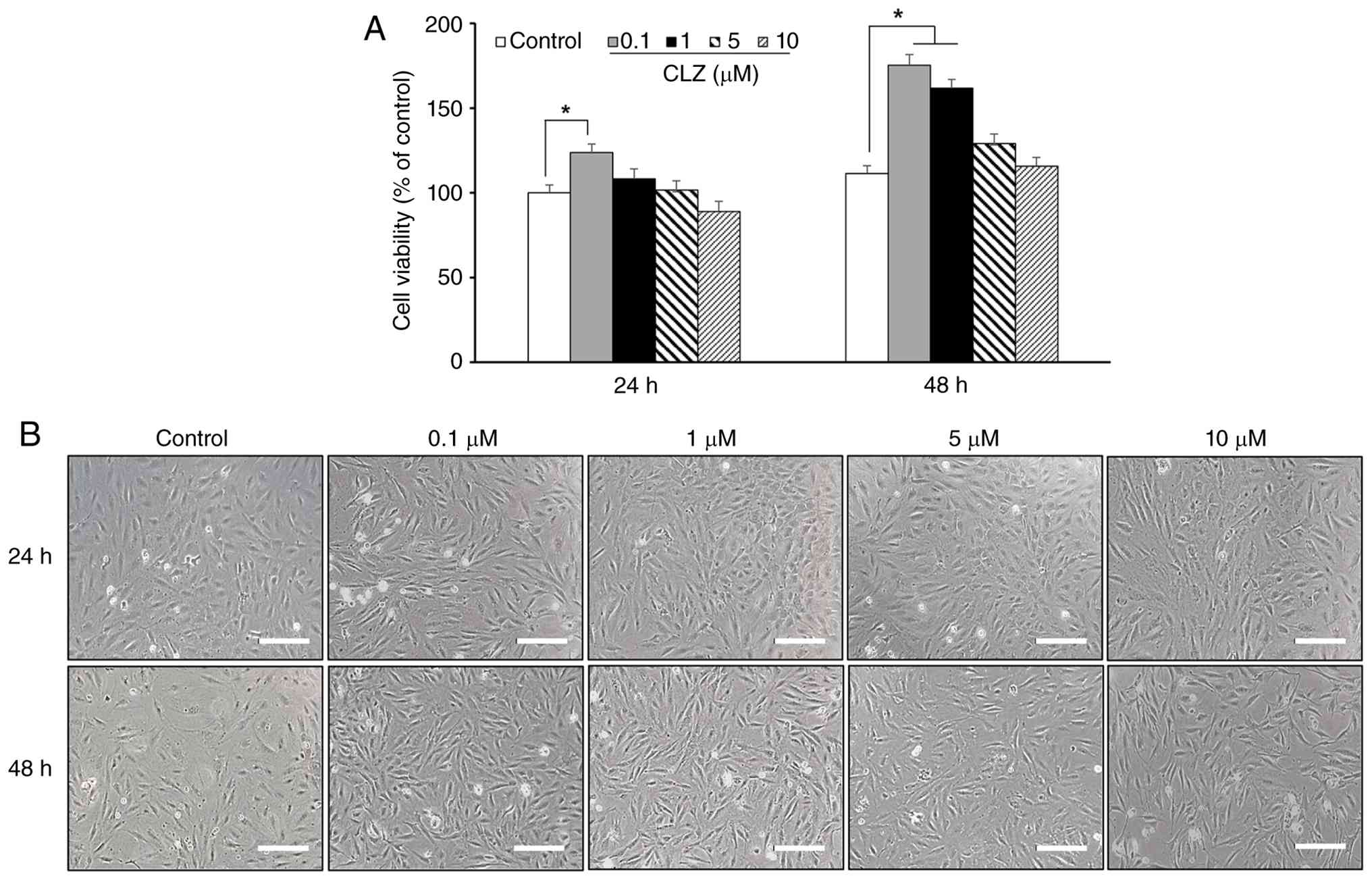
MC3T3-E1 cells were treated with various
concentrations of CLZ (0.1, 1, 5 and 10 µM) for 24 and 48 h to
assess its potential cytotoxicity in osteoblasts. CCK-8 assay
results showed that CLZ did not reduce cell viability at any of the
tested concentrations or time points; however, cell viability was
significantly increased following treatment with 0.1 µM CLZ at both
24 and 48 h. This indicated high biocompatibility of CLZ with
osteoblasts (Fig. 1A).
Subsequently, the effect of CLZ treatment on cell morphology was
examined. Morphological analysis using inverted microscopy revealed
that CLZ-treated cells retained their typical spindle-shaped
fibroblastic appearance, similar to that of the control group
(Fig. 1B). These findings indicate
that CLZ exhibited no detectable cytotoxicity within the tested
concentration range. Therefore, 0.1 and 1 µM were selected for
subsequent experiments because these concentrations showed no
evidence of cytotoxicity and were supported by previous in
vitro studies demonstrating their biological activity (23,24).
CLZ restores HG/GOx-induced
downregulation of osteogenic markers and OPG/RANKL expression
To evaluate the effect of CLZ on osteoblast
differentiation and bone remodeling balance under diabetic-like
conditions, the expression of key proteins was measured. Western
blot analysis revealed that HG/GOx exposure markedly reduced the
protein levels of RUNX2, Osterix, BMP-2, BMP-7 and COL1A1 (Fig. 2A). Semi-quantitative densitometric
analysis indicated that CLZ (0.1 and 1 µM) significantly increased
the expression of these osteogenic markers following HG/GOx-induced
oxidative stress (Fig. 2B).
Subsequently, the expression of OPG and RANKL was assessed. HG/GOx
treatment decreased OPG expression and increased RANKL expression
(Fig. 2C). However, CLZ treatment
increased OPG expression and reduced RANKL expression following
HG/GOx exposure (Fig. 2D). These
findings suggest that CLZ may contribute to the restoration of
osteoblast differentiation and the OPG/RANKL balance under
HG-induced stress.
 | Figure 2Effects of CLZ on osteogenic marker,
OPG and RANKL expression under HG/GOx-induced stress. MC3T3-E1
cells were exposed to HG (50 mM) and GOx (5 mU/ml) for 4 h. After
stimulation, the medium was replaced with either normal medium or
medium containing CLZ (0.1 or 1 µM), followed by incubation for an
additional 48 h before protein extraction. (A) Representative
western blot analysis of osteogenic markers (RUNX2, Osterix, BMP-2,
BMP-7 and COL1A1). (B) Semi-quantitative densitometric analysis of
osteogenic markers, normalized to β-actin. (C) Western blot
analysis of OPG and RANKL protein levels. (D) Semi-quantitative
densitometric analysis of OPG and RANKL protein expression,
normalized to β-actin. Data are presented as the mean ± SD of three
independent experiments. #P<0.05 Control vs. HG/Gox
groups and *P<0.05 HG/GOx vs. HG/GOx + CLZ groups.
BMP, bone morphogenetic protein; CLZ, cilostazol; COL1A1, collagen
type I α1 chain; GOx, glucose oxidase; HG, high glucose; OPG,
osteoprotegerin; RANKL, receptor activator of NF-κB ligand; RUNX2,
Runt-related transcription factor 2. |
CLZ modulates HG/GOx-induced MAPK
signaling dysregulation
To investigate the association between CLZ treatment
and MAPK signaling, MAPK phosphorylation was assessed. Western blot
analysis revealed that HG/GOx treatment markedly decreased p38 and
ERK phosphorylation and increased JNK phosphorylation (Fig. 3A). Semi-quantitative densitometric
analysis of the phosphorylated-to-total protein ratios showed that
HG/GOx treatment decreased the p-ERK/ERK and p-p38/p38 ratios and
increased the p-JNK/JNK ratio compared with the Control group,
whereas CLZ treatment partially reversed these changes (Fig. 3B). CLZ also attenuated the
HG/GOx-induced increase in p-JNK levels (Fig. 3B). These findings suggest that
CLZ-mediated modulation of MAPK phosphorylation, characterized by
increased p38 and ERK phosphorylation and decreased JNK
phosphorylation, may contribute to restoring signaling conditions
favorable for osteoblast maturation under diabetic-like
conditions.
CLZ restores HG/GOx-induced
suppression of ALP activity and mineralized nodule formation
To assess the functional effect of CLZ on osteoblast
differentiation, ALP activity and calcium deposition were assessed.
HG/GOx treatment markedly reduced ALP staining (Fig. 4A) and activity (Fig. 4B). However, treatment with CLZ (0.1
and 1 µM) significantly restored ALP activity (Fig. 4B). Subsequently, ARS staining was
performed to assess late-stage osteogenesis by evaluating
mineralized nodule formation. HG/GOx exposure markedly reduced
calcium-rich deposits, whereas CLZ treatment increased mineralized
nodule formation (Fig. 4C).
Quantitative analysis of the ARS staining also indicated
significantly increased mineralization following CLZ treatment in
HG/GOx-exposed cells (Fig. 4D).
Overall, these findings suggest that CLZ may restore the osteogenic
potential of MC3T3-E1 cells under diabetic-like conditions.
Schematic model of the protective
effect of CLZ against HG/GOx-induced osteogenic dysfunction
Based on the aforementioned findings, a schematic
model summarizing the proposed effects of CLZ on osteoblast
dysfunction under diabetic-like conditions is presented in Fig. 5. HG/GOx treatment was associated
with decreased p38 and ERK phosphorylation and increased JNK
phosphorylation. These changes were accompanied by decreased
expression of key osteogenic factors, including RUNX2, Osterix,
BMP-2, BMP-7 and COL1A1, decreased OPG expression, increased RANKL
expression, reduced ALP activity and decreased mineralized nodule
formation. CLZ treatment was associated with partial restoration of
these changes, including altered MAPK phosphorylation, increased
osteogenic marker expression and improved osteogenic function in
MC3T3-E1 cells.
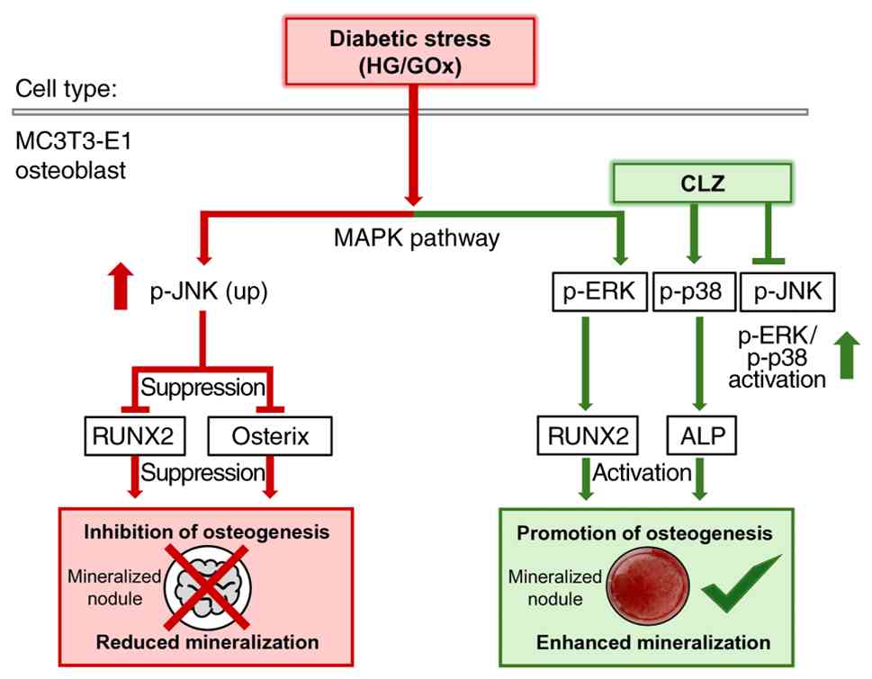 | Figure 5Schematic illustration of the effects
of CLZ on osteogenesis under HG/GOx-induced oxidative stress.
HG/GOx disrupts MAPK signaling by suppressing p-p38 and p-ERK while
activating p-JNK, accompanied by suppression of RUNX2 and Osterix
and reduced mineralization. CLZ modulates MAPK signaling,
accompanied by restoration of RUNX2 expression, ALP activity and
mineralization in MC3T3-E1 cells. This schematic summarizes the
proposed mechanism based on the findings of the present study.
Solid arrows indicate activation and T-shaped lines indicate
inhibition. Red indicates HG/GOx-induced detrimental changes,
whereas green indicates CLZ-associated restorative changes. ALP,
alkaline phosphatase; CLZ, cilostazol; GOx, glucose oxidase; HG,
high glucose; MAPK, mitogen-activated protein kinase; p-,
phosphorylated; RUNX2, Runt-related transcription factor 2. |
Discussion
The effects of CLZ on osteoblast dysfunction and the
potential underlying molecular mechanisms were investigated in
MC3T3-E1 cells under HG/GOx-induced diabetic-like conditions.
Excessive ROS production contributes to osteoblast dysfunction and
impaired bone formation in diabetic osteopathy (25). In the present study, CLZ restored
the expression of osteogenic markers suppressed under
HG/GOx-induced stress, suggesting that CLZ may contribute to the
recovery of osteoblast function under diabetic-like conditions. CLZ
increased the expression of key osteogenic markers, including BMP-2
and BMP-7, along with the master transcription factors RUNX2 and
Osterix. BMP-2 and BMP-7 trigger osteogenesis by activating
Smad-dependent and -independent signaling pathways, directly
enhancing RUNX2 transcriptional activity (26). Furthermore, the recovery of RUNX2
and Osterix, which regulate downstream genes including ALP and
COL1A1, suggests that CLZ may support osteoblastic differentiation
(12). The simultaneous increase
in these factors indicates that CLZ may contribute to the
restoration of the molecular network involved in osteoblast
differentiation under diabetic-like conditions.
The present findings suggested that the protective
effects of CLZ are associated with modulation of MAPK signaling
pathways. Previous studies have shown that p38 MAPK is essential
for late osteoblast maturation, while ERK1/2 signaling regulates
early osteoblast commitment and proliferation (13,14).
By contrast, excessive JNK activation under oxidative stress has
been reported to be associated with osteoblast dysfunction and
apoptosis (15). In the present
study, CLZ restored p38 and ERK phosphorylation while attenuating
JNK activation, suggesting that modulation of MAPK signaling may
contribute to the restoration of osteogenic function under
diabetic-like conditions. However, the precise upstream mechanism
by which CLZ regulates MAPK phosphorylation remains to be fully
elucidated. It has been demonstrated that CLZ exerts antioxidant
and anti-inflammatory effects through phosphodiesterase 3
inhibition and the subsequent elevation of intracellular cAMP
levels (27). These protective
actions, including the suppression of mitochondria-dependent
apoptotic signaling and activation of ERK1/2 and p38 MAPK pathways
in endothelial cells, have been previously reported and may be
associated with the MAPK signaling changes observed in the present
study (28). Such modulation of
MAPK signaling, together with the attenuation of JNK activation
observed in the present study, may ultimately facilitate the
recovery of osteogenic gene expression and osteoblast function
under diabetic-like conditions.
Moreover, the OPG/RANKL axis is important in
regulating the bone formation-resorption imbalance in diabetic
osteopathy (29). A low OPG/RANKL
ratio, a hallmark of bone loss in patients with diabetes, often
promotes osteoclastogenesis (30).
By increasing OPG expression and decreasing RANKL expression, CLZ
may support osteoblast function and contribute to the regulation of
bone remodeling under diabetic-like conditions.
Despite these results, several limitations should be
acknowledged. First, although changes in MAPK phosphorylation were
observed, the present study did not directly evaluate intracellular
oxidative stress using assays measuring ROS, superoxide dismutase,
malondialdehyde and glutathione peroxidase levels. Therefore,
further studies incorporating these oxidative stress-related
analyses, as well as specific MAPK inhibitors or activators, are
required to determine whether CLZ directly modulates MAPK signaling
or exerts its protective effects indirectly through the attenuation
of oxidative stress. Second, although JNK signaling has been
implicated in oxidative stress-induced apoptosis, the present study
did not directly assess osteoblast apoptosis using
apoptosis-specific assays, such as TUNEL staining, Annexin V-FITC
flow cytometry or the expression of apoptosis-related proteins,
including Bax, Bcl-2 and cleaved caspase-3. Therefore, whether CLZ
influences JNK-mediated apoptosis remains to be determined through
additional apoptosis-specific assays. Third, although CLZ increased
OPG expression and decreased RANKL expression, the present study
did not directly evaluate osteoclast differentiation or function.
Therefore, additional studies using tartrate-resistant acid
phosphatase staining, osteoclast functional assays or
osteoblast-osteoclast co-culture models are warranted to determine
whether these molecular changes translate into functional
regulation of bone remodeling. Finally, the present study was
conducted using a single osteoblastic cell line (MC3T3-E1), which
limits the direct clinical translation of the findings. Therefore,
further validation in diabetic animal models and clinical studies
is required to determine the potential relevance of CLZ in
vivo.
In conclusion, CLZ may ameliorate HG/GOx-induced
osteoblast dysfunction by modulating MAPK phosphorylation and
altering OPG and RANKL expression under diabetic-like conditions.
These findings provide insight into the effects of CLZ on
osteoblast function under diabetic-like conditions and set a basis
for future in vivo studies.
Acknowledgements
Not applicable.
Funding
Funding: The present research was supported by the Basic Science
Research Program through the National Research Foundation of Korea
funded by the Ministry of Education (grant no.
NRF-2022R1I1A1A01065439). The present study was also supported by
Wonkwang University in 2026 (grant no. not applicable).
Availability of data and materials
The data generated in the present study may be
requested from the corresponding author.
Authors' contributions
JEK conceived and designed the study, performed the
experiments, analyzed and interpreted the data and drafted the
manuscript. HP supervised the study, contributed to data
interpretation and critically revised the manuscript for important
intellectual content. JEK and HP confirm the authenticity of all
the raw data. Both authors read and approved the final manuscript
and agree to be accountable for all aspects of the work.
Ethics approval and consent to
participate
Not applicable.
Patient consent for publication
Not applicable.
Competing interests
The authors declare that they have no competing
interests.
Use of artificial intelligence tools
During the preparation of this work, artificial
intelligence tools (GPT-5.5; OpenAI) were used to improve the
readability and language of the manuscript, and subsequently, the
authors revised and edited the content produced by the artificial
intelligence tools as necessary, taking full responsibility for the
ultimate content of the present manuscript.
References
|
1
|
Hofbauer LC, Busse B, Eastell R, Ferrari
S, Frost M, Müller R, Burden AM, Rivadeneira F, Napoli N and Rauner
M: Bone fragility in diabetes: novel concepts and clinical
implications. Lancet Diabetes Endocrinol. 10:207–220.
2022.PubMed/NCBI View Article : Google Scholar
|
|
2
|
Napoli N, Chandran M, Pierroz DD,
Abrahamsen B, Schwartz AV and Ferrari SL: IOF Bone and Diabetes
Working Group. Mechanisms of diabetes mellitus-induced bone
fragility. Nat Rev Endocrinol. 13:208–219. 2017.PubMed/NCBI View Article : Google Scholar
|
|
3
|
Janghorbani M, Van Dam RM, Willett WC and
Hu FB: Systematic review of type 1 and type 2 diabetes mellitus and
risk of fracture. Am J Epidemiol. 166:495–505. 2007.PubMed/NCBI View Article : Google Scholar
|
|
4
|
Vestergaard P: Discrepancies in bone
mineral density and fracture risk in patients with type 1 and type
2 diabetes-a meta-analysis. Osteoporos Int. 18:427–444.
2007.PubMed/NCBI View Article : Google Scholar
|
|
5
|
Bai J, Gao Q, Wang C and Dai J: Diabetes
mellitus and risk of low-energy fracture: A meta-analysis. Aging
Clin Exp Res. 32:2173–2186. 2020.PubMed/NCBI View Article : Google Scholar
|
|
6
|
Oei L, Zillikens MC, Dehghan A, Buitendijk
GHS, Castaño-Betancourt MC, Estrada K, Stolk L, Oei EH, van Meurs
JB, Janssen JA, et al: High bone mineral density and fracture risk
in type 2 diabetes as skeletal complications of inadequate glucose
control: The Rotterdam study. Diabetes Care. 36:1619–1628.
2013.PubMed/NCBI View Article : Google Scholar
|
|
7
|
Yamamoto M and Sugimoto T: Advanced
glycation end products, diabetes, and bone strength. Curr
Osteoporos Rep. 14:320–326. 2016.PubMed/NCBI View Article : Google Scholar
|
|
8
|
Saito M and Marumo K: Collagen cross-links
as a determinant of bone quality: A possible explanation for bone
fragility in aging, osteoporosis, and diabetes mellitus. Osteoporos
Int. 21:195–214. 2010.PubMed/NCBI View Article : Google Scholar
|
|
9
|
Riegger J, Schoppa A, Ruths L,
Haffner-Luntzer M and Ignatius A: Oxidative stress as a key
modulator of cell fate decision in osteoarthritis and osteoporosis:
A narrative review. Cell Mol Biol Lett. 28(76)2023.PubMed/NCBI View Article : Google Scholar
|
|
10
|
Takanche JS, Kim JE, Han SH and Yi HK:
Effect of gomisin A on osteoblast differentiation in high
glucose-mediated oxidative stress. Phytomedicine.
66(153107)2020.PubMed/NCBI View Article : Google Scholar
|
|
11
|
Chen G, Deng C and Li YP: TGF-β and BMP
signaling in osteoblast differentiation and bone formation. Int J
Biol Sci. 8:272–288. 2012.PubMed/NCBI View Article : Google Scholar
|
|
12
|
Komori T: Functions of BMPs, Runx2, and
osterix in the development of bone and cartilage. Nihon Rinsho.
63:1671–1677. 2005.PubMed/NCBI
|
|
13
|
Johnson GL and Lapadat R:
Mitogen-activated protein kinase pathways mediated by ERK, JNK, and
p38 protein kinases. Science. 298:1911–1912. 2002.PubMed/NCBI View Article : Google Scholar
|
|
14
|
Ge C, Xiao G, Jiang D and Franceschi RT:
Critical role of the extracellular signal-regulated kinase-MAPK
pathway in osteoblast differentiation and skeletal development. J
Cell Biol. 176:709–718. 2007.PubMed/NCBI View Article : Google Scholar
|
|
15
|
Xu R, Zhang C, Shin DY, Kim JM, Lalani S,
Li N, Yang YS, Liu Y, Eiseman M, Davis RJ, et al: c-Jun N-terminal
kinases (JNKs) are critical mediators of osteoblast activity in
vivo. J Bone Miner Res. 32:1811–1815. 2017.PubMed/NCBI View Article : Google Scholar
|
|
16
|
Robless P, Mikhailidis DP and Stansby GP:
Cilostazol for peripheral arterial disease. Cochrane Database Syst
Rev: CD003748, 2008.
|
|
17
|
Kawabe T, Ichimura A, Yasue T, Li J,
Ishikawa H, Kim GE, Nagatomo H, Minamino N, Ueda Y, Ito H, et al:
Phosphodiesterase 3 inhibitors boost bone outgrowth. Br J
Pharmacol. 182:4327–4342. 2025.PubMed/NCBI View Article : Google Scholar
|
|
18
|
Lee YJ, Shu MS, Kim JY, Kim YH, Sim KH,
Sung WJ and Eun JR: Cilostazol protects hepatocytes against
alcohol-induced apoptosis via activation of AMPK pathway. PLoS One.
14(e0211415)2019.PubMed/NCBI View Article : Google Scholar
|
|
19
|
Park SY, Lee SW, Shin HK, Chung WT, Lee
WS, Rhim BY, Hong KW and Kim CD: Cilostazol enhances apoptosis of
synovial cells from rheumatoid arthritis patients with inhibition
of cytokine formation via Nrf2-linked heme oxygenase 1 induction.
Arthritis Rheum. 62:732–741. 2010.PubMed/NCBI View Article : Google Scholar
|
|
20
|
Ota H, Eto M, Kano MR, Ogawa S, Iijima K,
Akishita M and Ouchi Y: Cilostazol inhibits oxidative
stress-induced premature senescence via upregulation of Sirt1 in
human endothelial cells. Arterioscler Thromb Vasc Biol.
28:1634–1639. 2008.PubMed/NCBI View Article : Google Scholar
|
|
21
|
Yang Y, Li R, Wang P, Zhao Y, Li J, Jiao J
and Zheng H: Osteoking prevents bone loss and enhances osteoblastic
bone formation by modulating the AGEs/IGF-1/β-catenin/OPG pathway
in type 2 diabetic db/db mice. Cell Biol Int. 48:1507–1519.
2024.PubMed/NCBI View Article : Google Scholar
|
|
22
|
Mitra I, Potes MA, Shafi M, Tilton M,
Elder BD and Lu L: Enzyme-delivery metal-organic framework
composite coatings for restoration of hyperglycemia-damaged
osteoblast differentiation. Biomater Adv.
166(214055)2025.PubMed/NCBI View Article : Google Scholar
|
|
23
|
Etli M, Karahan O, Akkaya Ö and Savaş HB:
Cilostazol induces angiogenesis and regulates oxidative stress in a
dose-dependent manner: A chorioallantoic membrane study. Turk Gogus
Kalp Damar Cerrahisi Derg. 29:449–456. 2021.PubMed/NCBI View Article : Google Scholar
|
|
24
|
Tseng SY, Chang HY, Li YH and Chao TH:
Effects of cilostazol on angiogenesis in diabetes through
adiponectin/adiponectin receptors/sirtuin1 signaling pathway. Int J
Mol Sci. 23(14839)2022.PubMed/NCBI View Article : Google Scholar
|
|
25
|
Hamada Y, Fujii H and Fukagawa M: Role of
oxidative stress in diabetic bone disorder. Bone. 45 (Suppl
1):S35–S38. 2009.PubMed/NCBI View Article : Google Scholar
|
|
26
|
Wang L, Li JY, Zhang XZ, Liu L, Wan ZM, Li
RX and Guo Y: Involvement of p38MAPK/NF-κB signaling pathways in
osteoblasts differentiation in response to mechanical stretch. Ann
Biomed Eng. 40:1884–1894. 2012.PubMed/NCBI View Article : Google Scholar
|
|
27
|
Kherallah RY, Khawaja M, Olson M,
Angiolillo D and Birnbaum Y: Cilostazol: A review of basic
mechanisms and clinical uses. Cardiovasc Drugs Ther. 36:777–792.
2022.PubMed/NCBI View Article : Google Scholar
|
|
28
|
Lim JH, Woo JS and Shin YW: Cilostazol
protects endothelial cells against lipopolysaccharide-induced
apoptosis through ERK1/2- and P38 MAPK-dependent pathways. Korean J
Intern Med. 24:113–122. 2009.PubMed/NCBI View Article : Google Scholar
|
|
29
|
Kostenuik PJ: Osteoprotegerin and RANKL
regulate bone resorption, density, geometry and strength. Curr Opin
Pharmacol. 5:618–625. 2005.PubMed/NCBI View Article : Google Scholar
|
|
30
|
de Amorim FPLG, Ornelas SS, Diniz SF,
Batista AC and da Silva TA: Imbalance of RANK, RANKL and OPG
expression during tibial fracture repair in diabetic rats. J Mol
Histol. 39:401–408. 2008.PubMed/NCBI View Article : Google Scholar
|