Introduction
Intraductal oncocytic papillary neoplasm (IOPN) is a
rare, genetically distinct intraductal epithelial neoplasm of the
pancreas with malignant potential. Under previous classification
systems, IOPN was grouped as a morphological variant of intraductal
papillary mucinous neoplasm (IPMN); however, the 2019 World Health
Organization (WHO) Classification of Digestive System Tumors
reclassified IOPN as an independent diagnostic entity due to
profound discrepancies in histomorphology and molecular signatures
(1). Epidemiological analysis has
indicated that IOPN accounts for only 1-8% of all pancreatic
IPMN-spectrum lesions (1). Adsay
et al (2) first published a
series of 11 cases of pancreatic IOPN in 1996, noting clinical and
imaging phenotypes that overlapped with conventional IPMN.
Subsequent genomic research has identified core molecular
differences between these conditions: Unlike IPMN, which frequently
harbors KRAS and GNAS driver mutations, pancreatic
IOPN is characterized by recurrent protein kinase cAMP-activated
catalytic subunit alpha (PRKACA) or PRKACB gene
fusions, with minimal mucin secretion representing a key
morphological distinction (3,4).
Accumulating evidence from clinical series indicates
that the long-term prognosis of IOPN is generally favorable
(4-6).
Nevertheless, precise preoperative radiological diagnosis is
critical for risk stratification and individualized surgical
planning. The present study reported a case of surgically resected
pancreatic IOPN that initially manifested as recurrent pancreatitis
and described its evolution on CT and MRI findings acquired over a
2-year period. The recent literature was also reviewed to improve
multidisciplinary recognition of this rare pancreatic neoplasm.
Case report
Case presentation
A 54-year-old female patient presented to the
Ningcheng County Central Hospital (Chifeng, China) in March 2024
for further diagnosis and treatment of recurrent, spontaneous,
distending upper abdominal pain without shoulder-back radiation,
that had been reported for the past 3 months. The patient had been
previously admitted to Ningcheng County Central Hospital (Chifeng,
China) multiple times for acute pancreatitis. An association
between the recurrent pancreatitis and a cystic intraductal lesion
was suspected, although a definite causal relationship could not be
established.
The medical history of the patient did not include
hypertension, coronary artery disease, type 2 diabetes, viral
hepatitis, tuberculosis or other infectious disorders. Furthermore,
the patient had no prior abdominal surgery, trauma, blood
transfusion, known drug allergies, occupational heavy metal
exposure, familial pancreatic neoplasia or hereditary digestive
tract tumors. The vaccination records of the patient were
unavailable.
Physical examination
Physical examination revealed tenderness on
palpation of the left upper quadrant, without rebound tenderness or
muscular guarding. The liver and spleen were non-palpable below the
costal margin. Abdominal percussion was tympanic, shifting dullness
was absent, hepatic dullness was preserved and there was no
tenderness to percussion over the liver, spleen or bilateral
kidneys. Bowel sounds were normal, with no succussion splash or
other signs indicating an air-fluid level. Bilateral lower limb
edema or focal neurological deficits were not observed.
Ultrasound and laboratory
findings
Abdominal ultrasound revealed a mixed cystic-solid
pancreatic mass, a separate cyst in the pancreatic tail, dilatation
of the segmental pancreatic duct and concurrent hepatic steatosis.
Serum biochemistry and tumor marker test results exhibited a mildly
elevated CA19-9 level of 40.27 U/ml (reference range: 0.00-27.00
U/ml). CEA, serum amylase, lipase, liver function panel,
total/direct bilirubin and systemic inflammatory markers (including
CRP and white blood cell count) were also assessed during episodes
of pancreatitis. Serum amylase was reported to be 1504.80 U/l
(reference range: 35.00-135.00 U/l) and lipase was reported to be
2614.30 U/l (reference range: 13.00-60.00 U/l). All other
laboratory parameters were within their normal limits.
Sequential contrast-enhanced CT
imaging
All CT scans were performed using a multi-detector
row CT with uniform slice thickness, non-ionic intravenous contrast
medium and four-phase acquisition (unenhanced, arterial, portal
venous and delayed equilibrium phase). Initial unenhanced and
contrast-enhanced abdominal CT was performed in March 2022, during
the patient's first hospitalization for acute pancreatitis. It
revealed a well-circumscribed round hypoattenuating lesion within
the pancreatic body, measuring 1.7x2.0x2.5 cm. An internal solid
mural nodule measuring 1.1x0.7x1.1 cm was also present.
Perilesional peripancreatic inflammatory fat stranding was visible.
The main pancreatic duct exhibited no dilatation at this time
(Fig. 1).
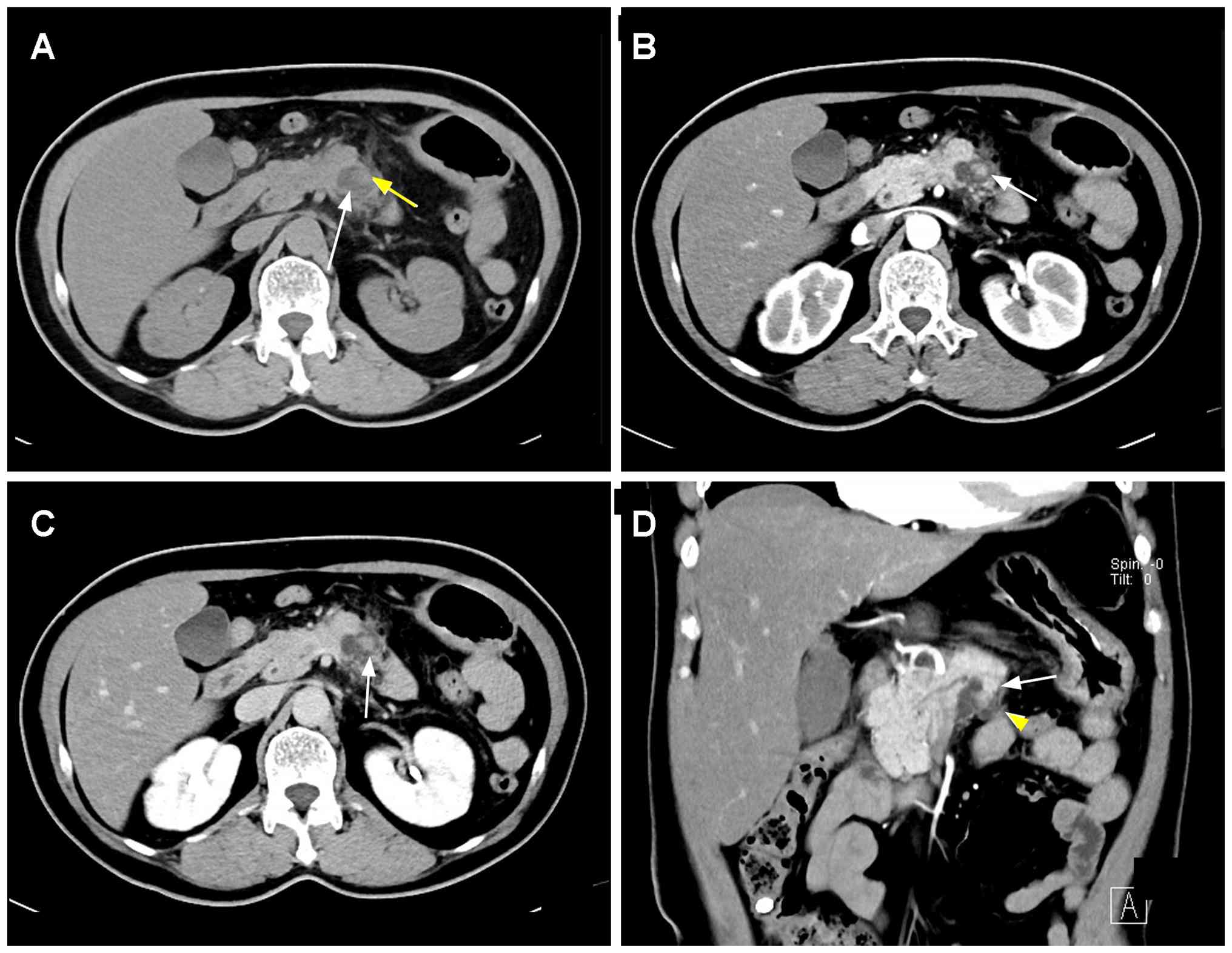
Follow-up contrast-enhanced CT performed in March
2024 revealed that the primary lesion had enlarged to 2.2x2.5x3.5
cm, forming an exophytic contour inferior to the pancreatic
parenchyma. The intralesional mural nodule had expanded to
1.0x1.0x2.1 cm. Segmental dilatation of the pancreatic body-tail
duct was also observed, with a maximum diameter of 0.7 cm.
Quantitative attenuation measurements determined that the mural
nodule demonstrated intrinsic contrast enhancement across all
post-contrast phases, but remained relatively hypoattenuating
compared with the adjacent normal pancreatic tissue in the arterial
and venous phases (Fig. 2).
Communication between the cystic lesion and the dilated pancreatic
duct was subsequently verified through magnetic resonance
cholangiopancreatography (Fig.
3).
MR plain and contrast-enhanced imaging
(Fig. 3)
Pancreatic MRI with gadolinium-based contrast and
dedicated MRCP sequences was performed, including T1-weighted
imaging (T1WI), T2WI, diffusion-weighted imaging (DWI) and apparent
diffusion coefficient (ADC) mapping. The pancreatic body lesion
appeared as a well-demarcated multicystic mass with prolonged T1/T2
signal intensity and focal exophytic bulging. The internal mural
nodules showed mild DWI hyperintensity with limited ADC diffusion
restriction. Direct communication between the lesion and the
segmentally dilated pancreatic body-tail duct was clearly
visualized on MRCP. The enhancement patterns of the mural nodule
were consistent with the CT findings. Peripancreatic inflammatory
fat stranding secondary to pancreatitis was clearly distinguished
from neoplastic invasion of the peripancreatic fat.
Surgical procedure, gross pathology
and histopathological diagnosis
The patient underwent distal pancreatectomy with
splenectomy. Microscopic examination of the resection margins was
negative for neoplastic epithelium. A number of peripancreatic
tissue samples were excised intraoperatively and processed for
histological examination; no dedicated lymph node dissection was
performed and no metastatic carcinoma was identified in the sampled
peripancreatic tissues. The resected specimen measured 9.5x3.0x2.5
cm, with a maximum lesion diameter of 3.5 cm. No gross imaging of
the resected specimen was available. Light microscopy revealed
complex arborizing papillary structures with central fibrovascular
cores, which were lined by oncocytic cuboidal and columnar
epithelial cells. These tumor cells exhibited abundant granular
eosinophilic cytoplasm, enlarged round nuclei and prominent
nucleoli, presenting typical architectural and cytological features
of high-grade dysplasia (Fig. 4).
No invasive neoplastic extension penetrating the pancreatic duct
wall was identified upon comprehensive histological
examination.
Histopathological processing
The resected specimen was fixed in 10% neutral
buffered formalin at room temperature for 24 h. Fixed tissues were
embedded in paraffin and serial sections of 4-µm thickness were
cut. The sections were dewaxed in xylene, rehydrated through a
graded ethanol series and stained with Harris hematoxylin at room
temperature for 5 min for nuclear counterstaining, followed by
differentiation in acid alcohol, bluing in running tap water, and
counterstaining with eosin Y at room temperature for 1-2 min.
Stained sections were dehydrated through graded ethanols, cleared
in xylene and mounted in neutral balsam (7).
The final standardized pathological diagnosis was
pancreatic IOPN with high-grade dysplasia, without associated
invasive adenocarcinoma. Immunohistochemical staining and molecular
testing were not performed in the present case. The diagnosis of
IOPN was rendered according to the original pathology report and
established on the basis of characteristic histological morphology
on H&E staining, including complex arborizing papillae with
fibrovascular cores lined by oncocytic epithelial cells with
abundant granular eosinophilic cytoplasm, enlarged round nuclei and
prominent nucleoli, consistent with the diagnostic criteria
established by the 2019 WHO Classification of Digestive System
Tumors (3). The absence of
invasive carcinoma was determined via thorough sampling of the
lesion.
Postoperative follow-up
Regular surveillance, including serum CA19-9
measurement, abdominal CT and MRCP, was scheduled every 6 months
postoperatively. No surgical complications, local recurrence or
distant metastasis occurred during the follow-up period. To date,
42 months have elapsed since surgery and the patient remains
disease-free without surgical-associated complications, local
recurrence or distant metastasis.
Discussion
IOPN is a rare intraductal pancreatic neoplasm
characterized by oncocytic epithelium that forms papillary
architectures. More than 200 cases of pancreatic and biliary IOPN
have been reported in the literature to date, with no significant
sex predilection. The reported age at onset ranges from 36 to 84
years, with a median age of 61 years (6). The clinical manifestations of IOPN
overlap substantially with conventional IPMN, including recurrent
pancreatitis, non-specific abdominal pain, palpable abdominal mass
and complete absence of clinical symptoms (8). In the present case, recurrent
pancreatitis served as the initial presentation. At present, serum
tumor markers lack diagnostic specificity for IOPN. Mild elevation
of CA19-9 and CEA have been reported in a minority of patients
(9), consistent with the mildly
elevated CA19-9 level in the patient of the present study.
IOPN may arise in any segment of the pancreas; the
pancreatic head is the most common site (67% of cases), followed by
the pancreatic body and tail. Approximately 5% of lesions involve
the entire pancreas. IOPN may be classified as main duct-type,
branch duct-type or mixed-type, analogous to IPMN classification
systems (10). The hallmark
imaging morphology of IOPN is mixed cystic-solid masses with
internal mural nodules, which are attributed to low mucin secretion
that limits diffuse pancreatic duct dilatation. Reported maximum
lesion diameters range from 1 to 15 cm, with a mean size of 5.5 cm
at diagnosis (6).
In the present case, serial imaging over a 2-year
period captured stepwise disease progression: An initially small
cystic-solid lesion with an isolated mural nodule without duct
dilatation, followed by lesion and nodule enlargement and secondary
pancreatic duct dilatation associated with high-grade dysplasia.
Prior research has confirmed mural nodules ≥5 mm in maximal
diameter as high-risk imaging signs associated with underlying
high-grade dysplasia or invasive carcinoma (11). However, it must be emphasized that
enhancing mural nodules are not pathognomonic for IOPN, as this
feature may occur in various cystic pancreatic neoplasms (4).
Regarding differential diagnosis, the following may
be considered: i) IPMN/mucinous intraductal papillary carcinoma
(IPMC): Clinical symptoms overlap broadly with those of IOPN,
although elevated CA19-9 and CEA levels are more pronounced in
malignant IPMC (12). Imaging
typically shows diffuse pancreatic duct dilatation secondary to
abundant mucin production, with smaller cystic locules and higher
rates of peripancreatic invasion, postoperative recurrence and
inferior overall survival compared with IOPN. International
consensus guidelines (1) stratify
IPMN risk based on mural nodule size and duct caliber. ii)
Intraductal tubulopapillary neoplasm (ITPN): Similar to IOPN, ITPN
produces minimal mucin secretion. However, ITPN is defined by
tubular rather than papillary epithelial proliferation. The imaging
hallmarks of ITPN include the two-tone duct sign and the wine-cork
filling defect sign on MRCP (13,14).
iii) Other entities in the differential diagnosis include mucinous
cystic neoplasm, serous cystic neoplasm, solid pseudopapillary
neoplasm and cystic pancreatic neuroendocrine neoplasm (5,11,12).
Accurate diagnosis requires comprehensive integration of the lesion
location, duct communication status, intralesional mucin volume,
internal architecture, mural nodule enhancement kinetics,
cytological sampling, histomorphology, and immunophenotype and
molecular profiles, rather than relying solely on duct dilatation
or mural nodule presence.
IOPN is recognized to have malignant potential, with
29% of resected cases complicated by concurrent invasive
adenocarcinoma. Surgical resection is indicated for all
radiologically resectable IOPN lesions with high-risk imaging
features. Operative planning must be individualized based on the
tumor location, lesion extension and patient functional status.
Pancreaticoduodenectomy (Whipple procedure) is typically performed
for pancreatic head IOPN, whereas distal pancreatectomy (with or
without splenectomy) is performed for body/tail lesions. Routine
adjuvant radiotherapy is not universally recommended for cases of
IOPN-associated invasive carcinoma. Decisions regarding adjuvant
management for patients with invasive disease should be
individualized and determined via multidisciplinary tumor board
discussion (5). Prognosis differs
significantly between non-invasive IOPN and IOPN with invasive
adenocarcinoma. Non-invasive IOPN is associated with excellent
long-term survival. A previous study (15) reported a 100% 5-year overall
survival rate and a 66% 5-year disease-free survival rate for
invasive IOPN, with 46% local recurrence within 10 years
post-resection. Repeat salvage surgical resection may be required
to maintain favorable long-term outcomes (15). Although the overall prognosis
remains favorable, invasive IOPN exhibits distinct biological
features that cannot be uniformly summarized as indolent without
stratification (5).
Several limitations of the present case report
should be acknowledged. First, immunohistochemical staining was not
performed, which precludes formal confirmation of oncocytic and
pancreatobiliary differentiation and limits direct immunophenotypic
comparison with previously reported IOPN series (4,14,15).
Although the histological morphology was sufficiently
characteristic to establish the diagnosis on H&E staining
alone, the addition of immunohistochemical markers (such as
hepatocyte paraffin 1, CD117, mucin 1 and cytokeratin 7) would have
strengthened diagnostic confidence, particularly in distinguishing
IOPN from its mimics. Second, molecular testing for PRKACA
or PRKACB gene fusions was not performed; these
rearrangements are highly characteristic of IOPN and could have
provided molecular confirmation. Third, the postoperative follow-up
duration was relatively limited and longer surveillance would be
needed to fully assess long-term recurrence risk. These limitations
should be considered when interpreting the findings and comparing
them with the published literature.
In conclusion, IOPN is a distinct pancreatic
intraductal epithelial neoplasm with malignant potential but a
generally favorable long-term prognosis. Nevertheless, preoperative
radiological diagnosis remains challenging. The patient of the
present study initially presented with recurrent pancreatitis.
Serial imaging over 2 years documented progressive enlargement of a
mixed cystic-solid lesion and an internal mural nodule, new-onset
pancreatic duct dilatation, persistent absence of peripancreatic
neoplastic invasion and mural nodules showing intrinsic contrast
enhancement with relative hypoattenuation/hypointensity compared
with normal pancreatic parenchyma on CT and MRI. Multidisciplinary
integration of clinical history, standardized cross-sectional
imaging, endoscopic ultrasound cytology, histopathology and
molecular testing is necessary for accurate preoperative
differentiation from other pancreatic cystic neoplasms and to
inform individualized surgical management.
Acknowledgements
Not applicable.
Funding
Funding: No funding was received.
Availability of data and materials
The data generated in the present study are included
in the figures and/or tables of this article.
Authors' contributions
YL was responsible for conceptualization, conducting
the investigation, data curation and writing the original draft. LZ
conducted the investigation, radiological and pathological review,
as well as being responsible for visualization and writing,
reviewing and editing the manuscript. HY was responsible for the
resources used, conducting patient follow-up, performing formal
analysis and writing, reviewing and editing the manuscript. LW
acquired and interpreted the CT and MRI data, radiological
measurements and imaging-pathological correlation. LW was also
responsible for verification of the raw imaging data, providing
supervision, conducting project administration and writing,
reviewing and editing the manuscript. All authors read and approved
the final version of the manuscript. YL and LZ confirm the
authenticity of all the raw data.
Ethics approval and consent to
participate
The present single retrospective case report was
conducted at Ningcheng County Central Hospital (Chifeng, China). As
the present study involved only one patient and used de-identified
retrospective clinical data, formal ethics committee approval was
not required. The present case was reviewed by the Ethics Committee
of Ningcheng County Central Hospital (Chifeng, China), which
confirmed that the study was exempt from formal ethical approval
and issued a letter confirming the authenticity of the patient's
informed consent statement. Written informed consent for
publication of all de-identified clinical data, laboratory results,
and imaging figures was obtained from the patient prior to
manuscript submission. All direct personal identifiers have been
removed from the manuscript and supplementary files. The present
manuscript is classified as a single-case case report rather than
original clinical research involving multiple study
participants.
Patient consent for publication
The patient consented to the publication of case
information and medical images.
Competing interests
The authors declare that they have no competing
interests.
References
|
1
|
van Huijgevoort NCM, Del Chiaro M,
Wolfgang CL, van Hooft JE and Besselink MG: Diagnosis and
management of pancreatic cystic neoplasms: Current evidence and
guidelines. Nat Rev Gastroenterol Hepatol. 16:676–689.
2019.PubMed/NCBI View Article : Google Scholar
|
|
2
|
Adsay NV, Longnecker DS and Klimstra DS
and Klimstra DS: Intraductal oncocytic papillary neoplasms of the
pancreas. Am J Surg Pathol. 20:980–994. 1996.PubMed/NCBI View Article : Google Scholar
|
|
3
|
Basturk O, Tan M, Bhanot U, Allen P, Adsay
V, Scott SN, Shah R, Berger MF, Askan G, Dikoglu E, et al: The
oncocytic subtype is genetically distinct from other pancreatic
intraductal papillary mucinous neoplasm subtypes. Mod Pathol.
29:1058–1069. 2016.PubMed/NCBI View Article : Google Scholar
|
|
4
|
Paolino G, Basturk O, Esposito I, Hong SM,
Brosens LA, Tarcan Z, Wood LD, Gkountakos A, Omori Y, Mattiolo P,
et al: Comprehensive characterization of intraductal oncocytic
papillary neoplasm of the pancreas: A systematic and critical
review. Mod Pathol. 37(100554)2024.PubMed/NCBI View Article : Google Scholar
|
|
5
|
Lucocq J, Haugk B, Joseph N, Hawkyard J,
White S, Mownah O, Menon K, Furukawa T, Inoue Y, Hirose Y, et al:
Invasive intraductal oncocytic papillary neoplasms (IOPN) and
adenocarcimoma arising from intraductal papillary mucinous
neoplasms (A-IPMN) of the pancreas: Comparative analysis of
clinicopathological features, patterns of recurrence and survival:
A multicentre study. HPB (Oxford). 26:1421–1428. 2024.PubMed/NCBI View Article : Google Scholar
|
|
6
|
Wang YZ, Lu J, Jiang BL and Guo JC:
Intraductal oncocytic papillary neoplasm of pancreas:A systematic
review. Pancreatology. 19:858–865. 2019.PubMed/NCBI View Article : Google Scholar
|
|
7
|
Fischer AH, Jacobson KA, Rose J and Pyles
R: Hematoxylin and eosin staining of tissue and cell sections. CSH
Protoc. 2008(pdb.prot4986)2008.PubMed/NCBI View Article : Google Scholar
|
|
8
|
Liszka L, Pajak J, Zielinska-Pajak E,
Krzych L, Gołka D, Mrowiec S and Lampe P: Intraductal oncocytic
papillary neoplasms of the pancreas and bile ducts: A destematic
survey of the literature. J Hepatobiliary Pancreat Sci. 17:246–261.
2010.PubMed/NCBI View Article : Google Scholar
|
|
9
|
Wu Y, Chang XY and Chen J: Clinical and
pathological features of intraductal oncocytic papillary neoplams
of the pancreas. Zhonghua Bing Li Xue Za Zhi. 48:829–832.
2019.PubMed/NCBI View Article : Google Scholar : (In Chinese).
|
|
10
|
Chunxue Y, Haimin X and Qian D:
Clinicopathological features of pancreatic intraductal oncocytic
papillary neoplasm: A report of 5 cases. J Surg Concepts Pract.
27:52–57. 2022.
|
|
11
|
Fang X, Jiang H, Wang L, Shao C, Lu J and
Bian Y: Analysis of clinical and imaging features of 12 cases of
pancreatic intraductal oncocytic papillary neoplasm. Chin J Dig.
12:458–463. 2022.
|
|
12
|
Nakaya M, Nakai Y, Takahashi M, Fukukura
Y, Sato K, Kameda A, Tashiro Y, Kageyama S, Sofue K, Nakano T, et
al: Intraductal nococytic papillary neoplasm the pancreas: Clinical
and radiological features compared to those of intraductal
papillary mucinous neoplasm. Abdom Radiol (NY). 48:2483–2493.
2023.PubMed/NCBI View Article : Google Scholar
|
|
13
|
Xu F, Yun B, Hui J, et al: Intraductal
tubulopapillary neoplasm of pancreas: Imaging features correlation
with pathological findings. Chin J Radiol. 55:551–554. 2021.
|
|
14
|
Qian Y, Mo S and Chang X: Deciphering
pancreatobiliary intraductal oncocytic papillary neoplasms:
Integrative analysis of histomorphologic patterns, immunophenotypic
markers, and emerging molecular biomarkers. World J Surg Oncol.
23(333)2025.PubMed/NCBI View Article : Google Scholar
|
|
15
|
Wang T, Askan G, Adsay V, Allen P,
Jarnagin WR, Memis B, Sigel C, Seven IE, Klimstra DS and Basturk O:
Intraductal oncocytic papillary neopasmsLclinical-pathologic
characterization of 24 cases,with an emphasis on associated
invasive carcinomas. Am J Surg Pathol. 43:656–661. 2019.PubMed/NCBI View Article : Google Scholar
|