Introduction
Severe brain injury (SBI) (1) often leads to severe complications,
such as paroxysmal sympathetic hyperactivity (PSH). The clinical
features of PSH include tachycardia, hypertension, shortness of
breath, elevated body temperature, profuse sweating and postural
rigidity. These symptoms may recur frequently, causing notable
distress to patients (2,3). Diagnosis relies on the PSH-assessment
measure (PSH-AM) score (4). The
PSH-AM consists of two components: The clinical feature scale (CFS)
and the diagnostic likelihood tool (DLT). The total score is the
sum of the two component scores, and a total score >17 supports
a definitive diagnosis of PSH. Other conditions, including
epilepsy, infection, pulmonary complications, pain and medication
effects, must first be systematically excluded (5). The first episode of PSH is typically
managed with oral clonazepam and propranolol. Gabapentin,
pregabalin and other agents can be used in the maintenance period,
with treatment lasting 3 to 6 months (6). The rehabilitation programme includes
passive joint mobilisation, tilt table training and hyperbaric
oxygen therapy (HBOT), which may reduce the risk of recurrence and
improve patient prognosis when initiated (7).
Using a case report of rehabilitation treatment for
a patient with PSH following SBI, the present study describes the
diagnostic process, clinical course and multidisciplinary
management strategy. Clinical observations are summarised regarding
the temporal associations between interventions, individualised
rehabilitation and patient outcomes, while acknowledging the
inherent limitations of single-case evidence.
Case report
A 31-year-old woman was found unconscious at home in
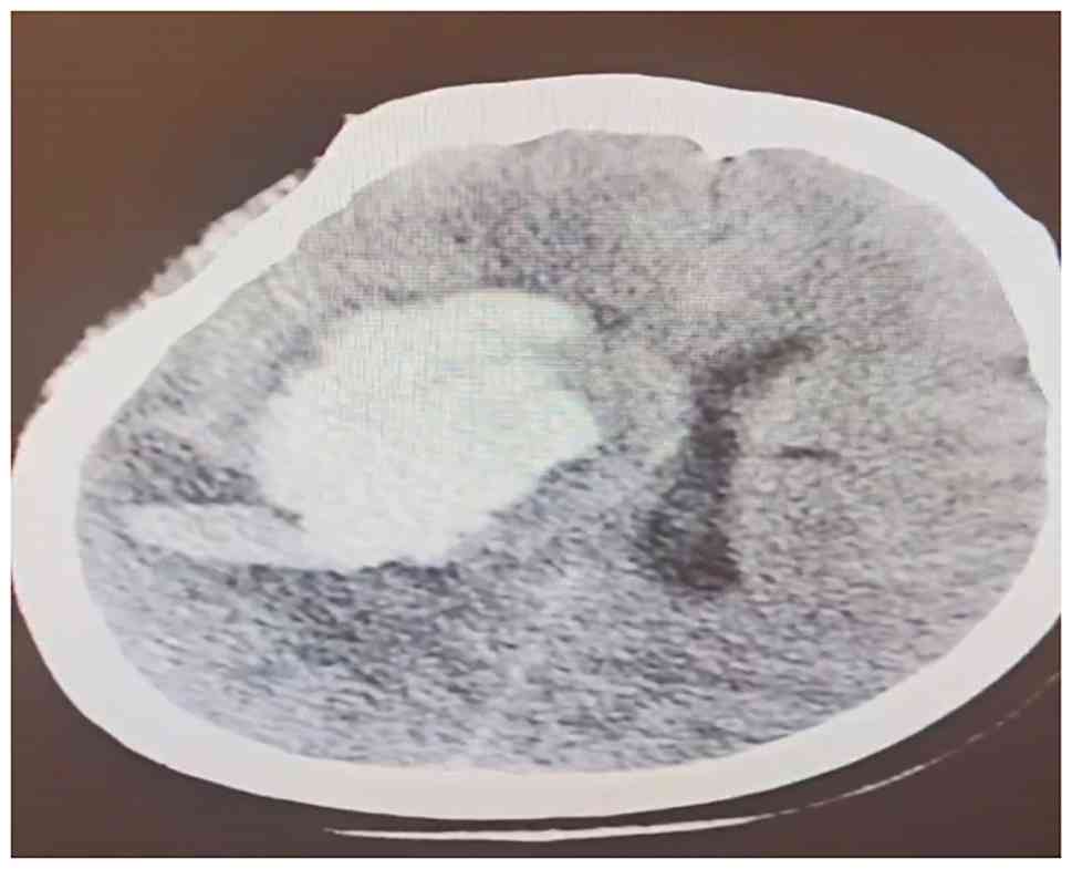
February 2024. Emergency head computed tomography (CT) revealed a
cerebral haemorrhage in the right basal ganglia (Fig. 1). The Glasgow Coma Scale (GCS)
score was E1VTM1 (total: 3 points; T indicates tracheal
intubation), meeting diagnostic criteria for SBI (8). On day 3 after admission, the patient
underwent an intracranial hematoma evacuation, decompressive
craniectomy and external ventricular drainage at the Department of
Neurosurgery, The First Affiliated Hospital, Zhejiang University
School of Medicine (Hangzhou, China). On the 27th day post-onset,
the patient suddenly developed a fever (39˚C; normal adult range,
36.0-37.2˚C), tachycardia (heart rate, 190 bpm; normal adult
resting heart rate, 60-100 bpm), hypertension (205/110 mmHg; normal
adult blood pressure, <130/80 mmHg), profuse sweating and
increased muscle tone. The attending physician systematically
excluded pulmonary oedema and severe pneumonia based on chest CT
findings, and epilepsy was excluded via electroencephalogram (EEG).
The differential diagnosis also considered and excluded infection,
medication side effects and pain as contributing causes. PSH was
diagnosed using the PSH-AM scoring system. The CFS of the patient
had a total score of 17, which was calculated as follows: Heart
rate (190 bpm; 3 points) + respiratory rate (42 breaths per min; 3
points) + systolic blood pressure (205 mmHg; 3 points) + body
temperature (39˚C; 3 points) + severe diaphoresis (3 points) +
moderate hypertonia during episodes (2 points). The DLT had a total
score of 9, which was calculated as follows: Presence of antecedent
acquired brain injury (1 point) + clinical features (fever,
tachycardia, hypertension, profuse sweating and increased muscle
tone) occurring simultaneously (1 point) + episodes were paroxysmal
in nature (1 point) + sympathetic over-reactivity to normally
non-painful stimuli (1 point) + features persisting for ≥2 weeks
post brain injury (1 point) + ≥2 episodes per day (1 point) +
medication administered to reduce sympathetic features (1 point) +
absence of parasympathetic features during episodes (1 point) +
absence of other presumed causes of the features (1 point).
Therefore, the total score of PSH-AM was 26, and a definitive
diagnosis of PSH was made. Treatment with clonazepam at 2 mg every
8 h and propranolol at 10 mg three times daily was initiated,
resulting in symptom improvement. On day 30, a PSH episode
recurred; 10 mg intravenous diazepam was administered resulting in
stabilisation. Over the subsequent weeks, PSH recurred
intermittently (~4 additional episodes). Maintenance medication was
adjusted to 2 mg clonazepam via nasogastric tube every 8 h, 10 mg
propranolol three times daily and 2.5 mg bromocriptine twice
daily.
After 5 months from the initial onset, the patient
was transferred to The First Affiliated Hospital of Zhejiang
Chinese Medical University (Zhejiang Provincial Hospital of Chinese
Medicine) for specialised care. On admission, a physical
examination revealed: i) A GCS score of E1VTM1; ii) no marked
increase in muscle tone in the extremities; and iii) Brunnstrom
stages I-I-I for the bilateral upper limbs, hands and lower limbs.
Barthel Index score is 0(9). The
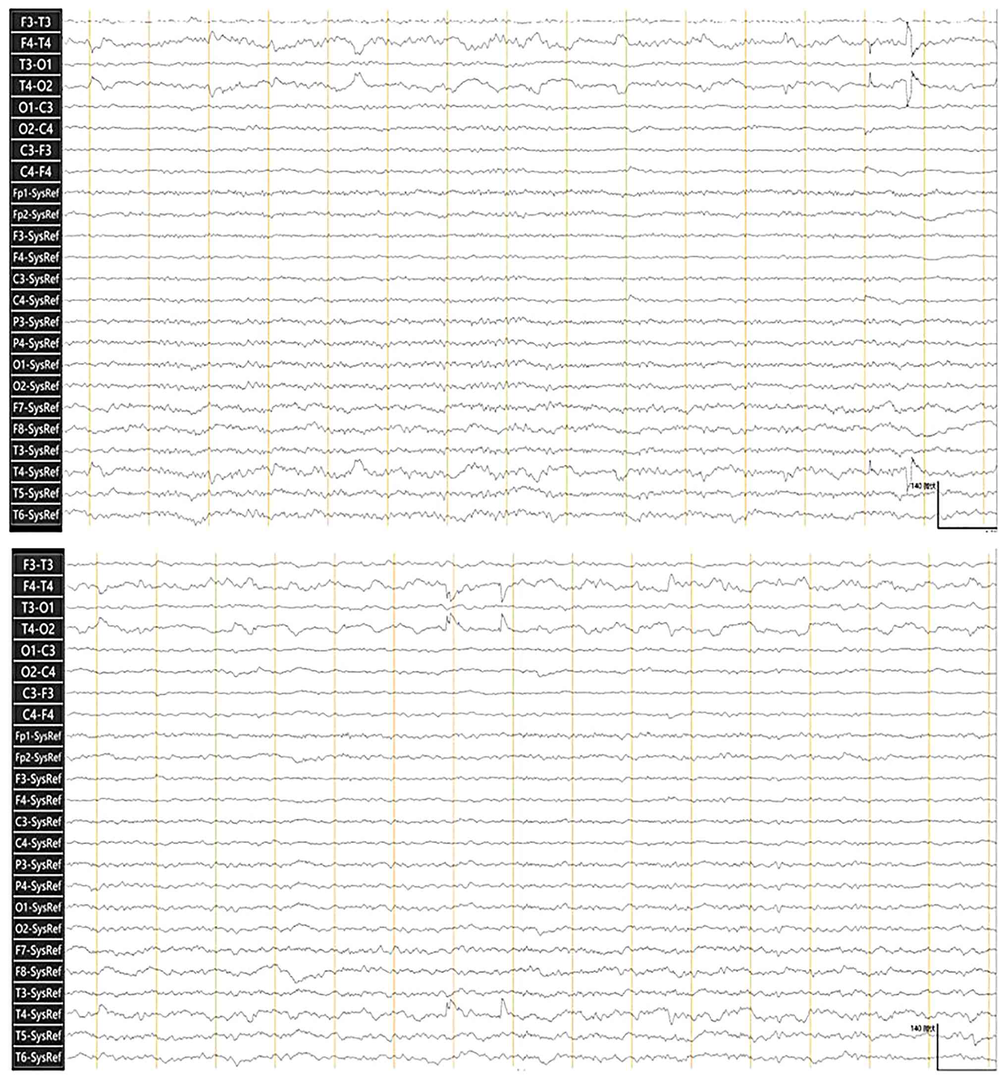
EEG (Fig. 2) indicated a severe
abnormality in brain function. Functional near-infrared
spectroscopy (fNIRS) at rest (10 min quiet-state recording;
Fig. 3) indicated an average
prefrontal cortex functional connectivity strength of 0.151. In
healthy adults, resting-state prefrontal functional connectivity
strength is typically reported at 0.40-0.70, and this low value
therefore indicates a severe decline in brain function. On the
third day of admission to The First Affiliated Hospital of Zhejiang
Chinese Medical University (Zhejiang Provincial Hospital of Chinese
Medicine), the patient experienced a PSH episode as follows: Body
temperature, 38.6˚C; heart rate, 190 bpm; blood pressure, 188/102
mmHg; limb muscle tone, grade 2; and generalised diaphoresis
lasting 11 min. Before diagnosing this episode as PSH, the
following alternative aetiologies were systematically excluded:
Infection (blood cultures and inflammatory markers reviewed),
seizures (concurrent EEG monitoring), pulmonary complications
(oxygen saturation trend and chest auscultation), pain response and
medication effects. The PSH-AM scoring system was applied: The CFS
had a total score of 16, which was calculated as follows: Heart
rate (190 beats per min; 3 points) + respiratory rate (38 breaths
per minute; 3 points) + systolic blood pressure (188 mmHg; 3
points) + body temperature (38.6˚C; 2 points) + severe diaphoresis
(3 points) + moderate hypertonia during episodes (2 points). The
DLT had a total score of 8, which was calculated as follows:
Presence of antecedent acquired brain injury (1 point) + clinical
features occurring simultaneously (1 point) + episodes were
paroxysmal in nature (1 point) + sympathetic over-reactivity to
normally non-painful stimuli (1 point) + ≥2 episodes per day (1
point) + medication administered to reduce sympathetic features (1
point) + absence of parasympathetic features during episodes (1
point) + absence of other presumed causes of the features (fever,
tachycardia, hypertension, generalised diaphoresis and increased
limb muscle tone) (1 point). Therefore, the total score of PSH-AM
was 24 and a definitive diagnosis of PSH was made. Immediate
treatment included 2 mg clonazepam, 50 mg metoprolol, 2.5 mg
bromocriptine mesylate and sodium valproate oral solution
containing 12 g of active pharmaceutical ingredient in a total
volume of 300 ml per bottle, yielding a standardized concentration
of 40 mg/ml (12 g/300 ml=0.04 g/ml=40 mg/ml). The clinical regimen
was administered via nasogastric tube feeding, with a dosing
interval of every 8 h and a single administered volume of 10 ml.
Accordingly, the single therapeutic dose of sodium valproate was
calculated as 400 mg (10 ml x 40 mg/ml). Following treatment, blood
oxygen saturation recovered to ≥95%, heart rate normalised to ~100
bpm, respiratory rate to 30-40 breaths/min, limb muscle tone
decreased and diaphoresis gradually resolved. Thereafter,
comprehensive rehabilitation therapy was combined with ongoing
pharmacological management, as detailed in Table I. The rehabilitation programme was
specifically differentiated as follows: i) PSH-targeted
pharmacotherapy consisting of clonazepam, propranolol/metoprolol,
sodium valproate and bromocriptine; ii) neurological rehabilitation
consisted of HBOT, high-frequency transcranial magnetic stimulation
(TMS), auricular vagus nerve electrical stimulation and motor
training; and iii) supportive/preventive care consisting of seizure
prophylaxis, spasticity management, consciousness promotion and
nutritional support. After comprehensive treatment, the frequency
of PSH episodes decreased to once every 1-2 months, with episode
duration shortened to <1 min.
 | Table IComprehensive treatment measures. |
Table I
Comprehensive treatment measures.
| Treatment method | Comprehensive
treatment measure |
|---|
| Environmental
requirements | Avoid strong light
and noise stimulation, maintain a constant room temperature of
22-24˚C and control humidity at 50-55%. |
| Sodium valproate oral
solution | 10 ml every 8 h,
administered via nasal feeding. |
| Metoprolol tartrate
tablets | 25 mg every 8 h,
administered via nasal feeding. |
| Clonazepam
tablets | 1 mg every 8 h,
administered via nasal feeding. |
| Bromocriptine
mesilate tablets | 2.5 mg twice daily,
administered via nasal feeding. |
| Levetiracetam
tablets | 0.5 g twice daily,
administered via nasal feeding. |
| Eperisone
hydrochloride tablets | 50 mg three times a
day, administered via nasal feeding. |
| Rehabilitation
therapy | Transcranial magnetic
stimulation uses 10 Hz high-frequency stimulation on the left
dorsolateral prefrontal cortex, delivering 1,200 pulses per
treatment session, lasting 15 min, 5 times a week. Vagus nerve
electrical stimulation in the left ear concha is performed at a
frequency of 25 Hz and a current of 0.5-1.0 mA for 20 min per
session, 5 times a week. The electric standing bed starts at an
initial tilt angle of 30˚ and gradually increases to 90˚, used once
daily for 20 min, 5 times a week. In joint mobilisation training,
the therapist supports the patient's elbow, knee, shoulder and hip
joints, and performs small range flexion and extension (5-30˚ along
the anatomical axis, speed ≤10˚/sec), twice daily for 30 min, 5
times a week. |
| Hyperbaric oxygen
therapy | 0.1 Megapascal for 60
min, once a day, 5 times a week. |
| Nursing
interventions | Turn the patient
every 2 h to prevent pressure sores, keep the skin dry and
breathable, maintain oral hygiene for local cleanliness, and
monitor vital signs daily. |
After 2 months of comprehensive rehabilitation in
the Department of Rehabilitation Medicine, The First Affiliated
Hospital of Zhejiang Chinese Medical University (Zhejiang
Provincial Hospital of Chinese Medicine), the re-examination
findings were as follows: Body temperature, 36.6˚C; respiratory
rate, 18 breaths per min; heart rate, 87 bpm; blood pressure,
116/76 mmHg; and limb muscle tone, grade 0. For the PSH-AM scoring:
The CFS had a total score of 0 and the DLT had a total score of 4,
which was calculated as follows: Presence of antecedent acquired
brain injury (1 point) + medication administered to reduce
sympathetic features (1 point) + absence of parasympathetic
features during episodes (1 point) + absence of other presumed
causes of the features (1 point). Therefore, the total score of
PSH-AM was 4. No PSH episodes were observed during the follow-up
period (2 months of in-hospital rehabilitation plus 1 month of
post-discharge observation). The GCS score improved to E4VTM3.
Direct and consensual pupillary light reflexes were intact. The
patient opened their eyes spontaneously without external
stimulation. No notable increase in limb muscle tone was observed.
Brunnstrom stages of the bilateral upper limbs, hands and lower
limbs progressed to II-II-II. Head CT re-examination (Fig. 4) indicated resolution of the right
basal ganglia haemorrhage with improvement compared with admission
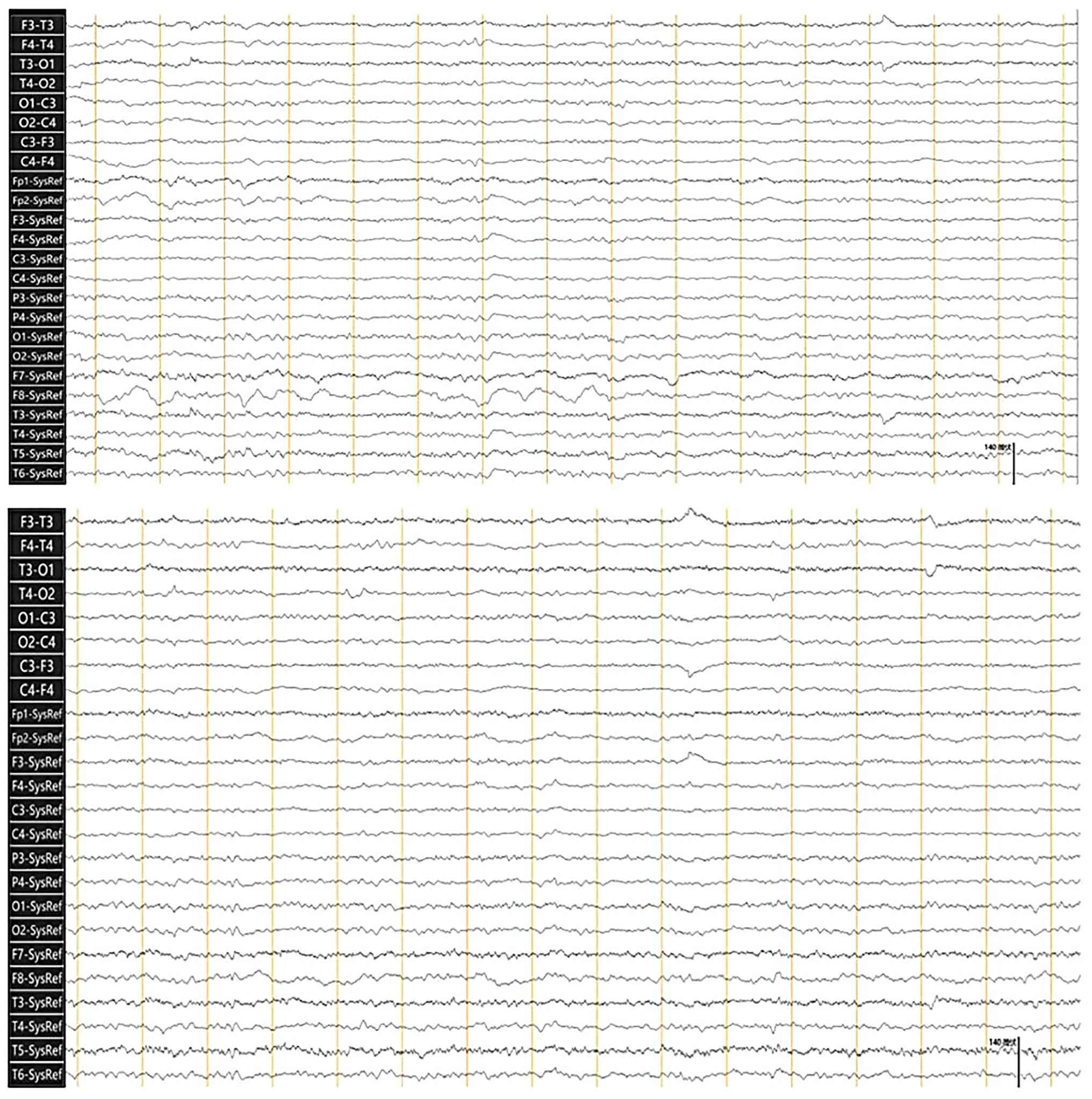
imaging. EEG re-examination (Fig.
5) revealed low-amplitude (≤35 µV) 6.5-7 Hz θ activity,
scattered low-amplitude (≤35 µV) 8 Hz α activity and decreased
scattered low-to-moderate-amplitude (peak 70 µV) 2-3 Hz δ activity.
In awake healthy adults, dominant posterior α rhythm ranges from
8-13 Hz with amplitudes of 20-100 µV; θ activity is minimal during
wakefulness and δ activity is typically absent. These
electrographic changes indicated improvement in brain function.
fNIRS re-examination (Fig. 6)
showed the average prefrontal cortex functional connectivity
strength increased to 0.551, a 0.4-unit increase from admission. At
the 2-month reassessment following admission to the rehabilitation
department (7 months after the initial brain injury), the patient
regained the ability to move the limbs independently and
demonstrated spontaneous emotional responses (tears and smiling).
The Barthel Index score improved from 0 at admission for
rehabilitation to 25 at the 2-month follow-up (7 months after
initial brain injury). Specifically, bowel and bladder control each
scored 10 points, and transfer ability scored 5 points, indicating
improvement in daily living activities. Major clinical,
neurophysiological and imaging parameters across the clinical
course are summarised in Fig. 7.
Key outcome indicators before and after comprehensive
rehabilitation are summarised in Table II.
 | Figure 7Clinical timeline of paroxysmal
sympathetic hyperactivity secondary to severe brain injury. SBI,
severe brain injury; PSH, paroxysmal sympathetic hyperactivity;
PSH-AM, paroxysmal sympathetic hyperactivity-assessment measure;
CFS, clinical feature scale; DLT, diagnostic likelihood tool; CT,
computed tomography; GCS, Glasgow Coma Scale; EEG,
electroencephalogram; fNIRS, functional near-infrared spectroscopy;
HBOT, hyperbaric oxygen therapy; TMS, transcranial magnetic
stimulation; BI, Barthel Index; T, body temperature; HR, heart
rate; bpm, beats per minute; BP, blood pressure; RR, respiratory
rate; ICU, intensive care unit; TID, three times daily; IV,
intravenous; NG, nasogastric (tube); q8h, every 8 h; BID, twice
daily; SpO2, peripheral oxygen saturation (pulse oxygen
saturation); PO, by mouth. |
| Table IIOutcome comparison before vs. after
comprehensive rehabilitation. |
Table II
Outcome comparison before vs. after
comprehensive rehabilitation.
| Parameter | At admission/first
episode | At
re-examination/follow-up |
|---|
| PSH-AM total
score | 26 (CFS 17 + DLT
9) | 4 (CFS 0 + DLT
4) |
| PSH episode
frequency | Several times per
week | ~Once every 1-2
months |
| PSH episode
duration | >11 min | <1 min |
| GCS score | E1VTM1 | E4VTM3 |
| Brunnstrom stage
(UL/hand/LL) | I-I-I | II-II-II |
| Limb muscle tone
(modified Ashworth scale) | Grade 2 (during
episodes) | Grade 0 |
| Body temperature | 38.6-39˚C (during
episodes) | 36.6˚C (stable) |
| Heart rate | 190 bpm (during
episodes) | 87 bpm (stable) |
| Blood pressure | 188/102-205/110
mmHg | 116/76 mmHg |
| Head CT | Right basal ganglia
haemorrhage | Haemorrhage
resolved |
| EEG | θ 6 Hz (≤35 µV);
severe abnormality | θ 6.5-7 Hz +
scattered α, improved |
| fNIRS (prefrontal
connectivity) | 0.151 (severe
decline) | 0.551 (+0.4
improvement) |
| Eye opening (0
points) | None (E1) | Spontaneous (E4) |
| Pupillary light
reflex | Not assessed | Intact (direct and
consensual) |
| Emotional
response | Absent | Present (tears and
smiling) |
| Limb movement | None | Independent
movement |
At the 2-month reassessment following admission for
rehabilitation (7 months after severe brain injury), the patient
demonstrated partial neurological and functional recovery. The
patient achieved spontaneous eye-opening with intact pupillary
light reflexes, exhibited primitive emotional responses and
regained limited independent limb movement. Motor function reached
Brunnstrom stage II in bilateral upper limbs, hands and lower
limbs, while activities of daily living remained severely impaired,
reflected by a Barthel Index score of 25. Although paroxysmal
sympathetic hyperactivity symptoms were well-controlled
pharmacologically, the patient still required ongoing
multidisciplinary rehabilitation support. Given that the patient
was within the sub-acute recovery phase of severe brain injury,
further functional improvements in consciousness, motor performance
and activities of daily living might be anticipated with continued
targeted rehabilitation intervention. Nevertheless, the long-term
prognosis remains uncertain, considering the substantial structural
brain damage secondary to right basal-ganglia haemorrhage.
Persistent neurological deficits, including disorders of
consciousness and motor impairment, cannot be excluded.
Discussion
The pathophysiological mechanisms of PSH mainly
include the following aspects: i) The central autonomic network
imbalance mechanism. In the present case, the right basal ganglia
haemorrhage may have affected the autonomic regulation pathway
between the diencephalon and brainstem. When damaged, the basal
ganglia, the core structure of the cortico-thalamic-basal ganglia
loop, can disrupt the inhibitory projections mediated by
γ-aminobutyric acid, leading to disinhibition of the sympathetic
excitation centre in the posterior hypothalamus. Functional
near-infrared imaging revealed that the connectivity strength of
the prefrontal cortex increased from 0.151 to 0.551 before and
after treatment, indicating that the functional reorganisation of
the prefrontal cortex and limbic system may improve the symptoms of
the patient by enhancing top-down sympathetic regulation (10). ii) The neurotransmitter storm
hypothesis (11-13).
The recurring sympathetic storms of the patient (such as increased
heart rate and blood pressure) may be related to the dysfunction of
presynaptic norepinephrine transporters. Animal experiments
indicate that after brain injury, the glutamate uptake capacity of
astrocytes decreases, leading to excessive activation of
N-methyl-D-aspartate receptors, triggering calcium influx and
promoting the release of large amounts of catecholamines. iii)
Neuro-immune-endocrine axis disorder. Previous studies found that
abnormal activation of the IL-6/Jak2/STAT 3 pathway in brain
endothelial cells can change blood-brain barrier permeability. This
allows peripheral inflammatory factors to directly affect the
paraventricular nucleus of the hypothalamus, thus forming an
‘inflammation-sympathetic hyperactivity’ cycle (14,15).
According to the auxiliary examination results, the
post-treatment head CT showed improved findings compared with the
previous CT, with resolution of the haemorrhage and low-density
shadows. The frequency of θ waves in the electroencephalogram was
higher than that at admission. The frequency of θ waves increased
from 6 to 6.5-7 Hz, and scattered α activity appeared. Previous
literature has shown that the θ wave primarily appears in the
medial part of the cerebral cortex, especially in the hippocampus,
and that it is the primary mode of brain activity during relaxation
and recovery. It is also closely related to cognitive and conscious
functions (16). Modulation of
hippocampal-medial cortical θ oscillations supports arousal
maintenance, attentional processing and memory-related neuronal
coordination; in patients with severe brain injury, an increase in
θ frequency and the emergence of α-band activity reflect partial
restoration of thalamocortical circuitry, which correlates with
improved consciousness and cognitive performance (17). Studies have shown that as patients
recover from brain injury, particularly with regard to motor and
cognitive functions, the frequency of θ waves on the EEG may
increase. The appearance of the α wave indicates that consciousness
has improved, reflecting the gradual recovery of the brain's
capacity for consciousness (18,19).
In the present case, the Brunnstrom stage of the
patient progressed from stage I to stage II, indicating some
functional recovery in the cortical-basal ganglia-thalamic loop.
The intracerebral haemorrhage affected the neuroregulatory pathway
between the cortex, thalamus and basal ganglia, thereby inducing
PSH. The GCS score increased from E1VTM1 to E4VTM3, reflecting
greater consciousness (20). The
emotional responses (such as crying and smiling) that appeared
later in the patient may indicate functional reorganisation of the
posterior nodes of the default mode network, consistent with
imaging evidence showing a 0.4-unit increase in resting-state
functional connectivity strength (21). HBOT may promote angiogenesis by
upregulating hypoxia-inducible factor-1α, thereby providing a
structural basis for improving consciousness and motor function
(22). High-frequency TMS can
inhibit the excessive activation of the amygdala (23). Motor training and electrical
stimulation of the auricular vagus nerve exert arousal-promoting
effects simultaneously (24).
Clonazepam enhances the inhibitory effect of central γ-aminobutyric
acid neurotransmission and reduces symptoms of sympathetic nervous
system hyperactivity (25).
β-blockers relieve tachycardia and hypertension by blocking
β-adrenergic receptors and reducing the effects of norepinephrine
and epinephrine on peripheral target organs (26). Sodium valproate inhibits excessive
activation of the central sympathetic pathway by enhancing the
inhibitory effect of γ-aminobutyric acid (27). Bromocriptine activates the
dopaminergic inhibitory pathway and indirectly inhibits the
overactivation of the midbrain sympathetic centre (28). Levetiracetam regulates synaptic
vesicle glycoprotein 2A, which may have unique value in preventing
the recurrence of a sympathetic storm (29).
Several key limitations of the present case report
must be acknowledged. First, since this is a single-patient report,
causal relationships between any specific intervention and the
observed clinical improvements cannot be established. Second, no
control condition was available; spontaneous neurological recovery,
known to occur following SBI (30), cannot be excluded as a contributor
to observed outcomes. Third, multiple rehabilitation and
pharmacological interventions were implemented simultaneously,
making it impossible to attribute improvements to any particular
treatment component. Fourth, the patient was transferred to The
First Affiliated Hospital of Zhejiang Chinese Medical University
(Zhejiang Provincial Hospital of Chinese Medicine) 5 months after
initial injury, limiting the validity of ‘rehabilitation’ claims as
applied to the period of early rehabilitation. Fifth, the
generalizability of single-case findings is inherently limited.
Sixth, formal quality-of-life instruments were not administered;
outcome data were based solely on objective clinical scales (such
as GCS and Brunnstrom). Future prospective, controlled studies with
larger cohorts and standardised protocols are needed to validate
these preliminary observations.
The present case report describes the clinical
course of PSH following SBI, from acute onset through chronic
management, with pathological mechanisms involving multi-level
neural circuit dysregulation. A stepwise, multidisciplinary
treatment strategy encompassing targeted pharmacotherapy,
neurological rehabilitation and supportive care was temporally
associated with symptomatic improvement, neurological recovery and
improved functional status. EEG frequency progression and changes
in fNIRS-measured prefrontal functional connectivity strength
provided objective biomarker data for monitoring treatment
response. These observations suggest the potential clinical value
of integrated multidisciplinary management for PSH following SBI.
Future research should prioritize: i) Prospective, controlled
studies with larger cohorts to evaluate the comparative
effectiveness of specific PSH treatment components; ii) serial
neuroimaging and neurophysiological monitoring at multiple time
points to characterize recovery trajectories; iii) biomarker
studies (such as neurotransmitter assays and inflammatory markers)
to test mechanistic hypotheses directly; iv) standardized
patient-reported outcome measures; and v) long-term follow-up
studies to assess PSH recurrence rates, medication tapering
strategies and ultimate functional outcomes.
In summary, the present case suggests the potential
clinical feasibility of an integrated, multidisciplinary approach
to PSH management following SBI, while underscoring the need for
higher-quality evidence to guide treatment decisions in this
challenging condition.
Acknowledgements
Not applicable.
Funding
Funding: No funding was received.
Availability of data and materials
The data generated in the present study may be
requested from the corresponding author.
Authors' contributions
ZZ, SX and ZG contributed to the conception and
design of the present case report, the analysis and interpretation
of data, and manuscript revision. ZZ and SX contributed to the
acquisition of the data. All authors have read and approved the
final manuscript. ZZ and SX confirm the authenticity of all the raw
data.
Ethics approval and consent to
participate
The study was conducted in accordance with the
guidelines of the Declaration of Helsinki and received ethical
approval on 24 April 2024 from the First Affiliated Hospital of
Zhejiang Chinese Medical University Ethics Committee (approval no.
2024-KLS-498-01).
Patient consent for publication
The patient's family provided written informed
consent during their first consultation, agreeing for the patient
to receive care and authorizing the use of their anonymized
clinical data, including images and videos, for medical, academic
and research purposes.
Competing interests
The authors declare that they have no competing
interests.
References
|
1
|
Vásquez-García S, Rubiano AM and Lee K:
Cerebral tissue oximetry monitoring for severe brain injury. J
Intensive Care Med. 8850666251357487:2025.PubMed/NCBI View Article : Google Scholar
|
|
2
|
Nancy F, Khowaja A and Khowaja P:
Paroxysmal sympathetic hyperactivity: A common consequence of
traumatic brain injury. Auton Neurosci. 257(103238)2025.PubMed/NCBI View Article : Google Scholar
|
|
3
|
Miyoshi T, Mizushima C, Noborio Y, Kimoto
Y, Nakaharu Y and Shimamoto S: Efficacy of combination therapy with
gabapentin and guanfacine for paroxysmal sympathetic hyperactivity
following hypoxic encephalopathy: A case report. J Int Med Res.
49(3000605211009721)2021.PubMed/NCBI View Article : Google Scholar
|
|
4
|
Panjaitan JAP, Ginting LF, Rajagukguk M,
Sibagariang DB and Siahaan AMP: Paroxysmal sympathetic
hyperactivity: Current update on diagnosis, treatments, and
outcomes. Surg Neurol Int. 16(163)2025.PubMed/NCBI View Article : Google Scholar
|
|
5
|
Huang Q, Zhou R, Zhang Y, Li J and Yu F:
Paroxysmal sympathetic hyperactivity syndrome caused by
Streptococcus intermedius meningoencephalitis in children: A
case report and literature review. Front Pediatr.
12(1480514)2024.PubMed/NCBI View Article : Google Scholar
|
|
6
|
Louraoui SM, Fliyou F, Aasfara J and El
Azhari A: Paroxysmal sympathetic hyperactivity after traumatic
brain injury: What is important to know? Cureus.
14(e24693)2022.PubMed/NCBI View Article : Google Scholar
|
|
7
|
Akira M, Yuichi T, Tomotaka U, Takaaki K,
Kenichi M and Chimi M: The outcome of neurorehabilitation efficacy
and management of traumatic brain injury. Front Hum Neurosci.
16(870190)2022.PubMed/NCBI View Article : Google Scholar
|
|
8
|
Aguilar-Fuentes V, Orozco-Puga P and
Jiménez-Ruiz A: The glasgow coma scale: 50-year anniversary. Neurol
Sci. 45:2899–2901. 2024.PubMed/NCBI View Article : Google Scholar
|
|
9
|
Chen X, Zhang CL and Jiang H: Barthel
index score at admission to predict 1-month and 1-year prognosis in
inpatients aged ≥75 years with multimorbidity. Int J Geriatr
Psychiatry. 40(e70147)2025.PubMed/NCBI View Article : Google Scholar
|
|
10
|
Zheng RZ, Lei ZQ, Yang RZ, Huang GH and
Zhang GM: Identification and management of paroxysmal sympathetic
hyperactivity after traumatic brain injury. Front Neurol.
11(81)2020.PubMed/NCBI View Article : Google Scholar
|
|
11
|
Miao H, Huang H, Chen W, Su YY and Zhang
Y: Clinical characteristics and prognosis of paroxysmal sympathetic
hyperactivity in patients with severe nontraumatic brain injury.
Brain Inj. 37:95–100. 2023.PubMed/NCBI View Article : Google Scholar
|
|
12
|
Muñoz-Ballester C and Robel S:
Astrocyte-mediated mechanisms contribute to traumatic brain injury
pathology. WIREs Mech Dis. 15(e1622)2023.PubMed/NCBI View Article : Google Scholar
|
|
13
|
Yuan M and Wu H: Astrocytes in the
traumatic brain injury: The good and the bad. Exp Neurol.
348(113943)2021.PubMed/NCBI View Article : Google Scholar
|
|
14
|
Ring M: An integrative approach to HPA
axis dysfunction: From recognition to recovery. Am J Med.
138:1451–1463. 2025.PubMed/NCBI View Article : Google Scholar
|
|
15
|
Guan X, Wang Q, Liu M, Sun A and Li X:
Possible involvement of the IL-6/JAK2/STAT3 pathway in the
hypothalamus in depressive-like behavior of rats exposed to chronic
mild stress. Neuropsychobiology. 80:279–287. 2021.PubMed/NCBI View Article : Google Scholar
|
|
16
|
Salimi-Nezhad N, Hasanlou M, Amiri M and
Keliris GA: A neuromimetic realization of hippocampal CA1 for theta
wave generation. Neural Netw. 142:548–563. 2021.PubMed/NCBI View Article : Google Scholar
|
|
17
|
Young CK, Ruan M and McNaughton N: Speed
modulation of hippocampal theta frequency and amplitude predicts
water maze learning. Hippocampus. 31:201–212. 2021.PubMed/NCBI View Article : Google Scholar
|
|
18
|
Gong WY, Sun J and Liu X: Status of CVEEG
and EEG reactivity grading and their evaluation value in patients
with severe traumatic brain injury. Hainan Med J (Chinese).
34:3384–3388. 2023.
|
|
19
|
Xu B, Yuan XD, Zhang PS, et al: Predictive
value of quantitative electroencephalographic indices for
consciousness status in patients with acute brain injury [in
Chinese]. J Pract Cardiac Cerebral Pneumal Vasc Dis. 30:52–59.
2022.
|
|
20
|
Xu PF, Bai YY, Zhao H, et al: Clinical
analysis of 5 cases of paroxysmal sympathetic hyperactivity
syndrome secondary to deep cerebral haemorrhage [in Chinese]. Chin
J Lab Diagn. 28:15–16. 2024.
|
|
21
|
Argyropoulos GPD, Moore L, Loane C,
Roca-Fernandez A, Lage-Martinez C, Gurau O, Irani SR, Zeman A and
Butler CR: Pathologic tearfulness after limbic encephalitis: A
novel disorder and its neural basis. Neurology. 94:e1320–e1335.
2020.PubMed/NCBI View Article : Google Scholar
|
|
22
|
Yamamoto N, Oyaizu T, Enomoto M, Horie M,
Yuasa M, Okawa A and Yagishita K: VEGF and bFGF induction by nitric
oxide is associated with hyperbaric oxygen-induced angiogenesis and
muscle regeneration. Sci Rep. 10(2744)2020.PubMed/NCBI View Article : Google Scholar
|
|
23
|
McCarthy B, Sesa-Ashton G, Rim D,
Henderson LA and Macefield VG: Single-pulse transcranial magnetic
stimulation of the dorsolateral prefrontal cortex does not directly
affect muscle sympathetic nerve activity in humans. Cereb Cortex.
34(bhae484)2024.PubMed/NCBI View Article : Google Scholar
|
|
24
|
Szulczewski MT: Transcutaneous auricular
vagus nerve stimulation combined with slow breathing: Speculations
on potential applications and technical considerations.
Neuromodulation. 25:380–394. 2022.PubMed/NCBI View Article : Google Scholar
|
|
25
|
Yang Y, Wang B, Zhong Z, Chen H, Ding W
and Hoi MPM: Clonazepam attenuates neurobehavioral abnormalities in
offspring exposed to maternal immune activation by enhancing
GABAergic neurotransmission. Biochem Pharmacol.
192(114711)2021.PubMed/NCBI View Article : Google Scholar
|
|
26
|
Esler M, Kjeldsen SE, Pathak A, Grassi G,
Kreutz R and Mancia G: Diverse pharmacological properties, trial
results, comorbidity prescribing and neural pathophysiology suggest
European hypertension guideline downgrading of beta-blockers is not
justified. Blood Press. 31:210–224. 2022.PubMed/NCBI View Article : Google Scholar
|
|
27
|
Li Q, Huang YH, Li QQ, Jia JN, Liu ZQ,
Zhou HH, Zhou XY, Jin WL and Mao XY: Sodium valproate ablates
ferroptosis in kainic acid-induced epileptic seizure via
suppressing lysyl oxidase. Neuroreport. 35:1090–1097.
2024.PubMed/NCBI View Article : Google Scholar
|
|
28
|
Abdulrazik SG, Attia TZ and Derayea SM:
The first spectrofluorimetric protocol for sensitive quantitative
analysis of bromocriptine in its pure and pharmaceutical forms:
Evaluation of the greenness of the method. RSC Adv. 13:35733–35740.
2023.PubMed/NCBI View Article : Google Scholar
|
|
29
|
Contreras-García IJ, Gómez-Lira G,
Phillips-Farfán BV, Pichardo-Macías LA, García-Cruz ME,
Chávez-Pacheco JL and Mendoza-Torreblanca JG: Synaptic vesicle
protein 2A expression in glutamatergic terminals is associated with
the response to levetiracetam treatment. Brain Sci.
11(531)2021.PubMed/NCBI View Article : Google Scholar
|
|
30
|
Xu H, Lu Y and Yuan P: Relationship
between changes of serum neurotransmitters and stress hormones and
secondary brain injury in acute phase of intracerebral hemorrhage.
J Pract Med. 40:3343–3348. 2024.
|