Introduction
Post-traumatic stress disorder (PTSD) is a serious
psychiatric condition that develops after exposure to traumatic or
highly stressful events, including violent crime, traffic
accidents, natural disasters, terrorist incidents, occupational
stress, and sexual violence (1).
PTSD affects approximately 7% of the population at some point
during life (2). In addition, the
COVID-19 pandemic was associated with a marked increase in PTSD
symptoms among healthcare workers and patients worldwide.
PTSD is associated with a range of pathological
symptoms, including anxiety, depression, intrusive memories, and
impairments in learning and memory (1). Unlike general chronic stress, PTSD is
characterized by persistent fear responses and difficulty
extinguishing traumatic memories, often accompanied by alterations
in cognitive function and brain activity (3).
At the biological level, PTSD is closely linked to
neuroinflammatory processes, including dysregulation of the
hypothalamic-pituitary-adrenal (HPA) axis and increased production
of pro-inflammatory cytokines such as interleukin-1β (IL-1β) and
tumor necrosis factor-α (TNF-α), in the brain (4). Neuroinflammation in the medial
prefrontal cortex (PFC) and hippocampus is particularly relevant
because these regions regulate cognitive and memory functions and
contribute to other neuropsychiatric symptoms (5).
Accordingly, reducing neuroinflammation may help
mitigate behavioral abnormalities, including PTSD-related memory
impairment, and may contribute to neuronal protection (5). Among the molecular mechanisms
involved, Toll-like receptor 4 (TLR4) is one of the most
extensively studied members of the TLR family. Activation of TLR4
triggers downstream signaling cascades that promote cytokine
release and activate the nuclear factor (NF)-κB pathway (6). These pathways are critically involved
in inflammation, apoptosis, and cognitive dysfunction, including
deficits in learning and memory.
Previous studies have shown that PTSD is associated
with synaptic loss and altered synaptic plasticity, both of which
are essential for memory formation and storage (7). PTSD has also been reported to affect
the expression of brain-derived neurotrophic factor (BDNF), a key
regulator of neuronal growth and survival, and its receptor,
tropomyosin receptor kinase B (TrkB) (8).
The BDNF-TrkB signaling pathway interacts with
intracellular cascades, including activation of the NF-κB pathway,
which plays a central role in regulating neuroinflammatory
responses such as pro-inflammatory cytokine production. These
processes are closely linked to neuronal survival and synaptic
plasticity (8). Therefore,
modulation of inflammatory cytokines and NF-κB activity, together
with regulation of the BDNF-TrkB signaling pathway, may improve
memory function by influencing synaptic plasticity (9).
Although commonly prescribed antidepressants for
PTSD can provide symptomatic relief, their therapeutic effects are
often temporary, and their clinical use is limited by significant
adverse effects. These limitations indicate the need for additional
therapeutic strategies (10).
Ginseng (Panax ginseng C.A. Meyer) has been
used for centuries as a traditional medicinal herb and is widely
recognized for its anti-fatigue, antioxidant, anti-allergic,
anti-tumor, and anti-inflammatory properties in chronic diseases
(11). More recently, ginseng has
attracted attention for its potential benefits in neurodegenerative
disorders, psychiatric conditions, and stress-related memory
impairment, with evidence suggesting improvements in learning and
cognitive performance (12).
The primary bioactive constituents of ginseng are
ginsenosides, a class of glycosides composed of an aglycone
backbone-either protopanaxadiol (PD) or protopanaxatriol (PT).
These compounds exhibit diverse pharmacological activities
(13,14). Previous studies have reported that
ginsenoside Rb1, a representative PD-type saponin, enhances
learning and memory in amyloid beta (Aβ)-induced Alzheimer's
disease (AD) mouse models, likely through its anti-inflammatory
effects (15). Similarly,
ginsenoside Rg3, another PD-type compound, has been shown to
attenuate cognitive deficits by suppressing inflammatory mediator
expression in lipopolysaccharide (LPS)-induced models of memory
impairment (16). In contrast,
ginsenoside Rg1, a PT-type saponin, has demonstrated efficacy in
improving cognitive function under postmenopausal conditions and
provides neuroprotective effects in the hippocampus in
ovariectomized and D-galactose-treated mouse models of AD (17). Collectively, these findings suggest
that both PD- and PT-type ginsenosides may have therapeutic
potential for alleviating neuroinflammation and memory deficits
associated with PTSD.
In this study, we compared the effects of PD and PT
on PTSD-induced memory impairment using a single prolonged stress
(SPS) rat model. We further examined whether these compounds exert
anti-inflammatory effects through modulation of the TLR4/NF-κB
signaling pathway, which plays a critical role in cognitive
function.
Materials and methods
Animals and PD or PT-type saponins
administration
Male Sprague-Dawley rats aged 7-8 weeks and weighing
200-220 g were obtained from Samtaco Animal Co. (Seoul, Korea). The
rats were housed in an animal room under a 12 h light/dark cycle
and were provided sterile drinking water and food ad
libitum. Animal experiments were conducted in accordance with
the Code of Ethics and guidelines for the management of laboratory
animals by the Animal Care and Use Committee of Kyung Hee
University (Seoul, South Korea), which also approved the
experimental protocol (approval no. KHSASP-21-268). Before
behavioral testing, the rats were allowed to acclimatize for 1 week
and were handled daily by the experimenter for at least 1 week to
minimize stress-related effects during the experiment. The rats
were randomly divided into 6 groups, with 6 to 7 rats per
group.
Panaxadiol (10 and 20 mg/kg, Merck, Darmstadt,
Germany) and panaxatriol (10 and 20 mg/kg, Merck), both extracted
from red ginseng and paroxetine (10 mg/kg, PAX; Sigma-Aldrich
Chemical Co., St. Louis, MO, USA, positive control) were
intraperitoneally injected once daily for 14 days. Based on
previous research findings, intraperitoneal administration was
selected to achieve rapid systemic absorption and ensure safe drug
delivery (8). These drugs were
dissolved in 0.9% physiological saline immediately before use. The
dosages of PD and PT were determined based on previous studies
(13,14). Forty-nine rats were randomly
divided into the following seven groups (n=7 rats/group). The
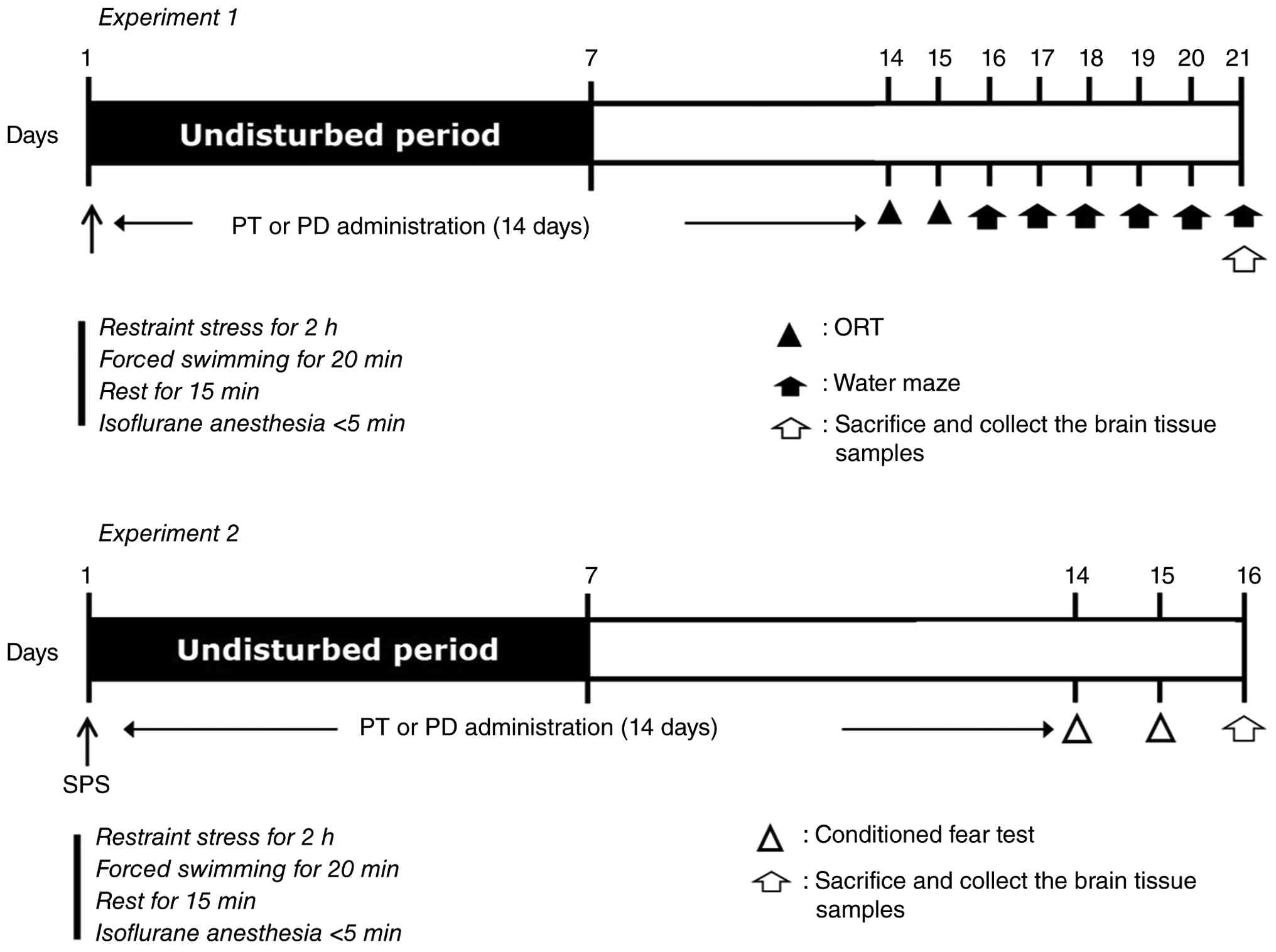
detailed experimental schedule is presented in Fig. 1. All behavioral measurements were
performed by an independent investigator who was blinded to the
experimental conditions.
Single prolonged stress
To establish a PTSD-like animal model, rats were
subjected to restraint stress for 2 h and forced swimming for 20
min. The rats were then allowed to rest for 15 min and were exposed
to isoflurane (2-3%) until loss of consciousness, as confirmed by
absence of righting reflex and lack of response to toe and tail
pinch (<5 min). Isoflurane was used only for induction at 2-3%
until loss of consciousness and was not maintained thereafter. This
procedure followed the SPS method (8). After SPS exposure, the rats were left
undisturbed for 7 days to allow sensitization.
Contextual fear conditioning and
extinction
The effects of PD and PT on learned fear were
assessed using the conditioned fear test. Conditioned fear is a
learned response in which animals associate sensory cues, such as
environmental context or sound, with a life-threatening or painful
stimulus (18). On day 1, rats
were placed in a computerized fear-conditioning chamber using the
Gemini Avoidance System (SD Instruments, San Diego, CA, USA). A
conditioned stimulus (CS; 30 sec, 85dB) was paired with an electric
shock as the unconditioned stimulus (US; 0.5 mA, 3 sec) to induce
fear memory. The white noise (85 dB) and chamber light were turned
on 28 sec before shock, delivery and were turned off when the shock
ended. Thus, the CS lasted 30 sec, and the electric shock was
delivered during the final 2 sec. This session was repeated 4
times, with an intertrial interval of 2 min. On day 2, rats
underwent extinction training consisting of 12 CS presentations
without shock. On day 3, rats received 3 CS-only presentations to
test extinction recall (in the same context). Fear responses were
observed for 5 min. Freezing was defined as the absence of movement
except respiration during video observation, and the total freezing
time was measured and compared among experimental groups.
Object recognition task
To assess object recognition memory, the object
recognition test (ORT) was performed (8). Rats were allowed to explore objects
in a black acrylic box (40x40x25 cm). Exploratory behavior was
measured as the basis for object discrimination. Two similar wooden
toys (familiar objects, A1 and A2) and one novel wooden toy (B)
with a completely different color and shape were used. The objects
were large enough that the rats could not easily move them. On the
first day, rats were acclimatized to the experimental environment
for 10 min. After 24 h, during the training phase, rats were placed
in the box with 2 similar toys (A1 and A2) and allowed to explore
for 5 min. After 2 h, during the test phase, rats were exposed to
one novel wooden toy (B) and one familiar object (A1) for 5 min.
Sniffing time directed toward novel or familiar objects was
measured. Exploration was defined as active sniffing of the object,
touching the object with the forepaw, or directing the snout toward
the object.
Morris water maze test
The Morris water maze (MWM) is widely used to
evaluate spatial learning and memory in rats (8). Acquisition and retention were
assessed using the MWM test. The apparatus consisted of a circular
plastic tank (diameter, 200 cm; height, 35 cm) filled with water a
depth of approximately 22 cm. The tank was divided into quadrants,
and an escape platform (height, 20 cm; diameter, 15 cm) was place
in a randomly selected quadrant. The platform was positioned 1.0 cm
below the water surface, and the water was made opaque with paint
to conceal the platform. Rats were placed into the tank from the
central wall of a quadrant that did not contain the platform.
Escape latency was measured from placement in the tank until the
rat found the platform. The acquisition test was conducted 3 times
per day for 5 days. When a rat reached the platform, it remained
there for 10 sec before the next trial. If the rat did not locate
the platform within 180 sec, the researcher guided it to the
platform and allowed it to remain there for 10 sec to learn the
platform location. Acquisition trials were repeated at 60 min
intervals. On day 6, the escape platform was removed, and spatial
memory retention was assessed during a 60 sec probe trial in the
quadrant where the platform had been located. All experimental
procedures were video-recorded using SMART software (version 2.5;
Panlab, Cornella, Spain) installed on a ceiling-mounted system.
Escape latency was analyzed from the recorded data using the image
analysis system.
Enzyme-linked immunosorbent assay
analysis of inflammatory mediators and NF-κB protein levels
After behavioral measurements, rats (n=4) were
deeply anesthetized with sodium pentobarbital (50 mg/kg, i.p.)
according to the approved IACUC protocol. Adequate anesthesia was
confirmed by the absence of the forelimb withdrawal reflex. Under
deep anesthesia, euthanasia was performed by cervical dislocation,
followed by thoracotomy, cardiac blood collection, and rapid
removal of the whole brain for subsequent analyses. Frontal cortex
was firstly taken out. After overlying temporal cortex was
unveiled, the hippocampus was exposed and collected for use. Blood
was sampled from the heart at the time of euthanasia. The brain of
rats was obtained, and pro-inflammatory cytokine levels in the PFC
and hippocampus were quantified using enzyme-linked immunosorbent
assay (ELISA). The concentrations of TNF-α and IL-1β (Abcam,
Cambridge, MA, USA), as well as interleukin-6 (IL-6; Cell Signaling
Technology, Danvers, MA, USA), were determined in both brain
regions according to previously established protocols. In addition,
hippocampal levels of TLR4 (NOVUS Biologicals, Chesterfield, MO,
USA) and NF-κB (MyBioSource, San Diego, CA, USA) were analyzed
using a sandwich ELISA method, following standard procedures
described in earlier studies.
Total RNA isolation and RT-PCR
analysis
After the rats were euthanized under anesthesia, the
hippocampus (n=3/group) was isolated, added to TRIzol®
reagent (Invitrogen; Thermo Fisher Scientific, Inc.) and vortexed
to recover total RNA. After RNA was reverse transcribed into cDNA,
PCR was performed using primers specific to each target, and PCR
reagents were selected according to previous reports (8). PCR was performed at 57˚C for 27
cycles for BDNF and at 58˚C for 28 cycles for TrkB using a thermal
cycler (MJ Research, Watertown, MA, USA). PCR produces were
confirmed by electrophoresis on 1.5% agarose gels. The following
primer sequences were used for PCR: GAPDH (228 bp), (forward)
5'-CTCCTGAACTTGAGGCAGTTTA-3' and (reverse)
5'-GTAACCAGTCATCAGCAAAATGT-3'; BDNF (227 bp):
5'-GCTGAGCGTGTGTGACAGTA-3' (sense) and 5'-ACTGGGTAGTTCGGCACTGG-3'
(antisense); TrkB (283 bp): 5'-GAGTTTCTGCCCCTGCTCTG-3' (sense) and
5'-GGACTCTTTGGGTCGCAGAA-3' (antisense).
Statistical analysis
All data was expressed as the mean ± SEM.
Statistical differences among groups were analyzed using one-way
analysis of variance (ANOVA) followed by Tukey's post hoc test in
SPSS software (version 23.0; SPSS Inc., Chicago, IL, USA).
P<0.05 was considered to indicate a statistically significant
difference.
Results
Effects of body weight and serum CORT
levels following single prolonged stress
Analysis of body weight over 14 days showed that the
saline-treated normal (SAL) group gained body weight over time
(Fig. 2A). In contrast, the SPS
group showed reduced body weight gain compared to the SAL group
(t=0.514, P<0.001). Although these body weight changes are not
directly related to SPS-induced memory impairment, body weight is
an important indicator of physiological status in rats. Therefore,
the reduced body weight gain in SPS-exposed rats suggests that the
animals underwent physiological changes in response to traumatic
stress.
After body weight measurement, serum CORT levels
were assessed in SPS-exposed rats (Fig. 2B). Serum CORT levels were
significantly higher in the SPS group than in the SAL group
(P<0.01). These results indicate that SPS induced a stress
response accompanied by changes in body weight.
Effects of PD and PT-type saponins on
SPS-induced freezing response
Following administration of PT and PD in the
SPS-induced animal model of memory impairment, fear responses were
assessed. Freezing response during extinction trials and short-term
extinction recall was used to determine whether SPS induced a
conditioned fear response (Fig.
3A). During fear conditioning, no difference in freezing
response was observed between the SAL and SPS groups (t=0.237,
P<0.01). However, freezing responses differed significantly
between the 2 groups during extinction learning (t=0.284,
P<0.05) and extinction recall (t=0.845, P<0.05). The PD20
group showed a significant decrease in freezing response during
extinction recall compared with the SPS group (P<0.05; Fig. 3B), whereas the PT group did not
affect freezing response during extinction recall. These results
indicate that SPS induced a stress-related conditioned fear
response in rats and that PD administration may inhibit SPS-induced
conditioned fear memory formation or promote fear-memory
extinction.
 | Figure 3Effect of PD and PT-type saponins on
SPS-induced memory impairment. Comparison of ginsenoside PD or PT
effects on (A) freezing behavior during conditioning, extinction,
and short-term recall and the (B) percentages of time spent
freezing during short-term recall in contextual fear conditioning.
Comparison of ginsenoside PD or PT effects on (C) exploration time
for two familiar objects, (D) exploration time for familiar and
novel objects, and the (E) ability to discriminate between familiar
and new objects in the novel object recognition test. Comparison of
ginsenoside PD or PT effects on (F) time to escape (latency) from
the water onto a submerged platform during acquisition trials, (G)
swimming speed, (H) percentage of time spent in the target
quadrant, (I) percentage of distance traveled in the target
quadrant, (J) path efficiency to reach the target zone and (K)
number of entries into the target zone in the Morris water maze.
*P<0.05, **P<0.01,
***P<0.001 vs. SAL group; #P<0.05,
##P<0.01 vs. SPS group. PD, protopanaxadiol; PT,
protopanaxatriol; SAL, saline; SPS, single prolonged stress, PAX,
paroxetine; C1-3, fear conditioning sessions; E1-6, extinction
training sessions; R1, extinction recall session. |
Effects of PD and PT-type saponins on
SPS-induced impairments in cognitive and spatial memory
The effects of PT and PD on cognitive function were
evaluated in the SPS-induced animal model of memory impairment. In
the ORT, no significant differences were observed among groups in
the exploration time of familiar objects (Fig. 3C). However, the SPS group showed a
marked reduction in sniffing time for novel objects compared with
the SAL group (P<0.001; Fig.
3D). In contrast, the PD20-treated group showed a significant
increase in novel-object exploration time compared with the SPS
group (P<0.05), whereas no significant change was observed in
the PT-treated group. The discrimination index was also
significantly higher in the PD20 group than in the SPS group
(P<0.05; Fig. 3E). These
findings indicate that SPS markedly impaired object recognition
memory in rats, whereas PD administration ameliorated SPS-induced
cognitive deficits.
The effects of PT and PD on spatial learning and
memory were evaluated using the SPS-induced model of spatial
cognitive impairment. During the 5-day acquisition phase of the MWM
test, the SPS group required significantly more time to locate the
hidden platform than the SAL group (P<0.01 on days 3-5; Fig. 3F), indicating impaired spatial
learning. In contrast, the PD20-treated group showed a
significantly shorter escape latency than the SPS group (P<0.05
on days 3 and 4 and P<0.01 on day 5), whereas the PT-treated
group did not show a meaningful reduction in escape latency. No
significant differences in mean swimming speed was observed among
groups (Fig. 3G), suggesting that
the observed differences in escape latency were not attributable to
altered motor function.
In the probe test assessing spatial memory, the SPS
group showed significant reductions in time spent in the target
quadrant, distance traveled within the target area, and number of
entries into the target zone compared with the SAL group (P<0.01
for all comparisons; Fig. 3H-K).
Conversely, the PD20 group showed significant increases in time
spent in the target area and number of target entries compared with
the SPS group (P<0.05 for each measure). These findings indicate
that PD administration improved SPS-induced deficits in spatial
learning and memory.
Effects of PD and PT-type saponins on
pro-inflammatory cytokines levels in the PFC and hippocampus
following SPS
After PT or PD administration in the SPS-induced
model of memory impairment, inflammatory cytokine levels were
measured in the PFC and hippocampus. The SPS group showed
significantly elevated concentrations of pro-inflammatory
cytokines, including TNF-α, IL-1β, and IL-6, in both brain regions
compared with the SAL group (P<0.01 for all comparisons;
Fig. 4A-F). In contrast, PD20
treatment significantly reduced TNF-α levels in both the PFC and
hippocampus (P<0.05) compared with the SPS group. IL-1β levels
in the hippocampus were also significantly decreased in the PD20
group (P<0.05). PT treatment did not significantly change
inflammatory cytokine levels in either the PFC or hippocampus.
 | Figure 4Effect of PD and PT-type saponins on
SPS-induced neuroinflammation. Comparison of ginsenoside PD or PT
effects on (A) TNF-α, (B) IL-1β and (C) IL-6 levels in the medial
prefrontal cortex, and (D) TNF-α, (E) IL-1β and (F) IL-6 levels in
the hippocampus of rats exposed to SPS as measured by enzyme-linked
immunosorbent assay. **P<0.01 vs. SAL group;
#P<0.05, ##P<0.01 vs. SPS group. IL,
interleukin; PD, protopanaxadiol; PT, protopanaxatriol; SAL,
saline; SPS, single prolonged stress; PAX, paroxetine; TNF-α, tumor
necrosis factor-α. |
Effects of PD and PT-type saponins on
BDNF and TrkB mRNA expression in the hippocampus following SPS
After PT or PD treatment in the SPS-induced model of
memory impairment, the mRNA expression levels of BDNF and TrkB were
analyzed in the hippocampus. The SPS group showed significantly
reduced BDNF mRNA expression in the hippocampus compared with the
SAL group (P<0.01; Fig. 5). By
contrast, PD20 administration significantly increased BDNF mRNA
expression in the hippocampus compared with the SPS group
(P<0.05). No significant changes in BDNF mRNA expression were
observed in the PT-treated group.
Effects of PD and PT-type saponins on
the TLR4 and NF-κB levels in the hippocampus following SPS
To clarify the molecular mechanisms underlying these
findings, hippocampal levels of TLR4 and NF-κB were measured after
PT or PD administration in the SPS-induced model of memory
impairment. The SPS group showed significantly elevated TLR4 and
NF-κB protein levels compared with the SAL group (P<0.01;
Fig. 6). By contrast, PD20
treatment significantly reduced both proteins in the hippocampus
(P<0.05). These results suggest that the anti-inflammatory
effects of PD are associated with modulation of the TLR4/NF-κB
signaling pathway.
Discussion
This study compared the effects of PT and PD on
PTSD-induced impairments in learning and memory. Unlike PT, PD
improved SPS-induced cognitive deficits, reduced pro-inflammatory
cytokine production in the PFC and hippocampus, and restored the
SPS-induced decrease in BDNF mRNA expression in the hippocampus.
These findings suggest that the memory-enhancing and
anti-inflammatory effects of PD are associated, at least in part
with modulation of the TLR4/NF-κB signaling pathway.
After SPS exposure, reduced body weight gain and
increased serum CORT levels were observed. These changes indicate
that the PTSD-like model was successfully established using the SPS
paradigm and are consistent with physiological responses commonly
associated with acute trauma and chronic stress. Accumulating
evidence suggests that dysregulation of the HPA axis leads to
sustained elevations in circulating CORT levels, which are closely
linked to the development of memory impairments (19).
Several studies have reported that animals exposed
to contextual fear paradigms, such as a single traumatic stimulus
combined with contextual recall, exhibit enhanced fear conditioning
and altered extinction learning (18). In this study, SPS-exposed rats
displayed increased freezing behavior during contextual fear
conditioning and impaired extinction recall. Short-term extinction
recall was also significantly elevated. PD administration
attenuated these effects by reducing freezing responses during
short-term extinction retrieval. These findings suggest that PD may
alleviate maladaptive fear responses associated with traumatic
memory, which can interfere with normal memory processing and
consolidation. Fear-related memories are known to suppress
extinction learning by inhibiting neural activity in key brain
regions, including the PFC and hippocampus (20). The present results therefore
suggest that PD may contribute to the restoration of impaired fear
extinction and memory regulation, potentially through reduced
neuroinflammation in the PFC.
SPS exposure also induced in object recognition
memory deficits, as shown by reduced novel-object exploration time
and decreased discrimination indices (21). These results indicate impaired
discrimination between familiar and novel stimuli, reflecting
deficits in short-term and reference memory. PD administration
significantly improved these cognitive deficits.
SPS-exposed rats exhibited significantly prolonged
escape latency in the MWM, indicating impaired spatial learning
(22). Deficits in spatial memory
consolidation and retrieval were also observed. By contrast, PD
administration improved learning performance, as shown by reduced
escape latency during the acquisition phase. Spatial memory
consolidation was also enhanced, as indicated by increased time
spent in the target quadrant and a higher number of platform
crossings. These results suggest that PD ameliorates SPS-induced
impairments in spatial memory retrieval and recall. Overall, the
findings indicate that PD treatment can alleviate SPS-induced
deficits in fear memory, spatial learning, and cognitive function.
Rapid and persistent neuroinflammatory responses can disrupt memory
networks, elevate pro-inflammatory cytokine levels, and alter
synaptic plasticity (23,24). Accordingly, SPS-induced
inflammatory responses in the PFC and hippocampus may contribute to
the observed cognitive and memory impairments (5). PD administration appeared to exert
anti-inflammatory effects by suppressing pro-inflammatory cytokine
expression in these brain regions.
BDNF has been reported to promote neuronal survival
by modulating NF-κB signaling cascades and interacting with
inflammatory mediators through the BDNF-TrkB pathway (9). Accordingly, BDNF is an important
neurotrophic regulator linking neuroinflammation and memory
function. Several studies have suggested that modulation of BDNF
expression may alleviate neuroinflammation-induced memory deficits
in chronic stress and PTSD (25).
In this study, PD administration significantly reversed the
SPS-induced reduction in BDNF mRNA expression. This effect was
accompanied by a decreases in pro-inflammatory cytokines, including
TNF-α and IL-1β, in both the PFC and hippocampus.
TLR4, a key pattern recognition receptor, activates
downstream signaling cascades that converge on the NF-κB pathway,
leading to the upregulation of multiple inflammatory mediators
(6). After phosphorylation and
activation, NF-κB promotes the expression of pro-inflammatory
cytokines, including TNF-α, IL-1β, and IL-6, thereby contributing
to inflammatory responses and tissue injury (6). In this study, SPS-induced PTSD-like
animals exhibited elevated levels of TLR4, phosphorylated NF-κB,
TNF-α, and other inflammatory markers. These increases were
significantly attenuated after PD administration. These findings
suggest that suppression of the TLR4/NF-κB signaling pathway may
underlie the anti-inflammatory effects of PD. Taken together, these
results indicate that targeting the TLR4/NF-κB signaling axis may
represent a promising therapeutic strategy for alleviating
neuroinflammation and cognitive dysfunction associated with
PTSD.
Numerous studies have reported beneficial effects of
ginsenosides in animal models of memory impairment induced by
chronic stress (26). For example,
ginsenoside Rb1, a representative PD-type saponin, has been shown
to significantly improve cognitive deficits induced by chronic
restraint stress (CRS) in both reward-based instrumental
conditioning and MWM tasks. These effects are thought to be
mediated through suppression of oxidative stress and apoptosis,
thereby enhancing synaptic plasticity (27,28).
Similarly, ginsenoside Rg3 has demonstrated neuroprotective
properties and cognitive benefits in AD, a condition characterized
by excessive accumulation of β-amyloid peptide (Aβ1-42) (29). In addition, ginsenoside Rg1, a
PT-type saponin, has been reported to alleviate learning and memory
deficits in CRS models by preventing reduction in BDNF and TrkB
expression in the PFC (30). The
PD-type ginsenoside group mainly includes Rb1, Rb2, Rc, and Rd,
whereas the PT-type group consists primarily of Rg1, Re, and Rf
(13,14). Although both PD- and PT-type
ginsenosides have been reported to improve cognitive impairment in
various studies (31,32), the present findings indicate that
PD-type ginsenosides exert a more pronounced effect on SPS-induced
deficits in spatial and cognitive memory. In contrast, PT-type
ginsenosides did not produce significant improvements under the
same conditions. Taken together, these results suggest that PD-type
ginsenosides may be more effective than PT-type ginsenosides in
restoring spatial, cognitive, and fear-related memory impairments
associated with SPS-induced PTSD-like conditions, rather than
chronic stress alone.
In conclusion, this study demonstrated that PD-type
ginsenoside administration improved multiple aspects of memory
function, including learning acquisition, memory retention, and
fear extinction, in an SPS-induced rat model of memory impairment.
These beneficial effects appear to be associated with
anti-inflammatory mechanisms involving modulation of the TLR4/NF-κB
signaling pathway. Taken together, PD-type ginsenosides may
represent promising bioactive compounds for preventing or
alleviating memory decline associated with neuropsychiatric
disorders such as PTSD.
Acknowledgements
Not applicable.
Funding
Funding: This research was supported by a grant from the
National Research Foundation of Korea funded by the Korean
government (grant no. RS-2023-00241316).
Availability of data and materials
The data generated in the present study may be
requested from the corresponding author.
Authors' contributions
BL performed conceptualization, formal analysis,
funding acquisition, investigation, methodology, project
administration, visualization and wrote the original draft. BL
confirms the authenticity of all the raw data, and read and
approved the final manuscript.
Ethics approval and consent to
participate
All animal experiments were performed in accordance
with the National Institutes of Health guidelines for the care and
use of laboratory animals and were approved by the Animal Care and
Use Committee of Kyung Hee University (approval no.
KHSASP-21-268).
Patient consent for publication
Not applicable.
Competing interests
The author declares that they no competing
interests.
References
|
1
|
Javidi H and Yadollahie M: Post-traumatic
stress disorder. Int J Occup Environ Med. 3:2–9. 2012.PubMed/NCBI
|
|
2
|
Yehuda R, Hoge CW, McFarlane AC, Vermetten
E, Lanius R, Lanius RA, Nievergelt CM, Hobfoll SE, Koenen KC,
Neylan TC and Hyman SE: Post-traumatic stress disorder. Nat Rev Dis
Primers. 1(15057)2015.PubMed/NCBI View Article : Google Scholar
|
|
3
|
Parsons RG and Ressler KJ: Implications of
memory modulation for post-traumatic stress and fear disorders. Nat
Neurosci. 16:146–153. 2013.PubMed/NCBI View
Article : Google Scholar
|
|
4
|
Bommaraju S, Dhokne MD, Rakeshkumar PP and
Datusalia AK: Memantine alleviates PTSD-like symptoms and improves
dendritic arborization through modulation of the HPA axis and
neuroinflammation in rats. Neurochem Res. 50:58–72. 2024.PubMed/NCBI View Article : Google Scholar
|
|
5
|
Rossignoli MT, Lopes-Aguiar C, Ruggiero
RN, Do Val da Silva RA, Bueno-Junior LS, Kandratavicius L,
Peixoto-Santos JE, Crippa JA, Cecilio Hallak JE, Zuardi AW, et al:
Selective post-training time window for memory consolidation
interference of cannabidiol into the prefrontal cortex: Reduced
dopaminergic modulation and immediate gene expression in limbic
circuits. Neuroscience. 350:85–93. 2017.PubMed/NCBI View Article : Google Scholar
|
|
6
|
Liu M, Xie J and Sun Y:
TLR4/MyD88/NF-κB-mediated inflammation contributes to cardiac
dysfunction in rats of PTSD. Cell Mol Neurobiol. 40:1029–1035.
2020.PubMed/NCBI View Article : Google Scholar
|
|
7
|
Liu Q, Ding X, Wang Y, Chu H, Guan Y, Li M
and Sun K: Artemisinin reduces PTSD-like symptoms, improves
synaptic plasticity, and inhibits apoptosis in rats subjected to
single prolonged stress. Front Pharmacol.
15(1303123)2024.PubMed/NCBI View Article : Google Scholar
|
|
8
|
Lee B, Sur B and Oh S: Neuroprotective
effect of Korean Red Ginseng against single prolonged
stress-induced memory impairments and inflammation in the rat brain
associated with BDNF expression. J Ginseng Res. 46:435–443.
2022.PubMed/NCBI View Article : Google Scholar
|
|
9
|
Green CR, Corsi-Travali S and Neumeister
A: The role of BDNF-TrkB signaling in the pathogenesis of PTSD. J
Depress Anxiety. 2013 (S4)(006)2013.PubMed/NCBI View Article : Google Scholar
|
|
10
|
Albucher RC and Liberzon I:
Psychopharmacological treatment in PTSD: A critical review. J
Psychiatr Res. 36:355–367. 2002.PubMed/NCBI View Article : Google Scholar
|
|
11
|
Park SK, Hyun SH, In G, Park CK, Kwak YS,
Jang YJ, Kim B, Kim JH and Han CK: The antioxidant activities of
Korean Red Ginseng (Panax ginseng) and ginsenosides: A
systemic review through in vivo and clinical trials. J Ginseng Res.
45:41–47. 2021.PubMed/NCBI View Article : Google Scholar
|
|
12
|
Sanghavi CR, Barhate SA, Mahajan MS, Mohan
M and Kasture SB: Korean ginseng extract attenuates
reserpine-induced orofacial dyskinesia and improves cognitive
dysfunction in rats. Nat Prod Res. 25:704–715. 2011.PubMed/NCBI View Article : Google Scholar
|
|
13
|
Kim TH and Lee SM: The effects of ginseng
total saponin, panaxadiol and panaxatriol on ischemia/reperfusion
injury in isolated rat heart. Food Chem Toxicol. 48:1516–1520.
2010.PubMed/NCBI View Article : Google Scholar
|
|
14
|
Oh HA, Kim DE, Choi HJ, Kim NJ and Kim DH:
Anti-stress effects of 20(S)-protopanaxadiol and
20(S)-protopanaxatriol in immobilized mice. Biol Pharm Bull.
38:331–335. 2015.PubMed/NCBI View Article : Google Scholar
|
|
15
|
Lin J, Gao S, Wang T, Shen Y, Yang W, Li Y
and Hu H: Ginsenoside Rb1 improves learning and memory ability
through its anti-inflammatory effect in Aβ1-40 induced
Alzheimer's disease of rats. Am J Transl Res. 11:2955–2968.
2019.PubMed/NCBI
|
|
16
|
Lee B, Sur B, Park J, Kim SH, Kwon S, Yeom
M, Shim I, Lee H and Hahm DH: Ginsenoside rg3 alleviates
lipopolysaccharide-induced learning and memory impairments by
anti-inflammatory activity in rats. Biomol Ther (Seoul).
21:381–390. 2013.PubMed/NCBI View Article : Google Scholar
|
|
17
|
Zhang X, Wang J, Xing Y, Gong L, Li H, Wu
Z, Li Y, Wang J, Wang Y, Dong L and Li S: Effects of ginsenoside
Rg1 or 17β-estradiol on a cognitively impaired, ovariectomized rat
model of Alzheimer's disease. Neuroscience. 220:191–200.
2012.PubMed/NCBI View Article : Google Scholar
|
|
18
|
Vimalanathan A, Gidyk DC, Diwan M, Gouveia
FV, Lipsman N, Giacobbe P, Nobrega JN and Hamani C: Endocannabinoid
modulating drugs improve anxiety but not the expression of
conditioned fear in a rodent model of post-traumatic stress
disorder. Neuropharmacology. 166(107965)2020.PubMed/NCBI View Article : Google Scholar
|
|
19
|
Hauer D, Kaufmann I, Strewe C, Briegel I,
Campolongo P and Schelling G: The role of glucocorticoids,
catecholamines and endocannabinoids in the development of traumatic
memories and posttraumatic stress symptoms in survivors of critical
illness. Neurobiol Learn Mem. 112:68–74. 2014.PubMed/NCBI View Article : Google Scholar
|
|
20
|
Omura J, Fuchikami M, Araki M, Miyagi T,
Okamoto Y and Morinobu S: Chemogenetic activation of the mPFC
alleviates impaired fear memory extinction in an animal model of
PTSD. Prog Neuropsychopharmacol Biol Psychiatry.
108(110090)2021.PubMed/NCBI View Article : Google Scholar
|
|
21
|
Lee B, Shim I, Lee H and Hahm DH:
Melatonin ameliorates cognitive memory by regulation of
cAMP-response element-binding protein expression and the
anti-inflammatory response in a rat model of post-traumatic stress
disorder. BMC Neurosci. 19:38–48. 2018.PubMed/NCBI View Article : Google Scholar
|
|
22
|
Wang HN, Peng Y, Tan QR, Chen YC, Zhang
RG, Qiao YT, Wang HH, Liu L, Kuang F, Wang BR and Zhang ZJ:
Quetiapine ameliorates anxiety-like behavior and cognitive
impairments in stressed rats: Implications for the treatment of
posttraumatic stress disorder. Physiol Res. 59:263–271.
2010.PubMed/NCBI View Article : Google Scholar
|
|
23
|
Feng L, Gao J, Wang Y, Cheong YK, Ren G
and Yang Z: Etidronate-zinc complex ameliorated cognitive and
synaptic plasticity impairments in 2-vessel occlusion model rats by
reducing neuroinflammation. Neuroscience. 390:206–217.
2018.PubMed/NCBI View Article : Google Scholar
|
|
24
|
Zhang D, Rodríguez-Kirby LAR, Lin Y, Wang
W, Song M, Wang L, Wang L, Kanatani S, Jimenez-Beristain T, Dang Y,
et al: Spatial dynamics of brain development and neuroinflammation.
Nature. 647:213–227. 2025.PubMed/NCBI View Article : Google Scholar
|
|
25
|
Xie P, Chen L, Wang J, Wang X, Yang S and
Zhu G: Polysaccharides from Polygonatum cyrtonema Hua prevent
post-traumatic stress disorder behaviors in mice: Mechanisms from
the perspective of synaptic injury, oxidative stress, and
neuroinflammation. J Ethnopharmacol. 319(117165)2024.PubMed/NCBI View Article : Google Scholar
|
|
26
|
Rinwa P and Kumar A: Modulation of
nitrergic signalling pathway by American ginseng attenuates chronic
unpredictable stress-induced cognitive impairment,
neuroinflammation, and biochemical alterations. Naunyn
Schmiedebergs Arch Pharmacol. 387:129–141. 2014.PubMed/NCBI View Article : Google Scholar
|
|
27
|
Jiang N, Wang K, Zhang Y, Huang H, Lv JW,
Wang Q, Wang HX, Xia TJ and Liu XM: Protective effect of
ginsenoside Rb1 against chronic restraint stress (CRS)-induced
memory impairments in rats. Behav Brain Res.
405(113146)2021.PubMed/NCBI View Article : Google Scholar
|
|
28
|
Liu Y, DiStasio M, Su G, Asashima H,
Enninful A, Qin X, Deng Y, Nam J, Gao F, Bordignon P, et al:
High-plex protein and whole transcriptome co-mapping at cellular
resolution with spatial CITE-seq. Nat Biotechnol. 41:1405–1409.
2023.PubMed/NCBI View Article : Google Scholar
|
|
29
|
Alinkeel R, Kutscher HL, Singh A,
Cwiklinski K, Khechen N, Schwartz SA, Prasad PN and Mahajan SD:
Neuroprotective effects of a biodegradable poly(lactic-co-glycolic
acid)-ginsenoside Rg3 nanoformulation: A potential nanotherapy for
Alzheimer's disease? J Drug Target. 26:182–193. 2018.PubMed/NCBI View Article : Google Scholar
|
|
30
|
Wang Y, Kan H, Yin Y, Wu W, Hu W, Wang M
and Li W and Li W: Protective effects of ginsenoside Rg1 on chronic
restraint stress induced learning and memory impairments in male
mice. Pharmacol Biochem Behav. 120:73–81. 2014.PubMed/NCBI View Article : Google Scholar
|
|
31
|
Tohda C, Matsumoto N, Zou K, Meselhy MR
and Komatsu K: Abeta(25-35)-induced memory impairment, axonal
atrophy, and synaptic loss are ameliorated by M1, A metabolite of
protopanaxadiol-type saponins. Neuropsychopharmacology. 29:860–868.
2004.PubMed/NCBI View Article : Google Scholar
|
|
32
|
Wang YZ, Chen J, Chu SF, Wang YS, Wang XY,
Chen NH and Zhang JT: Improvement of memory in mice and increase of
hippocampal excitability in rats by ginsenoside Rg1's metabolites
ginsenoside Rh1 and protopanaxatriol. J Pharmacol Sci. 109:504–510.
2009.PubMed/NCBI View Article : Google Scholar
|