Introduction
CHARGE syndrome is a rare genetic, multisystem
disorder with numerous malformations and distinctive diagnostic
criteria. The term ‘CHARGE’ describes a group of clinical features
that include coloboma, heart defects, choanal atresia, retardation
of growth and/or development, genitourinary malformation and ear
abnormalities (1). A renewed
definition of the diagnostic criteria for CHARGE syndrome was
proposed by Blake et al (2)
and later updated by Verloes (3).
According to the most recent criteria, the major diagnostic
features are coloboma, choanal atresia and hypoplasia of the
semicircular canals. Minor diagnostic features include
rhombencephalic dysfunction (brainstem dysfunction and cranial
nerve III-XII anomalies, including neurosensory deafness),
hypothalamo-hypophyseal dysfunction (including growth hormone and
gonadotrophin defects), ear malformation (internal or external),
malformation of mediastinal organs (such as the heart and
esophagus) and mental retardation.
The primary causes of CHARGE syndrome are mutations
in the chromodomain helicase DNA binding protein 7 (CHD7)
gene (accession no. NM_017780.4), which encodes a protein that is
part of the chromodomain helicase DNA-binding (CHD) protein family
(4). The CHD protein family is a
highly conserved group of nuclear proteins that consists of nine
members in vertebrates. The functions of this protein family are
diverse; however, all proteins in this family serve a role in
transcriptional regulation (5-7).
CHD7 in particular functions as an ATP-dependent nucleosome
remodeler (8). In order to remodel
nucleosomes, CHD7 functions as part of a multiprotein complex,
binding to <10,000 sites across the mammalian genome. CHD7 is
also an important regulator of developmental transcription factor
genes and signaling pathways (such as p53 and SOX2)
(9,10).
Therefore, the present report outlines the case of a
fetus who exhibited a number of ultrasound findings, including
persistent left superior vena cava (LSVC), delay in the fusion of
the cerebellar vermis, inferior vermis hypoplasia and borderline
mega cisterna magna (MCM). To further investigate these findings,
fetal exome sequencing was performed in the present study,
revealing a novel mutation in CHD7.
Case report
In January 2024, a 30-year-old pregnant woman
(gravida: 1 and parity: 0) was referred to Access to Genome P.C.
(Athens, Greece) following the detection of increased nuchal
translucency at 12 + 1 weeks (3.13 mm). The personal and family
medical history of the patient was unremarkable.
Chorionic villi sampling (CVS) was performed at 12 +
1 weeks. DNA was extracted from the CVS sample and high-resolution
chromosomal microarray analysis was performed using the SurePrint
G3 CGH ISCA v2 Microarray Kit, 8x60K (cat. no. G5955A; Agilent
Technologies). The analysis resolution of this platform is ~500 kb
along the genome and ~50 kb in ~240 regions of known clinical
importance which are associated with ~512 genetic diseases,
according to the International Standard Cytogenetic Consortium
(11). This analysis showed a
normal male genomic pattern with no imbalances.
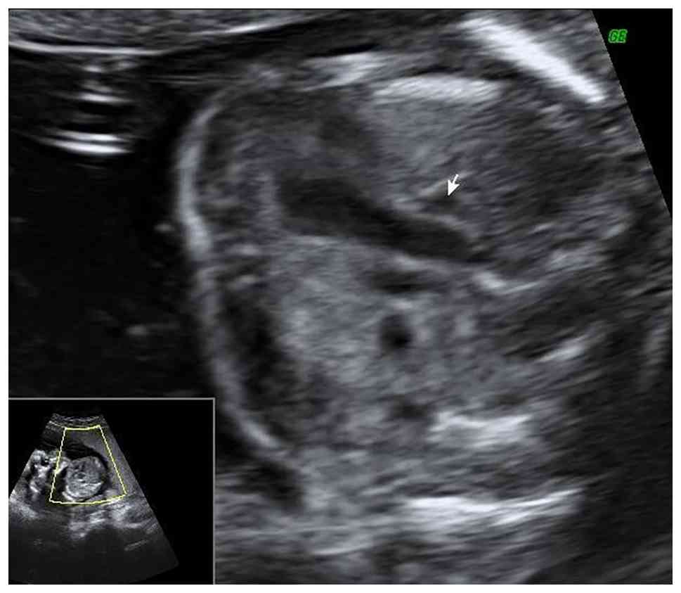
During a routine ultrasound examination performed at
18 + 1 weeks, the presence of persistent LSVC was observed
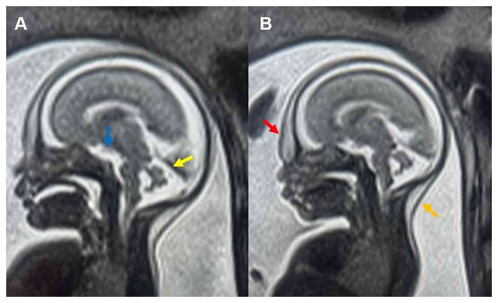
(Fig. 1). At 21 + 6 weeks, a fetal
brain MRI was performed, exhibiting borderline enlargement of the
cisterna magna (essentially, MCM; 10.5 mm; Fig. 2A). Delay in the fusion of the
cerebellar vermis was also observed (Fig. 2A). Marked upward rotation of the
cerebellar vermis axis was not observed. The fourth ventricle
appeared to have lost its triangular shape. The normal
communication of the 4th ventricle with the cisterna magna was
occluded. Elevation of the cerebellar tentorium was not observed.
In a sagittal section, the primary fissure of the cerebellar vermis
was recognized, while the ratio of the anterior to the posterior
lobe of the vermis appeared to be disturbed and equalized. The
inferior part of the cerebellar vermis appeared to be thin and
small. Cerebellar vermis biometry was 12 mm in height and 5 mm in
anteroposterior diameter (normal values for this gestational age
are 8.5-11.5 and 5.5-8 mm, respectively) (12). Inferior vermis hypoplasia
(Dandy-Walker variant) was observed without ruling out atypical
vermis hypoplasia of Joubert Syndrome (Fig. 2A). Examination of other anatomical
structures of the fetus revealed the presence of a small-sized
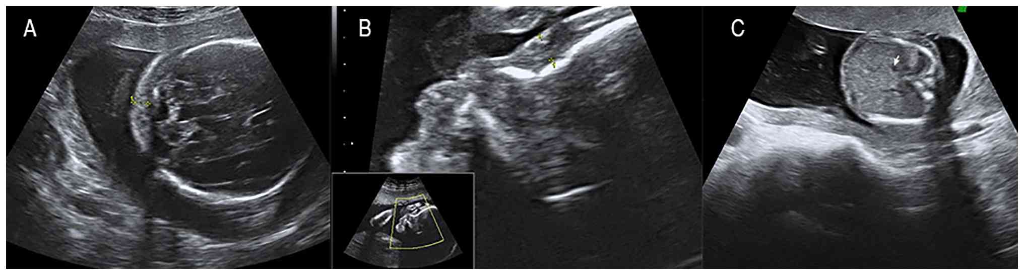
stomach, as well as mild prenasal and nuchal edema (Fig. 2B). At 23 + 5 weeks, a level II
ultrasound was performed, which further demonstrated the MRI
findings. Specifically, it demonstrated the borderline MCM and
inferior vermis hypoplasia with delay in the fusion of the
cerebellar vermis (Fig. 3A), the
mild prenasal edema (Fig. 3B) and
the small-sized stomach (Fig.
3C).
Although microarray analysis, performed following
the detection of increased nuchal translucency, appeared normal,
the presence of multiple sonographic and MRI findings raised
concern for an underlying genetic condition and fetal exome
sequencing was therefore performed on fetal DNA from the CVS
sample. DNA was extracted from the CVS sample using the NucleoSpin
Tissue kit (Macherey-Nagel GmbH & Co. KG), according to the
manufacturer's instructions. Exome amplification and library
preparation was performed using the VAHTS Universal Plus DNA
Library Prep Kit for Illumina V2 (cat. no. ND627-01; Illumina
Inc.), according to the manufacturer's instructions. The samples
were qualified using the Qubit Fluorometric Quantification system
(Thermo Fisher Scientific, Inc.). Next-generation sequencing was
performed using the DNBSEQ-T7 MGI Tech genetic analyzer (MGI Tech
Co., Ltd.) according to the manufacturer's instructions, using
paired-end 150 bp reads (2x150 bp). The loading concentration of
the final library was 10 pM. Data analysis was performed using the
VarSome Clinical (Saphetor SA) bioinformatics platform (v11.18.0;
https://clinical.varsome.com/) (13) and SpliceAI (version 1.3.1;
https://spliceailookup.broadinstitute.org/) (14), and was performed for all genes that
are associated with known genetic diseases and syndromes, according
to the Online Mendelian Inheritance in Man database (https://www.omim.org/). All findings from the
aforementioned analysis were evaluated in accordance with the
international literature and the American College of Medical
Genetics and Genomics (ACMG) guidelines (15). The reference genome was UCSC hg19.
Bioinformatics analysis revealed the presence of the novel
c.2836-3C>G splicing mutation in CHD7. According to the
ACMG guidelines, this mutation is categorized as likely pathogenic
based on the PP3 (multiple lines of computational evidence support
a deleterious effect on the gene or gene product), PM2 (absent from
controls or at extremely low frequency in the gnomAD database), and
PS2 (de novo occurrence in a patient with the disease and no
family history with both maternity and paternity confirmed)
criteria (15).
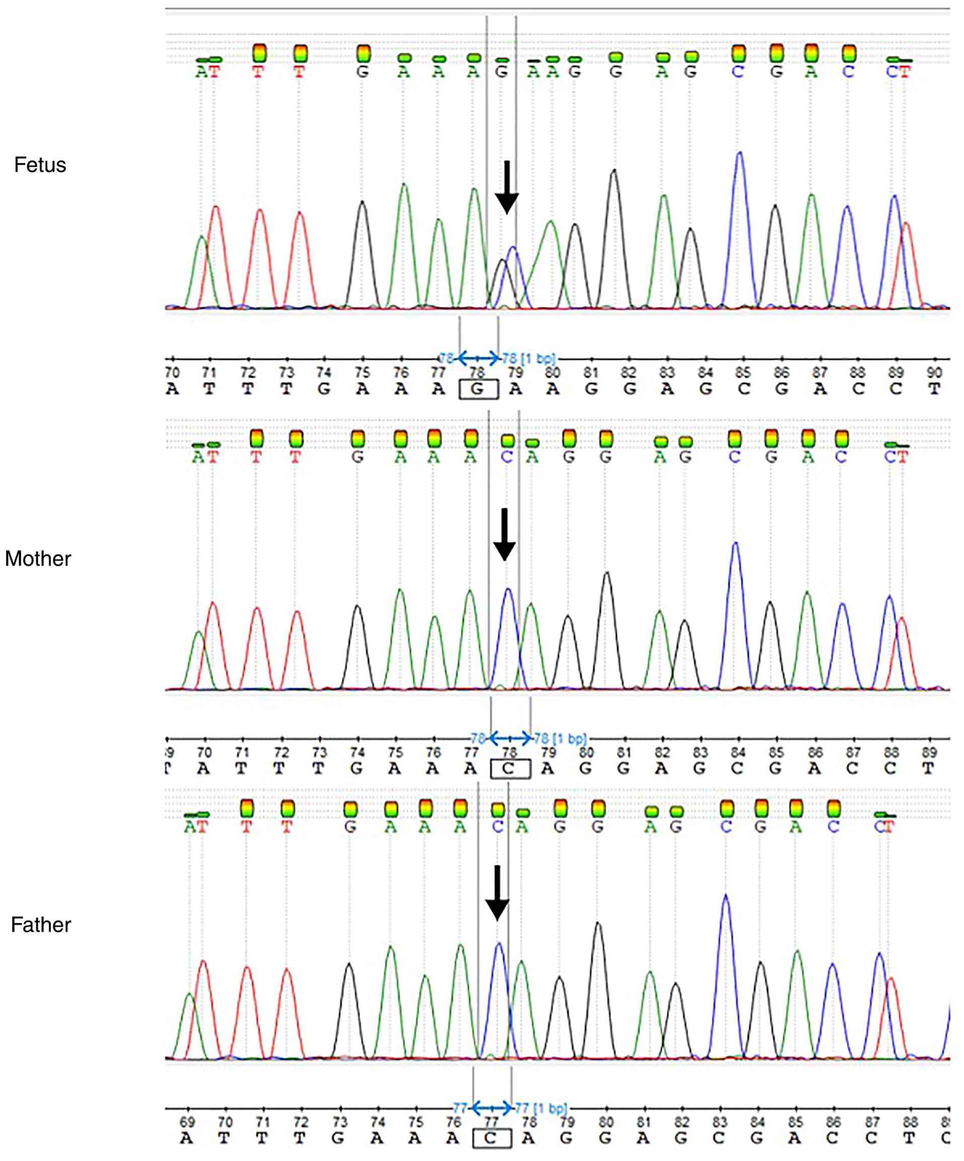
Sanger sequencing was performed in the fetus and the
parents in order to determine the presence of the c.2836-3C>G
mutation and to establish whether the mutation was inherited or
whether it occurred de novo. For the fetal sample, Sanger
sequencing was performed using the DNA already isolated for exome
sequencing. For the parental samples, DNA was extracted from
peripheral blood samples using the NucleoSpin Blood kit
(Macherey-Nagel GmbH & Co. KG), according to the manufacturer's
instructions. DNA amplification was performed via PCR using GoTaq
DNA Polymerase (Promega Corporation). The PCR thermocycling
conditions were as follows: Initial denaturation at 95˚C for 5 min,
followed by 35 cycles of denaturation at 95˚C for 30 sec, annealing
at 60˚C for 30 sec and extension at 72˚C for 30 sec, with a final
extension at 72˚C for 5 min. Following amplification, part of the
CHD7 gene was sequenced and visualized using the ABI 3500
Genetic Analyzer (Thermo Fisher Scientific, Inc.), and was then
compared with the control sequence (NM_017780.4). The sequences of
the primers used were: 5'-CCGGAAACAGAGCGTGTGGTAA-3' (forward
primer) and 5'-TGTACCCACAAATGCATACCCAAAG-3' (reverse primer).
Results confirmed the presence of the novel mutation in the fetus,
while it was not detected in either of the parents, indicating that
this was a de novo mutation (Fig. 4).
In the present study, in silico analysis was
performed using SpliceAI using genome version hg38 and default
maximum distance window of 500 bp. The SpliceAI Δ score range is
0-1, indicating the probability of a splicing effect, with high
probability defined as >0.5 and highly confident defined as
>0.8. The results showed a high probability of disruption of the
canonical splice acceptor site (Δ score: 0.81) and the potential
activation of a nearby cryptic splice acceptor site (Δ score:
0.68). The scSNV-ADA database (v1.1, hg38) was also used via the
VarSome Clinical platform. The scSNV-ADA score range is 0-1,
indicating the probability of altered splicing, with supporting
pathogenic defined as >0.957813. The analysis yielded a high
probability of altered splicing, with a score of 0.995936.
A thorough literature search was performed using
PubMed (https://pubmed.ncbi.nlm.nih.gov/) and Google Scholar
(https://scholar.google.com/) databases
to evaluate whether the phenotypic findings observed in the present
case have been previously reported in other prenatal cases of
CHARGE syndrome. Keywords used included ‘CHD7’, ‘CHARGE syndrome’,
‘persistent left superior vena cava’, ‘mega cisterna magna’,
‘prenatal diagnosis’ and other related terms. The literature search
demonstrated that the observed combination of sonographic, MRI, and
genetic findings was consistent with CHARGE syndrome. Additionally,
it demonstrated that, even though persistent LSVC and borderline
MCM are related to CHARGE syndrome, they have not been reported in
a fetus with CHARGE syndrome thus far. Due to the combination of
all aforementioned clinical findings and the presence of the novel,
likely pathogenic mutation in CHD7, the prognosis was poor
due to the high likelihood of a multisystem disorder consistent
with CHARGE syndrome, which is associated with a number of
malformations, encompassing the major and minor diagnostic criteria
detailed previously. The aforementioned literature search findings
were incorporated into the genetic counseling provided to the
couple; they were informed of the molecular diagnosis of CHARGE
syndrome, the associated poor prognosis, the de novo nature
of the detected mutation, and the low recurrence risk for future
pregnancies. Following genetic counseling, the couple opted for
termination of pregnancy, which was performed at 24+5 weeks. The
couple also received post-termination follow-up care 2 weeks later,
including physical evaluation, psychological support and counseling
regarding future reproductive planning.
Discussion
In the present report, the case of a fetus with
persistent LSVC, delay in the fusion of the cerebellar vermis, a
borderline MCM, inferior vermis hypoplasia, small-sized stomach, as
well as mild prenasal and nuchal edema, is outlined. Prenatal whole
exome sequencing revealed the presence of the novel, splice site
c.2836-3C>G mutation in CHD7 of de novo origin,
suggesting a CHARGE syndrome diagnosis.
As aforementioned, CHARGE syndrome is a rare
multisystem disorder with a number of clinical characteristics. The
majority of cases are diagnosed in the neonatal or childhood
period. So far, prenatally diagnosed cases of CHARGE syndrome are
limited (16). This low number of
prenatally diagnosed cases may be attributed to the fact that the
majority of abnormalities, especially the primary defects such as
coloboma, choanal atresia and auditory deficit, cannot be
visualized through prenatal ultrasound (16). Furthermore, abnormalities that may
be observed prenatally may be subtle or may seem as an isolated
abnormality or phenotypic diversity (16). In a retrospective study of 13 cases
with CHARGE syndrome, it was observed that 2 of 13 cases (15.4%)
exhibited normal ultrasound scans during pregnancy, 1 of 13 (7.7%)
exhibited cystic hygroma in the first trimester and presented
intrauterine fetal demise at 16 weeks and the remaining 10 of 13
cases (76.9%) exhibited abnormal ultrasound features in
utero. Among these 10, 1 case showed increased nuchal
translucency in the first trimester and 5 cases exhibited abnormal
ultrasound features in the second trimester (including
micrognathia, cardiac defects and facial defects). A further 4
cases showed abnormal ultrasound findings in the third trimester,
which included micrognathia, isolated fetal growth restriction and
polyhydramnios (17).
In the present case, there were numerous prenatal
sonographic features and brain MRI findings that were consistent
with CHARGE syndrome. For instance, persistent LSVC was observed at
18 + 1 weeks, a characteristic that is often observed in patients
with CHARGE syndrome. More specifically, in a retrospective study
by Goldenberg et al (18)
involving 8 patients with CHARGE syndrome, persistent LSVC was
observed in 50% of the patients. Persistent LSVC is a congenital
heart anomaly that is found in 0.3-0.5% of the general population
and it can be associated with congenital heart diseases in up to 8%
of cases globally (19). However,
to the best of our knowledge, it has not yet been detected
prenatally in fetuses with CHARGE syndrome. In addition, vermis
hypoplasia was observed in the brain MRI at 21 + 6 weeks. Although
it is not included in the diagnostic criteria, cerebellar vermis
hypoplasia may be a clinical diagnostic feature of CHARGE syndrome
(20). In a retrospective study by
Sohn et al (20) 29.4% of
the patients had cerebellar vermis hypoplasia (5 of 17 patients).
There is also 1 prenatal case report of cerebellar vermis
hypoplasia in a fetus with CHARGE syndrome in the international
literature (21). An additional
feature observed in the prenatal brain MRI at 21 + 6 weeks is
borderline MCM. In a study by Shiohama et al (22), the brain morphology of 10 patients
with CHARGE syndrome was investigated and, in 1 case, MCM was
observed. The present report therefore appears to be the first case
of prenatal detection of borderline MCM in a fetus with CHARGE
syndrome.
As aforementioned, prenatal diagnosis of CHARGE
syndrome is challenging as the characteristic features of the
syndrome cannot be visualized by ultrasound. However, certain
prenatal findings may serve as early indicators. Increased nuchal
translucency in the first trimester may represent an early
non-specific marker associated with increased risk of genetic
abnormalities and warrants careful follow-up (17,23).
During the second and third trimester, detailed anatomical
ultrasound and, when indicated, fetal MRI, may reveal structural
anomalies, including congenital heart defects and brain
abnormalities (16,17). The identification of numerous
anomalies, even when subtle, should raise suspicion for syndromic
conditions, such as CHARGE syndrome and advanced genetic testing
should be considered.
Within this context, the clinical importance of
prenatal findings such as persistent LSVC or MCM should be
carefully interpreted. When LSVC or MCM is prenatally detected as
an isolated finding, it is often considered a benign variant when
no other anomalies are present (24,25).
However, when such findings co-occur or are accompanied by other
subtle abnormalities, the index of suspicion for an underlying
syndromic condition should increase. In such cases, consideration
of advanced genetic testing, such as exome sequencing, may be
warranted.
CHARGE syndrome is primarily caused by mutations in
CHD7, a gene that is involved in transcriptional activation
and is an important regulator of developmental transcription factor
genes and signaling pathways (9,10).
The role of CHD7 has also been demonstrated using animal models.
Mouse mutants have been shown to develop similar abnormalities as
described in CHARGE syndrome, such as inner ear, choanal, heart,
eye, genital and palate defects (26). It has also been demonstrated that
as in humans, normal eye development in mice is sensitive to
Chd7 haploinsufficiency (27). Further studies in zebrafish have
shown that chd7 mutants display craniofacial, cardiac,
visual, auditory and locomotive defects (28).
In the present case, the novel c.2836-3C>G
mutation that was detected, is located in the splicing region of
intron 10 of the CHD7 gene and, according to bioinformatic
analysis tools, it is expected to affect the correct mRNA splicing.
Mutations that affect the splice site region are expected to impair
normal splice-site recognition and lead to aberrant splicing, such
as exon skipping or activation of a cryptic acceptor site (29). For instance, in silico
analysis using SpliceAI predicted a high probability of disruption
of the canonical splice acceptor site and the potential activation
of a nearby cryptic splice acceptor site. These would result in a
frameshift and the introduction of a premature termination codon,
potentially leading to the production of a truncated protein or
triggering non-sense-mediated mRNA decay. Therefore, the detected
mutation is expected to impact the form and function of the
produced CHD7 protein. Although RNA studies would be required to
further demonstrate the exact splicing outcome, these predictions
support a loss-of-function mechanism and, consequently,
haploinsufficiency, which is consistent with the established
pathogenicity mechanism for CHD7 (30,31).
Notably, to the best of our knowledge, the c.2836-3C>G mutation
in CHD7 has not been reported in the Genome Aggregation
Database (https://gnomad.broadinstitute.org/), ClinVar database
(https://www.ncbi.nlm.nih.gov/clinvar/) or the
international scientific literature. As aforementioned, according
to the ACMG guidelines, this mutation is classified as likely
pathogenic based on the PP3, PM2 and PS2 criteria (15). Collectively, this suggested that
the detected novel splicing mutation in CHD7 was responsible
for the sonographic and MRI findings in the present fetus.
Overall, in the present report, the case of a fetus
with CHARGE syndrome caused by a novel splicing mutation in
CHD7 is described. To the best of our knowledge, this is the
first report of the likely pathogenic c.2836-3C>G mutation in
CHD7. The present case report therefore adds to the limited
number of prenatal CHARGE syndrome diagnoses. In addition, it
describes the clinical characteristics and the sonographic and MRI
findings of the fetus, which is of marked importance given the
rarity of prenatal diagnoses of CHARGE syndrome. The present report
emphasizes the importance of integrating numerous anomalies that
may appear minor or borderline into prenatal risk assessment and
counseling. The co-occurrence of subtle anomalies should raise
suspicion for syndromic conditions (such as CHARGE syndrome) and
support comprehensive genetic testing. To the best of our
knowledge, this is the first report of prenatal detection of
persistent LSVC and borderline MCM in a fetus with CHARGE syndrome.
Finally, the present case description could serve as a valuable
reference for evaluating similar findings in other prenatal cases
and for providing genetic counseling in cases of prenatal detection
of the c.2836-3C>G mutation in CHD7.
Acknowledgements
Not applicable.
Funding
Funding: No funding was received.
Availability of data and materials
The data generated in the present study may be found
in the European Nucleotide Archive under accession number
ERR15604719 or at the following URL: (persistent, https://www.ebi.ac.uk/ena/browser/view/ERR15604719).
Authors' contributions
EP was primarily responsible for the design of the
present study and critically revised the manuscript. CK contributed
to the design of the present study and prepared the manuscript. EM
and IP were in charge of patient management, project supervision
and critically revising the manuscript. ES, CE and SO performed
exome sequencing and Sanger sequencing. MR, SS, GKP and MM were
responsible for medical treatment and assessment. CC performed the
MRI examination. MR performed the ultrasound examination. EM and IP
confirm the authenticity of all raw data. All authors read and
approved the final version of the manuscript.
Ethics approval and consent to
participate
Not applicable.
Patient consent for publication
Written informed consent was obtained from the
parents for the inclusion of their data as well as the fetal data
and images in the present case report. All information revealing
the family members identities has been removed. All procedures
followed were conducted according to The Declaration of Helsinki,
as revised in 2008.
Competing interests
The authors declare that they have no competing
interests.
References
|
1
|
Hsu P, Ma A, Wilson M, Williams G, Curotta
J, Munns CF and Mehr S: CHARGE syndrome: A review. J Paediatr Child
Health. 50:504–511. 2014.PubMed/NCBI View Article : Google Scholar
|
|
2
|
Blake KD, Davenport SL, Hall BD, Hefner
MA, Pagon RA, Williams MS, Lin AE and Graham JM Jr: CHARGE
association: An update and review for the primary pediatrician.
Clin Pediatr (Phila). 37:159–173. 1998.PubMed/NCBI View Article : Google Scholar
|
|
3
|
Verloes A: Updated diagnostic criteria for
CHARGE syndrome: A proposal. Am J Med Genet A. 133A:306–308.
2005.PubMed/NCBI View Article : Google Scholar
|
|
4
|
Usman N and Sur M: CHARGE Syndrome: In
StatPearls. StatPearls Publishing, Treasure Island, FL, 2024.
|
|
5
|
Hall JA and Georgel PT: CHD proteins: A
diverse family with strong ties. Biochem Cell Biol. 85:463–476.
2007.PubMed/NCBI View
Article : Google Scholar
|
|
6
|
Marfella CGA and Imbalzano AN: The Chd
family of chromatin remodelers. Mutat Res. 618:30–40.
2007.PubMed/NCBI View Article : Google Scholar
|
|
7
|
Zentner GE, Hurd EA, Schnetz MP, Handoko
L, Wang C, Wang Z, Wei C, Tesar PJ, Hatzoglou M, Martin DM and
Scacheri PC: CHD7 functions in the nucleolus as a positive
regulator of ribosomal RNA biogenesis. Hum Mol Genet. 19:3491–3501.
2010.PubMed/NCBI View Article : Google Scholar
|
|
8
|
Bouazoune K and Kingston RE: Chromatin
remodeling by the CHD7 protein is impaired by mutations that cause
human developmental disorders. Proc Natl Acad Sci USA.
109:19238–19243. 2012.PubMed/NCBI View Article : Google Scholar
|
|
9
|
Basson MA and van Ravenswaaij-Arts C:
Functional Insights into chromatin remodelling from studies on
CHARGE syndrome. Trends Genet. 31:600–611. 2015.PubMed/NCBI View Article : Google Scholar
|
|
10
|
van Ravenswaaij-Arts CM, Hefner M, Blake K
and Martin DM: CHD7 Disorder: In GeneReviews®. Adam MP, Bick S,
Mirzaa GM, Pagon RA, Wallace SE and Amemiya A (eds). University of
Washington, Seattle, WA, 2006.
|
|
11
|
Miller DT, Adam MP, Aradhya S, Biesecker
LG, Brothman AR, Carter NP, Church DM, Crolla JA, Eichler EE,
Epstein CJ, et al: Consensus statement: Chromosomal microarray is a
first-tier clinical diagnostic test for individuals with
developmental disabilities or congenital anomalies. Am J Hum Genet.
86:749–764. 2010.PubMed/NCBI View Article : Google Scholar
|
|
12
|
Kyriakopoulou V, Vatansever D, Davidson A,
Patkee P, Elkommos S, Chew A, Martinez-Biarge M, Hagberg B,
Damodaram M, Allsop J, et al: Normative biometry of the fetal brain
using magnetic resonance imaging. Brain Struct Funct.
222:2295–2307. 2017.PubMed/NCBI View Article : Google Scholar
|
|
13
|
Kopanos C, Tsiolkas V, Kouris A, Chapple
CE, Albarca Aguilera M, Meyer R and Massouras A: VarSome: The human
genomic variant search engine. Bioinformatics. 35:1978–1980.
2019.PubMed/NCBI View Article : Google Scholar
|
|
14
|
Jaganathan K, Panagiotopoulou SK, McRae
JF, Darbandi SF, Knowles D, Li YI, Kosmicki JA, Arbelaez J, Cui W,
Schwartz GB, et al: Predicting splicing from primary sequence with
deep learning. Cell. 176:535–548.e24. 2019.PubMed/NCBI View Article : Google Scholar
|
|
15
|
Richards S, Aziz N, Bale S, Bick D, Das S,
Gastier-Foster J, Grody WW, Hegde M, Lyon E, Spector E, et al:
Standards and guidelines for the interpretation of sequence
variants: A joint consensus recommendation of the American college
of medical genetics and genomics and the association for molecular
pathology. Genet Med. 17:405–424. 2015.PubMed/NCBI View Article : Google Scholar
|
|
16
|
Traisrisilp K, Chankhunaphas W,
Sittiwangkul R, Phokaew C, Shotelersuk V and Tongsong T: Prenatal
sonographic features of CHARGE syndrome. Diagnostics (Basel).
11(415)2021.PubMed/NCBI View Article : Google Scholar
|
|
17
|
Yu QX, Zhen L and Li DZ: Fetal phenotype
of CHARGE syndrome with a molecular confirmation: A series of 13
cases. Fetal Diagn Ther. 51:388–394. 2024.PubMed/NCBI View Article : Google Scholar
|
|
18
|
Goldenberg P, Shikany A, Parrott A, Ware
SM and Hinton RB: persistent left superior vena cava: An overlooked
feature of CHARGE syndrome? Cardiogenetics. 5(5511)2015.
|
|
19
|
Poenaru MO, Hamoud BH, Sima RM, Valcea ID,
Chicea R and Ples L: Persistent left superior vena cava
significance in prenatal diagnosis-case series. J Clin Med.
11(4020)2022.PubMed/NCBI View Article : Google Scholar
|
|
20
|
Sohn YB, Ko JM, Shin CH, Yang SW, Chae JH
and Lee KA: Cerebellar vermis hypoplasia in CHARGE syndrome:
Clinical and molecular characterization of 18 Unrelated Korean
patients. J Hum Genet. 61:235–239. 2016.PubMed/NCBI View Article : Google Scholar
|
|
21
|
Lok IW, Kong C, Kan AS, Yeung K, Yu MH,
Chung BH and To WW: Whole exome sequencing for prenatal diagnosis
of CHARGE syndrome: A case report. Hong Kong J Gynaecol Obstet
Midwifery. 19:125–128. 2019.
|
|
22
|
Shiohama T, McDavid J, Levman J and
Takahashi E: Quantitative brain morphological analysis in CHARGE
syndrome. Neuroimage Clin. 23(101866)2019.PubMed/NCBI View Article : Google Scholar
|
|
23
|
Choy KW, Wang H, Shi M, Chen J, Yang Z,
Zhang R, Yan H, Wang Y, Chen S, Chau MHK, et al: Prenatal diagnosis
of fetuses with increased nuchal translucency by genome sequencing
analysis. Front Genet. 10(761)2019.PubMed/NCBI View Article : Google Scholar
|
|
24
|
Lopes KRM, Bartsota M, Doughty V and
Carvalho JS: Single left superior vena cava: Antenatal diagnosis,
associated anomalies and outcomes. Ultrasound Obstet Gynecol.
60:640–645. 2022.PubMed/NCBI View Article : Google Scholar
|
|
25
|
Pishjoo M, Kheradmand D, Safdari M,
Safdari Z and Mashhadinejad M: Mega cisterna magna: Current
perspectives and future directions-a literature review. Radiol Res
Pract. 2025(9591364)2025.PubMed/NCBI View Article : Google Scholar
|
|
26
|
Bosman EA, Penn AC, Ambrose JC,
Kettleborough R, Stemple DL and Steel KP: Multiple mutations in
mouse Chd7 provide models for CHARGE syndrome. Hum Mol Genet.
14:3463–3476. 2005.PubMed/NCBI View Article : Google Scholar
|
|
27
|
Gage PJ, Hurd EA and Martin DM: Mouse
models for the dissection of CHD7 functions in eye development and
the molecular basis for ocular defects in CHARGE syndrome. Invest
Ophthalmol Vis Sci. 56:7923–7930. 2015.PubMed/NCBI View Article : Google Scholar
|
|
28
|
Hodorovich DR, Lindsley PM, Berry AA,
Burton DF and Marsden KC: Morphological and sensorimotor phenotypes
in a zebrafish CHARGE syndrome model are domain-dependent. Genes
Brain Behav. 2022(e12839)2022.PubMed/NCBI View Article : Google Scholar
|
|
29
|
Anna A and Monika G: Splicing mutations in
human genetic disorders: Examples, detection, and confirmation. J
Appl Genet. 59:253–268. 2018.PubMed/NCBI View Article : Google Scholar
|
|
30
|
Zentner GE, Layman WS, Martin DM and
Scacheri PC: Molecular and phenotypic aspects of CHD7 mutation in
CHARGE syndrome. Am J Med Genet A. 152A:674–686. 2010.PubMed/NCBI View Article : Google Scholar
|
|
31
|
Vissers LELM, van Ravenswaaij CMA,
Admiraal R, Hurst JA, de Vries BBA, Janssen IM, van der Vliet WA,
Huys EHLPG, de Jong PJ, Hamel BCJ, et al: Mutations in a new member
of the chromodomain gene family cause CHARGE syndrome. Nat Genet.
36:955–957. 2004.PubMed/NCBI View
Article : Google Scholar
|