Introduction
Intraductal papillary mucinous neoplasm (IPMN) of
the pancreas is among the most well-established precursors of
pancreatic cancer (1-3).
A meta-analysis included 51 studies mainly from Japan, the USA and
France reveled that the prevalence rates of cancer in IPMN arising
in main duct and mixed type IPMN can reach 39% (4). IPMN can be divided into main duct
IPMN, branch duct type and mixed type (5) based on the location of the lesion
site. The transformation of IPMNs in pancreatic cancer is
progressive and occurs through low-grade dysplasia, high-grade
dysplasia and ultimately invasive cancer. Jaundice, enhancing mural
nodules measuring >5 mm, main pancreatic duct (MPD) dilation
>10 mm and the cytological conformation of high-grade dysplasia
represent high-risk stigmata (HRS) of malignant transformation
(6). IPMN diagnosis is primarily
based on noninvasive and invasive imaging techniques. Modalities,
including MRI, CT and endoscopic ultrasound (EUS) are extensively
applied for evaluating disease activity and tracking disease
progression (7,8). In recent years (9), endoscopic retrograde
cholangiopancreatography (ERCP) has been recognized as one of the
principal approaches used in IPMN diagnosis (10), especially with the development of
eyeMAX. The eyeMAX (Micro-Tech; Nanwei Medical Technology Co.,
Ltd.) is a novel disposable biliary imaging system. It can pass the
working channel of endoscopes to reach bile duct and pancreatic
duct. Pancreatic duct diseases, such as pancreatobiliary stricture,
pancreatic stone are indications for eyeMAX (11). Given the malignant potential of
IPMNs (9), the main treatment
method is surgical excision. Despite this, a proportion of these
patients who fulfill surgical criteria may be considered ineligible
for surgery owing to surgical contraindications or considerations
associated with high surgical risks, such as pancreatic fistula and
postoperative bleeding. Therefore, defining the pathology of IPMNs
before determining treatment could help doctors develop precise and
personalized treatments; however, this remains a clinical
challenge. In the present study, visualization of the lesion was
possible and biopsy was performed with the aid of eyeMAX during
ERCP, which helped achieve more accurate treatment for the present
patient.
Case report
A male patient aged 71 years was admitted to
Shandong Provincial Hospital Affiliated to Shandong First Medical
University (Jinan, China) in August 2025 with symptomatic abdominal
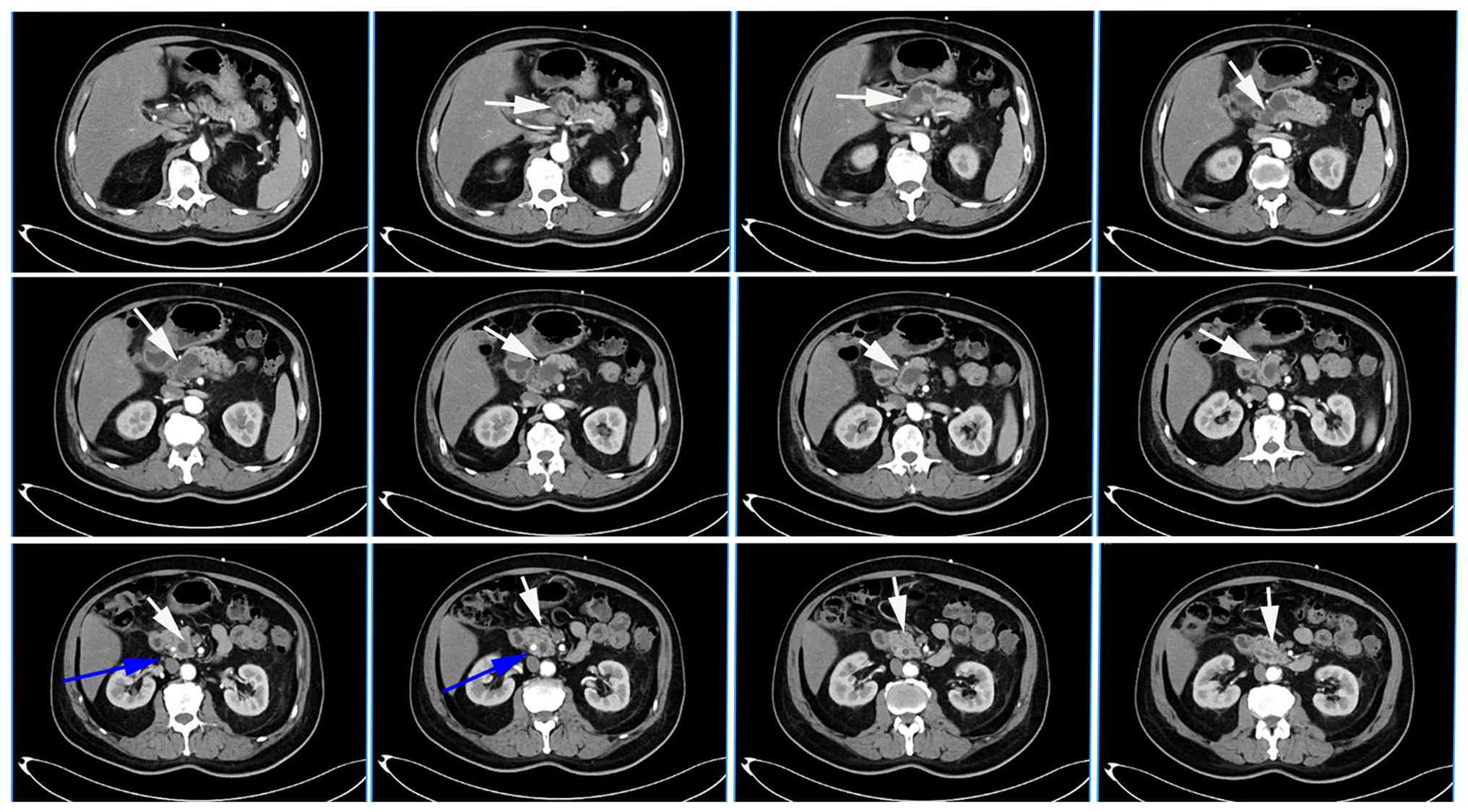
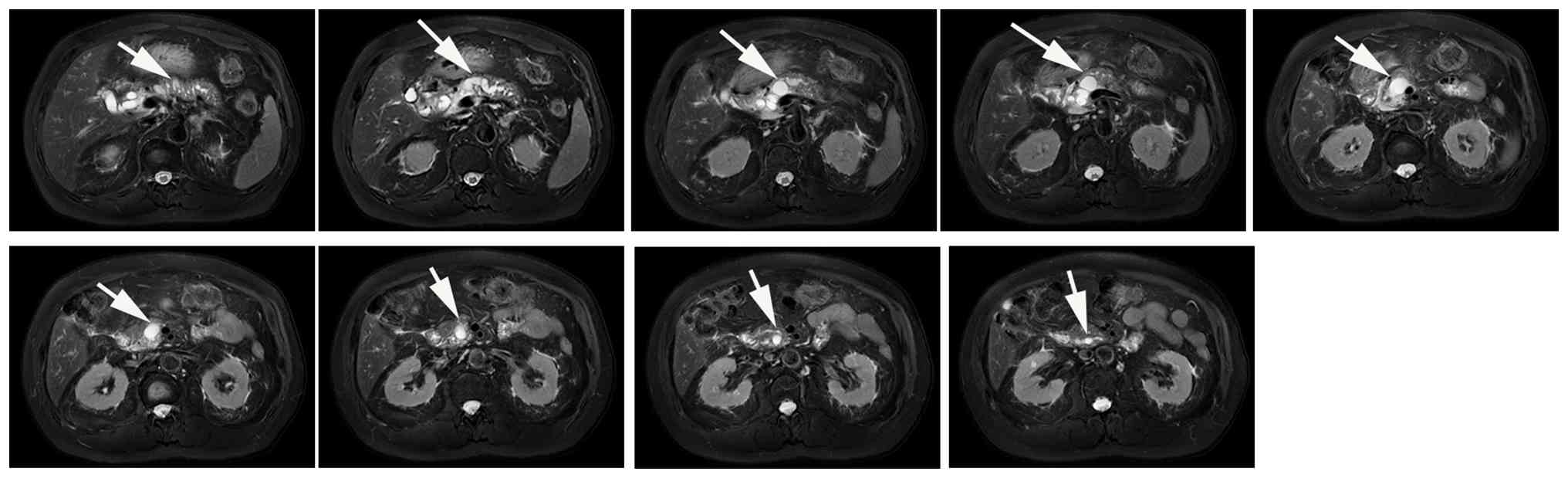
distress and abdominal distension. An initial CT scan (Fig. 1) and MRI (Fig. 2) exhibited dilatation of the MPD
and branch pancreatic duct (BPD) with small cystic changes, meaning
IPMNs could not be completely excluded. In addition, the CT scan
revealed the presence of gallstones in the common bile duct (CBD).
The CA19-9 (normal reference range, 0-37 IU/ml) concentration was
>1,000 IU/ml and CEA was 4.0 ng/ml (normal reference range, 0-5
ng/ml). CA19-9 was measured by electrochemiluminescence
immunoassay. Sample was incubated with biotin labeled anti-CA19-9
specific monoclonal antibody and ruthenium (Ru)a-labeled
anti-CA19-9 specific monoclonal antibody to form an
antigen-antibody sandwich complex. Streptavidin-coated magnetic
beads were added for incubation, and the complex bound to the
magnetic beads via biotin-streptavidin interaction. The reaction
mixture was aspirated into the measuring cell, and magnetic beads
were captured onto the electrode surface by electromagnetic force.
Unbound substances were removed using ProCell II M. A defined
voltage was applied to the electrode to trigger chemiluminescence
of the complex, and luminescence intensity was measured by a
photomultiplier tube. The test result was automatically calculated
by the instrument using a two-point calibrated calibration curve
and the master curve obtained via cobas link. The patient was
therefore diagnosed with a pancreatic mass and suspected IPMN. An
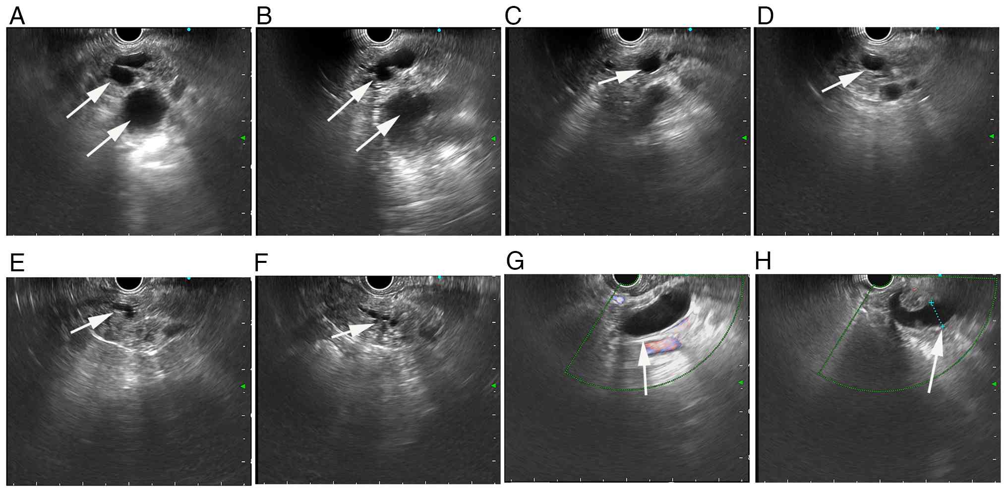
EUS (Fig. 3) was subsequently
performed, revealing that the cystic lesion and MPD of the patient
was dilated (Fig. 3A-F), with the
widest point being 11.5 mm (Fig.
3H). There was no obvious dilation of CBD (Fig. 3G).
After the preoperative surgical conversation, the
patient eventually refused surgery due to concerns regarding the
potential risks. In accordance with a multidisciplinary team
discussion, it was decided that an attempt would be made to obtain
the pathology of the IPMN and subsequently develop a further
treatment plan. ERCP was subsequently adopted in August 2025 to
remove gallstones in the CBD and to further clarify the lesions of
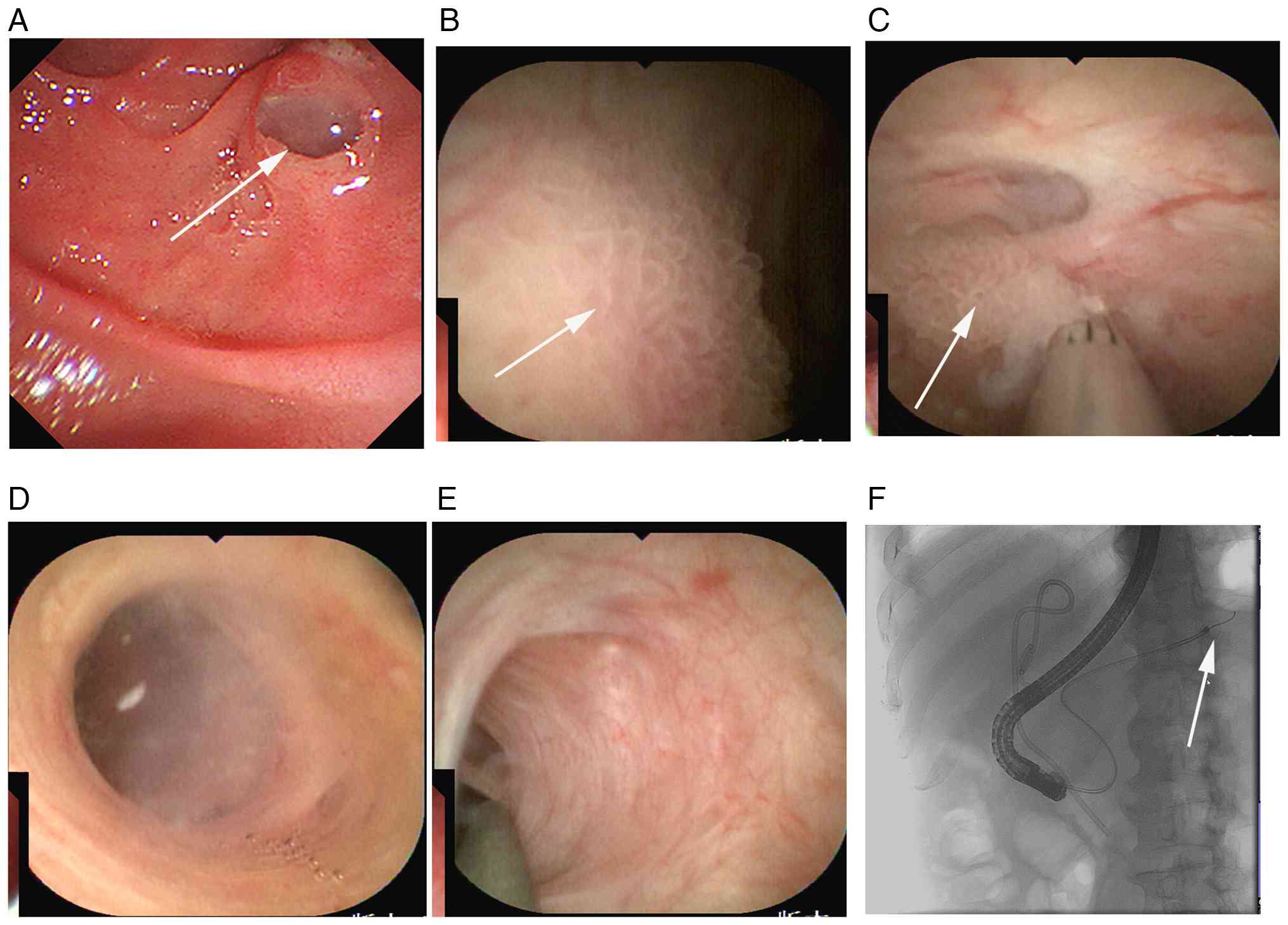
the pancreas (Fig. 4). The
duodenal papilla exhibited signs of a fish mouth (the opening of
duodenal papilla was significantly enlarged and the shape was round
and blunt, similar to the ‘fish mouth’) covered with mucus through
observation using a duodenoscope (Fig.
4A). On the basis of the ERCP using eyeMAX, villus-like and
papillary protrusions were visualized intuitively in the MPD wall
of the pancreatic head segment (Fig.
4B) and a total of four pieces of tissue were taken for biopsy
(Fig. 4C). The wall of the
body-tail portion of the MPD was smooth (Fig. 4D-F). A HistoCore SPECTRA H&E
Staining System S2 (filing no. 20170724; Leica Biosystems) with
fully automatic programming was used for H&E staining. This
system worked based on the HistoCore SPECTRA ST Stainer. Furnished
with a built-in barcode system, it enables accurately tracking of
different slides. Through an integrated dual translational robotic
arm, it can store and parallelly execute H&E staining
procedures. An integrated oven was incorporated in the device,
which simplifies the workflow by removing the need of the external
heating unit. The stainer can be directly integrated with Leica
HistoCore SPECTRA CV coverslip (12). Finally, the biopsy sections were
observed through OLYMPUS microscope (BX53) and images were
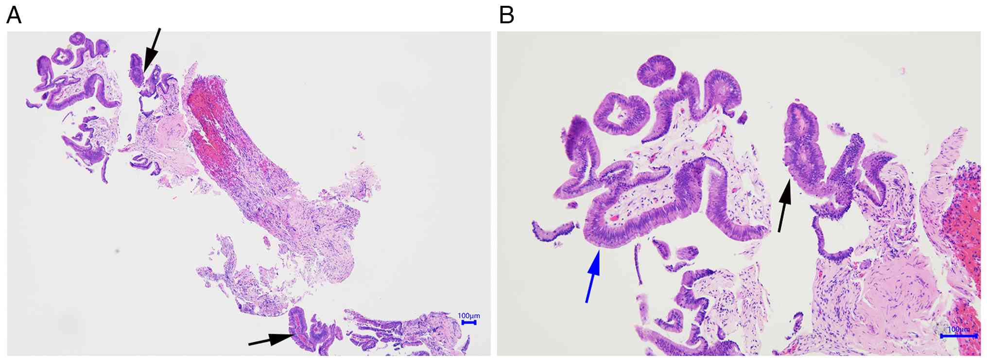
captured. Furthermore, the pathology revealed low-grade dysplasia
(Fig. 5). No adverse events
regarding the patient occurred following ERCP or biopsy. Given that
the pathology revealed a low malignant transformation tendency, it
was decided that a follow-up strategy, including monitoring
abdominal imaging and CA19-9 would be conducted for the patient.
The patient was discharged at the end of August, 2025 and has
remained in good condition. The most recent follow-up occurred in
July 2026. The patient was reported to be in a good general
condition without jaundice, pancreatitis or abdominal pain. The
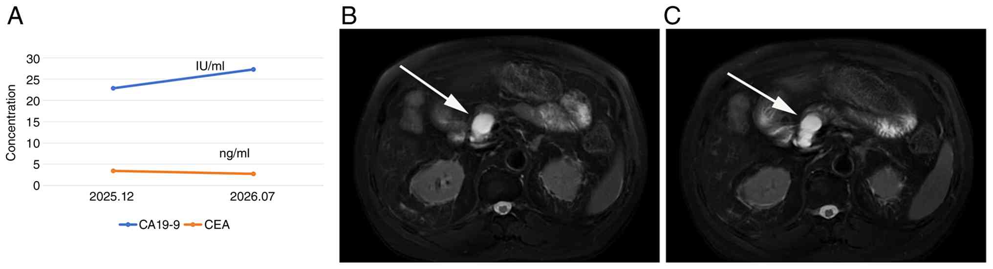
CA19-9 concentration of the patient was reported to be 27.284 IU/ml
and CEA was 2.751 ng/ml at the latest follow-up (Fig. 6A). The patient underwent an
enhanced MRI examination again in July 2026. Compared with the
previous scan, the MPD was shown to be slightly dilated. No notably
enhanced mural nodules or other malignant features were detected
(Fig. 6B and C).
Discussion
IPMNs are a mucin-producing subtype of the
pancreatic cyst lesions arising from the pancreatic duct system
(6) and are difficult for
diagnosis and treatment. It has been estimated that up to 49.1% of
individuals who have undergone cross-sectional imaging have
incidentally detected pancreatic cystic lesions (13). CT and MRI serve as the main imaging
modalities for the diagnosis of IPMNs which have typical and
characteristic CT and MRI features (14). CT provides excellent spatial
resolution and is especially efficient in evaluating pancreatic
ductal adenocarcinoma (PDAC), vascular invasion and metastatic
diseases. On the contrary, MR provides superior soft tissue
contrast and ductal delineation, allowing detailed assessment of
subtle stenosis, cystic lesions, and intraductal abnormalities
(15). Notably, CT serves a
dominant role in the evaluation of IPMNs; however, delineating the
connection between the cystic lesion and duct system is difficult
partly because the intravenous contrast cannot opacify the duct
lumen (16). MRI/MRCP is the
preferred initial method because it has advantages in
distinguishing benign from malignant IPMNs and similarly, MRI/MRCP
is the preferred choice for long-term follow-up (17). In accordance with the European
evidence-based Guidelines, EUS is recommended as an auxiliary
imaging modality when clinical or radiological findings cannot
exclude malignant transformation of IPMNs (18). EUS-fine-needle aspiration should be
performed when conventional radiological and EUS features fail to
yield a confirmed diagnosis (19);
however, its accuracy is low to a certain extent. Peroral
pancreatoscopy based on ERCP facilitates the diagnosis of IPMN,
especially with the development of eyeMAX in recent years (20). By benefitting from image processing
algorithms and eyeMAX, eyeMAX bite, not only can high-resolution
images be obtained, but also biopsy can be performed (21,22).
In addition, compared with previous pancreatoscopy methodologies,
eyeMAX allows more detailed observation of blood vessels and
glandular ducts and provides more representative images for further
analysis. In patients with suspected IPMN, who exhibit a dilated
MPD but no obvious mural nodule detected by EUS, the eyeMAX clearly
displays the microstructure of the lesion and acquires sufficient
pathological tissue. These factors also help to differentiate IPMN
from other pancreatic cystic lesions such as mucinous cystic
neoplasms and serous cystadenomas. In addition, eyeMAX more easily
reveals lesions of the distal pancreatic duct and has more economic
advantages, which is important for the majority of patients
(21,23). In general, the combination of CT,
MRI, EUS and ERCP using eyeMAX resulted in a more accurate
diagnosis of IPMN and helped to determine a more precise treatment
for the present patient. In addition, biopsy, improved cell block
processing, and marker identification were helpful for the
diagnosis and selection of individualized treatment (24-26).
The biological behavior of IPMNs ranges from benign
to invasive malignancy. Despite clinicians making their best
efforts to evaluate the lesions as extensively as possible, a
portion of patients with benign cysts may receive unnecessary
surgical resection, whereas lesions that are deemed appropriate for
surveillance can eventually progress to malignancy. In accordance
with Kyoto guidelines for managing pancreatic IPMN, an MPD diameter
≥10 mm constitutes one of the HRSs indicating elevated malignant
potential (7). In addition, a
previous retrospective cohort study suggested that an MPD diameter
of 7.2 mm or greater had the highest diagnostic accuracy and was an
independent predictor of malignancy in patients with MD or mixed
IPMNs (27). This study further
argued that a lower MPD diameter threshold should be recognized as
a criterion for surgical management (27). However, surgical resection is
associated with notable risks of morbidity and mortality (28).
In the present case, before the originally planned
surgery, the patient underwent CT, MRI and EUS examinations. Thus,
surgery was recommended first. As the patient refused resection of
the lesion and at the same time, a stone in the CBD was found, ERCP
was performed and the eyeMAX was also used. Unlike CT, MRI, EUS and
ERCP, lesions can only be evaluated through indirect imaging.
Through eyeMAX, images of the lesion were directly obtained and
targeted biopsy was performed under direct visualization. Pathology
of the lesion revealed low-grade dysplasia, and eventually, the
patient accepted a follow-up plan and was in good condition for ~1
year after the biopsy.
Despite the biopsy result being reassuring in the
present patient, a limited biopsy from the pancreatic duct cannot
necessarily exclude high-grade dysplasia or invasive carcinoma
elsewhere in the lesion, particularly in the context of main
pancreatic duct dilatation >10 mm. Therefore, the pathology
acquired through biopsy may not be able to represent the entirety
of the lesion. In general, it is necessary for clinicians to
perform multiple or regular biopsies; however, the diagnosis of
IPMN in the present patient was based on biopsy, imaging
examination (including CT and MRI) and endoscopy (including EUS and
ERCP) and given that complete pathological specimens of the lesion
were unobtainable, other cystic diseases of the pancreas could not
be completely excluded. Notably, in the present case, the elevated
CA19-9 and the pancreatic duct width greater than 10 mm were
considered HRSs of IPMN. However, firstly, the present patient
could not bear the risk of surgery and secondly, the pathology
revealed low-grade dysplasia and no obviously tortuous or dilated
blood vessels were detected by eyeMAX. Thus, a follow-up strategy
was adopted for the present patient. Importantly, the decision to
pursue follow-up should not be presented as a generally preferable
option for patients with low-grade dysplasia. In the present case,
it was chosen as an individualized approach after multidisciplinary
discussion because the patient declined surgical resection. CA19-9
is an important biomarker for malignant pancreatic tumors (29); however during biliary tract
inflammation, CA19-9 may be elevated and usually declines after
infection is controlled. In the present patient, the serum CA19-9
concentration was markedly high before stone removal from the CBD
and returned to the normal level after the operation. Thus, in the
present case, the increase in CA19-9 was considered to be
associated with inflammation and obstruction of the CBD secondary
to stones.
Overall, the findings from the present case
highlight the importance of biopsy acquired through eyeMAX based on
ERCP without causing high risks, which exhibited an important
diagnostic value for guiding the formulation of a treatment plan
for the present patient. Despite there being many sampling
limitations, more patients with pancreatic diseases may benefit
from the development of eyeMAX on the basis of CT, MR, EUS and
ERCP. In the future, the guidelines may be more optimized and
meticulous with the development of technology to obtain biopsies of
IPMNs.
Acknowledgements
The present authors would like to thank Dr. Zhixin
Cao (Department of Pathology, Shandong Provincial Hospital
Affiliated to Shandong First Medical University). The doctor
provided the patient's pathological images and reports.
Funding
Funding: No funding was received.
Availability of data and materials
The data generated in the present study may be
requested from the corresponding author.
Authors' contributions
WW and TL designed and conceptualized the present
study. Data acquisition and curation were implemented by WW and JH.
The initial draft was written by WW, with ZL and YY contributing to
manuscript revision, reviewing and editing. All authors were
responsible for all aspects of the research in order to ensure the
accuracy and integrity of the work. WW and YY confirm the
authenticity of all the raw data. All authors read and approved the
final version of the manuscript.
Ethics approval and consent to
participate
Not applicable.
Patient consent for publication
The present patient provided written informed
consent to publish the present case report and the associated
images. The patient was notified that all identifiable personal
data would be deleted to protect their privacy.
Competing interests
The authors declare that they have no competing
interests.
References
|
1
|
Omori Y, Furukawa T, Scarpa A and Luchini
C: Co-occurring IPMN and pancreatic cancer: The same or different?
An overview from histology to molecular pathology. J Clin Pathol.
76:734–739. 2023.PubMed/NCBI View Article : Google Scholar
|
|
2
|
Sato M, Kitahata Y, Nakamura Y,
Motobayashi H, Takemoto N, Nakai T, Hyo T, Ueno M, Hayami S,
Miyamoto A, et al: Urine Cell-Free DNA in the diagnosis of
intraductal papillary mucinous neoplasms: A prospective
observational study. Ann Surg Oncol: Jul 31, 2026 doi:
10.1245/s10434-026-20286-z (Epub ahead of print).
|
|
3
|
Cui M, Mo S, Bai J, Javed AA, Habib JR,
Yang S, Chen T, Xiao R, Diao W, Jiang D, et al: Spatial
transcriptomics defines the molecular progression, invasion and
immune landscape of IPMN and IPMN-derived pancreatic cancer. Gut.
75:801–814. 2026.PubMed/NCBI View Article : Google Scholar
|
|
4
|
Mahmud O, Fatimi AS, Grewal M, DiMaggio C,
Hewitt DB, Javed AA, Wolfgang CL and Sacks GD: Risk of pancreatic
cancer and high-grade dysplasia in resected main-duct and
mixed-type intraductal papillary mucinous neoplasms: A prevalence
meta-analysis. Eur J Surg Oncol. 51(109742)2025.PubMed/NCBI View Article : Google Scholar
|
|
5
|
Tanaka M, Fernández-del Castillo C, Adsay
V, Chari S, Falconi M, Jang JY, Kimura W, Levy P, Pitman MB,
Schmidt CM, et al: International consensus guidelines 2012 for the
management of IPMN and MCN of the pancreas. Pancreatology.
12:183–197. 2012.PubMed/NCBI View Article : Google Scholar
|
|
6
|
Nista EC, Schepis T, Candelli M, Giuli L,
Pignataro G, Franceschi F, Gasbarrini A and Ojetti V: Humoral
predictors of malignancy in IPMN: A review of the literature. Int J
Mol Sci. 22(12839)2021.PubMed/NCBI View Article : Google Scholar
|
|
7
|
Ohtsuka T, Fernandez-Del Castillo C,
Furukawa T, Hijioka S, Jang JY, Lennon AM, Miyasaka Y, Ohno E,
Salvia R, Wolfgang CL and Wood LD: International evidence-based
Kyoto guidelines for the management of intraductal papillary
mucinous neoplasm of the pancreas. Pancreatology. 24:255–270.
2024.PubMed/NCBI View Article : Google Scholar
|
|
8
|
Vege SS, Ziring B, Jain R and Moayyedi P:
American gastroenterological association institute guideline on the
diagnosis and management of asymptomatic neoplastic pancreatic
cysts. Gastroenterology. 148:819–822. 2015.PubMed/NCBI View Article : Google Scholar
|
|
9
|
Qing Q, Deng X, Deng X, Mou T, Li B, Tan Y
and Wu Q: A single-center study examining the safety and
effectiveness of ERCP with pancreatoscopy and endoluminal
radiofrequency ablation for main-duct IPMN treatment. Sci Rep.
15(5420)2025.PubMed/NCBI View Article : Google Scholar
|
|
10
|
Jung MK, Jang YJ, Cho CM, Kim GC, Cho SH,
Lee D, Ryeom HK and Shin KM: Iatrogenic pancreatitis in patients
with IPMN after ERCP: Incidence and predictive signs. Abdom
Imaging. 39:949–954. 2014.PubMed/NCBI View Article : Google Scholar
|
|
11
|
Toyonaga H, Tanisaka Y, Masaki M, Oka A,
Okita A, Osaki F, Matsuno Y, Takayama S, Nakata H, Takayama T, et
al: Efficacy and safety of balloon Endoscopy-Assisted peroral
cholangiopancreatoscopy (POCPS) using a novel 9Fr cholangioscope in
patients with surgically altered anatomy: A Dual-center
retrospective study (With Video). Dig Endosc.
38(e70237)2026.PubMed/NCBI View Article : Google Scholar
|
|
12
|
Kucarov MD, Takács M, Czakó BG, Molnár B
and Kozlovszky M: Mapping manual laboratory tasks to robot
movements in digital pathology workflow. Sensors (Basel).
25(6830)2025.PubMed/NCBI View Article : Google Scholar
|
|
13
|
Kromrey ML, Bülow R, Hübner J, Paperlein
C, Lerch MM, Ittermann T, Völzke H, Mayerle J and Kühn JP:
Prospective study on the incidence, prevalence and 5-year
pancreatic-related mortality of pancreatic cysts in a
population-based study. Gut. 67:138–145. 2018.PubMed/NCBI View Article : Google Scholar
|
|
14
|
Barron MR, Roch AM, Waters JA, Parikh JA,
DeWitt JM, Al-Haddad MA, Ceppa EP, House MG, Zyromski NJ, Nakeeb A,
et al: Does preoperative cross-sectional imaging accurately predict
main duct involvement in intraductal papillary mucinous neoplasm? J
Gastrointest Surg. 18:447–456. 2014.PubMed/NCBI View Article : Google Scholar
|
|
15
|
Maino C, Castagnoli F, Vernuccio F,
Cannella R, Bali MA, Koh DM, Zamboni G and Ippolito D: CT and MR
evaluation of main pancreatic duct dilation: A radiologist's guide
to differentiating benign from malignant causes. Eur J Radiol.
199(112803)2026.PubMed/NCBI View Article : Google Scholar
|
|
16
|
Machado NO, Al Qadhi H and Al Wahibi K:
Intraductal papillary mucinous neoplasm of pancreas. N Am J Med
Sci. 7:160–175. 2015.PubMed/NCBI View Article : Google Scholar
|
|
17
|
Liu H, Cui Y, Shao J, Shao Z, Su F and Li
Y: The diagnostic role of CT, MRI/MRCP, PET/CT, EUS and DWI in the
differentiation of benign and malignant IPMN: A meta-analysis. Clin
Imaging. 72:183–193. 2021.PubMed/NCBI View Article : Google Scholar
|
|
18
|
European Study Group on Cystic Tumours of
the Pancreas. European evidence-based guidelines on pancreatic
cystic neoplasms. Gut. 67:789–804. 2018.PubMed/NCBI View Article : Google Scholar
|
|
19
|
Al-Taee AM and Taylor JR: Endoscopic
imaging of pancreatic cysts. Gastrointest Endosc Clin N Am.
33:583–598. 2023.PubMed/NCBI View Article : Google Scholar
|
|
20
|
Miwa H, Sugimori K, Irie K, Goda Y,
Shibasaki K, Ishino Y, Tsunoda S, Endo K, Oishi R, Suzuki Y, et al:
Feasibility of novel slim peroral cholangiopancreatoscopy for the
diagnosis of pancreatobiliary disease. DEN Open.
6(e70152)2026.PubMed/NCBI View Article : Google Scholar
|
|
21
|
Ogura T, Ueno S, Hakoda A, Aboelezz AF,
Okuda A, Nishioka N, Sakamoto J, Matsuno J, Uba Y, Tomita M, et al:
Diagnostic yield of a novel 11-Fr digital cholangioscope for
indeterminate biliary disease using Macroscopic-On-Site evaluation:
Prospective comparative study. J Gastroenterol Hepatol.
40:1307–1314. 2025.PubMed/NCBI View Article : Google Scholar
|
|
22
|
Tanisaka Y, Mizuide M, Fujita A, Shin T,
Sugimoto K, Jinushi R and Ryozawa S: Successful
cholangioscopy-guided biopsy using a novel thin cholangioscope
under balloon enteroscopy in a patient with Roux-en-Y gastrectomy.
Endoscopy. 56:E256–E257. 2024.PubMed/NCBI View Article : Google Scholar
|
|
23
|
Karsenti D, Sportes A, Leblanc S, Béchet
S, Derman J, Fassler I and Brieau B: Contribution of the EyeMAX™
11Fr (Micro-Tech) digital single-operator cholangioscope with a
wide working channel: A multicenter pilot study on the first French
experiences. Ther Adv Gastroenterol.
18(17562848251360117)2025.PubMed/NCBI View Article : Google Scholar
|
|
24
|
Policardo F, Tralongo P, Arciuolo D,
Fiorentino V, Cardasciani L, Pierconti F, Carlino A, Curatolo M,
Pontecorvi A, Fadda G, et al: p53 expression in cytology samples
may represent a marker of early-stage cancer. Cancer Cytopathol.
131:392–401. 2023.PubMed/NCBI View Article : Google Scholar
|
|
25
|
Kirbis IS, Kholova I, Huhtala H, Flezar
MS, Roque RR, Mäkinen HH, Litmanen M, Vrdoljak-Mozetič D, Harabajsa
S, Hewer E, et al: Cellularity of routinely prepared cell blocks:
Insights from an international study. Cytopathology. 37:214–221.
2026.PubMed/NCBI View Article : Google Scholar
|
|
26
|
Fiorentino V, Giordano W, Pizzimenti C,
Zuccalà V, Ieni A, Molinario C, Cannavò S, Campennì A, Tralongo P,
Martini M, et al: Molecular profiling of thyroid nodules on
cytologic samples: Findings from an Italian multi-institutional
cohort. Cancer Cytopathol. 134(e70065)2026.PubMed/NCBI View Article : Google Scholar
|
|
27
|
Sugimoto M, Elliott IA, Nguyen AH, Kim S,
Muthusamy VR, Watson R, Hines OJ, Dawson DW, Reber HA and Donahue
TR: Assessment of a revised management strategy for patients with
intraductal papillary mucinous neoplasms involving the main
pancreatic duct. JAMA Surg. 152(e163349)2017.PubMed/NCBI View Article : Google Scholar
|
|
28
|
Kleeff J, Michalski C, Kong B, Erkan M,
Roth S, Siveke J, Friess H and Esposito I: Surgery for cystic
pancreatic lesions in the post-sendai era: A single institution
experience. HPB Surg. 2015(847837)2015.PubMed/NCBI View Article : Google Scholar
|
|
29
|
Månsson C, Nilsson A, Urdzik J and Karlson
BM: The Value of CA19-9 after irreversible electroporation for
pancreatic cancer. Anticancer Res. 39:6193–6196. 2019.PubMed/NCBI View Article : Google Scholar
|