Introduction
Crowned dens syndrome (CDS) was first defined in
1985 as a clinico-radiological syndrome resulting from the
deposition of periodontoid calcium pyrophosphate dihydrate (CPPD)
or hydroxyapatite (HA) crystals (1). The characteristic imaging features
include a halo-like or coronal calcification located posterior to
the odontoid process (2). Clinical
manifestations can be categorized into two types: The acute type,
which is more prevalent, often presenting with fever and elevated
levels of inflammatory markers, such as erythrocyte sedimentation
rate (ESR), C-reactive protein (CRP) and leukocyte count (white
blood cells) (3). Conversely, the
chronic type is rare and is characterized by substantial calcified
lesions that form retroodontoid pseudotumors, leading to the
compression of the cervical spinal cord. This may result in
cervical spondylotic myelopathy, limb muscle weakness, paresthesia
and positive pathological signs (2,4,5).
The management of CDS typically depends on the
symptoms of the patient. The majority of cases respond favorably to
conservative treatment approaches. Currently, primary medical
interventions involve non-steroidal anti-inflammatory drugs
(NSAIDs) or short courses of corticosteroids. Patients frequently
experience significant symptom relief within a few days of
administration (6,7).
However, for those suffering from cervical
spondylotic myelopathy due to prolonged pain associated with spinal
cord compression, accompanied by limb muscle weakness, regional
paresthesia and positive pathological indicators of spinal nerve
injury, surgical intervention becomes imperative before
irreversible damage occurs to the spinal cord (8). Various surgical techniques are
employed based on individual patient conditions, hospital resources
available for surgery and the clinical expertise of the attending
physicians. Presently utilized surgical methods primarily include
the following: Anterior approaches via microsurgery (transoral) or
endoscopy (transnasal), as well as posterior approaches through
epidural access involving the direct resection of lesions, while
simultaneously performing posterior internal fixation (2,5,9,10).
Therefore, the present study reports two cases of CDS with distinct
clinical presentations and therapeutic strategies, one successfully
managed conservatively and the other with CDS-related high cervical
myelopathy treated by isolated resection of the posterior arch of
the atlas under microscopic assistance, a surgical approach rarely
documented in the literature. The study aims to elucidate the
clinical spectrum of this rare disorder and to highlight that
posterior atlas arch resection alone may serve as an effective and
less invasive option for patients with CDS complicated by cervical
cord compression.
Case reports
Case 1
A 72-year-old man was admitted to the Department of
orthopedics, Guangzhou Red Cross Hospital of Jinan University
(Guangzhou, China) in April 2022, presenting with occipital and
cervical pain that had persisted for >1 month prior to
admission, accompanied by restricted mobility of the cervical
spine. The pain was exacerbated in the morning and was associated
with episodes of dizziness and occasional numbness in both hands.
The patient had no notable previous medical history, including
rheumatoid arthritis or pseudoarthritis, or any other predisposing
factors.
A physical examination revealed tenderness upon
palpation of the spinous processes and interspinous spaces of the
cervical spine, as well as limited extension and rotation of the
neck. All other aspects of the physical examination did not reveal
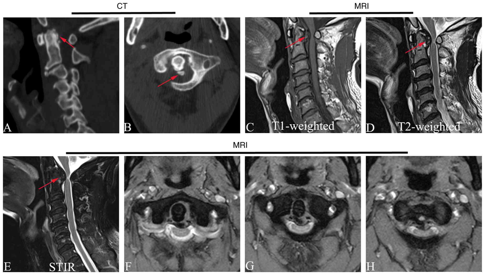
any notable findings. A computed tomography (CT) scan of the
cervical spine demonstrated thickening of the posterior ligament at
the odontoid process along with coronal calcification, which
resulted in the narrowing of the adjacent spinal canal (Fig. 1). Magnetic resonance imaging (MRI)
showed thickening of the soft tissue posterior to the odontoid
process, with compression and deformation of the anterior margin of
the spinal cord (Fig. 1).
Laboratory tests did not show an increase in inflammatory markers
such as C-reactive protein, interleukin-6, erythrocyte
sedimentation and rheumatoid factor (data not shown).
Following a thorough assessment to exclude
contraindications for medication use, treatment was initiated with
diclofenac sodium sustained-release tablets (75 mg once daily) and
prednisone tablets (5 mg three times daily) for a duration of 1
week. This regimen led to symptomatic relief. Upon discharge,
medication was continued for an additional week to reinforce
therapeutic outcomes. The relevant clinical information for case 1
is presented in Table I.
 | Table ISummary of clinical data of case 1 and
case 2. |
Table I
Summary of clinical data of case 1 and
case 2.
| Clinical
information | Case 1 (conservative
treatment) | Case 2 (surgical
treatment) |
|---|
| Age, years | 72 | 81 |
| Sex | Male | Male |
| Main symptoms | Isolated neck pain
and restricted mobility | Neck pain accompanied
by spinal cord compression symptoms such as neck stiffness,
radiating pain and numbness in both upper extremities, unsteady
gait and dysuria |
| Neurological
examination | Tenderness and
percussion pain over each cervical spinous process and interspinous
space, with limited neck extension and rotation. | Limited neck
mobility, spastic gait and impaired fine motor skills of both
hands; decreased muscle strength and palmar sensation in the left
upper extremity, positive Hoffmann's sign on the left |
| Laboratory
examination | No elevation of
inflammatory markers | No elevation of
inflammatory markers |
| CT/MRI findings | CT: Thickening and
calcification of the posterior ligament of the odontoid process,
secondary spinal canal stenosis. MRI: Marked thickening of the soft
tissue posterior to the odontoid process, with compression and
deformation of the anterior margin of the spinal cord | CT: Multiple bone
erosions of the odontoid process and the left atlanto-occipital
articular surface, with surrounding irregular density shadows. MRI:
Spinal canal stenosis posterior to the odontoid process,
degenerative compression of the cervical spinal cord at the C1-C2
segment |
| Treatment | Diclofenac sodium
sustained-release tablets (75 mg orally once daily) combined with
prednisone tablets (5 mg orally three times daily) for conservative
treatment for 2 weeks | Microscope-assisted
posterior atlas posterior arch resection; postoperative maintenance
treatment with NSAIDs for 2 weeks |
| Follow-up
duration | 2 years | 3 years |
| Recurrence | Neck pain has not
recurred | Symptoms of spinal
cord compression were markedly relieved, the patient regained
self-care ability and the symptoms did not recur |
| Complications | None | No complications such
as anterior arch stress fracture, atlantoaxial instability or
enlargement of calcified foci |
Prior to treatment initiation, the visual analog
scale (VAS) score (11) of the
patient was recorded at 5 points; however, a post-treatment
evaluation indicated a reduction in the VAS score to 1 point.
During 2 years of subsequent follow-up visits, as the patient
reported no recurrence of neck pain and was unwilling to undergo
further cervical spine imaging, no imaging analyses were
performed.
Case 2
An 81-year-old man was admitted to the Department of
Orthopedics, Guangzhou Red Cross Hospital of Jinan University in
December 2021, presenting with progressive neck and shoulder pain
for 3 months, accompanied by restricted neck mobility, radiating
pain in the upper limbs, numbness, gait instability, dysuria and
impaired fine finger movements indicative of nerve compression. The
patient had no prior history of rheumatoid arthritis,
pseudoarthritis or cervical spine trauma, but had a 6-year history
of hypertension and a 20-year history of chronic bronchitis. The
patient had previously undergone knee joint replacement surgery and
percutanous coronary drug-coated balloon angioplasty.
A physical examination revealed limited neck
movement, a spastic gait, diminished fine motor function in both
hands, reduced muscle strength and superficial sensation in the
left upper limb, as well as a positive left Hoffman sign.
Laboratory tests indicated no elevated levels of inflammatory
markers. Dynamic X-rays of the cervical spine revealed no signs of
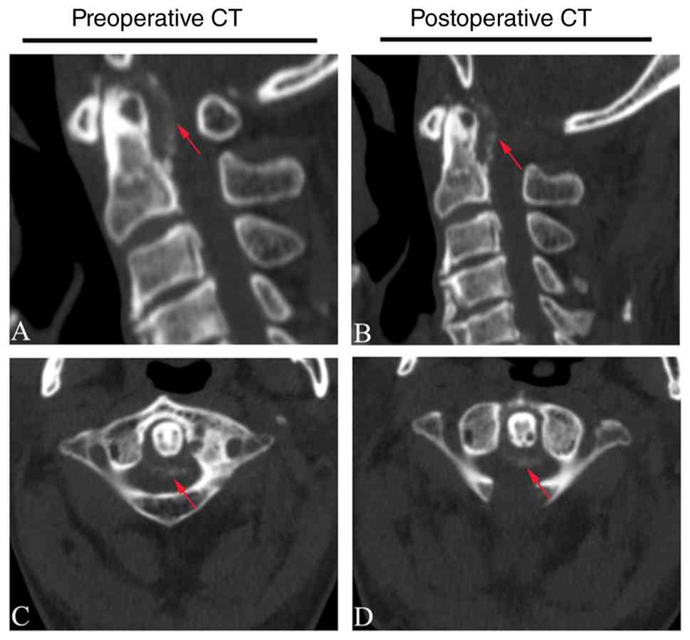
instability. A CT scan revealed multiple bone erosions with
irregular density surrounding the odontoid process and the left
atlantooccipital joint surface (Fig.
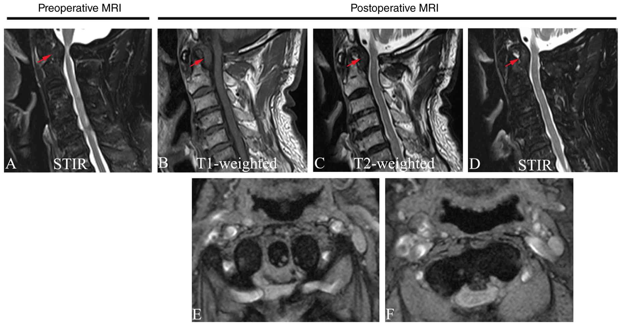
2). MRI demonstrated spinal canal stenosis posterior to the
odontoid process along with cervical spinal cord compression at the
C1-C2 level (Fig. 3). The relevant
clinical information for case 2 is presented in Table I.
The patient presented with obvious symptoms of
spinal cord compression, and exhibited a poor response to
conservative treatment (200 mg oral celecoxib every 12 h for 2
weeks), with neurological function gradually deteriorating. Surgery
had to be performed prior to the occurrence of irreversible spinal
cord damage. However, considering the advanced age of the patient
and the potential complex complications associated with surgery, a
microscope-assisted posterior arch resection of the atlas via the
posterior approach was performed. The surgery was performed under
general anesthesia with endotracheal intubation. Following
successful anesthesia, the patient was placed in the prone
position, and cranial traction was applied to the cervical
vertebrae using a cranial traction device. Following satisfactory
positioning, routine disinfection and draping of the neck were
performed. After locating the C1 vertebral segment on the body
surface, a midline posterior incision of ~4 cm was made. The skin,
subcutaneous tissue and supraspinous ligament were incised
longitudinally. The erector spinae muscles on both sides were
stripped using a periosteal elevator to expose the posterior arch
of C1. With the assistance of a neurosurgical microscope, the
posterior arch of the atlas was precisely resected using a rongeur
and the width of the space created by the arch removal was ~3 cm.
The dural sac was gradually exposed, and adequate decompression was
achieved to provide sufficient space for the compressed spinal cord
to shift. During the surgery, two anti-adhesion membranes were
placed, and hemostatic suturing was performed. Intraoperative blood
loss was ~30 ml. The total duration of the surgery was 2 h and 10
min.
Postoperatively, NSAIDs were administered for 2
weeks. The preoperative Japanese Orthopedic Association (JOA) score
(12) was 8, and the VAS score was
5. Post-operatively, the JOA score improved to 14, and the VAS
score decreased to 1. At 3 years post-surgery, the JOA score had
reached 16, and the VAS score was 0. The post-operative
neurological function improvement rate was 66.7%, which increased
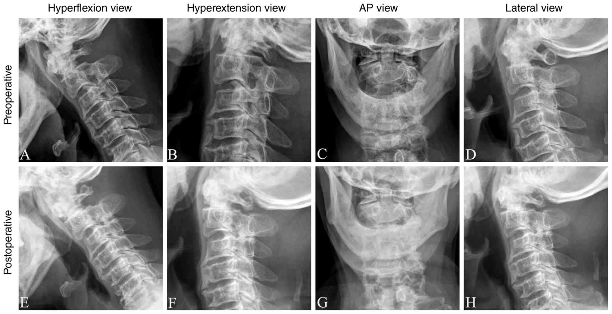
to 88.9% at 3 years post-surgery. During the 3-year follow-up, the
symptoms of the patient did not recur, and digital radiography
revealed no increase or absorption of calcified foci around the
odontoid process, no stress fracture of the anterior arch, no
atlantoaxial instability and no other complications (Fig. 4).
Discussion
The prevalence of CPPD crystal disease in
individuals presenting with acute neck pain is ~2% (13). Due to the limited understanding of
CDS, a misdiagnosis or a missed diagnosis frequently occurs in
routine clinical practice (14).
In the two cases presented in the current study, both patients were
elderly men at >70 years of age who sought medical attention for
acute neck pain accompanied by restricted neck movement; both were
subsequently diagnosed with CDS following admission. Consequently,
it is imperative for clinicians, particularly those working in
Emergency Departments, to enhance their awareness of CDS. This
condition should be considered in patients experiencing acute neck
pain alongside elevated inflammatory markers. Timely cervical spine
CT scans are essential to improve the diagnostic rate of CDS and to
prevent unnecessary examinations or treatments that could delay
appropriate diagnosis and management.
The pathological mechanism underlying CDS is closely
associated with the deposition of CPPD or HA crystals. While CPPD
deposition typically affects peripheral joints, such as the knees
and wrists, spinal involvement remains rare (15,16).
Conversely, HA crystal deposition predominantly occurs at tendon
attachment sites. Existing literature indicates that CPPD-related
CDS is more commonly reported than HA crystal deposition-induced
CDS, which is consistently recognized as a rare subtype
predominantly affecting middle-aged women. Although precise
incidence rates are unavailable due to the rarity of these
conditions and the absence of large-scale epidemiological data, the
number of published cases and case series for CPPD-related CDS
substantially exceeds that for HA crystal deposition-related CDS
(15,16). The imaging characteristics
exhibited by these two conditions are similar; therefore,
differentiation based solely on imaging findings may not be
feasible (14,17).
CDS may manifest with a range of clinical symptoms;
however, the typical presentations during the acute phase often
include acute neck pain, neck stiffness and elevated levels of
inflammatory markers such as ESR, CRP and white blood cell count.
As the disease progresses, substantial calcified lesions can evolve
into space-occupying masses that progressively compress the
cervical spinal cord, ultimately resulting in cervical spondylotic
myelopathy. Patients may exhibit signs of nerve damage, including
diminished muscle strength in the limbs, paresthesia, abnormal
bowel and urinary function, as well as positive pathological
findings (18). Consequently, it
is essential to differentiate CDS from various other conditions
such as trauma, meningitis, cervical discitis or giant cell
arteritis, epidural abscesses, osteomyelitis, polymyalgia
rheumatica, metastatic bone disease and tumors. Clinically relevant
differential diagnoses can be established through a thorough
patient medical history review coupled with meticulous physical
examinations and appropriate auxiliary investigations (4).
A neck CT scan is considered the gold standard for
diagnosing CDS due to its high resolution, which allows for the
clear visualization of calcification morphology and distribution
along with assessing spinal canal stenosis severity (19). In contrast to CT scans, while MRI
cannot directly identify crystal deposition, it plays a crucial
role in evaluating the extent of spinal cord compression and
excluding tumors or infections among other diseases (20). Dynamic X-ray imaging serves a key
purpose by aiding in the assessment of cervical spine stability
prior to surgical intervention while also aiding in the prevention
of secondary deformities postoperatively (5). In the present study, the diagnosis of
CDS in case 2 was established based on a combination of
characteristic clinical features, pathognomonic imaging findings
and systematic exclusion of differential diagnoses. The diagnosis
of CDS in case 2 fulfilled the currently established diagnostic
criteria, including: i) Acute severe neck pain; ii) marked
limitation of cervical motion, particularly rotation; iii) elevated
inflammatory markers, such as elevated CRP levels; iv) CT evidence
of calcium deposition around the odontoid process; and v) absence
of trauma history, with exclusion of other inflammatory diseases
and tumors (21,22). Among these, cervical CT scan
remains the gold standard for diagnosing CDS, as its high
resolution enables clear visualization of the morphology and
distribution of calcifications and accurate assessment of spinal
canal stenosis. The most characteristic imaging finding of CDS is
the coronal or horseshoe-shaped calcification deposited around the
top and lateral aspects of the odontoid process, which was clearly
demonstrated on the cervical CT scan of case 2. Although MRI cannot
directly identify crystal deposits, it plays an irreplaceable role
in evaluating the degree of spinal cord compression and excluding
neoplastic or infectious conditions, while dynamic cervical
radiographs are valuable for preoperative assessment of cervical
stability to prevent postoperative secondary deformity.
Notably, the CT scans in case 2 revealed multiple
bone erosions around the odontoid process and atlantooccipital
joint. Such erosive changes are recognized manifestations in
patients with CDS, likely resulting from chronic inflammatory
stimulation caused by CPPD or HA crystal deposition. However, these
erosions exhibit a non-aggressive, localized pattern and lack the
permeative or destructive characteristics typical of neoplastic or
infectious diseases, thereby supporting the diagnosis of CDS rather
than other invasive pathologies.
In clinical practice, CDS must be differentiated
from trauma, meningitis, cervical myofascial pain syndrome,
polymyalgia rheumatica (PMR), epidural abscess, osteomyelitis,
metastatic bone disease and tumors through detailed history taking,
meticulous physical examination and appropriate ancillary
investigations (23). In the
present study, trauma was excluded on clinical and imaging grounds,
as case 2 had no history of recent neck trauma and cervical CT
revealed no evidence of atlantoaxial dislocation or fracture.
Meningitis, one of the most critical differential diagnoses, was
also excluded on clinical and imaging grounds; although meningitis
closely mimics CDS in clinical presentation, an important
diagnostic criterion for meningitis is the demonstration of nodular
or thickened linear meningeal enhancement on MRI, with meningeal
enhancement >3 cm raising strong suspicion for meningeal
pathology, yet no such meningeal changes were observed in case 2.
Cervical myofascial pain syndrome was excluded on clinical and
physical examination grounds, as this condition typically results
from overuse of cervical myofascial tissues or acute trauma, with
palpable cord-like structures at the affected site and symptomatic
relief achievable through manual massage, whereas case 2 had no
predisposing factors, no abnormal cord-like structures were
palpated and symptoms could not be relieved by manual massage.
Polymyalgia rheumatica was excluded on clinical and laboratory
grounds, as the typical manifestations of PMR, including stiffness
and pain in the neck, shoulder and hip girdles, shoulder girdle
fatigue in nearly all patients, neck girdle fatigue in ~70% of
cases, and non-specific systemic features such as low-grade fever,
fatigue, somnolence, generalized myalgia, anorexia and weight loss,
were absent in case 2. Infectious conditions such as epidural
abscess and osteomyelitis were excluded on laboratory and imaging
grounds, as peripheral white blood cell and neutrophil counts were
within normal limits upon admission, and cervical MRI showed no
evidence of spinal canal abscess or vertebral body infection.
Similarly, metastatic bone disease and primary cervical tumors were
excluded on clinical and imaging grounds, as case 2 had no history
of malignancy and cervical MRI revealed no malignant
space-occupying lesions. In conclusion, the diagnosis of CDS in
case 2 is well established based on the characteristic clinical
presentation, pathognomonic imaging findings and systematic
exclusion of relevant differential diagnoses through clinical
evaluation, laboratory investigations and imaging studies.
As no pharmacological therapy is currently available
to eliminate or prevent intra-articular crystal deposition, the
management strategy for CDS emphasizes inflammation control and
individualized treatment. For acute CDS, NSAIDs combined with
short-term corticosteroids can effectively inhibit the inflammatory
response and alleviate pain and stiffness. Colchicine may serve as
a second-line option in refractory cases; however, its
gastrointestinal side-effects could limit its use among elderly
patients. Since there is no direct drug therapy available to
eliminate or prevent the deposition of crystal deposits in the
joints, the current focus of medication treatment is to reduce the
associated inflammation, resulting in a certain probability of
symptom recurrence. In different studies, the recurrence rate of
symptoms following conservative treatment is mostly concentrated in
the range of 20 to 35%, with follow-up periods typically ranging
from 6 to 12 months. In the study by Goto et al (18), 40 patients with symptomatic CDS
confirmed by CT were included, and the treatment regimen included
NSAID monotherapy, prednisone monotherapy or a combination of both.
At the 9-month follow-up, the symptom recurrence rate was 22.5%
(9/40). In another study, the overall recurrence rate after
conservative treatment was 34.62% (9/26); the recurrence time span
ranged from 0.5 to 12 months, and patients who experienced
recurrence could still achieve rapid symptom relief with the same
type of medication (24). Within
the present study, in case 1, after excluding relevant drug
contraindications for the patient, a combination of diclofenac
sodium sustained-release tablets and prednisone tablets was used
for treatment. The symptoms of the patient were markedly relieved,
consistent with the treatment outcomes reported in other cases, and
no recurrence occurred during subsequent follow-up.
As observed in case 2 of the present study, the
patient ultimately required surgical intervention due to the
compression of the cervical spinal cord caused by the formation of
a posterior odontoid pseudotumor (8). Currently, the most commonly employed
surgical methods include: The anterior approach, with microsurgical
(transoral) and endoscopic (transnasal) techniques; and the
posterior approach, wherein the lesion is directly excised through
an epidural route during surgery, accompanied by posterior internal
fixation. These surgical approaches have demonstrated favorable
treatment outcomes for patients. Nonetheless, there remains no
consensus on the optimal surgical strategy to date (25).
Considering elderly patients with complex underlying
conditions, they may not tolerate the high risks, prolonged
operative times and potential postoperative complications
associated with transoral resection of calcification or isolated
posterior fusion procedures. Within the present study, in case 2, a
posterior approach was used for an atlas posterior arch resection.
Before proceeding with the resection, a dynamic fluoroscopic
evaluation was performed under anesthesia, including
flexion-extension views, to confirm the absence of pathological
translation or angulation at the atlantoaxial joint. During the
surgical exposure, the integrity of the transverse atlantal
ligament and the surrounding soft tissues was also directly
inspected. Stability was defined as the absence of >3 mm of
anterior translation of the atlas relative to the axis on lateral
fluoroscopy and no evidence of ligamentous disruption on direct
visualization. Preoperative imaging assessment revealed no
atlantoaxial instability in the patient, so internal fixation was
not performed. Simple atlas resection (without fusion) is a
surgical procedure applicable to conditions such as Arnold-Chiari
malformation (often combined with suboccipital craniectomy),
intraspinal tumors at the C1 level and retrodental pseudotumors
without instability. The total width of the resected atlas
posterior arch was 3 cm (below the safe exposure width of ~3.5 cm
for the atlas posterior arch), which avoided intraoperative injury
to the vertebral artery (26). The
postoperative neurological improvement rate was ~66.7%, and the
improvement rate at 3 years postoperatively reached ~88.9%.
Long-term stress fractures of the atlas anterior arch after an
atlas posterior arch resection are rare, but severe cases may
result in atlantoaxial joint instability, necessitating a
reoperation for internal fixation (26-28).
In the present case, the 3-year follow-up imaging assessment
revealed no enlargement or absorption of calcified lesions around
the odontoid process, no anterior arch stress fracture, no cervical
instability and no other complications.
In conclusion, the management of cervical
degenerative stenosis should be tailored to the extent of
calcification and nerve compression present in each patient. For
the majority of individuals with acute CDS, conservative treatment
is deemed appropriate; however, resection of the posterior arch of
the atlas has been shown to effectively alleviate symptoms
associated with chronic myelopathy. Based on the single surgically
treated case described within the present study, a posterior arch
resection appears to be a potential treatment option; however, its
effectiveness cannot be validated at this stage. Further reports
with a greater number of cases are necessary to draw definitive
conclusions.
Acknowledgements
Not applicable.
Funding
Funding: The present study was financially supported by the
Guangzhou Municipal Science and Technology Project (grant nos.
2025A03J3363, 2023A03J0571 and 2023A03J0574), the Guangzhou
Traditional Chinese Medicine and Integrated Traditional Chinese and
Western Medicine Project (grant no. 20242A011014), and the
Technology Project of Guangzhou Municipal Health Commission (grant
no. 20261A010032).
Availability of data and materials
The data generated in the present study are included
in the figures and/or tables of this article.
Authors' contributions
YL contributed to study conception and design,
collated clinical cases and wrote the original manuscript. XL
obtained medical images (MRI and CT scans), analyzed patient data
and drafted the manuscript. ZW obtained X-ray images, and performed
the diagnostic analysis. XX contributed to study conception and
design, and revised the manuscript. SH and HM contributed to study
conception and design, and provided critical revision and writing
guidance. XX and HM confirm the authenticity of all the raw data.
All authors have read and approved the final version of the
manuscript.
Ethics approval and consent to
participate
Informed consent for the use of their samples in
scientific research was obtained from all patients. The present
study was conducted after the protocol was approved by the Medical
Ethics Committee of Guangzhou Red Cross Hospital of Jinan
University (Guangzhou, China; approval no. 2022-091-01).
Patient consent for publication
Written informed consent was obtained from the
patients for publication of the present case report and associated
images.
Competing interests
The authors declare that they have no competing
interests.
References
|
1
|
Bouvet JP, le Parc JM, Michalski B,
Benlahrache C and Auquier L: Acute neck pain due to calcifications
surrounding the odontoid process: The crowned dens syndrome.
Arthritis Rheum. 28:1417–1420. 1985.PubMed/NCBI View Article : Google Scholar
|
|
2
|
Haas P, Hauser TK, Kandilaris K, Skardelly
M, Tatagiba M and Adib SD: Case report: Posterolateral epidural
supra-C2-root approach (PESCA) for biopsy of a retro-odontoid
lesions in same sitting after occipitocervical fixation and
decompression in a case of crowned dens syndrome with brainstem
compression and displacement. Front Surg. 9(797495)2022.PubMed/NCBI View Article : Google Scholar
|
|
3
|
Younis AAR: Crowned dens syndrome as a
cause of acute neck pain: A case report and review of the
literature. Mediterr J Rheumatol. 28:101–105. 2017.PubMed/NCBI View Article : Google Scholar
|
|
4
|
Michel J, Bedel JB, Pinson C and Joly LM:
Crowned dens syndrome: An uncommon mimic of meningitis in the
emergency department. Am J Emerg Med. 63:180.e1–180.e3.
2023.PubMed/NCBI View Article : Google Scholar
|
|
5
|
Adada B, Vera Silva M, Darwish H and
Dakwar E: Far-lateral trans-atlas extradural resection of
retro-odontoid synovial cyst: Surgical technique and review of
literature. Interdiscip Neurosurg. 17:28–35. 2019.
|
|
6
|
Zeng L, Liao H, Xie M, Ren F, Zhang Y and
Wang Q: Images in neuroscience: Crowned dens syndrome. Neurol
india. 70:1329–1330. 2022.PubMed/NCBI View Article : Google Scholar
|
|
7
|
Otaki K, Takahashi T, Kai R and Horii A:
Crowned dens syndrome: A differential diagnosis of postoperative
neck pain. Eur Ann Otorhinolaryngol Head Neck Dis. 140:199–200.
2023.PubMed/NCBI View Article : Google Scholar
|
|
8
|
Chang DG, Park JB, Jung HY and Seo KJ:
Cervical myelopathy due to subaxial calcium pyrophosphate dihydrate
(CPPD) deposition with simultaneous asymptomatic crowned dens
syndrome: Two case reports. BMC Musculoskelet Disord.
21(713)2020.PubMed/NCBI View Article : Google Scholar
|
|
9
|
Visocchi M, Signorelli F, Liao C, Rigante
M, Paludetti G, Barbagallo G and Olivi A: Transoral versus
transnasal approach for craniovertebral junction pathologies: Never
say never. World Neurosurg. 110:592–603. 2018.PubMed/NCBI View Article : Google Scholar
|
|
10
|
Riley K, Singh H, Meyer SA and Jenkins AL
III: Minimally invasive surgical approach for odontoid lesions: A
technical description in a case of high cervical osteomyelitis and
abscess. World Neurosurg. 91:332–339. 2016.PubMed/NCBI View Article : Google Scholar
|
|
11
|
Marjamaa J, Huttunen J, Kankare J,
Malmivaara A, Pernaa K, Salmenkivi J and Pekkanen L: The Finnish
spine register (FinSpine): Development, design, validation and
utility. Eur Spine J. 32:3731–3743. 2023.PubMed/NCBI View Article : Google Scholar
|
|
12
|
Kawakami M, Takeshita K, Inoue G,
Sekiguchi M, Fujiwara Y, Hoshino M, Kaito T, Kawaguchi Y, Minetama
M, Orita S, et al: Japanese orthopaedic association (JOA) clinical
practice guidelines on the management of lumbar spinal stenosis,
2021-secondary publication. J Orthop Sci. 28:46–91. 2023.PubMed/NCBI View Article : Google Scholar
|
|
13
|
Naim T, Khan D, Ali M and Fanciullo J:
Crowned dens syndrome: A challenging diagnosis in older adults
presenting with acute neck pain. Cureus. 15(e37101)2023.PubMed/NCBI View Article : Google Scholar
|
|
14
|
Nogueira Gomes T, Camelo Pereira M,
Pinheiro Alves A and Madeiro M: Crowned dens syndrome: A rare
complication of calcium pyrophosphate crystal deposition disease.
Cureus. 14(e25593)2022.PubMed/NCBI View Article : Google Scholar
|
|
15
|
Ea HK and Lioté F: Diagnosis and clinical
manifestations of calcium pyrophosphate and basic calcium phosphate
crystal deposition diseases. Rheum Dis Clin North Am. 40:207–229.
2014.PubMed/NCBI View Article : Google Scholar
|
|
16
|
Lee GS, Kim RS, Park HK and Chang JC:
Crowned dens syndrome: A case report and review of the literature.
Korean J Spine. 11:15–17. 2014.PubMed/NCBI View Article : Google Scholar
|
|
17
|
Koyfman A and Yaffe D: Crowned dens
syndrome. A case report. Neuroradiol J. 27:495–497. 2014.PubMed/NCBI View Article : Google Scholar
|
|
18
|
Goto S, Umehara J, Aizawa T and Kokubun S:
Crowned dens syndrome. J Bone Joint Surg Am. 89:2732–2736.
2007.PubMed/NCBI View Article : Google Scholar
|
|
19
|
Bansal A and Gupta M: Crowned dens
syndrome presenting as pyrexia of unknown origin (PUO). Rom J
Intern Med. 57:266–269. 2019.PubMed/NCBI View Article : Google Scholar
|
|
20
|
Godfrin-Valnet M, Godfrin G, Godard J,
Prati C, Toussirot E, Michel F and Wendling D: Eighteen cases of
crowned dens syndrome: Presentation and diagnosis. Neurochirurgie.
59:115–120. 2013.PubMed/NCBI View Article : Google Scholar
|
|
21
|
Zheng B, Zhu Z, Cheng Q and Liu H: Crowned
dens syndrome detected by positron emission tomography-computed
tomography (PET-CT): A case description. Quant Imaging Med Surg.
14:4243–4248. 2024.PubMed/NCBI View Article : Google Scholar
|
|
22
|
Haikal A, Everist BM, Jetanalin P and Maz
M: Cervical CT-dependent diagnosis of crowned dens syndrome in
calcium pyrophosphate dihydrate crystal deposition disease. Am J
Med. 133:e32–e37. 2020.PubMed/NCBI View Article : Google Scholar
|
|
23
|
Huang X, Hu W, Li C, Lai X, Zheng J and
Liu N: Crowned dens syndrome: Recent progress on diagnosis and
treatment. Int J Clin Med. 13:452–460. 2022.
|
|
24
|
Wei YF, Cao JJ, Shi M, Wang T, Liu JP and
Yang CW: Crowned dens syndrome. Spine Res. 1:173–179. 2025.
|
|
25
|
Takahata M, Hyakkan R, Oshima S, Oda I,
Kanayama M, Hyakumachi T, Fujita R, Endo T, Kajino T and Iwasaki N:
Cervical myelopathy caused by non-rheumatic retro-odontoid
pseudotumor: An investigation of underlying mechanisms and optimal
surgical strategy. Glob Spine J. 13:2053–2062. 2023.PubMed/NCBI View Article : Google Scholar
|
|
26
|
Naito N, Miyamoto T, Yamashita H, Ueda S
and Toyoshima A: Anterior arch fracture of the atlas on
postoperative day 6 following unilateral C1 laminectomy requiring
occiput-to-C2 fusion: A case report. Cureus.
18(e113373)2026.PubMed/NCBI View Article : Google Scholar
|
|
27
|
Shimizu T, Otsuki B, Fujibayashi S,
Takemoto M, Ito H, Sakamoto T, Adachi T and Matsuda S: Spontaneous
anterior arch fracture of the atlas following C1 laminectomy
without fusion: A report of three cases and finite element
analysis. J Orthop Sci. 21:306–315. 2016.PubMed/NCBI View Article : Google Scholar
|
|
28
|
Shimizu T, Otsuki B, Fujibayashi S,
Kumamoto S, Hijikata Y, Shimizu Y, Takemoto M, Ota M, Ito H and
Matsuda S: Incidence and risk factors of anterior arch fracture of
the atlas following C1 laminectomy without fusion. Spine (Phila Pa
1976). 43:667–674. 2018.PubMed/NCBI View Article : Google Scholar
|