1. Introduction
Infantile hemangioma (IH) is the most prevalent
benign vascular tumor of infancy, characterized by a unique life
cycle of rapid postnatal proliferation, stabilization and
spontaneous involution with fibro-fatty replacement (1,2).
However, the mechanisms driving IH involution remain poorly
understood (3,4). Existing studies, based on human
tissues, in vitro models and animal systems, have implicated
processes such as reduced endothelial proliferation, immune
microenvironment remodeling and adipogenic differentiation.
Nevertheless, the evidence remains fragmented and lacks a coherent
synthesis (5). Recent work has
further highlighted stage-dependent cell-fate transitions in
hemangioma stem cells, including differentiation toward
endothelial, perivascular and adipocytic lineages across IH
development and involution (6).
Single-cell profiling has also revealed marked cellular
heterogeneity and intercellular communication between endothelial
and mural-cell populations, supporting a multicellular model of IH
progression (7). More recent
evidence further emphasizes dynamic immune-vascular crosstalk
during the transition from proliferation to involution,
particularly changes in macrophage states and stromal remodeling
(8). In this narrative review, the
key mechanisms underlying IH involution were summarized and current
evidence was organized according to its relative strength, with an
emphasis on pathways most consistently supported across studies.
For many pathways, however, available evidence remains largely
associative and their causal roles in IH involution have yet to be
fully established.
2. Methods
Evidence appraisal and grading
To evaluate the strength of evidence for candidate
mechanisms of IH involution, a modified grading system based on the
Oxford Centre for Evidence-based Medicine and the U.S. Preventive
Services Task Force guidelines was adopted (9), focusing on the integrity and causal
clarity of IH-specific evidence: Grade A requires a complete closed
loop of clinical, in vitro, molecular mechanism and in
vivo validation, confirming the direct causal relationship of
the pathway in mediating IH involution; Grade B necessitates
clinical or in vitro functional evidence plus validation of
key pathway nodes, but lacks a full causal chain; Grade C only
involves expression associations between molecules and IH
involution without direct mechanistic validation; Grade I has no
IH-specific experimental support and relies solely on indirect
speculation or extrapolation from other disease models, with
insufficient evidence to support mechanistic relevance (Table I). This grading framework is
intended to provide a structured overview of relative evidence
strength rather than a formal systematic evaluation.
 | Table IRelative evidentiary weight of
representative mechanisms proposed in IH involution based on
modified Oxford Centre for Evidence-based Medicine and the U.S.
Preventive Services Task Force criteria. |
Table I
Relative evidentiary weight of
representative mechanisms proposed in IH involution based on
modified Oxford Centre for Evidence-based Medicine and the U.S.
Preventive Services Task Force criteria.
| Mechanism | IH-specific
evidence | Grade | Interpretation for
a mechanistic review |
|---|
| Adipogenic
differentiation of hemangioma-derived progenitors | HemSCs/Hem-MSCs
adipogenic in vitro; IGF-1/PI3K/AKT/PPARγ axis validated;
clinical fibro-fatty replacement; animal model correlation | A | Strong
recommendation; core histological endpoint with complete causal
chain, high translational potential. |
| KIAA1429-associated
endothelial reprogramming | Involution
lesions/HemECs high expression; suppresses endothelial function,
enhances stemness/adipogenesis; animal model microvessel reduction;
Non-m6A dependent | A | Strong
recommendation; definitive endothelial de-escalation node,
consistent in vitro/in vivo/clinical evidence. |
|
Propranolol-mediated PI3K/Akt/eNOS/VEGF
attenuation | Clinical VEGF/eNOS
downregulation; inhibits HemEC function via p-Akt/p-eNOS
suppression; rescue with LY294002; animal model vascular density
reduction. | A | Strong
recommendation; clinically validated pathway that explains
propranolol's therapeutic effect. |
| p16INK4a/cell-cycle
restraint | Low in
proliferation, high in involution; correlates with reduced
endothelial cycling; consistent across cohorts | B | Strong
recommendation; critical for growth restraint, lacks tissue
replacement causal validation. |
|
Macrophage-associated inflammation and
EndMT | Involution M1
infiltration; TNF-α/IL-1β induces EndMT in HemECs; functional
phenotypic switch correlation | B | Moderate
recommendation; immune remodeling supported, EndMT causality needs
lineage tracing. |
| HIF-1α/Notch/bFGF
attenuation | High in
proliferation, reduced in involution; inhibits HemEC
proliferation/angiogenesis; xenograft anti-angiogenic effect. | B | Moderate
recommendation; supports growth suppression, insufficient for full
fibro-fatty replacement. |
| IDO/immune
tolerance axis | High in
proliferation, declined in involution; T-cell surveillance
dampening; immune cell infiltration correlation | C | Weak
recommendation; plausible, lacks IH-specific
knockout/overexpression validation. |
| ECM remodeling and
exosomal signaling | Altered matrix
turnover/integrin signaling; exosomal miRNAs/lncRNAs regulate
communication; correlates with vascular collapse/adipogenesis. | C | Weak-moderate
recommendation; contributory, instructive vs. reactive role
unclear. |
|
Pyroptosis/necroptosis/ferroptosis | Signature-level
gene upregulation; indirect oxidative stress/immune activation; no
direct in vivo/lineage validation | I | Insufficient
evidence; hypothesis-generating, associative/extrapolated from
non-IH models. |
| Endocrine signaling
(estrogen/FSH) | FSHR expression;
estrogen supports HemSC self-renewal; FSH promotes
proliferation/inhibits apoptosis; phase correlation | I | Insufficient
evidence; plausible phase modulator, no involution-triggering
validation. |
Scope of the review. Studies using human
tissue, in vitro experiments and animal models suggest that
reduced endothelial proliferation, immune remodeling and adipogenic
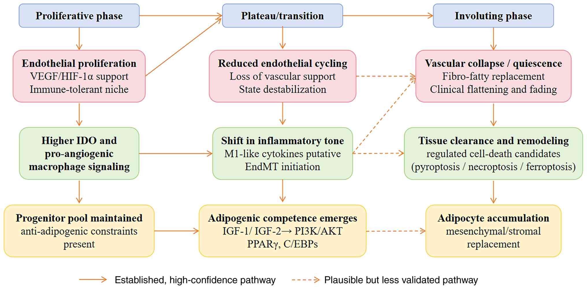
differentiation contribute to regression. Key candidate mechanisms
are outlined in Table I and
illustrated in Fig. 1, including
endothelial proliferation restraint, immune remodeling and
adipogenic replacement.
Inclusion and exclusion criteria. The
inclusion criteria were as follows: i) Original research with
experimental validation (clinical, in vitro, in vivo
or bioinformatics analyses specific to IH); ii) studies focusing on
infantile hemangioma (human tissues, IH-derived cells or
IH-specific animal models); iii) research on molecular or cellular
mechanisms underlying IH involution; iv) publications reporting
outcomes related to IH involution (molecular changes, cellular
phenotypes, histology or lesion regression); and v) full-text
articles in English or Chinese with accessible methods and data.
Priority was given to original research, while selected reviews
were used for contextual support.
The following exclusion criteria were applied: i)
Case reports, editorials, letters or abstracts without experimental
validation; ii) studies on non-IH vascular anomalies or unrelated
diseases without IH-specific mechanistic evidence; iii) descriptive
studies without investigating involution mechanisms; iv)
duplicates, inaccessible full texts or studies with critical design
flaws; and v) research extrapolating from non-IH models without
IH-specific verification.
3. Core mechanisms of IH Involution
Mechanisms that may restrain
endothelial proliferation and initiate phenotypic change
Attenuation of endothelial proliferation is a
consistent feature of involuting IH, indicating active suppression
of aberrant endothelial growth accompanied by phenotypic
remodeling. KIAA1429 is best considered part of a broader network
of post-transcriptional and cell-cycle regulators rather than an
independent driver of involution.
Post-transcriptional and cell-cycle mechanisms
constraining endothelial proliferation. KIAA1429 is an
RNA-binding protein associated with the N6-methyladenosine (m6A)
methyltransferase complex, with potential additional roles in
transcript-specific regulation (10). In IH, KIAA1429 expression is
increased in involuting lesions and is enriched in hemangioma
endothelial cells (11).
Functional studies show that KIAA1429 overexpression suppresses
endothelial proliferation, migration and tube formation, while
promoting a fibroblast-like phenotype (12).
Mechanistically, KIAA1429 binds target RNAs in
hemangioma endothelial cells (HemECs) and downregulates Cyclin D1
and Cyclin E, leading to cell-cycle arrest and reduced endothelial
activity, thereby facilitating phenotypic transition and priming
cells for adipogenic differentiation. However, the relative
contribution of m6A-dependent vs. m6A-independent regulation
remains elusive (13). Current
evidence supports a role for KIAA1429 in endothelial reprogramming
rather than as a primary initiator of involution.
p16 inhibitor of cyclin-dependent kinase 4 A
(INK4a) and the broader growth-arrest program. The
p16INK4a-retinoblastoma protein (Rb) pathway is a canonical
regulator of the G1-S transition and provides a mechanistic basis
for endothelial growth arrest during IH involution (14,15).
In IH, p16 expression is low in proliferative lesions and increases
during involution, consistent with reduced endothelial cycling and
angiogenic activity (16).
Mechanistically, p16INK4a upregulation activates the Rb pathway,
inducing G1/S arrest and limiting endothelial proliferation. This
growth-arrest program contributes to suppression of vascular
expansion but does not account for downstream tissue
remodeling.
Accordingly, p16INK4a should be interpreted as part
of a broader endothelial de-escalation program. Together with
regulators such as KIAA1429, it constrains endothelial
proliferation, while additional processes are required to drive
immune remodeling, vascular regression and fibro-fatty
replacement.
Immune remodeling and regulated
cell-death programs
Immune remodeling is a consistent feature of IH
involution and links vascular regression with tissue clearance.
Compared with proliferative lesions, involuting IH exhibits altered
immune composition and increased inflammatory signaling. Available
evidence suggests a shift from immune tolerance toward inflammatory
activation, involving reduced immune suppression, macrophage
polarization, cytokine-mediated endothelial disruption and
activation of regulated cell-death pathways. These processes
collectively contribute to endothelial destabilization and tissue
remodeling, rather than representing a single dominant trigger.
Indoleamine 2,3-dioxygenase (IDO) and the waning
of an immune-tolerant niche. IDO regulates local immune
tolerance by metabolizing tryptophan and activating the kynurenine
pathway. In proliferative IH, elevated IDO expression is associated
with suppressed T-cell activity and the maintenance of an
immune-permissive microenvironment (17). During involution, reduced IDO
expression likely restores T-cell surveillance, particularly
CD8+ T-cell activity, facilitating clearance of abnormal
vascular cells. This shift suggests a transition from an
immune-tolerant to an immune-reactive state.
However, current evidence is primarily associative.
IDO is therefore best interpreted as a modulator of immune context
rather than a primary initiator of involution.
Macrophage-associated inflammation and
endothelial fate transition. Macrophages are a major immune
component in IH and exhibit functional plasticity between
pro-angiogenic and pro-inflammatory states. During involution,
available evidence supports a shift from an M2-like, pro-angiogenic
phenotype toward an M1-like, inflammatory phenotype (18-20).
Mechanistically, M1-associated cytokines, including TNF-α, IL-1β
and IFN-γ, disrupt endothelial stability and activate TGF-β/Smad2/3
signaling in HemECs, upregulating transcription factors such as
Snail and Twist. This is associated with reduced expression of
endothelial markers (e.g., vascular endothelial cadherin, CD31),
morphological changes and features consistent with
endothelial-to-mesenchymal transition (EndMT) (21).
These findings support a role for macrophage-driven
inflammation in endothelial destabilization and phenotypic
transition. However, whether EndMT represents a required mechanism
in IH involution remains to be established (22,23).
Pyroptosis as a candidate contributor to
inflammatory tissue clearance. Pyroptosis is an
inflammasome-mediated form of inflammatory cell death characterized
by caspase-1 activation and release of pro-inflammatory cytokines.
In IH, its involvement has been suggested primarily by
gene-expression signatures associated with immune infiltration
(23,24). Mechanistically, pyroptosis may
promote inflammatory amplification and immune cell recruitment,
contributing to the clearance of vascular cells and tissue
remodeling. However, direct functional evidence in IH remains
lacking.
Accordingly, pyroptosis is best regarded as a
candidate mechanism of inflammatory clearance rather than an
established driver of involution.
Necroptosis-related signatures: Signal or
by-product? Necroptosis-related gene signatures have been
identified in IH and are associated with immune-related features
and lesion heterogeneity. Mechanistically, necroptosis may
contribute to inflammatory signaling by activating the receptor
interacting serine/threonine kinase (RIPK1)/RIPK3/ mixed lineage
kinase domain like pseudokinase pathway, potentially enhancing
immune-mediated tissue remodeling.
However, current evidence is limited to associative
analyses without functional validation. Necroptosis should
therefore be regarded as a hypothesis-generating pathway rather
than a confirmed contributor to IH involution.
Ferroptosis, oxidative stress and macrophage
function. Ferroptosis has been proposed as a mechanism linking
oxidative stress and lipid peroxidation to endothelial
vulnerability during IH involution. Observations involving
glutathione peroxidase 4-related pathways, redox imbalance and
macrophage-associated changes suggest that involution occurs within
an oxidatively altered microenvironment (25). Mechanistically, ferroptosis may
promote selective endothelial cell loss through lipid peroxidation
and reactive oxygen species accumulation, potentially interacting
with inflammatory signaling pathways.
However, supporting evidence in IH remains indirect.
Ferroptosis is therefore best interpreted as a potential
contributor to oxidative stress-mediated vascular regression rather
than a primary driver of involution.
Extracellular matrix (ECM) remodeling
and intercellular communication
Regression of IH requires not only changes in
endothelial cell fate but also structural reorganization of the
tumor microenvironment. During involution, ECM remodeling
contributes to vascular destabilization and tissue replacement
(26). Mechanistically, altered
integrin signaling and ECM turnover disrupt focal adhesion kinase
and Rho GTPase pathways, weakening endothelial adhesion and
destabilizing the vascular scaffold. In parallel, extracellular
vesicles carrying microRNAs and long non-coding RNAs mediate
intercellular communication among HemECs, hemangioma stem cells
(HemSCs) and stromal cells, coordinating endothelial regression and
stromal remodeling (27-29).
These processes likely facilitate structural
reorganization required for involution. However, whether ECM
remodeling functions as an active driver of phase transition or
primarily reflects downstream adaptation remains elusive.
Endocrine and hormonal influences
Endocrine signaling may modulate the cellular
context in which IH develops and regresses. Compared with
involution-specific mechanisms, current evidence supports a role in
regulating proliferative capacity rather than initiating
regression. Mechanistically, estradiol activates estrogen receptor
(ER)α/ERβ signaling in HemSCs, leading to PI3K/Akt activation and
upregulation of VEGFA, basic fibroblast growth factor (bFGF) and
Cyclin D1, thereby promoting cell proliferation and survival
(30-32).
Similarly, follicle-stimulatory hormone (FSH) signaling through FSH
receptor activates the cyclic AMP/protein kinase A pathway,
increasing Cyclin D1 and Bcl-2 expression, which enhances
cell-cycle progression and resistance to apoptosis (33,34).
These pathways contribute to the maintenance of a
proliferative niche and may indirectly influence the timing of
involution. However, direct evidence supporting a role in
initiating regression remains lacking.
Adipogenic differentiation and
fibro-fatty replacement
Fibro-fatty replacement represents the defining
histological endpoint of IH involution, placing adipogenic
differentiation at the center of regression models. Compared with
other proposed mechanisms, this process is supported by relatively
strong experimental evidence (35).
Insulin-like growth factor (IGF)-related
signaling and adipogenic competence. The IGF axis regulates
progenitor-cell fate in IH through IGF-1R-mediated PI3K/Akt
signaling. Mechanistically, IGF-1 and IGF-2 activate PI3K/Akt
signaling, upregulating adipogenic transcription factors including
peroxisome proliferator-activated receptor (PPAR)γ,
CCAAT-enhancer-binding protein (C/EBP)α and C/EBPβ, thereby driving
differentiation of HemSCs into adipocytes (36-38).
IGF-2 may also contribute to propranolol-associated adipogenesis
through the same pathway (39,40).
These findings support a role for IGF signaling in
establishing adipogenic competence. However, given its broad
physiological functions, IGF signaling is more likely to enable
lineage transition after vascular regression rather than act as a
primary initiating trigger.
Platelet-derived growth factor (PDGF) signaling
as a counterweight to adipogenesis. PDGF signaling has been
proposed as a counter-regulatory pathway opposing adipogenic
differentiation. Mechanistically, PDGF activates PI3K/Akt, ERK1/2
and STAT3 signaling via PDGFR-β, which suppresses adipogenic
transcription factors such as PPARγ and C/EBPα/β, thereby
inhibiting HemSC differentiation into adipocytes.
Reduced PDGF signaling may therefore shift the
balance toward adipogenic differentiation during involution.
However, current evidence remains limited and its position within
the causal hierarchy of IH regression remains elusive.
Hemangioma-derived mesenchymal populations and
terminal tissue replacement. Hemangioma-derived mesenchymal and
stem-like populations exhibit adipogenic differentiation potential,
providing a cellular basis for fibro-fatty replacement (41). This supports a model in which
involution involves active lineage transition rather than passive
vascular loss. These progenitor populations likely serve as the
primary cellular source of adipocytes during tissue remodeling.
T-box transcription factor 2 (TBX2) and
additional regulators of adipogenic transition. Transcriptional
regulators, including TBX2 and related factors, have been
implicated in modulating adipogenic differentiation in IH-derived
progenitors (42,43). Mechanistically, these factors
regulate the promoter activity of adipogenic genes, such as PPARγ
and C/EBP family members, and may interact with regulators,
including Kruppel-like factor 4 and sterol regulatory element
binding transcription factor 1, to fine-tune lineage
commitment.
However, current evidence is limited and these
regulators should be interpreted as modulators of adipogenic
transition rather than as central drivers of involution.
Anti-angiogenic and hypoxia-related
pathways
Attenuation of pro-angiogenic signaling is a
consistent feature of IH involution but does not alone define a
regression-specific program. Mechanistically, reduced activity of
hypoxia-inducible factor (HIF)-1α, Notch and bFGF/FGFR1 signaling
is associated with decreased expression of VEGFA and angiopoietin
2, suppression of the MAPK/ERK and PI3K/Akt pathways, and reduced
endothelial proliferation and survival (44-48).
These changes contribute to diminished angiogenic support and
facilitate vascular regression. However, growth suppression alone
does not account for coordinated tissue remodeling and fibro-fatty
replacement (49).
Accordingly, anti-angiogenic and hypoxia-related
pathways are best interpreted as creating a permissive environment
for involution rather than acting as primary drivers.
4. Summary of key mechanisms, current
limitations and future directions
Core findings and limitations of
existing research
Through a systematic review of 150 original studies
(clinical specimens, primary cell experiments and animal models),
the present study summarized three core mechanisms underlying the
spontaneous regression of IH. Several key pathways within these
mechanisms carry Grade A evidence and represent key components of
the current mechanistic understanding.
The PI3K/Akt/PPARγ axis-mediated adipogenic
differentiation of HemSCs represents the terminal phase of
involution and the only tissue-replacement mechanism with Grade A
evidence. IGF-1 and IGF-2 activate AKT phosphorylation, thereby
upregulating adipogenic transcription factors, including PPARγ,
C/EBPα and C/EBPβ. Histological observations and animal data
support this pathway. Notably, IGF-2 also mediates
propranolol-induced adipogenesis, directly linking clinical therapy
to endogenous involution programs.
Endothelial proliferation restraint and phenotypic
reprogramming are mediated by KIAA1429-associated endothelial
reprogramming and propranolol-mediated attenuation of
PI3K/Akt/endothelial nitric oxide synthase/VEGF signaling (both
Grade A) (50). These pathways
inhibit endothelial function and reduce vascular density,
consistent with propranolol's therapeutic effects (20,51,52).
In addition, HIF-1α/Notch/bFGF signaling decay and
p16INK4a-mediated cell cycle arrest (Grade B) further contribute to
growth suppression. Macrophage-mediated immune remodeling (Grade B)
shifts the microenvironment from pro-angiogenic M2-like toward
pro-inflammatory M1-like macrophages, which promote EndMT and
vascular breakdown. Additional mechanisms, such as IDO-related
immune tolerance and regulated cell death pathways, remain largely
unsupported by causal evidence (Grades C/I).
While these findings provide a useful overview, key
gaps remain that hinder the full understanding of the molecular
underpinnings of IH regression (53). Specifically, the upstream triggers
of core pathways, the molecular crosstalk between complementary
mechanisms and the integration of mechanistic findings with
clinical needs-such as predicting individual regression
trajectories and optimizing propranolol efficacy-remain unresolved
(54,55).
Future mechanistic research directions.
Future studies should prioritize mechanisms supported by
high-evidence data to establish causal relationships rather than
mere descriptive associations. First, upstream regulators of the
PI3K/Akt/PPARγ axis and KIAA1429 remain to be identified.
Phosphoproteomic and transcriptomic profiling of paired
proliferative and regressing IH specimens can help pinpoint
candidate kinases (e.g., pyruvate dehydrogenase kinase 1, mTOR
complex 2) or phosphatases (e.g., phosphatase and tensin homolog)
for subsequent functional validation (56).
Second, key downstream effectors should be defined,
including fate-determining transcription factors governing HemSC
adipogenesis and direct molecular targets of KIAA1429 in
angiogenesis and adipogenic transition, using integrated sequencing
strategies (57).
Third, the molecular basis of macrophage-mediated
recognition and clearance of abnormal vascular structures remains
to be investigated. Patient-derived organoids and immune-competent
mouse models will be valuable to dissect these cellular
interactions (58).
Translational value and technical pathway
recommendations. These research directions carry clear
translational potential. By advancing these pathways, clinical
applications could be significantly improved, with potential for
more precise, personalized therapies. Identifying upstream
regulators of the PI3K/Akt/PPARγ axis may enable the development of
small-molecule inhibitors that accelerate IH regression, thereby
reducing treatment duration and systemic side effects (59). Targeting regulators of HemSC
differentiation may offer alternative strategies for patients with
propranolol resistance (60,61).
KIAA1429 expression may serve as a predictive biomarker, with high
levels likely associated with favorable responses to
propranolol.
A three-step validation strategy (clinical specimens
→ primary cell experiments → animal models) is recommended. This
three-step validation strategy ensures that mechanistic findings
are biologically relevant and can be effectively translated into
clinical applications. Incorporating patient-derived models will
further improve preclinical predictive value and strengthen
translational outcomes (62).
5. Conclusion
Spontaneous involution of IH is best understood as
an active process of coordinated immunovascular remodeling rather
than passive vascular regression. This review summarizes current
evidence into three interconnected mechanistic modules, endothelial
proliferation restraint, immune remodeling and adipogenic
replacement, while distinguishing pathways by their level of
support. Key challenges remain in defining upstream triggers,
resolving interactions between pathways and linking mechanistic
insights to clinical variability. Addressing these gaps through
longitudinal human studies and functional models will be essential
for improving mechanistic understanding and informing more targeted
therapeutic strategies.
Acknowledgements
Not applicable.
Funding
Funding: This work was supported by the Regional Science
Foundation Project of the National Natural Science Foundation of
China (grant no. 82460560); the General Program of Jiangxi Natural
Science Foundation Committee (grant no. 20242BAB25523); the Science
and Technology Project of Jiangxi Provincial Health Commission
(grant no. 202410057); and the Guided Science and Technology
Project of Ganzhou Science and Technology Bureau (grant no.
GZ2024ZSF183).
Availability of data and materials
Not applicable.
Authors' contributions
All authors contributed to the study conception and
design. Material preparation, data collection, analysis and
administrative support were performed by XZ, YZ, JL, YW, XW, WP,
WF, HL, QL and FC. The first draft of the manuscript was written by
XZ, and all authors commented on and revised previous versions of
the manuscript for important intellectual content. Data
authentication is not applicable. All authors read and approved the
final manuscript.
Ethics approval and consent to
participate
Not applicable.
Patient consent for publication
Not applicable.
Competing interests
The authors declare that they have no competing
interests.
References
|
1
|
Takahashi K, Mulliken JB, Kozakewich HP,
Rogers RA, Folkman J and Ezekowitz RA: Cellular markers that
distinguish the phases of hemangioma during infancy and childhood.
J Clin Invest. 93:2357–2364. 1994.PubMed/NCBI View Article : Google Scholar
|
|
2
|
Yuan SM, Guo Y, Wang Q, Xu Y, Wang M, Chen
HN and Shen WM: Over-expression of PPAR-γ2 gene enhances the
adipogenic differentiation of hemangioma-derived mesenchymal stem
cells in vitro and in vivo. Oncotarget. 8:115817–115828.
2017.PubMed/NCBI View Article : Google Scholar
|
|
3
|
Ji Y, Chen S, Li K, Li L, Xu C and Xiang
B: Signaling pathways in the development of infantile hemangioma. J
Hematol Oncol. 7(13)2014.PubMed/NCBI View Article : Google Scholar
|
|
4
|
Mitra R, Fitzsimons HL, Hale T, Tan ST,
Gray C and White MPJ: Recent advances in understanding the
molecular basis of infantile haemangioma development. Br J
Dermatol. 191:661–669. 2024.PubMed/NCBI View Article : Google Scholar
|
|
5
|
Holm A, Mulliken JB and Bischoff J:
Infantile hemangioma: The common and enigmatic vascular tumor. J
Clin Invest. 134(e172836)2024.PubMed/NCBI View Article : Google Scholar
|
|
6
|
Chen Q, Zheng J and Bian Q: Cell fate
regulation during the development of infantile hemangioma. J Invest
Dermatol. 145:266–279. 2025.PubMed/NCBI View Article : Google Scholar
|
|
7
|
Chen ZT, Cui MJ, Guo SK, Wang ZL, Zhai QL,
Duan YF, Zhou T, Yang RM, Zheng JW, Liu BQ and Liu C: Single-cell
RNA sequencing reveals bidirectional development of infantile
hemangioma. J Invest Dermatol. 146:1344–1353. 2026.PubMed/NCBI View Article : Google Scholar
|
|
8
|
Yang L, Gong X, Xu Y, Zhang K, Xiang S,
Zhang Y and Ji Y: Immune microenvironment of infantile hemangioma:
Cellular dynamics, molecular networks, and emerging
immunomodulatory strategies. Front Immunol.
17(1908287)2026.PubMed/NCBI View Article : Google Scholar
|
|
9
|
Harris RP, Helfand M, Woolf SH, Lohr KN,
Mulrow CD, Teutsch SM and Atkins D: Methods Work Group, Third U.S.
Preventive Services Task Force. REPRINT oF: Current methods of the
U.S. preventive services task force: A review of the process. Am J
Prev Med. 58:316–331. 2020.PubMed/NCBI View Article : Google Scholar
|
|
10
|
Zhang X, Dai XY, Qian JY, Xu F, Wang ZW,
Xia T, Zhou XJ, Li XX, Shi L, Wei JF and Ding Q: SMC1A regulated by
KIAA1429 in m6A-independent manner promotes EMT progress in breast
cancer. Mol Ther Nucleic Acids. 27:133–146. 2021.PubMed/NCBI View Article : Google Scholar
|
|
11
|
Wang L, Zou Y, Huang Z, Wang W, Li J, Bi J
and Huo R: KIAA1429 promotes infantile hemangioma regression by
facilitating the stemness of hemangioma endothelial cells. Cancer
Sci. 114:1569–1581. 2023.PubMed/NCBI View Article : Google Scholar
|
|
12
|
Yu Y, Flint AF, Mulliken JB, Wu JK and
Bischoff J: Endothelial progenitor cells in infantile hemangioma.
Blood. 103:1373–1375. 2004.PubMed/NCBI View Article : Google Scholar
|
|
13
|
Li Y, Zhu X, Kong M, Chen S, Bao J and Ji
Y: Three-dimensional microtumor formation of infantile
hemangioma-derived endothelial cells for mechanistic exploration
and drug screening. Pharmaceuticals (Basel).
15(1393)2022.PubMed/NCBI View Article : Google Scholar
|
|
14
|
Serrano M, Hannon GJ and Beach D: A new
regulatory motif in cell-cycle control causing specific inhibition
of cyclin D/CDK4. Nature. 366:704–707. 1993.PubMed/NCBI View
Article : Google Scholar
|
|
15
|
Li J, Poi MJ and Tsai MD: Regulatory
mechanisms of tumor suppressor P16(INK4A) and their relevance to
cancer. Biochemistry. 50:5566–5582. 2011.PubMed/NCBI View Article : Google Scholar
|
|
16
|
Lorusso B, Nogara A, Fioretzaki R,
Corradini E, Bove R, Roti G, Gherli A, Montanaro A, Monica G,
Cavazzini F, et al: CD26 is differentially expressed throughout the
life cycle of infantile hemangiomas and characterizes the
proliferative phase. Int J Mol Sci. 25(9760)2024.PubMed/NCBI View Article : Google Scholar
|
|
17
|
Ritter MR, Moreno SK, Dorrell MI, Rubens
J, Ney J, Friedlander DF, Bergman J, Cunningham BB, Eichenfield L,
Reinisch J, et al: Identifying potential regulators of infantile
hemangioma progression through large-scale expression analysis: A
possible role for the immune system and indoleamine 2,3 dioxygenase
(IDO) during involution. Lymphat Res Biol. 1:291–299.
2003.PubMed/NCBI View Article : Google Scholar
|
|
18
|
Zhang W, Chen G, Wang FQ, Ren JG, Zhu JY,
Cai Y, Zhao JH, Jia J and Zhao YF: Macrophages contribute to the
progression of infantile hemangioma by regulating the proliferation
and differentiation of hemangioma stem cells. J Invest Dermatol.
135:3163–3172. 2015.PubMed/NCBI View Article : Google Scholar
|
|
19
|
Wang FQ, Chen G, Zhu JY, Zhang W, Ren JG,
Liu H, Sun ZJ, Jia J and Zhao YF: M2-polarised macrophages in
infantile haemangiomas: Correlation with promoted angiogenesis. J
Clin Pathol. 66:1058–1064. 2013.PubMed/NCBI View Article : Google Scholar
|
|
20
|
Zhu Z, Jiang X, Wang F, Zhao L and Song Q:
Decoding infantile hemangioma: Cellular dynamics, molecular
signals, and microenvironmental influences. Front Oncol.
15(1675194)2025.PubMed/NCBI View Article : Google Scholar
|
|
21
|
Wu KQ, Muratore CS, So EY, Sun C,
Dubielecka PM, Reginato AM and Liang OD: M1 macrophage-induced
endothelial-to-mesenchymal transition promotes infantile hemangioma
regression. Am J Pathol. 187:2102–2111. 2017.PubMed/NCBI View Article : Google Scholar
|
|
22
|
Medici D, Shore EM, Lounev VY, Kaplan FS,
Kalluri R and Olsen BR: Conversion of vascular endothelial cells
into multipotent stem-like cells. Nat Med. 16:1400–1406.
2010.PubMed/NCBI View
Article : Google Scholar
|
|
23
|
Greenberger S, Yuan S, Walsh LA, Boscolo
E, Kang KT, Matthews B, Mulliken JB and Bischoff J: Rapamycin
suppresses self-renewal and vasculogenic potential of stem cells
isolated from infantile hemangioma. J Invest Dermatol.
131:2467–2476. 2011.PubMed/NCBI View Article : Google Scholar
|
|
24
|
Liu L, Lin S, Bai J and Zhang B:
Integrative bioinformatics analysis of pyroptosis-related genes and
analysis of immune cell infiltration in infantile hemangioma
regression. Clin Cosmet Investig Dermatol. 18:291–302.
2025.PubMed/NCBI View Article : Google Scholar
|
|
25
|
Liu J, Zhong W, Wang R, Wang P, Tong G,
Chai M, Sun Y, Zhu T, Huang C, Yang S, et al: Macrophage
ferroptotic resistance is required for the progression of infantile
hemangioma. J Am Heart Assoc. 14(e034261)2025.PubMed/NCBI View Article : Google Scholar
|
|
26
|
Tan ST, Velickovic M, Ruger BM and Davis
PF: Cellular and extracellular markers of hemangioma. Plast
Reconstr Surg. 106:529–538. 2000.PubMed/NCBI View Article : Google Scholar
|
|
27
|
Wu M, Chen Y, Feng L, Dai H, Fang S and Xu
J: MiR-206 promotes extracellular matrix accumulation and relieves
infantile hemangioma through targeted inhibition of DNMT3A. Cell
Cycle. 20:978–992. 2021.PubMed/NCBI View Article : Google Scholar
|
|
28
|
Zhao ZL, Liu C, Wang QZ, Wu HW and Zheng
JW: Engineered exosomes for targeted delivery of miR-187-3p
suppress the viability of hemangioma stem cells by targeting Notch
signaling. Ann Transl Med. 10(621)2022.PubMed/NCBI View Article : Google Scholar
|
|
29
|
Li Z, Cao Z, Li N, Wang L, Fu C, Huo R, Xu
G, Tian C and Bi J: M2 macrophage-derived exosomal lncRNA
MIR4435-2HG promotes progression of infantile hemangiomas by
targeting HNRNPA1. Int J Nanomedicine. 18:5943–5960.
2023.PubMed/NCBI View Article : Google Scholar
|
|
30
|
Johnson A, Zhang H, Gonzalez SR, Lee M,
Wei T and Richter G: Presence of estrogen and progesterone
receptors in proliferating and involuting infantile hemangiomas. J
Plast Reconstr Aesthet Surg. 74:3061–3065. 2021.PubMed/NCBI View Article : Google Scholar
|
|
31
|
Munabi NCO, Tan QK, Garzon MC, Behr GG,
Shawber CJ and Wu JK: Growth hormone induces recurrence of
infantile hemangiomas after apparent involution: Evidence of growth
hormone receptors in infantile hemangioma. Pediatr Dermatol.
32:539–543. 2015.PubMed/NCBI View Article : Google Scholar
|
|
32
|
Hou F, Dai Y, Fan CY, Suen JY and Richter
GT: Estrogen is involved in hemangioma regression associated with
mast cells. Orphanet J Rare Dis. 13(181)2018.PubMed/NCBI View Article : Google Scholar
|
|
33
|
Maclellan RA, Konczyk DJ, Goss JA and
Greene AK: Analysis of follicle-stimulating hormone receptor in
infantile hemangioma. Ann Plast Surg. 80 (4 Suppl 4):S211–S213.
2018.PubMed/NCBI View Article : Google Scholar
|
|
34
|
Hong Z, Kuang J, Guo Y, Zhou G, Zhu Z and
Jiang L: Effects of follicle-stimulating hormone on the
proliferation and apoptosis of infantile hemangioma stem cells.
Biochem Biophys Rep. 35(101551)2023.PubMed/NCBI View Article : Google Scholar
|
|
35
|
Khan ZA, Boscolo E, Picard A, Psutka S,
Melero-Martin JM, Bartch TC, Mulliken JB and Bischoff J:
Multipotential stem cells recapitulate human infantile hemangioma
in immunodeficient mice. J Clin Invest. 118:2592–2599.
2008.PubMed/NCBI View Article : Google Scholar
|
|
36
|
Arasiewicz H and Dec M: Targeted therapies
in infantile hemangiomas and vascular malformations: From
β-blockers to PI3K/AKT/mTOR inhibitors. J Cell Mol Med.
30(e71103)2026.PubMed/NCBI View Article : Google Scholar
|
|
37
|
Yang K, Qiu T, Zhou J, Gong X, Zhang X,
Lan Y, Zhang Z and Ji Y: Blockage of glycolysis by targeting PFKFB3
suppresses the development of infantile hemangioma. J Transl Med.
21(85)2023.PubMed/NCBI View Article : Google Scholar
|
|
38
|
Wang F, Li H, Lou Y, Xie J, Cao D and
Huang X: Insulin-like growth factor I promotes adipogenesis in
hemangioma stem cells from infantile hemangiomas. Mol Med Rep.
19:2825–2830. 2019.PubMed/NCBI View Article : Google Scholar
|
|
39
|
Ritter MR, Dorrell MI, Edmonds J,
Friedlander SF and Friedlander M: Insulin-like growth factor 2 and
potential regulators of hemangioma growth and involution identified
by large-scale expression analysis. Proc Natl Acad Sci USA.
99:7455–7460. 2002.PubMed/NCBI View Article : Google Scholar
|
|
40
|
Zhang K, Wang F, Huang J, Lou Y, Xie J, Li
H, Cao D and Huang X: Insulin-like growth factor 2 promotes the
adipogenesis of hemangioma-derived stem cells. Exp Ther Med.
17:1663–1669. 2019.PubMed/NCBI View Article : Google Scholar
|
|
41
|
Harbi S, Wang R, Gregory M, Hanson N,
Kobylarz K, Ryan K, Deng Y, Lopez P, Chiriboga L and Mignatti P:
Infantile hemangioma originates from a dysregulated but not fully
transformed multipotent stem cell. Sci Rep. 6(35811)2016.PubMed/NCBI View Article : Google Scholar
|
|
42
|
Madsen MS, Siersbæk R, Boergesen M,
Nielsen R and Mandrup S: Peroxisome proliferator-activated receptor
γ and C/EBPα synergistically activate key metabolic adipocyte genes
by assisted loading. Mol Cell Biol. 34:939–954. 2014.PubMed/NCBI View Article : Google Scholar
|
|
43
|
Choi SK, Park S, Jang S, Cho HH, Lee S,
You S, Kim SH and Moon HS: Cascade regulation of PPARγ(2) and
C/EBPα signaling pathways by celastrol impairs adipocyte
differentiation and stimulates lipolysis in 3T3-L1 adipocytes.
Metabolism. 65:646–654. 2016.PubMed/NCBI View Article : Google Scholar
|
|
44
|
Maimaiti A, Aierken Y, Zhou L, He J,
Abudureyimu A and Li SX: Inhibiting interleukin-6/signal
transducers and activators of transduction-3/hypoxia-inducible
factor-1α signaling pathway suppressed the growth of infantile
hemangioma. Eur J Pediatr Surg. 33:158–166. 2023.PubMed/NCBI View Article : Google Scholar
|
|
45
|
Sun ZJ, Cai Y, Chen G, Wang R, Jia J, Chen
XM, Zheng LW and Zhao YF: LMO2 promotes angiogenesis probably by
up-regulation of bFGF in endothelial cells: An implication of its
pathophysiological role in infantile haemangioma. Histopathology.
57:622–632. 2010.PubMed/NCBI View Article : Google Scholar
|
|
46
|
Wu JK, Adepoju O, De Silva D, Baribault K,
Boscolo E, Bischoff J and Kitajewski J: A switch in Notch gene
expression parallels stem cell to endothelial transition in
infantile hemangioma. Angiogenesis. 13:15–23. 2010.PubMed/NCBI View Article : Google Scholar
|
|
47
|
Wang W, Chen S, Wang Y, Jie C and Shen W:
Effects of Notch signaling on proliferation, angiogenesis, and
adipogenesis of hemangioma-derived stem cells. Eur J Histochem.
69(4241)2025.PubMed/NCBI View Article : Google Scholar
|
|
48
|
Zhang H, Wei T, Johnson A, Sun R, Richter
G and Strub GM: NOTCH pathway activation in infantile hemangiomas.
J Vasc Surg Venous Lymphat Disord. 9:489–496. 2021.PubMed/NCBI View Article : Google Scholar
|
|
49
|
Kowalska M, Dębek W and Matuszczak E:
Infantile hemangiomas: An update on pathogenesis and treatment. J
Clin Med. 10(4631)2021.PubMed/NCBI View Article : Google Scholar
|
|
50
|
Chen S, Zhuang K, Sun K, Yang Q, Ran X, Xu
X, Mu C, Zheng B, Lu Y, Zeng J, et al: Itraconazole induces
regression of infantile hemangioma via downregulation of the
platelet-derived growth factor-D/PI3K/Akt/mTOR pathway. J Invest
Dermatol. 139:1574–1582. 2019.PubMed/NCBI View Article : Google Scholar
|
|
51
|
Munabi NCO, England RW, Edwards AK,
Kitajewski AA, Tan QK, Weinstein A, Kung JE, Wilcox M, Kitajewski
JK, Shawber CJ and Wu JK: Propranolol targets hemangioma stem cells
via cAMP and mitogen-activated protein kinase regulation. Stem
Cells Transl Med. 5:45–55. 2016.PubMed/NCBI View Article : Google Scholar
|
|
52
|
Pan WK, Li P, Guo ZT, Huang Q and Gao Y:
Propranolol induces regression of hemangioma cells via the
down-regulation of the PI3K/Akt/eNOS/VEGF pathway. Pediatr Blood
Cancer. 62:1414–1420. 2015.PubMed/NCBI View Article : Google Scholar
|
|
53
|
Cai B, Lin Q, Ke R, Chen L, Wan X, Ni X,
Shan X and Wang B: Determinants of proliferation and involution in
infantile hemangioma: Macrophage crosstalk, endothelial plasticity,
and metabolic rewiring. Front Pediatr. 14(1847957)2026.PubMed/NCBI View Article : Google Scholar
|
|
54
|
Zaaroura H, Tibi A, Avitan-Hersh E and
Khamaysi Z: Our experience in treating infantile hemangioma:
Prognostic factors for relapse after propranolol discontinuation.
Adv Ther. 42:537–547. 2025.PubMed/NCBI View Article : Google Scholar
|
|
55
|
Li Q, Xiao L, Su M, Chen H, Zhang H, Cao
N, Jiang L, Liu X and Zhang G: Factors influencing treatment
duration of oral propranolol in infantile hemangioma: A five-year
retrospective analysis. Front Pediatr. 13(1693660)2026.PubMed/NCBI View Article : Google Scholar
|
|
56
|
Li M, Wang X, Yang E, Li Y, Geng Y, Chen
Z, Huang S, Zhang D and Wu H: OTUB1 catalytic-independently
deubiquitinates TGFBI and mediates the angiogenesis in infantile
hemangioma by regulating glycolysis. Arterioscler Thromb Vasc Biol.
43:654–673. 2023.PubMed/NCBI View Article : Google Scholar
|
|
57
|
Chen Q, Rong H, Zhang L, Wang Y, Bian Q
and Zheng J: KLF2 orchestrates pathological progression of
infantile hemangioma through hemangioma stem cell fate decisions. J
Invest Dermatol. 144:1850–1864.e9. 2024.PubMed/NCBI View Article : Google Scholar
|
|
58
|
Kong M, Li Y, Wang K, Zhang S and Ji Y:
Infantile hemangioma models: Is the needle in a haystack? J Transl
Med. 21(308)2023.PubMed/NCBI View Article : Google Scholar
|
|
59
|
Holm A, Graus MS, Wylie-Sears J, Tan JWH,
Alvarez-Harmon M, Borgelt L, Nasim S, Chung L, Jain A, Sun M, et
al: An endothelial SOX18-mevalonate pathway axis enables
repurposing of statins for infantile hemangioma. J Clin Invest.
135(e179782)2025.PubMed/NCBI View Article : Google Scholar
|
|
60
|
Yang E, Wang X, Huang S, Li M, Li Y, Geng
Y, Liu X, Chen Z, Zhang D and Wu H: Shikonin reverses pyruvate
kinase isoform M2-mediated propranolol resistance in infantile
hemangioma through reactive oxygen species-induced autophagic
dysfunction. Cancer Sci. 114:806–821. 2023.PubMed/NCBI View Article : Google Scholar
|
|
61
|
Shah SD, Baselga E, McCuaig C, Pope E,
Coulie J, Boon LM, Garzon MC, Haggstrom AN, Adams D, Drolet BA, et
al: Rebound growth of infantile hemangiomas after propranolol
therapy. Pediatrics. 137(e20151754)2016.PubMed/NCBI View Article : Google Scholar
|
|
62
|
Mitra R, Hale TK, Fitzsimons HL, Gray C
and White MPJ: A novel three-dimensional model of infantile
haemangioma. Br J Dermatol. 192:874–882. 2025.PubMed/NCBI View Article : Google Scholar
|