Introduction
According to the World Health Organization,
tuberculosis (TB) affected 10.8 million people and caused 1.25
million mortalities in 2024(1).
One-third of the global population has been infected with
Mycobacterium tuberculosis (MTB), and 5-10% of these
individuals develop active TB disease, with clinical symptoms such
as a persistent cough, fever, night sweats and weight loss
(2). Despite the widespread use of
the Bacillus Calmette-Guérin (BCG) vaccine, its efficacy is limited
in adults, partly because of the absence of immunogenic proteins
(3). Therefore, the development
and evaluation of an efficacious TB vaccine remains a major
challenge.
Through comparative genomic hybridization between
the MTB strain H37Rv and BCG, ~129 open reading frames were found
to be distributed among H37Rv-specific genomic regions of deletion
(RDs), designated RD1 to RD16 (4,5).
These regions exist in H37Rv but not in BCG, suggesting that they
may encode potential virulent antigens for bacterial pathogenesis
and may be suitable for the development of new vaccine targets. TB
vaccine candidates currently under study include mainly live
attenuated vaccines, DNA vaccines, recombinant viral vector
vaccines and protein subunit vaccines (6-8).
Considering the potential risks of both whole-cell and virus-based
vaccines, protein subunit vaccines appear to be more valuable for
reagent application (9). However,
the biggest problem with subunit vaccines is their insufficient
immunogenicity. Therefore, it is important to optimize the vaccine
components to trigger a stronger and more lasting immune response.
Subunit vaccines formed by fusing immunodominant antigens from
different stages of TB infection are beneficial for enhancing
immunogenicity. Moreover, new vaccine adjuvants can prolong the
immune stimulation response (10,11).
In recent years, chitosan-based nanoparticles have
attracted considerable attention as safe and effective vaccine
delivery systems (12,13). Chitosan is biocompatible,
biodegradable and has low toxicity, and its cationic nature allows
for efficient encapsulation of protein antigens. Furthermore,
surface modification with targeting ligands, such as mannose,
enables active delivery to antigen-presenting cells (APCs) via
mannose receptors, thereby enhancing antigen uptake and
cross-presentation. These properties make mannosylated chitosan
(MCS) an ideal platform for subunit vaccines, especially against
intracellular pathogens such as MTB.
Pro-Glu (PE) and Pro-Pro-Glu (PPE) family proteins
serve increasingly important roles in the pathogenesis and chronic
protracted infection observed in TB (14,15).
The PE/PPE proteins of mycobacteria, defined by highly conserved
N-terminal PE or PPE motifs, represent a unique and highly diverse
group of proteins (16). The
PE/PPE protein family constitutes 7-10% of the MTB genome and
serves key roles in both bacterial physiology and host-pathogen
interactions. They are involved in nutrient uptake, metabolism,
enzymatic activity and drug resistance (17,18).
PE/PPE proteins also modulate processes such as phagosome
maturation, antigen presentation, autophagy, cell death regulation
and immune invasion (19). In
addition, PE/PPE proteins are rich in T-cell and B-cell epitopes,
eliciting strong immune responses and thus representing promising
candidates for subunit vaccine development (20). Different PE/PPE proteins may serve
different roles in modulating host immunity, and the flexible
interactions between these proteins contribute to their functional
diversity, adding to the complexity of their study. Vaccines
targeting PPE proteins, such as M72/AS01E and ID93/GLA-SE, have
shown promise in clinical trials (21,22).
MTB infects macrophages and persists in human macrophages for a
prolonged period by escaping the host immune defence system
(23). Preventing MTB from
invading macrophages aids in the early clearance of bacteria and
inhibits their colonization and proliferation. This may represent a
novel approach for the development of TB vaccines.
In the present study, two proteins from the PE/PPE
family were selected, namely, Rv1768 and Rv1983, and were combined
to form a fusion protein. The fusion protein was subsequently
prepared into MCS nanoparticles. The immune effects of the fusion
protein nanoparticles were verified both in vitro and in
vivo, especially their ability to inhibit the invasion of MTB
into macrophages.
Materials and methods
Bacteria, cells and mice
Escherichia coli DH5α and BL21 (Thermo Fisher
Scientific, Inc.), RAW264.7 macrophages (Mouse; ATCC), THP-1 cells
(Human; ATCC), the pET32a plasmid (Novagen; Merck KGaA), and the
virulent MTB H37Rv (ATCC 93009) and its genome were preserved.
H37Rv was cultured in 7H9 Middlebrook broth (cat. no. 271310;
Becton, Dickinson and Company) supplemented with 10% oleic acid,
albumin, dextrose and catalase (Becton, Dickinson and Company).
THP-1 cells were differentiated with 100 ng/ml phorbol 12-myristate
13-acetate (Sigma-Aldrich; Merck KGaA) for 48 h before the
experiments. Female C57BL/6 specific pathogen-free mice at
6-weeks-old were purchased from the Hubei Center for Disease
Control and Prevention. The mice were maintained under infection
barrier conditions in a negative-pressure animal room and fed a
sterile commercial mouse diet. All the studies were reviewed and
approved by the Animal Ethics and Welfare Committee of Wuhan
Pulmonary Hospital (approval no. 2024006).
Cloning of the Rv1768 and Rv1983 genes
and construction of the vector
The DNA sequences used in the present study were
obtained from the GenBank database (accession no. NC_000962.3). All
sequences were verified by Sangon Biotech Co., Ltd. sequencing to
ensure accuracy. The amplification primers for Rv1768 (F:
5'-AAATGGGATGCCCGGGCCGCTCGAGCACCACCACCAC-3'; R:
5'-AGAAATGACACACCAGCACCGGCGCCC-3') and for Rv1983 (F: 5'-ATGGGTCGCG
GATCCGTGTCATTTCTGGTCGTGGTTCC-3'; R:
5'-GCTTCCGCCACCGCCGCTTCCCGCCGGATGATCAAAGACT-3') were synthesized by
Sangon Biotech Co., Ltd. PCR was performed using PrimeSTAR Max DNA
Polymerase (Takara Bio, Inc.) under the following conditions:
Initial denaturation at 95˚C for 5 min; 30 cycles of 95˚C for 10
sec, 55˚C for 15 sec and 72˚C for 1 min; and a final extension at
72˚C for 5 min. The PCR products were analyzed by 1% agarose gel
electrophoresis and visualized by GelRed staining under UV light.
Using the H37Rv genome as the template, the Rv1768 and Rv1983 genes
were amplified, cloned and inserted into the pET32a vector by
homologous recombination using a ClonExpress II One Step Cloning
Kit (Vazyme Biotech Co., Ltd.) according to the manufacturer's
instructions. The vector primers used for Rv1768 gene insertion
were F: 5'-TGGTGTGTCATTTCTGGTCGTGGT-3'; R:
5'-TGGTGGTGGTGGTGCTCGAGCGGCCCGGGCATCC-3', whereas for Rv1983 gene
insertion, the primers were F: 5'-GCCGTGAGAATTCGAGCTCCGTCGA-3';
R:5'-GCTTCCGCCACCGCCGCTTCCGGATCCGCGACCCA-3'. Finally, the
expression vector pET32a-Rv1768/Rv1983 was constructed and
transformed into E. coli DH5α competent cells. Briefly, 10
µl of the recombination reaction was added to 100 µl of competent
cells, incubated on ice for 30 min, heat-shocked at 42˚C for 45 sec
and placed on ice for 2 min. Next, 900 µl of LB broth was added,
and the cells were recovered at 37˚C with agitation at 220 rpm for
1 h. The cells were plated onto LB agar containing 100 µg/ml
ampicillin and incubated at 37˚C overnight.
Bioinformatics analysis
The amino acid sequences encoded by Rv1768 and
Rv1983 were retrieved from the NCBI database (accession no.
NC_000962.3). Transmembrane helices were predicted using the TMHMM
2.0 server (DTU Health Tech, Technical University of Denmark) with
default parameters (https://services.healthtech.dtu.dk/service.php?TMHMM-2.0).
Expression and purification of fusion
proteins
A single colony of BL21 cells carrying the
expression vector was picked from an LB agar plate containing 100
µg/ml ampicillin and inoculated into 5 ml LB broth containing 100
µg/ml ampicillin. The culture was incubated overnight at 37˚C with
agitation at 220 rpm. The overnight culture was then diluted 1:100
into fresh LB broth containing 100 µg/ml ampicillin and cultivated
at 37˚C with agitation at 220 rpm until the optical density at 600
nm (OD600) reached 0.6. Subsequently, 0.5 mM isopropyl
β-D-1-thiogalactopyranoside (Beijing Solarbio Science &
Technology Co., Ltd.) was added, and expression was induced at 16˚C
with agitation at 220 rpm for 18 h. The bacterial cultures before
and after induction were collected, and the bacterial cells were
harvested after induction. The bacterial cells were disrupted using
an ultrasonicator (JY92-IIN; Ningbo Scientz Biotechnology Co.,
Ltd.) at a frequency of 20 kHz with 3 sec on/5 sec off cycles for
10 min in an ice bath, and then centrifuged at 12,000 x g for 30
min at 4˚C, after which the supernatant and precipitate were
collected. The component containing the fusion protein was
collected, and refolding was performed by sequential dialysis
against renaturation buffer (50 mM Tris-HCl, pH 8.0, 0.5 M
L-arginine, 5% glycerol, 1 mM dithiothreitol and 0.5 mM EDTA)
containing decreasing urea concentrations [0.5 M for 6 h, then 0 M
for 12 h with one buffer change, using a 10-kDa molecular weight
cut-off (MWCO) dialysis membrane at 4˚C]. Afterwards, affinity
chromatography on a nickel-nitrilotriacetic acid (Ni-NTA) column
(GE Healthcare) was used for purification, and the target protein
was eluted with 20, 80, 100, 150 and 300 mM imidazole solutions.
Finally, proteins were separated by SDS-PAGE using a 12% separating
gel and a 5% stacking gel, and then transferred onto a PVDF
membrane (MilliporeSigma) at 100 V for 1 h at 4˚C. The membrane was
blocked with 5% skimmed milk powder in TBST (20 mM Tris-HCl, pH
7.5, 150 mM NaCl and 0.1% Tween-20) for 1 h at room temperature.
Next, membranes were incubated with a mouse anti-His tag monoclonal
antibody (cat.no. ab18184; 1:2,000 dilution; Abcam) for 3 h at 4˚C,
followed by three washes with TBST for 10 min each. Subsequently,
the membrane was incubated with an HRP-labelled goat anti-mouse
antibody (cat. no. 115-035-003; 1:5,000 dilution; Jackson
ImmunoResearch Laboratories, Inc.) for 1 h at room temperature,
followed by three washes with TBST for 10 min each. Protein bands
were visualized using a DAB substrate kit (cat. no. DA1015; Beijing
Solarbio Science & Technology Co., Ltd.) according to the
manufacturer's instructions.
Preparation of MCS nanoparticles
First, 0.25 g chitosan (Sigma-Aldrich; Merck KGaA)
was weighed and placed in a 150 ml beaker and 50 ml 5% acetic acid
aqueous solution (pH 5.0) was added to it. The beaker was then
placed on a magnetic stirrer and stirred at 400 rpm and 40˚C for 1
h. Subsequently, 0.25 g mannose (Sigma-Aldrich; Merck KGaA) and
0.25 g sodium triacetoxyborohydride (Sigma-Aldrich; Merck KGaA)
were weighed, dissolved in 10 ml pure water, and the mixed aqueous
solution was added dropwise into the chitosan solution using a
dropper. The mixture was stirred at room temperature for 24 h. The
resulting liquid was transferred into a 10 kDa dialysis bag
(Repligen) and dialyzed against pure water on a magnetic stirrer at
room temperature for 3 days to obtain an aqueous solution of MCS.
After vacuum freeze-drying, the MCS was ready for use. The fusion
protein was mixed with the MCS solution in a beaker (1:10) and
stirred at 500 rpm on a magnetic stirrer for 30 min. Then, 1 mg/ml
tripolyphosphate sodium (TPP; Sigma-Aldrich; Merck KGaA) was slowly
added dropwise, and the mixture was stirred for 1 h to obtain
MCS-Rv1768-Rv1983. The optimal mass ratio of MCS to TPP (C/T),
optimal pH and optimal stirring time were determined through
single-factor experiments (Figs.
S1 and S2).
Particle size and appearance of
nanoparticles
Potassium bromide was removed from the drying oven,
placed under a tungsten filament lamp to dry, ground into powder
form and mixed evenly with the sample.
For Fourier transform infrared spectroscopy (FTIR)
analysis, the samples (MCS, chitosan and mannose) were ground with
potassium bromide powder at a ratio of 1:100 and pressed into
translucent pellets. FTIR spectra were recorded in the range of
4,000-400 cm-1 with a resolution of 4 cm-1
using a Nicolet iS10 spectrometer (Thermo Fisher Scientific,
Inc.).
A particle size analyzer (ZS90; Malvern Instruments,
Inc) was used to determine the particle size, polydispersity index
and zeta potential of the nanoparticles at 25˚C. The nanoparticles
were freeze-dried and dispersed into pure water. A pipette was used
to drop the dispersed sample onto a sample carrier copper grid and
allowed to air dry at room temperature. Then, the sample was placed
under a transmission electron microscope (Libra 120; Zeiss AG) to
observe the morphology of the nanoparticles.
Drug loading capacity
The prepared MCS-Rv1768-Rv1983 solution was
centrifuged at 13,684 x g for 30 min at 4˚C. The supernatant was
collected, and the precipitate was resuspended in 1 ml of pure
water to ensure complete resuspension. The resuspended precipitate
was then placed in a freeze-dryer and dried for 24 h to form
nanoparticle powder. The protein concentration of the nanoparticle
solution before centrifugation and the concentration of
unencapsulated protein in the supernatant were measured using a
Nanodrop 8000 spectrophotometer (Thermo Scientific, Ltd.). The
supernatant of the blank MCS nanoparticles was used as a control.
Drug loading (DL; %) was calculated according to the following
formula: DL
(%)=[((Ct-Cb)-(Cs-Cbs))
x V/M] x100, where Ct is the protein concentration of
the nanoparticle solution before centrifugation, Cb is
the protein concentration of the blank nanoparticle solution,
Cs is the protein concentration in the supernatant after
centrifugation, Cbs is the protein concentration of the
blank nanoparticle supernatant, V is the volume of the nanoparticle
solution and M is the total mass of the dried nanoparticles.
Release determination
The prepared nanoparticle solution was centrifuged
at 13,684 x g for 30 min, after which the precipitate was
collected, resuspended in 15 ml pure water, placed in a dialysis
bag (10 kDa MWCO), sealed at both ends and immersed in 100 ml PBS
(pH 7.4) on a magnetic stirrer at 37˚C and 100 rpm. Samples were
taken at 0, 0.5, 1, 3, 6, 12, 24, 36, 48, 56 and 72 h. Each sample
was replaced with an equal volume of fresh dialysis medium. The
protein concentration at each time point was determined using the
Nanodrop. The cumulative release rate Q was calculated as follows:
Qn=C1V1 + C2V2 +…+
CnVn; Q=Qn/M x 100, where C1 and
Cn are the sample concentrations at different time
points; V1 and Vn are the sample volumes
taken each time; Qn is the cumulative release amount;
and M is the total mass of protein in the nanoparticles.
Cell cytotoxicity
The cytotoxic effects of the MCS nanoparticles on
THP-1 cells were assessed using the Cell Counting Kit-8 Cell
Proliferation and Cytotoxicity Assay Kit (cat. no. CA1210; Beijing
Solarbio Science & Technology Co., Ltd.). Cells were cultured
in 96-well plates at 5x10⁴ cells/well for 24 h at 37˚C
in a 5% CO2 incubator, containing 200 µl RPMI-1640
medium supplemented with 10% FCS (Gibco; Themo Fisher Scientific,
Inc.), 100 U/ml penicillin and 100 µg/ml streptomycin (Gibco; Themo
Fisher Scientific, Inc.). The cells were treated with MCS,
Rv1768-Rv1983 and MCS-Rv1768-Rv1983 at 50, 100, 150 and 200 µM for
24 and 48 h, respectively. PBS-treated cells served as controls.
Cell viability was determined using an ELISA reader (K6600; Beijing
Kaiao Technology Development Co., Ltd.).
Animal model
A total of 24 female C57BL/6 mice were randomly
divided into four groups (n=6 per group) as follows: Control group,
injected with 200 µl PBS only; MCS group, 0.50 mg MCS dissolved in
200 µl PBS; Rv1768-Rv1983 protein group, 40 µg Rv1768-Rv1983
dissolved in 200 µl PBS; and MCS-Rv1768-Rv1983 group, 0.50 mg MCS
encapsulating 40 µg Rv1768-Rv1983 dissolved in 200 µl PBS. Sample
size was determined using a power analysis based on the Schoenfeld
formula for the log-rank test, with a two-sided significance level
of α=0.05, power=80% and an expected hazard ratio of 0.5. Each
group was subcutaneously injected once at weeks 0, 2 and 4. Blood
samples (~100 µl per mouse per time point) were collected from the
orbital sinus using heparinized capillary tubes under isoflurane
anesthesia (3-4% for induction, 1-1.5% for maintenance, by
inhalation). The first collection was performed 2 weeks after the
first immunization (week 2, for T-cell activity analysis), and the
second collection was performed at 2 weeks after the last
immunization (week 6, for antibody detection), with an interval of
4 weeks between the two draws. To minimize local tissue damage, the
two collections were performed from the contralateral eye (left eye
for the first draw, right eye for the second draw). Afterwards, the
mice were challenged with 5x10⁷ colony-forming units
(CFUs) of H37Rv via tail vein injection. Mice were monitored twice
daily for signs of morbidity. The humane endpoints established for
the present study included: i) ≥20% loss of initial body weight;
ii) persistent lethargy, hunched posture or reluctance to move;
iii) respiratory distress (labored breathing); or iv) inability to
reach food or water. Any mouse meeting at least one of these
criteria was immediately euthanized. During the experiment, none of
24 mice reached these humane endpoints. After all the mice in the
control group died, all surviving mice were euthanized by cervical
dislocation under deep anesthesia induced by intraperitoneal
injection of pentobarbital sodium (150 mg/kg). Lungs and spleens
were dissected and ground in 1 ml PBS, serially diluted 10-fold and
cultured on Löwenstein-Jensen medium plates (BD Biosciences) at
37˚C for 4 weeks. MTB colonies were counted, and the results were
expressed as log10 CFU per organ. Portions of lungs from
three mice in each group were fixed with 4% paraformaldehyde for 24
h. The tissues were embedded in paraffin and 4-µm sections were
cut. After deparaffinisation in xylene twice for 10 min each and
rehydration through a graded ethanol series (100, 95, 80 and 70%; 5
min each), sections were examined via H&E and acid-fast
staining (Ziehl-Neelsen method). H&E staining was performed
with haematoxylin for 5 min and eosin for 2 min at room
temperature. Ziehl-Neelsen staining was performed with carbol
fuchsin for 10 min and methylene blue for 1 min at room
temperature. Sections were observed under a Leica CW4000
microscope, and images were captured using Leica Microsystems
GmbH.
Cytokine analysis
Blood cells were isolated from the mice 2 weeks
after the first immunization. After the red blood cells were lysed
in ammonium chloride potassium buffer (Gibco; Thermo Fisher
Scientific Inc.), 5x10⁵ peripheral blood mononuclear
cells (PBMCs) isolated from mouse blood and cultured for 48 h in
DMEM (Sigma-Aldrich; Merck KGaA) supplemented with 10% FCS, 100
U/ml penicillin and 100 mg/ml streptomycin. Single-cell suspensions
were stimulated for 16 h at 37˚C and 5% CO2 using the
fusion protein (5 µg/ml). The cells were continuously cultured for
another 5 h in the presence of GolgiPlug (BD Biosciences) according
to the manufacturer's instructions. Non-specific binding was
blocked by incubating cells with 5 µl FcR blocking reagent (cat.
no. 422302; BioLegend, Inc.) per 106 cells in 100 µl
staining buffer for 10 min at room temperature. The activated cells
were then surface-stained with an FITC-labelled anti-CD4 antibody
(cat. no. 340133; 1:20 dilution; BD Biosciences) and a
PerCP-Cy5.5-anti-CD8 antibody (cat. no. 341050; 1:40 dilution; BD
Biosciences). After surface staining, cells were fixed and
permeabilized using the BD Cytofix/Cytoperm
Fixation/Permeabilization Kit (cat. no. 554714; BD Biosciences)
according to the manufacturer's instructions. Briefly, cells were
resuspended in 250 µl BD Cytofix/Cytoperm solution and incubated
for 20 min at 4˚C, followed by two washes with 1 ml BD Perm/Wash
buffer (cat. no. 554723; BD Biosciences). The cells were then
intracellularly stained with PE-anti-IFN-γ (cat. no. 340452; 1:50
dilution; BD Biosciences) and APC-anti-IL-4 (cat. no. 554436; 1:100
dilution; BD Biosciences) in BD Perm/Wash buffer. The numbers of
specific cell types were quantified using a FACSAria flow cytometer
(BD Biosciences), and data were analysed using FlowJo™
software version 10.10 (BD Biosciences).
Analysis of antibody response
To measure the antibody levels of IgG1 and IgG2a,
ELISA was used 2 weeks after the last vaccination, using
recombinant Rv1768-Rv1983 fusion protein as a coating antigen. In
brief, 2 µg/ml of the fusion protein was coated onto ELISA plates
(Costar; Corning, Inc.) overnight at 4˚C in NaHCO3
buffer (pH 9.6). Next, 5% BSA (Sigma-Aldrich; Merck KGaA) in PBS
was added for 2 h at 37˚C. Afterwards, the plates were incubated
for 2 h with serum samples at a 1:8 dilution. Subsequently, the
bound antibodies were detected with HRP-conjugated goat anti-mouse
IgG1 and IgG2a (cat. nos. A2554 and A2664; 1:3,000; Sigma-Aldrich;
Merck KGaA) for 1 h at 37˚C. Between each step, plates were washed
three times with PBS containing 0.05% Tween-20 (200 µl/well) for 3
min each. After being washed, the assay was developed in
o-phenylenediamine dihydrochloride-H2O2
substrate solution (Sigma-Aldrich; Merck KGaA) for 15 min at room
temperature in the dark. The reaction was stopped by adding 50 µl
of 2 M H2SO4 per well,and the absorbance was
measured at 492 nm using a microplate ELISA reader (K6600, Beijing
Kaiao Technology Development Co., Ltd.).
Phagocytic index detection
RAW264.7 macrophages were cultured in RPMI-1640
supplemented with 10% FBS and penicillin/streptomycin. After 48 h,
the cells were counted by Trypan blue staining. To evaluate the
effects of antibodies against H37Rv invasion of macrophages,
1x10⁶ CFU of H37Rv were first incubated with serum
(1:10) from different groups at 37˚C for 15 min. Then, the samples
were centrifuged at 13,684 x g for 5 min at 4˚C, after which the
supernatant was discarded. RAW264.7 macrophages (1x10⁶)
were mixed with opsonized H37Rv in 2 ml medium. Phagocytosis was
allowed to occur for 1 h at 37˚C in 6-well plates, each well
containing a coverslip pre-treated with polylysine (0.1 mg/ml).
RAW264.7 cells were seeded at a density of 1x10⁶
cells/well in 2 ml medium. The coverslip was removed, and the cells
were fixed with ice-cold acetone at -20˚C for 10 min. The cells
were then stained with auramine O (Sigma-Aldrich; Merck KGaA) for
15 min at room temperature and washed with PBS. Finally, the
coverslips were observed under a fluorescence microscope (CW4000;
Leica Microsystems GmbH). A total of 100 cells were counted from
each coverslip. The phagocytic index was calculated as follows:
Phagocytic index=total MTB captured by macrophages/number of
macrophages capable of capturing bacteria.
Statistical analysis
The experimental data were analysed with GraphPad
Prism 10.1 (Dotmatics). For normally distributed data, comparisons
among multiple groups were performed by one-way analysis of
variance (ANOVA) followed by Student-Newman-Keuls (SNK) post hoc
test. For the cytotoxicity assay, data at each time point (24 and
48 h) were analyzed separately using one-way ANOVA with SNK post
hoc test to compare differences among concentration groups; no
statistical comparison was performed between the two time points.
Survival curves were analysed using the Kaplan-Meier method with
the log-rank (Mantel-Cox) test. Mycobacterial counts were
log10-transformed and analysed by one-way ANOVA.
P<0.05 was considered to indicate a statistically significant
difference.
Results
Expression and purification of the
Rv1768-Rv1983 fusion protein
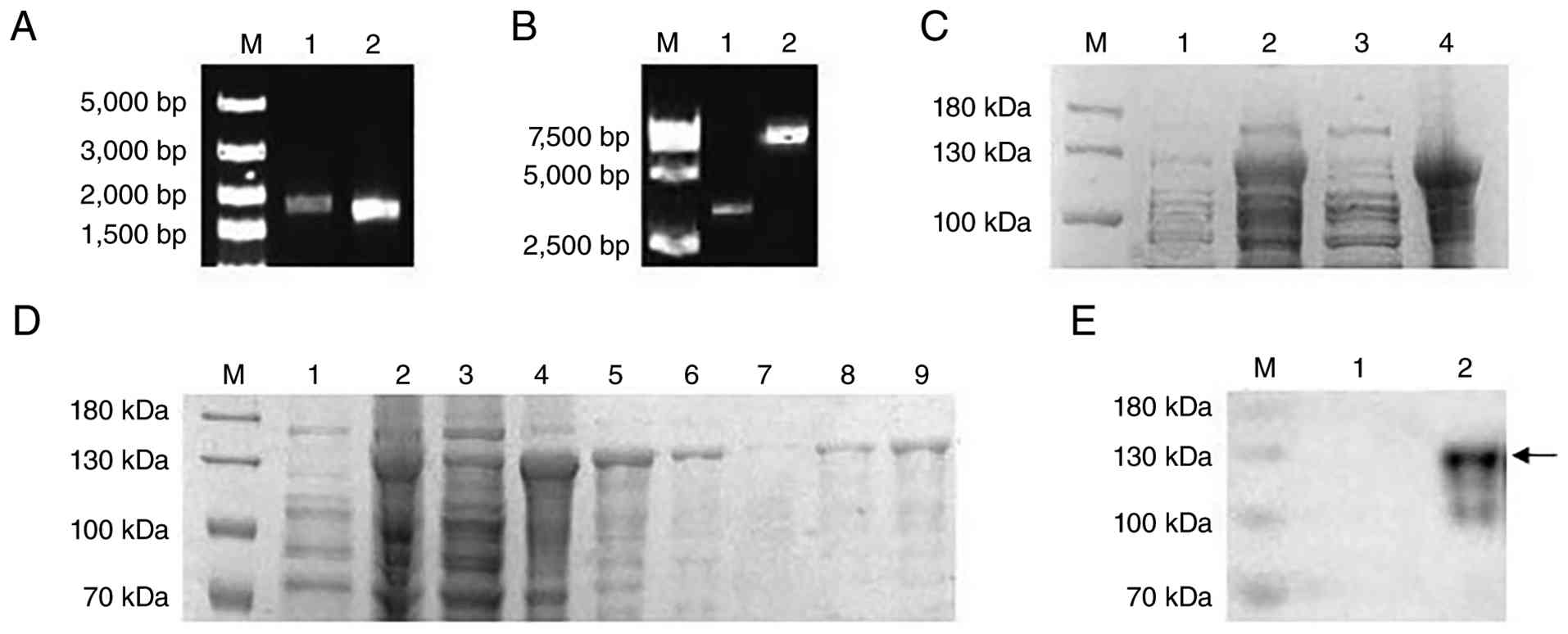
Fragments of the Rv1768 and Rv1983 genes were
amplified separately, with sizes of 1,854 and 1,674 bp,
respectively (Fig. 1A). Using
homologous arm recombination, Rv1768 and Rv1983 were ligated into
the pET32a vector. The two genes were connected by a 21 bp flexible
linker composed of alternating glycine and serine residues.
Successful construction of the fusion expression vector was
confirmed by electrophoresis and sequencing (Fig. 1B). After the induction, the
Rv1768-Rv1983 fusion protein, with a molecular mass of ~129 kDa,
was expressed in the form of inclusion bodies (Fig. 1C). Purification was performed using
a Ni-NTA affinity chromatography column, and the target protein was
obtained with a relatively high purity under 150 mM imidazole
gradient elution (Fig. 1D).
Western blot analysis further confirmed the successful expression
and purification of the fusion protein (Fig. 1E). Bioinformatics analysis revealed
no transmembrane regions in Rv1983 and Rv1768, suggesting that they
are both extracellular proteins (Fig.
S1).
Physical properties of the fusion
protein nanoparticles
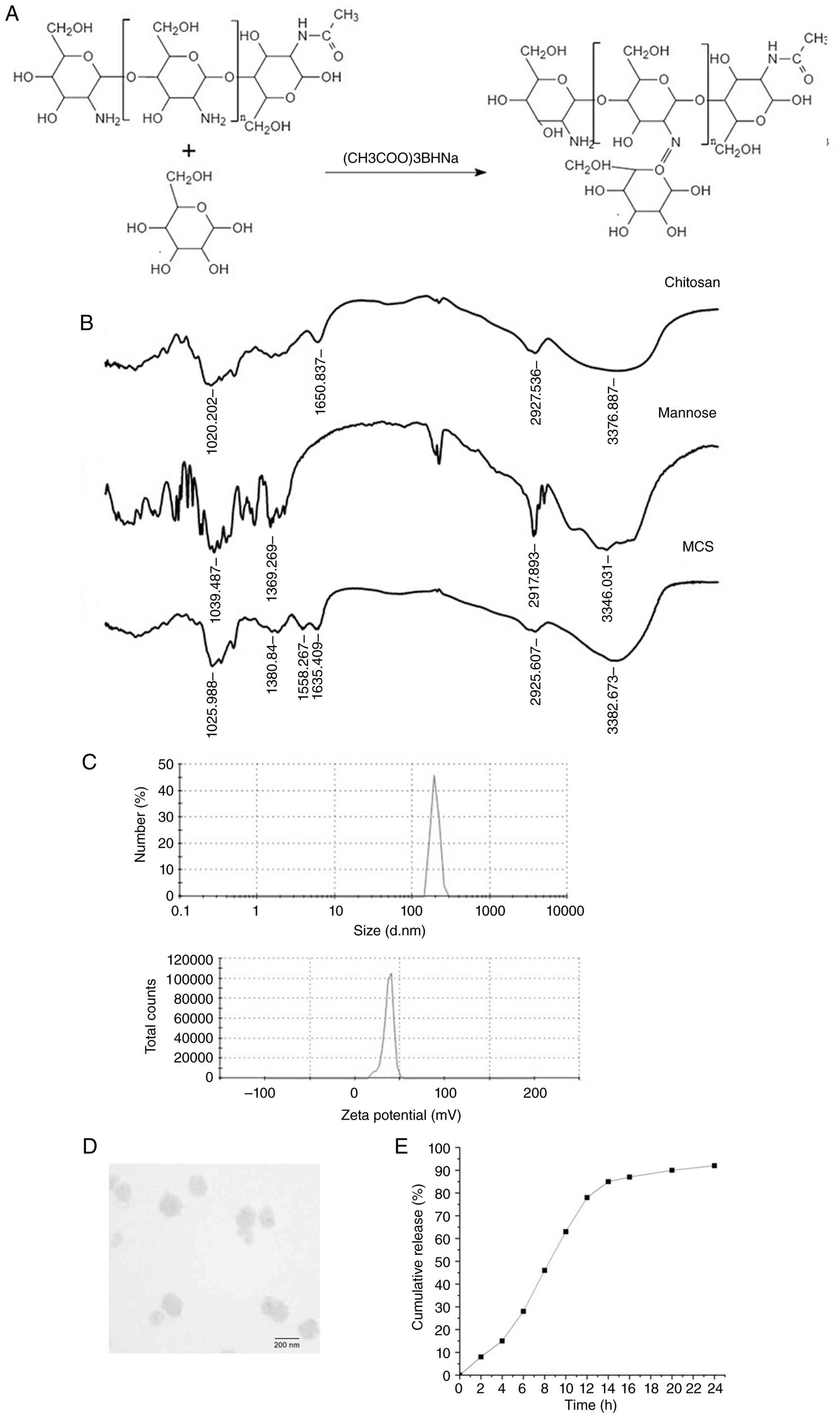
In Fig. 2A, the
synthesis pathway of MSC is illustrated. FTIR analysis revealed
characteristic absorption peaks at the hydroxyl stretching
vibration at 3,376 cm-1, C-H stretching vibration at
2,927 cm-1 and N-H bending vibration at 1,650
cm-1, confirming the presence of primary amine groups.
Additionally, the N-H bending vibration at 1,558 cm-1
and C-N stretching vibration at 1,380 cm-1 in the MCS
indicated the formation of imine groups through reductive amination
between mannose and chitosan, confirming the successful grafting of
mannose onto chitosan (Fig. 2B).
The optimal conditions for preparing MCS nanoparticles were
determined as follows: MCS concentration of 1 mg/ml, TPP
concentration of 1 mg/ml, C/T ratio of 5:1, pH of 5.0 and stirring
time of 50 min (Figs. S2 and
S3). The resulting
MCS-Rv1768-Rv1983 nanoparticles had an average particle size of
196.3 nm, a polydispersity index of 0.297 and a zeta potential of
-12.7 mV (Fig. 2C). When the
concentration of the fusion protein was 0.4 mg/ml, the drug loading
capacity was 8.02% (Fig. S4).
Transmission electron microscopy revealed that the nanoparticles
were elliptical in shape, uniform in morphology and well dispersed
(Fig. 2D). The release profile of
MCS-Rv1768-Rv1983 revealed an initial rapid release phase, reaching
~80% within 12 h, followed by a sustained release phase entering a
plateau, with a cumulative release of 95% over 24 h (Fig. 2E). These data indicate that the
encapsulation of the fusion protein by MCS provides a sustained
release effect and efficient protein utilization.
Effect of MCS on cell viability
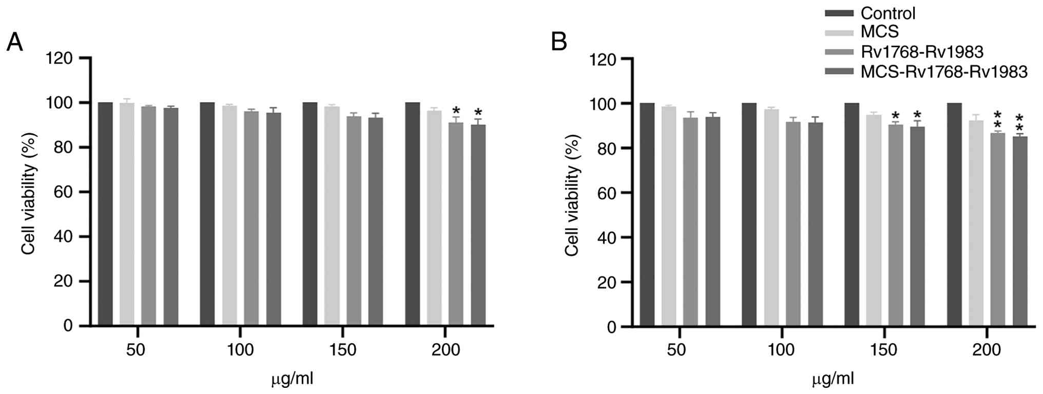
Cytotoxicity analysis results revealed that the
toxicity of MCS to THP-1 cells was extremely low. Compared with the
PBS treatment group, after 24 h of interaction with different
concentrations of MCS (50-200 µM), cell viability remained
essentially unchanged (Fig. 3A).
After 48 h, the cell viability remained >90% for all the tested
MCS concentrations. Treatment with Rv1768-Rv1983 or
MCS-Rv1768-Rv1983 for 24 and 48 h led to a moderate decrease in
cell viability, but it remained >85% (Fig. 3B; P<0.05 for Rv1768-Rv1983 and
P<0.01 for MCS-Rv1768-Rv1983 vs. PBS control). According to the
ISO 10993-5:2009 guidelines for biological evaluation of medical
devices, a cell viability of >70% is considered non-cytotoxic
(24). The present results,
showing cell viability consistently >85%, are well above this
threshold.
Cytokine production in PBMCs
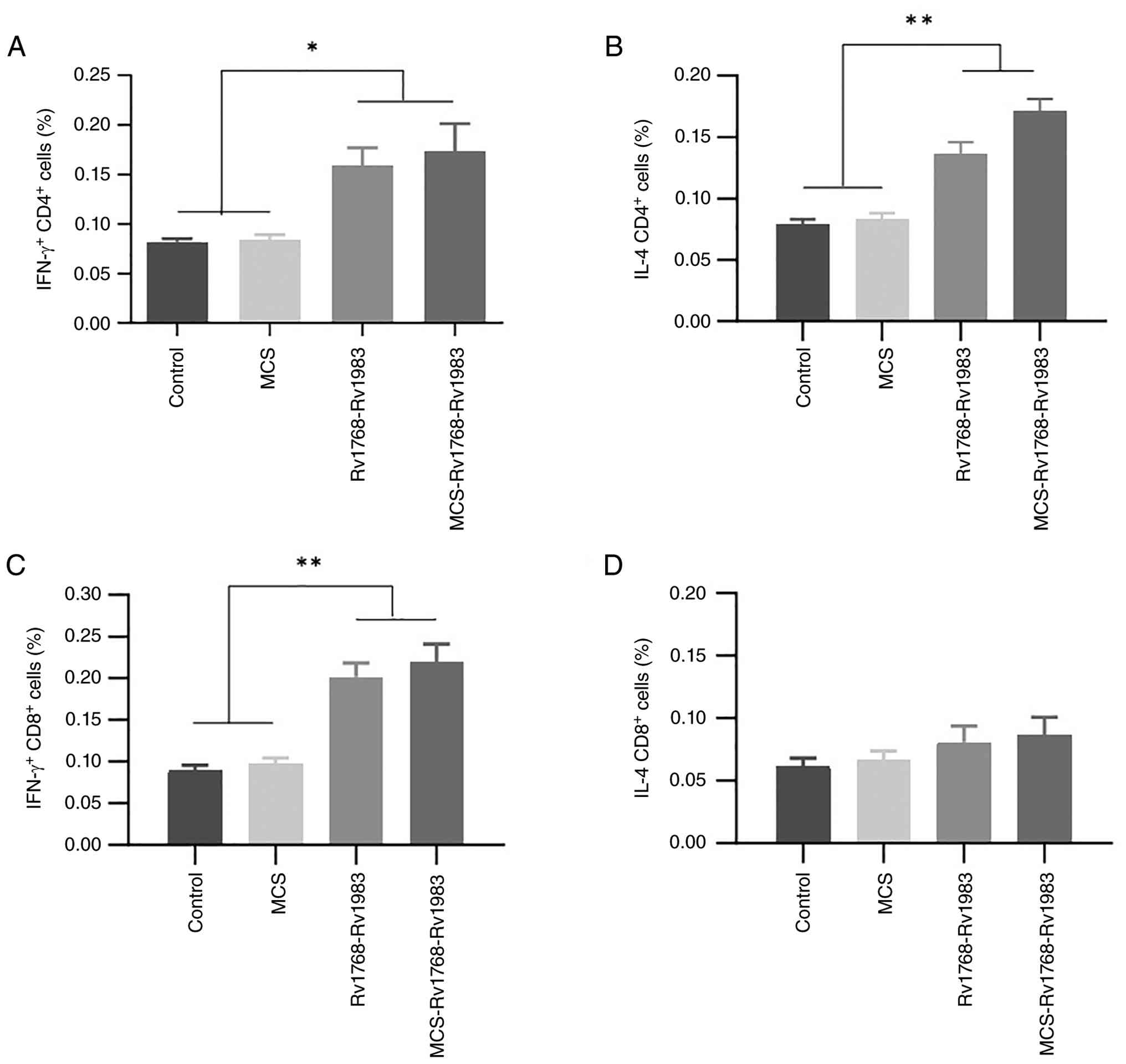
To evaluate T-cell activation, the percentages of
IFN-γ- and IL-4-producing CD4+ and CD8+ T
cells in PBMCs were measured 2 weeks after the first vaccination.
After ex vivo stimulation with the fusion protein for 16 h,
the percentages of IFN-γ- and IL-4-producing CD4+ T
cells were significantly greater in the Rv1768-Rv1983 and
MCS-Rv1768-Rv1983 groups than in the control and MCS groups
(Fig. 4A and B). One-way ANOVA with an SNK post hoc
test revealed significant differences between the two immunization
groups and the control groups (both P<0.01). For CD8+
T cells, the percentage of IFN-γ-producing cells was also
significantly greater in the immunization groups than in the
control groups (Fig. 4C;
P<0.05), whereas the percentage of IL-4-producing
CD8+ T cells was not significantly different among the
groups (Fig. 4D; P>0.05).
Antibody responses and inhibition of
macrophage invasion
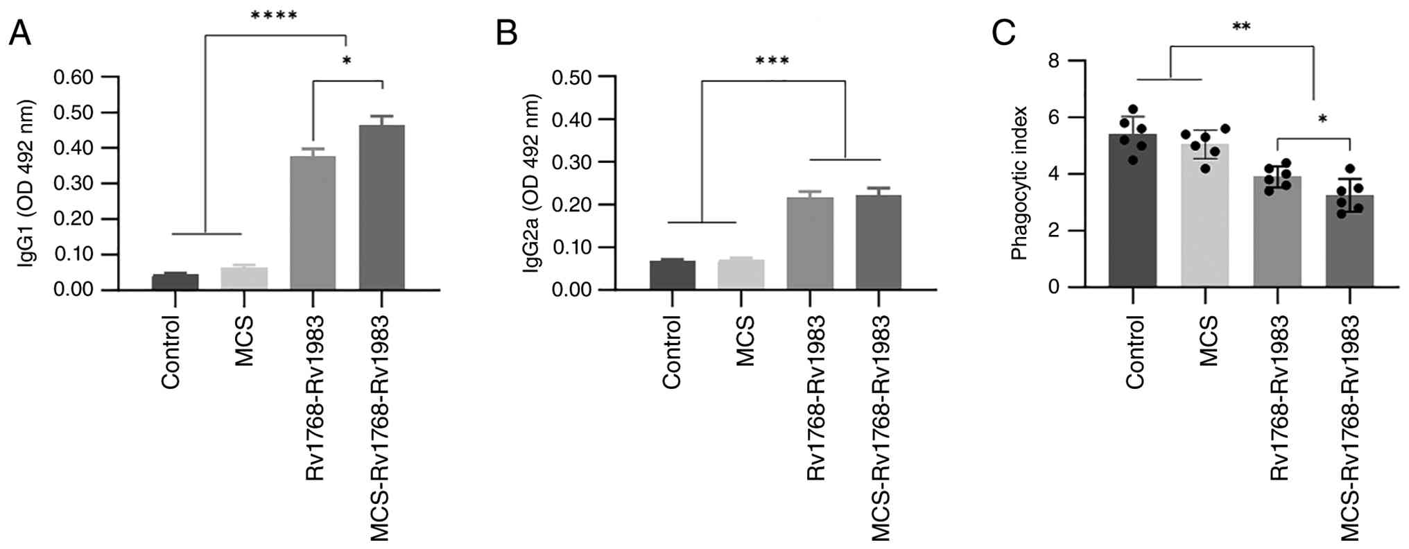
To determine the induced humoral immune response,
serum IgG1 and IgG2a subclasses were analysed. Compared with those
in the control and MCS groups, the IgG1 titres in both the
Rv1768-Rv1983 and MCS-Rv1768-Rv1983 groups were significantly
greater; moreover, compared with the Rv1768-Rv1983 group, the
MCS-Rv1768-Rv1983 group exhibited higher IgG1 levels (Fig. 5A; P<0.05 for MCS-Rv1768-Rv1983
vs. Rv1768-Rv1983). IgG2a levels were also significantly higher in
both the immunization groups than in the control and MCS groups,
but no significant difference was found between the Rv1768-Rv1983
and MCS-Rv1768-Rv1983 groups (Fig.
5B; P>0.05). After different anti-sera were incubated with
H37Rv, the ability of the bacteria to invade macrophages was
notably altered. Compared with the Rv1768-Rv1983 group, the
MCS-Rv1768-Rv1983 group had a significantly lower phagocytic index
(Fig. 5C; P<0.01), indicating
that MSC encapsulation enhances antibody-mediated inhibition of MTB
invasion.
In vivo protective efficacy
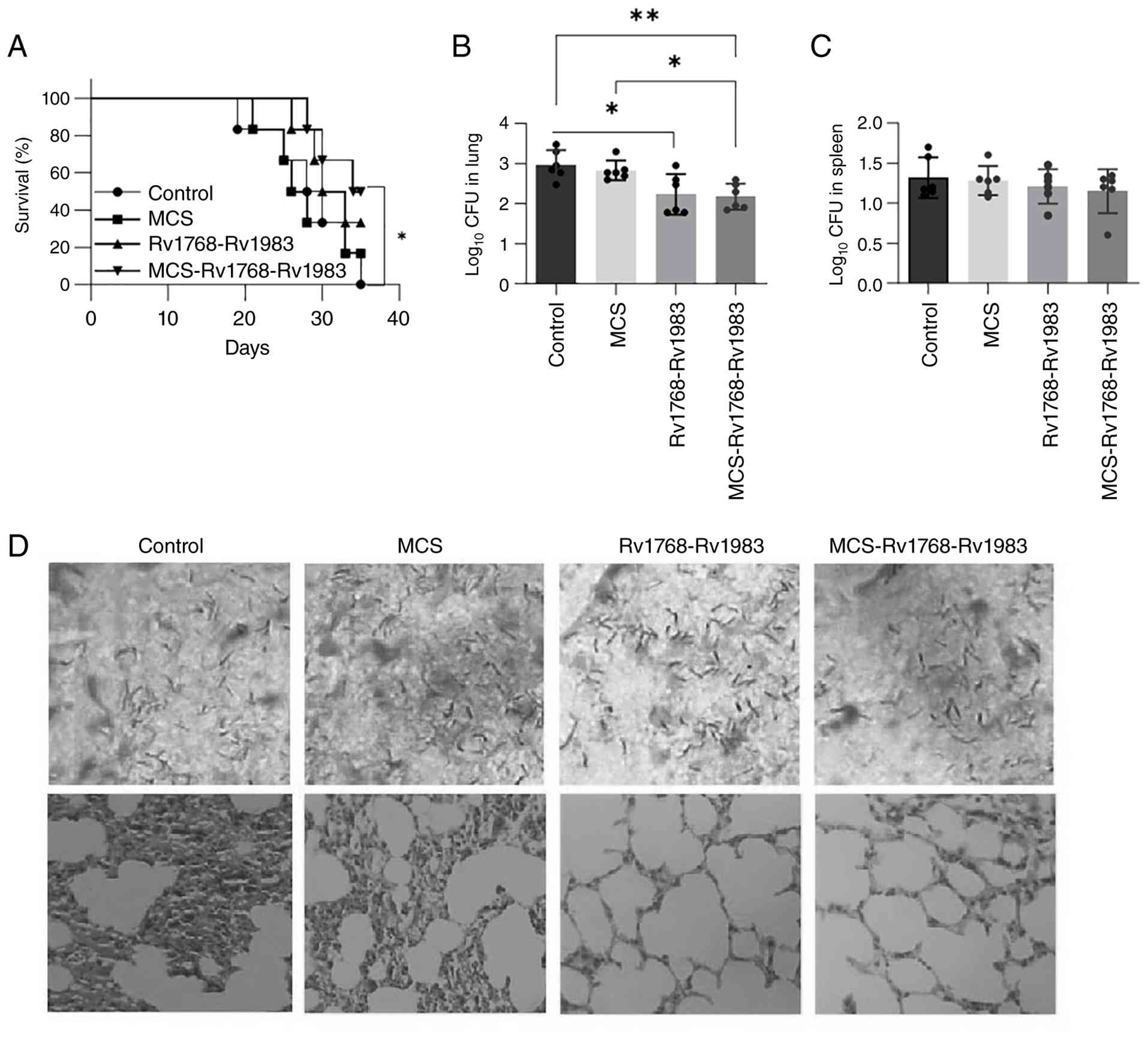
Survival analysis revealed that compared with the
control group, the MCS-Rv1768-Rv1983 group had significantly longer
survival (log-rank test, P<0.05), whereas the survival rate of
the Rv1768-Rv1983 group did not significantly change (Fig. 6A; P>0.05). Bacterial loads in
murine lungs and spleens were determined at 4 weeks after
sacrifice. Compared with the control groups, bacterial counts in
the lungs were significantly lower in both the MCS-Rv1768-Rv1983
(P<0.01) and Rv1768-Rv1983 groups (P<0.05), while compared
with the MCS group, bacterial counts in the lungs were
significantly lower in the MCS-Rv1768-Rv1983 group (P<0.05)
(Fig. 6B). Notably, compared with
the Rv1768-Rv1983 group, the MCS-Rv1768-Rv1983 group exhibited a
stronger inhibitory effect on bacterial proliferation. In the
spleen, the bacterial counts did not significantly differ among the
groups (Fig. 6C; P>0.05).
Histopathological examination of lung sections revealed that the
control group had marked inflammatory reactions, including alveolar
consolidation and fusion. By contrast, both the MCS-Rv1768-Rv1983
and the Rv1768-Rv1983 groups showed less alveolar fusion, reduced
swelling and more prominent air spaces (Fig. 6D). Acid-fast staining also revealed
fewer acid-fast bacilli in the lungs of MCS-Rv1768-Rv1983-immunized
mice than in those of control mice (Fig. 6D; upper panels).
Discussion
The immune evasion mechanism of MTB has not been
fully elucidated. However, if the invasion of MTB into macrophages
is prevented, the host immune system may be able to clear the
bacteria at an early stage (25).
With the continuous deepening understanding of MTB pathogenicity, a
number of new vaccine candidates have been discovered and some are
already in preclinical trials aiming to replace or enhance the
efficacy of BCG (26). Among the
TB vaccine candidates currently in clinical trials, protein subunit
formulations serve a pivotal role. However, optimizing antigen
combinations is important for overcoming the limited immunogenicity
of protein subunit vaccines (27).
The present successful cloning, expression and
purification of the Rv1768-Rv1983 fusion protein provides a solid
basis for its evaluation as a vaccine antigen. Inclusion body
formation and subsequent refolding by gradient dialysis ensured
adequate protein yield and purity. Furthermore, the physicochemical
characterization of MCS-encapsulated nanoparticles revealed
favourable properties, an average size of ~200 nm, a negative zeta
potential and a sustained release profile, which are known to
favour lymphatic drainage and antigen presentation. The excellent
biocompatibility of the MCS confirms its safety as a vaccine
carrier, supporting its use in subsequent in vivo studies.
Rv1768 belongs to RD14 and can induce IFN-γ production by mouse
PBMCs, as well as the generation of specific IgG and IgM against
Rv1768; hence, it is a promising candidate antigen (28). Studies have also shown that Rv1768
promotes MTB survival in macrophages by regulating the NF-κB-TNF-α
signalling pathway and by modulating arachidonic acid metabolism
through S100A9 (29,30). Rv1983 is an immunodominant antigen
that belongs to RD2. Rv1983 can induce IFN-γ responses and has been
identified as a potential diagnostic agent or subunit vaccine
candidate (31,32). In the present study, the fusion of
these two antigens elicited robust T-cell responses.
In addition to the cellular immune responses
dominated by IFN-γ, the present data revealed a marked increase in
the level of IL-4 secreted by CD4+ T cells in mice
immunized with Rv1768-Rv1983 and MCS-Rv1768-Rv1983. IL-4 is a
signature Th2 cytokine that promotes B cell activation, antibody
class switching and the production of IgG1, which is consistent
with the robust IgG1 response observed in the present study. The
balanced induction of both IFN-γ (Th1) and IL-4 (Th2) indicates a
mixed Th1/Th2 immune profile, which is favourable for protective
anti-TB immunity through the coordination of cellular clearance and
humoral neutralization. Previous studies have confirmed that Th2
responses support antibody-mediated inhibition of mycobacterial
invasion into macrophages, thereby complementing Th1-dependent
bactericidal activity (33-35).
Thus, the upregulation of IL-4 further supports the notion that
MCS-encapsulated Rv1768-Rv1983 induces a well-balanced protective
immune response rather than a polarized Th1 or Th2 reaction.
A major novelty of the present work is demonstrated
by antibodies induced by MCS-Rv1768-Rv1983 markedly blocking MTB
invasion into macrophages. This antibody-mediated ‘neutralizing’
effect is distinct from that of classical opsonophagocytosis. We
hypothesize that by preventing MTB from entering its preferred
intracellular niche, bacteria remain extracellular where they
become susceptible to a broader array of host defences. First,
extracellular bacteria are more readily recognized and lysed by
natural killer (NK) cells and complement components. Second,
compared with mannose receptor-mediated entry exploited by MTB,
antibody-opsonized bacteria can be captured by macrophages via Fcγ
receptors, which may trigger a more robust pro-inflammatory
response and enhanced phagosome lysosomal fusion. Thus, the present
strategy does not simply bypass macrophages; rather, it redirects
the pathogen to a more destructive phagocytic route while also
exposing it to extracellular immune effectors. This dual-mechanism
prevention of invasion (humoral) and the killing of intracellular
bacteria (cellular) represent a paradigm shift from the traditional
focus solely on cellular immunity and highlights the potential of
this vaccine strategy.
Nanocarrier vaccines, as immunological adjuvants,
can increase the immunogenicity of protein antigens (36). Chitosan is the most widely used
nanoadjuvant material, but further improvements are still needed
before its clinical application (37). Macrophages express mannose
receptors on their surface; thus, mannose-modified chitosan
nanoparticles are more likely to be engulfed by macrophages,
thereby triggering an immune response (38). Studies have shown that
tri-mannose-grafted chitosan nanoparticles affect mitochondrial
structure, increase cellular metabolism and upregulate the
expression of numerous immune-related genes in macrophages
(39,40). In the present study, the
synergistic effect of MCS and Rv1768-Rv1983 likely stems from i)
targeted delivery to mannose receptors on APCs, promoting efficient
antigen uptake and cross-presentation and ii) the intrinsic
immunostimulatory properties of chitosan, which promote APC
maturation and cytokine release, creating a favourable milieu for
T-cell polarization. This dual action explains the superior
antibody and T-cell responses observed with MCS-encapsulated
antigen compared with the protein alone.
For a long time, the anti-TB immunity has been
focussed mainly on cellular immunity, particularly the cytotoxic
effect induced by CD8+ T cells (41); however, the anti-TB effect of
antibodies has not received sufficient attention. In the present
study, a novel subunit vaccine for TB based on the Rv1768-Rv1983
fusion protein and MCS nanoparticles was successfully developed and
evaluated. This vaccine not only elicits a balanced Th1/Th2 mixed
immune response but also uniquely induces the production of
antibodies with ‘neutralizing activity’, effectively blocking the
initial invasion of macrophages by MTB. Bacteria that do not enter
macrophages are more likely to be eliminated by NK cells and other
extracellular mechanisms (42). In
addition, the presence of antibodies alters the way macrophages
engulf bacteria, which can activate the complement system and
inhibit the immune escape of MTB associated with macrophage
inflammation and glycolytic activation (43,44).
This mechanism offers a new direction for TB vaccine development:
Shifting from the traditional ‘enhanced bactericidal’ strategy to a
preventive ‘block invasion’ approach.
From a translational research perspective, the
present study validates the use of MCS as a versatile and robust
platform for delivering complex PE/PPE antigens, which are
notoriously difficult to handle. Given that the MTB genome encodes
a large number of these immunogenic proteins, the present work
establishes a replicable methodological pipeline. Researchers can
adapt this MCS nanocapsule system to test other combinations of MTB
antigens, particularly those with poor solubility or those that
require specific targeting to APCs. The present proof-of-concept
study thus provides a valuable tool for accelerating the discovery
of next-generation subunit vaccines against TB.
There are a few limitations to acknowledge. First,
the degree of mannose substitution on chitosan was not measured in
the exact batches used for the animal experiments because of sample
depletion; however, the protocol was based on well-established
methods, and FTIR confirmed successful conjugation. Second, the
sample size for survival analysis was based on a power calculation,
but larger cohorts would further strengthen the findings. Third,
while the present flow cytometry gating strategy is a
well-established method consistent with our previous work (45), the absence of anti-CD3 staining
could be considered a technical limitation. Future studies will
include this additional marker to further validate T-cell
identification. Future studies should also investigate the
long-term durability of the immune response and the protective
efficacy against different MTB strains.
In conclusion, the present findings demonstrate that
the MCS-Rv1768-Rv1983 nanovaccine is a highly promising candidate
for TB prevention, warranting further in-depth preclinical and
clinical studies.
Supplementary Material
TMHMM 2.0 tool was used to predict the
transmembrane regions of the (A) Rv1768 and (B) Rv1983 proteins
respectively.
Effects of different MCS/TPP mass
ratios on the (A) size and (B) zeta potential of MCS. MCS,
mannosylated chitosan; TPP, tripolyphosphate sodium.
Effects of pH of different MCS
solutions on the (A) size and (B) zeta potential of MCS. MCS,
mannosylated chitosan.
The effects of different
concentrations of fusion proteins on drug loading of protein
mannosylated chitosan nanoparticles.
Representative flow cytometry plots
corresponding to Fig. 4. (A)
Gating strategy: Lymphocytes were first gated based on FSC and SSC,
followed by gating on CD4+ or CD8+ T cell
populations. (B) Representative plots of IFN-γ+ and
IL-4+ CD4+ T cells for each experimental
group [Control (PBS), MCS, Rv1768-Rv1983 and MCS-Rv1768-Rv1983].
(C) Representative plots of IFN-γ+ and IL-4+
CD8+ T cells for each group. The percentages of positive
cells are indicated in each plot. Peripheral blood mononuclear
cells were isolated from mice 2 weeks after the first immunization
and stimulated ex vivo with the Rv1768-Rv1983 fusion protein
(5 μg/ml) for 16 h. Cells were surface-stained with anti-CD4
or anti-CD8 antibodies and intracellularly stained with anti-IFN-γ
and anti-IL-4 antibodies. FSC, forward scatter; SSC, side scatter;
MCS, mannosylated chitosan.
Acknowledgements
Not applicable.
Funding
Funding: The present work was supported by the Wuhan Municipal
Health Commission Scientific Research Project (grant no.
WX23A82).
Availability of data and materials
The data generated in the present study may be
requested from the corresponding author.
Authors' contributions
JZ wrote the original draft, and performed data
curation and funding acquisition. MA was responsible for
methodology and data curation. SZ performed the acquisition,
analysis and interpretation of data. YS performed the acquisition,
analysis and interpretation of data. ZY performed study conception
and design, and the acquisition of data. YX was responsible for the
analysis and interpretation of data, and critical revision of the
manuscript. FC performed study conception and design, was
responsible for the analysis and interpretation of data, critically
revised the manuscript and gave final approval of the version to be
published. All authors have read and approved the final version of
the manuscript. JZ and FC confirm the authenticity of all the raw
data.
Ethics approval and consent to
participate
All animal experiments were reviewed and approved by
the Animal Ethics and Welfare Committee of Wuhan Pulmonary Hospital
(approval no. 2024006).
Patient consent for publication
Not applicable.
Competing interests
The authors declare that they have no competing
interests.
References
|
1
|
World Health Organization (WHO). Global
tuberculosis report 2024, 2024.
|
|
2
|
Cobelens F, Suri RK, Helinski M, Makanga
M, Weinberg AL, Schaffmeister B, Deege F and Hatherill M: TB
Vaccine Roadmap Stakeholder Group. Accelerating research and
development of new vaccines against tuberculosis: A global roadmap.
Lancet Infect Dis. 22:e108–e120. 2022.PubMed/NCBI View Article : Google Scholar
|
|
3
|
Lange C, Aaby P, Behr MA, Donald PR,
Kaufmann SHE, Netea MG and Mandalakas AM: 100 Years of
mycobacterium bovis bacille calmette-Guérin. Lancet Infect Dis.
22:e2–e12. 2022.PubMed/NCBI View Article : Google Scholar
|
|
4
|
O'Toole RF and Gautam SS: Limitations of
the Mycobacterium tuberculosis reference genome H37Rv in the
detection of virulence-related loci. Genomics. 109:471–474.
2017.PubMed/NCBI View Article : Google Scholar
|
|
5
|
Behr MA, Wilson MA, Gill WP, Salamon H,
Schoolnik GK, Rane S and Small PM: Comparative genomics of BCG
vaccines by whole-genome DNA microarray. Science. 284:1520–1523.
1999.PubMed/NCBI View Article : Google Scholar
|
|
6
|
Gonzalo-Asensio J, Marinova D, Martin C
and Aguilo N: MTBVAC: Attenuating the human pathogen of
tuberculosis (TB) toward a promising vaccine against the TB
epidemic. Front Immunol. 8(1803)2017.PubMed/NCBI View Article : Google Scholar
|
|
7
|
Dockrell HM: Towards new TB vaccines: What
are the challenges? Pathog Dis. 74(ftw016)2016.PubMed/NCBI View Article : Google Scholar
|
|
8
|
Li J, Zhao A, Tang J, Wang G, Shi Y, Zhan
L and Qin C: Tuberculosis vaccine development: From classic to
clinical candidates. Eur J Clin Microbiol Infect Dis. 39:1405–1425.
2020.PubMed/NCBI View Article : Google Scholar
|
|
9
|
Metcalfe HJ, Biffar L, Steinbach S, Guzman
E, Connelley T, Morrison I, Vordermeier HM and Villarreal-Ramos B:
Ag85A-specific CD4+ T cell lines derived after boosting
BCG-vaccinated cattle with Ad5-85A possess both mycobacterial
growth inhibition and anti-inflammatory properties. Vaccine.
36:2850–2854. 2018.PubMed/NCBI View Article : Google Scholar
|
|
10
|
Suliman S, Luabeya AKK, Geldenhuys H,
Tameris M, Hoff ST, Shi Z, Tait D, Kromann I, Ruhwald M, Rutkowski
KT, et al: Dose optimization of H56:IC31 vaccine for
tuberculosis-endemic populations. A double-blind,
placebo-controlled, dose-selection trial. Am J Respir Crit Care
Med. 199:220–231. 2019.PubMed/NCBI View Article : Google Scholar
|
|
11
|
Moni SS, Abdelwahab SI, Jabeen A, Elmobark
ME, Aqaili D, Ghoal G, Oraibi B, Farasani AM, Jerah AA, Alnajai MMA
and Mohammad Alowayni AMH: Advancements in vaccine adjuvants: The
journey from Alum to Nano formulations. Vaccines (Basel).
11(1704)2023.PubMed/NCBI View Article : Google Scholar
|
|
12
|
Nakashima M, Suga N, Ikeda Y, Yoshikawa S
and Matsuda S: Recent progress of chitosan nanoparticles for the
development of superior delivery of vaccines. Discov Med.
36:457–466. 2024.PubMed/NCBI View Article : Google Scholar
|
|
13
|
Sadeghi Z, Fasihi-Ramandi M, Davoudi Z and
Bouzari S: Multi-epitope vaccine candidates associated with
mannosylated chitosan and LPS conjugated chitosan nanoparticles
against brucella infection. J Pharm Sci. 112:991–999.
2023.PubMed/NCBI View Article : Google Scholar
|
|
14
|
Sayes F, Sun L, Di Luca M, Simeone R,
Degaiffier N, Fiette L, Esin S, Brosch R, Bottai D, Leclerc C and
Majlessi L: Strong immunogenicity and cross-reactivity of
Mycobacterium tuberculosis ESX-5 type VII secretion-encoded
PE-PPE proteins predicts vaccine potential. Cell Host Microbe.
11:352–363. 2012.PubMed/NCBI View Article : Google Scholar
|
|
15
|
Guo F, Wei J, Song Y, Li B, Qian Z, Wang
X, Wang H and Xu T: Immunological effects of the PE/PPE family
proteins of Mycobacterium tuberculosis and related vaccines.
Front Immunol. 14(1255920)2023.PubMed/NCBI View Article : Google Scholar
|
|
16
|
Gómez-González PJ, Grabowska AD, Tientcheu
LD, Tsolaki AG, Hibberd ML, Campino S, Phelan JE and Clark TG:
Functional genetic variation in pe/ppe genes contributes to
diversity in Mycobacterium tuberculosis lineages and
potential interactions with the human host. Front Microbiol.
14(1244319)2023.PubMed/NCBI View Article : Google Scholar
|
|
17
|
Xu T, Wang C, Li M, Wei J, He Z, Qian Z,
Wang X and Wang H: Mycobacterium tuberculosis PE_PGRS45
(Rv2615c) promotes recombinant mycobacteria intracellular survival
via regulation of innate immunity, and inhibition of cell
apoptosis. J Microbiol. 62:49–62. 2024.PubMed/NCBI View Article : Google Scholar
|
|
18
|
Kramarska E, Squeglia F, De Maio F, Delogu
G and Berisio R: PE_PGRS33, an important virulence factor of
Mycobacterium tuberculosis and potential target of host
humoral immune response. Cells. 10(161)2021.PubMed/NCBI View Article : Google Scholar
|
|
19
|
Dheenadhayalan V, Delogu G, Sanguinetti M,
Fadda G and Brennan MJ: Variable expression patterns of
Mycobacterium tuberculosis PE_PGRS genes: Evidence that
PE_PGRS16 and PE_PGRS26 are inversely regulated in vivo. J
Bacteriol. 188:3721–3725. 2006.PubMed/NCBI View Article : Google Scholar
|
|
20
|
Wang Q, Boshoff HIM, Harrison JR, Ray PC,
Green SR, Wyatt PG and Barry CE III: PE/PPE proteins mediate
nutrient transport across the outer membrane of Mycobacterium
tuberculosis. Science. 367:1147–1151. 2020.PubMed/NCBI View Article : Google Scholar
|
|
21
|
Rodo MJ, Rozot V, Nemes E, Dintwe O,
Hatherill M, Little F and Scriba TJ: A comparison of
antigen-specific T cell responses induced by six novel tuberculosis
vaccine candidates. PLoS Pathog. 15(e1007643)2019.PubMed/NCBI View Article : Google Scholar
|
|
22
|
Zhang Z, Dong L, Li X, Deng T and Wang Q:
The PE/PPE family proteins of Mycobacterium tuberculosis:
Evolution, function, and prospects for tuberculosis control. Front
Immunol. 16(1606311)2025.PubMed/NCBI View Article : Google Scholar
|
|
23
|
Deng W, Long Q, Zeng J, Li P, Yang W, Chen
X and Xie J: Mycobacterium tuberculosis PE_PGRS41 enhances
the intracellular survival of M. smegmatis within macrophages via
blocking innate immunity and inhibition of host defense. Sci Rep.
7(46716)2017.PubMed/NCBI View Article : Google Scholar
|
|
24
|
International Organization for
Standardization (ISO): ISO 10993-5:2009. Biological evaluation of
medical devices-Part 5: Tests for in vitro cytotoxicity. ISO,
Geneva, 2009.
|
|
25
|
Abebe F: Immunological basis of early
clearance of Mycobacterium tuberculosis infection: The role
of natural killer cells. Clin Exp Immunol. 204:32–40.
2021.PubMed/NCBI View Article : Google Scholar
|
|
26
|
Fatima S, Kumari A, Das G and Dwivedi VP:
Tuberculosis vaccine: A journey from BCG to present. Life Sci.
252(117594)2020.PubMed/NCBI View Article : Google Scholar
|
|
27
|
Chen Z, Zhang Y, Wu J, Xu J, Hu Z and Fan
XY: A multistage protein subunit vaccine as BCG-booster confers
protection against Mycobacterium tuberculosis infection in
murine models. Int Immunopharmacol. 139(112811)2024.PubMed/NCBI View Article : Google Scholar
|
|
28
|
Yuan CH, Zhang S, Xiang F, Gong H, Wang Q,
Chen Y and Luo W: Secreted Rv1768 from RD14 of Mycobacterium
tuberculosis activates macrophages and induces a strong
IFN-γ-releasing of CD4+ T cells. Front Cell Infect
Microbiol. 9(341)2019.PubMed/NCBI View Article : Google Scholar
|
|
29
|
Liu S, Xie Y, Luo W, Dou Y, Xiong H, Xiao
Z and Zhang XL: PE_PGRS31-S100A9 interaction promotes mycobacterial
survival in macrophages through the regulation of NF-κB-TNF-α
signaling and arachidonic acid metabolism. Front Microbiol.
11(845)2020.PubMed/NCBI View Article : Google Scholar
|
|
30
|
Poladian N, Orujyan D, Narinyan W, Oganyan
AK, Navasardyan I, Velpuri P, Chorbajian A and Venketaraman V: Role
of NF-κB during Mycobacterium tuberculosis infection. Int J
Mol Sci. 24(1772)2023.PubMed/NCBI View Article : Google Scholar
|
|
31
|
Cockle PJ, Gordon SV, Lalvani A, Buddle
BM, Hewinson RG and Vordermeier HM: Identification of novel
Mycobacterium tuberculosis antigens with potential as
diagnostic reagents or subunit vaccine candidates by comparative
genomics. Infect Immun. 70:6996–7003. 2002.PubMed/NCBI View Article : Google Scholar
|
|
32
|
Chen J, Ruan Q, Shen Y, Wang S, Shao L and
Zhang W: Assessing and screening for T-cell epitopes from
Mycobacterium tuberculosis RD2 proteins for the diagnosis of
active tuberculosis. Braz J Infect Dis. 22:462–471. 2018.PubMed/NCBI View Article : Google Scholar
|
|
33
|
Saikolappan S, et al: IL-4 promotes
antibody-mediated protection against Mycobacterium
tuberculosis. J Immunol. 202:3032–3042. 2019.
|
|
34
|
Wang Q, Nag D, Baldwin SL, Coler RN and
McNamara RP: Antibodies as key mediators of protection against
Mycobacterium tuberculosis. Front Immunol.
15(1430955)2024.PubMed/NCBI View Article : Google Scholar
|
|
35
|
Dheda K, et al: The immune response in
tuberculosis: An integrated view. Lancet Infect Dis. 20:e12–e26.
2020.
|
|
36
|
Kheirollahpour M, Mehrabi M, Dounighi NM,
Mohammadi M and Masoudi A: Nanoparticles and vaccine development.
Pharm Nanotechnol. 8:6–21. 2020.PubMed/NCBI View Article : Google Scholar
|
|
37
|
Gong X, Gao Y, Shu J, Zhang C and Zhao K:
Chitosan-based nanomaterial as immune adjuvant and delivery carrier
for vaccines. Vaccines (Basel). 10(1906)2022.PubMed/NCBI View Article : Google Scholar
|
|
38
|
Feng H, Yang X, Zhang L, Liu Q, Feng Y, Wu
D, Liu Y and Yang J: Mannose-modified chitosan
poly(lactic-co-glycolic acid) microspheres Act as a mannose
receptor-mediated delivery system enhancing the immune response.
Polymers (Basel). 13(2208)2021.PubMed/NCBI View Article : Google Scholar
|
|
39
|
Coya JM, De Matteis L, Giraud-Gatineau A,
Biton A, Serrano-Sevilla I, Danckaert A, Dillies MA, Gicquel B, De
la Fuente JM and Tailleux L: Tri-mannose grafting of chitosan
nanocarriers remodels the macrophage response to bacterial
infection. J Nanobiotechnology. 17(15)2019.PubMed/NCBI View Article : Google Scholar
|
|
40
|
Serrasqueiro F, Barbosa AI, Lima SAC and
Reis S: Targeting the mannose receptor with functionalized
fucoidan/chitosan nanoparticles triggers the classical activation
of macrophages. Int J Mol Sci. 24(9908)2023.PubMed/NCBI View Article : Google Scholar
|
|
41
|
Roy Chowdhury R, Valainis JR, Dubey M, von
Boehmer L, Sola E, Wilhelmy J, Guo J, Kask O, Ohanyan M, Sun M, et
al: NK-like CD8+ γδ T cells are expanded in persistent
Mycobacterium tuberculosis infection. Sci Immunol.
8(eade3525)2023.PubMed/NCBI View Article : Google Scholar
|
|
42
|
Dai Y, Wang X, Du W, Chen R, Ma F, Ma T,
Yue L, Fang T, Wang G, Geng L, et al: NK cell-derived exosomes
inhibit survival of Mycobacterium tuberculosis by promoting
apoptosis in mice. Cytokine. 185(156820)2025.PubMed/NCBI View Article : Google Scholar
|
|
43
|
Liu Y, Wang J, Pu Y and Tang S: Complement
C1q inhibits the immune escape of Mycobacterium tuberculosis
associated with macrophage inflammation levels and glycolytic
activation. Microb Pathog. 206(107757)2025.PubMed/NCBI View Article : Google Scholar
|
|
44
|
Velasco-Velázquez MA, Barrera D,
González-Arenas A, Rosales C and Agramonte-Hevia J:
Macrophage-Mycobacterium tuberculosis interactions: Role of
complement receptor 3. Microb Pathog. 35:125–131. 2003.PubMed/NCBI View Article : Google Scholar
|
|
45
|
Chen F, Yan Q, Yu Y and Wu MX: BCG vaccine
powder-laden and dissolvable microneedle arrays for lesion-free
vaccination. J Control Release. 255:36–44. 2017.PubMed/NCBI View Article : Google Scholar
|