Introduction
Endometriosis is a chronic condition where
endometrial-like tissue grows outside the uterus, leading to
symptoms such as persistent pelvic pain, painful periods, pain
during intercourse and infertility. The severity of the condition
varies; some women experience only mild symptoms, whereas others
face more severe issues such as ovarian cysts, adhesions and deep
lesions (1). Pelvic pain, often
worsened by menstruation and intercourse, is one of the primary
challenges for affected individuals. Infertility is a notable
concern, affecting ≤40% of women with endometriosis (2) and gastrointestinal problems are
common, especially in more advanced stages of the disease (3). Diagnosing endometriosis often
requires laparoscopy, as imaging methods are less effective,
particularly in detecting deep lesions (4,5).
Treatment typically involves hormonal therapies, including combined
oral contraceptives, dienogest and gonadotropin-releasing hormone
analogs, as well as surgical options, although symptoms often recur
and long-term medication use can lead to side effects (6-8).
Endometriosis can have a marked impact on quality of life,
contributing to social and economic challenges (9).
Paeoniae Radix Alba, or Bai Shao, is a crucial herb
in traditional Chinese medicine (TCM), known for its efficacy in
regulating menstruation. Notably, it is commonly used to address
gynecological concerns, such as dysmenorrhea, irregular periods and
pelvic pain. Paeoniae Radix Alba possesses cramp-relieving
properties and serves as a core herb in numerous TCM formulas
targeting female reproductive health (10).
Hedysarum multijugum Maxim, known as Huang
Qi, has been a cornerstone of TCM for centuries. It serves a
notable role in supporting immune function, and is commonly used to
treat conditions such as fatigue, spontaneous sweating and frequent
colds. Additionally, it helps reduce edema, promotes wound healing
and accelerates recovery after illness (11).
Hedysarum multijugum Maxim and Paeoniae Radix
Alba both contain bioactive compounds that contribute to their
therapeutic effects. The active ingredients of Hedysarum
multijugum Maxim, such as saponins, polysaccharides and
flavonoids, are known for their immune-boosting, anti-inflammatory
and antioxidant properties (12).
These qualities are particularly important in treating conditions
such as endometriosis, where inflammation and tissue damage have an
important role (13). When used
together, Hedysarum multijugum Maxim and Paeoniae Radix Alba
address both systemic and localized symptoms of conditions such as
endometriosis, particularly pelvic pain and inflammation. Their
combined effects have been supported by modern research,
reinforcing their value in the treatment of gynecological
conditions (14). The herbs both
hold promise as complementary therapies for endometriosis,
alongside conventional treatments.
In our previous study, we investigated
hypoxia-related molecular mechanisms in endometriosis using
integrated bioinformatics analyses and identified discoidin
domain-containing receptor 2 as a potential diagnostic and
therapeutic biomarker (15).
However, this previous study focused primarily on
hypoxia-associated gene signatures, extracellular matrix (ECM)
remodeling and invasive behavior in endometriosis. By contrast, the
present study aimed to investigate the pharmacological basis of
Paeoniae Radix Alba and Hedysarum multijugum Maxim (BSHQ),
with particular emphasis on BSHQ-derived active ingredients and the
quercetin-serpin family E member 1 (SERPINE1) axis.
The present study aimed to identify BSHQ-related
therapeutic targets and active compounds for endometriosis through
network pharmacology, transcriptomics analysis, weighted gene
co-expression network analysis (WGCNA), machine learning and
reanalysis of published single-cell RNA sequencing (scRNA-seq)
datasets. Following the identification of candidate biomarkers and
active ingredients, in vitro experiments were performed to
evaluate the biological effects of quercetin and its interaction
with SERPINE1. The current study differs from our previous
hypoxia-focused work by emphasizing the pharmacological mechanisms
of BSHQ and experimentally validating the quercetin-SERPINE1
regulatory axis in endometriosis. The findings of the present study
may provide novel therapeutic targets and strategies for the
clinical treatment of endometriosis, offering a promising approach
to improving patient outcomes.
Materials and methods
Screening of active ingredients and
targets of BSHQ
The active components of BSHQ were initially
screened from the TCM Systems Pharmacology (TCMSP) database
(https://www.tcmsp-e.com/tcmsp.php)
using the criteria of oral bioavailability ≥20% and drug-likeness
≥0.10. Following this, the corresponding protein targets of the
selected active components were identified within the TCMSP
database. These protein targets were subsequently normalized using
the UniProt database (http://www.uniprot.org) by selecting the ‘Homo
sapiens’ taxonomy. Finally, Cytoscape 3.10.1 (https://cytoscape.org/) was employed to construct a
network that illustrates the relationships between the active
components and their target genes. Topological analysis of the
network was performed using the NetworkAnalyst plugin (https://www.networkanalyst.ca/) embedded in
Cytoscape to calculate and rank the degree of connectivity of each
active ingredient.
Data acquisition
The datasets GSE7305, GSE11691 and GSE58178 were
sourced from the Gene Expression Omnibus (GEO; https://www.ncbi.nlm.nih.gov/geo/) database and
normalization analyses were subsequently performed (16-18).
The GEO datasets used in the present study, GSE7305, GSE11691 and
GSE58178, were also included in our previous study on
hypoxia-related mechanisms in endometriosis (15); however, the present study was
independently designed to investigate BSHQ-related pharmacological
mechanisms and quercetin-associated therapeutic targets. All data
preprocessing, gene screening, downstream analyses and figure
generation were independently performed for the current study. The
GSE7305 dataset includes 10 normal endometrial samples (paired
eutopic endometrium from the same patients) and 10 endometriosis
samples, and was analyzed using the GPL570 [HG-U133_Plus_2]
Affymetrix Human Genome U133 Plus 2.0 Array. The GSE11691 dataset
consists of nine endometrial samples and nine matched ectopic
endometriotic lesion samples, which are paired tissues collected
from the same nine women with endometriosis. All samples were
analyzed with the GPL96 [HG-U133A] Affymetrix Human Genome U133A
Array. The GSE58178 dataset contains six normal endometrial samples
(from healthy women without endometriosis) and six endometriosis
samples, analyzed on the GPL6947 Illumina HumanHT-12 V3.0
expression beadchip. Endometriosis-related gene sets were retrieved
from GeneCards (https://www.genecards.org/) using ‘endometriosis’ as
the search key word. The GeneCards relevance score is an integrated
metric automatically computed by the database based on multi-source
evidence including disease-gene annotations, peer-reviewed
literature, genome-wide association study data and mutation
records, where a higher score indicates a stronger disease-gene
association. Genes with a relevance score >1 were screened as
candidate endometriosis-related genes, yielding a total of 1,359
genes. The GSE7846 dataset served as an independent validation
cohort to verify the diagnostic performance of the identified hub
genes. This dataset was also retrieved from the GEO database,
containing 5 samples of primarycultured human endometrial
endothelial cells (HEECs) isolated from the eutopic endometrium of
diseasefree premenopausal women and 5 HEEC samples from women with
endometriosis, analyzed with the GPL570 [HG-U133_Plus_2] Affymetrix
Human Genome U133 Plus 2.0 Array. This dataset was initially
reported in a previous study exploring the transcriptomic profile
of endometrial endothelial cells in endometriosis (19).
Differentially expressed genes (DEGs)
analysis
The DEGs between endometriosis samples and normal
endometrial control samples analysis was conducted using the limma
package (version 3.66.0; https://bioconductor.org/packages/limma/) in R
software (version 4.3.1; https://www.r-project.org/), which utilizes a linear
model to compute fold change (FC) values and P-values. The
Benjamini-Hochberg method was employed to control the false
discovery rate. Genes with |log2FC| >0.3 and P<0.05 were
considered significantly differentially expressed. To visualize the
expression profiles of the identified DEGs, heatmaps and volcano
plots were generated using the pheatmap (version 1.0.13; https://cran.r-project.org/package=pheatmapcran) and
ggplot2 (version 4.0.3; https://ggplot2.tidyverse.org) packages, respectively,
in R software. Principal component analysis (PCA) was additionally
performed to visualize the overall distribution of samples after
normalization, and to assess the separation tendency between normal
and endometriosis samples. Receiver operating characteristic (ROC)
curve analysis was performed to assess the diagnostic performance
of the key genes in differentiating ectopic from eutopic
endometrial tissues. ROC curves were plotted using the pROC package
(v1.18.0; https://cran.r-project.org/package=pROC) in R (v4.3.1)
and the area under the curve (AUC) was calculated to evaluate the
predictive accuracy of the model.
Gene ontology (GO) and kyoto
encyclopedia of genes and genomes (KEGG) enrichment analyses
Chromosomal localization analysis of candidate genes
was performed using the RIdeogram R package (version 0.2.2;
https://cran.r-project.org/web/packages/RIdeogram/index.html)
to visualize the chromosomal distribution of the genes. GO and KEGG
enrichment analyses were conducted to identify the biological
functions and pathways associated with the DEGs. Using the R
package clusterProfiler (version 4.8.3; https://bioconductor.org/packages/clusterProfiler/),
DEGs were analyzed for enrichment in biological processes (BP),
cellular components (CC), molecular functions (MF) and pathways.
Pearson correlation analysis was performed to calculate the
expression correlation among candidate genes. Correlation plots
were generated using the corrplot R package (version 0.92;
https://cran.r-project.org/web/packages/corrplot/index.html),
with color intensity indicating the strength of correlation. Chord
plots were constructed using the circlize R package (version
0.4.16; https://cran.r-project.org/web/packages/circlize/index.html)
to visualize the pairwise correlation relationships. The
protein-protein interaction (PPI) network of the candidate genes
was constructed using the STRING database (version 12.0; https://string-db.org/), with the organism limited to
Homo sapiens and a minimum interaction score of 0.4 as the
inclusion threshold. The network was then imported into Cytoscape
3.10.1, and the top 10 hub genes were identified via the maximal
clique centrality (MCC) algorithm using the cytoHubba plugin
(https://apps.cytoscape.org/apps/cytohubba).
WGCNA
WGCNA was performed to identify gene modules that
are significantly associated with endometriosis. The normalized
gene expression matrix derived from GEO datasets served as the
input, with low-variance genes excluded to improve network
stability and minimize noise. Sample clustering was conducted using
the flashClust R package (version 1.01-2; https://cran.r-project.org/web/packages/flashClust/index.html).
Using the WGCNA R package (version 1.72-1; https://cran.r-project.org/package=WGCNA), pairwise
gene correlations were calculated and subsequently transformed into
an adjacency matrix. A soft-thresholding power of eight was
selected according to the scale-free topology criterion, ensuring
the construction of a robust and biologically relevant network. The
topological overlap matrix (TOM) was then computed to assess gene
connectivity and similarity within the network. Hierarchical
clustering was employed to organize genes into distinct
co-expression modules, with each module assigned a unique color for
visualization purposes. Module eigengenes, which represent the
first principal component of each module, were calculated to
summarize the expression profiles of the modules.
scRNA-seq analysis
The single-cell transcriptomic dataset GSE213216 for
endometriosis was retrieved from the GEO database (20). All downstream analyses were
performed using the Seurat R package (version 4.3.0; https://satijalab.org/seurat/). First, quality control
was performed to filter low-quality cells. After quality control,
the filtered gene expression matrix was normalized using the
LogNormalize method, and highly variable genes were identified via
the FindVariableFeatures function. PCA was then conducted based on
highly variable genes for linear dimensionality reduction.
Subsequently, the uniform manifold approximation and projection
(UMAP) method was applied for non-linear dimensionality reduction
and 2D visualization of all cells, with the first 20 principal
components selected as input. Cell clustering was performed using
the FindNeighbors and FindClusters functions, and cell type
annotation was conducted based on canonical marker genes. The
expression patterns of target genes across cell types were
visualized via feature plots, violin plots and heatmaps generated
using the Seurat R package.
Machine learning methods
Machine learning techniques, specifically least
absolute shrinkage and selection operator (LASSO) and support
vector machine-recursive feature elimination (SVM-RFE), were
employed to identify key genes from the DEGs. LASSO regression was
conducted using the glmnet package in R (version 4.1-7; https://cran.r-project.org/package=glmnet), aiming to
select the most relevant genes by shrinking regression coefficients
and excluding non-informative features. Genes with non-zero
coefficients were retained for further analysis. Following this,
SVM-RFE was applied to rank the selected genes according to their
significance in classification. This method, implemented via the
e1071 package (version 1.7-12; https://cran.r-project.org/package=e1071), recursively
removes the least influential features to enhance the performance
of the model. Cross-validation was utilized to verify the stability
and reliability of the feature selection process.
Nomogram construction and decision
curve analysis (DCA)
A nomogram was constructed based on the expression
levels of the three identified hub genes (PTGER3, SERPINE1 and
VCAM1) using the rms R package (version 6.7-1; https://cran.r-project.org/web/packages/rms/index.html).
Each biomarker was assigned a weighted point according to its
regression coefficient, and the total point was calculated to
predict the risk of endometriosis. DCA was performed to evaluate
the clinical utility of the nomogram using the rmda R package
(version 1.6; https://github.com/mdbrown/rmda), by calculating the
net benefit of the model across different risk thresholds (21).
Molecular docking
Small molecules were selected from the TCMSP
database and their structures were prepared using
AutoDockTools-1.5.7 (https://ccsb.scripps.edu/mgltools/). This preparation
involves adding hydrogen atoms, calculating partial charges and
exporting the structures as PDBQT-format files. Protein structures
were obtained from the RCSB PDB database (version 2023; https://www.rcsb.org). These structures were processed
with PyMOL (version 2.5.2; https://pymol.org/) and AutoDockTools-1.5.7, where
water molecules were removed and hydrogen atoms were added.
AlphaFold-predicted structures (version 4; https://alphafold.ebi.ac.uk/) underwent similar
processing. Docking simulations were carried out using AutoDock
Vina (version 1.2.5; https://vina.scripps.edu/), generating nine
conformations for each ligand. The binding affinity was assessed
based on the docking score, where values <-7 kcal/mol were
considered indicative of strong binding. The conformations with the
lowest binding free energy were selected for subsequent analysis.
Non-covalent interactions were examined using Protein-Ligand
Interaction Profiler (PLIP; version 2.1.7; https://plip-tool.biotec.tu-dresden.de/), and
visualized with PyMOL. The docking models with a binding free
energy of <-7 kcal/mol were chosen for further study.
Gene set enrichment analysis
(GSEA)
GSEA was performed to identify enriched biological
pathways and MF terms associated with DEGs. The normalized gene
expression matrix of the merged GEO datasets were ranked based on
their association with the phenotype and used to generate a ranked
gene list. Predefined gene sets from the Molecular Signatures
Database (version 7.4; https://www.gsea-msigdb.org/gsea/msigdb/index.jsp) or
custom gene sets were selected for analysis. GSEA was conducted
using GSEA software (version 4.3.2; https://www.gsea-msigdb.org/gsea/index.jsp) and an
enrichment score was calculated for each gene set to determine its
significance. Statistical significance was assessed through
permutation testing. Enrichment score plots and heatmaps were
generated to visualize the results.
Cell lines and culture conditions
The VK2/E6E7 and 12Z endometriosis cell lines,
obtained from Procell Life Science & Technology Co., Ltd., were
cultured in high-glucose Dulbecco's Modified Eagle's Medium (cat.
no. PM15021; Procell Life Science & Technology Co., Ltd.),
supplemented with 10% fetal bovine serum (FBS; cat. no. 10099141C;
Gibco; Thermo Fisher Scientific, Inc.). The cells were grown in a
humidified incubator at 37˚C with 5% CO2 to ensure
optimal growth conditions and maintain their viability for
experimental purposes.
Cell transfection procedure
Transfection was performed on both 12Z and VK2/E6E7
endometriosis cell lines. Cells were seeded at a density of
2x10⁵ cells/well in 6-well plates 24 h prior to
transfection, reaching 70-80% confluence on the day of
transfection. Prior to transfection, 1.5-ml microcentrifuge tubes
were UV-irradiated and Opti-MEM medium (cat. no. 31985070; Gibco;
Thermo Fisher Scientific, Inc.) was pre-warmed to 37˚C. Cells were
washed twice with PBS and the culture medium was then replaced with
Opti-MEM. The transfection mixture was prepared by combining 125 µl
Opti-MEM, 2.5 µg SERPINE1 overexpression plasmid and 4 µl Lipo 8000
reagent (cat. no. C0533; Beyotime Biotechnology). The mixture was
added dropwise to the wells and incubated at 37˚C in a humidified
5% CO2 incubator for 6 h. After incubation, the medium
was replaced with fresh medium containing 10% FBS. For SERPINE1
overexpression experiments, the plasmid backbone used was
pcDNA3.1(+), an ampicillin-resistant vector purchased from Beijing
Tsingke Biotech Co., Ltd. Data S1
provides the complete coding sequence (consensus coding sequence
region) of the human SERPINE1 gene (transcript variant
NM_000602.5), which was used as the insert fragment for plasmid
construction, the complete plasmid map is shown in Fig. S1. The negative control group was
transfected with the corresponding empty pcDNA3.1 vector, which was
obtained from the same supplier and also carried ampicillin
resistance. For cell proliferation rescue assays, quercetin (cat.
no. HY18085; MedChemExpress) treatment at the respective
half-maximal inhibitory concentration (IC50)
concentration for each cell line was initiated 24 h after
transfection. Cells were incubated at 37˚C in a humidified 5%
CO2 incubator, and proliferation was measured at 0, 24,
48, 72 and 96 h after quercetin administration.
Reverse transcription-quantitative
polymerase chain reaction (RT-qPCR)
Total RNA was extracted from cells treated with
quercetin at the respective IC50 concentration for each
cell line for 24 and 48 h at 37˚C in a 5% CO2 humidified
incubator (only data obtained at the 24 h timepoint are presented
in the current study), with DMSO as the vehicle control or
SERPINE1-overexpression transfected cells using TRIzol®
reagent (Invitrogen; Thermo Fisher Scientific, Inc.), following the
standard RNA extraction protocol, and cDNA was synthesized using
the PrimeScript RT Reagent Kit (Takara Bio, Inc.) in accordance
with the manufacturer's recommended protocol. qPCR was performed on
the Applied Biosystems 7500 Real-Time PCR System (Applied
Biosystems; Thermo Fisher Scientific, Inc.) using 2X SYBR Green
qPCR Master Mix (cat. no. G3320-15; Wuhan Servicebio Technology
Co., Ltd.). Each 20-µl reaction contained 4 µl 2X SYBR Green Mix,
0.2 µl SERPINE1 forward primer (25 µM,
5'-CGCCTCTTCCACAAATCAGACG-3') and 0.2 µl SERPINE1 reverse primer
(10 µM, 5'-GGCAGTTCCAGGATGTCGTAGT-3'), or 0.2 µl GAPDH forward
primer (25 µM, 5'-ACCCACTCCTCCACCTTTGACG-3') and 0.2 µl GAPDH
reverse primer (25 µM, 5'-TCTCTTCCTCTTGTGCTCTTG-3'), 0.5 µl cDNA
and nuclease-free water to adjust the final volume. The SERPINE1
target gene and GAPDH housekeeping gene were detected in
independent reaction tubes, with only one pair of primers added per
reaction. The cycling conditions were set as follows: Initial
denaturation at 95˚C for 10 min, followed by 40 cycles at 95˚C for
15 sec and 60˚C for 1 min. Relative expression levels of SERPINE1
were normalized to GAPDH using the 2-ΔΔCq method
(22).Western blotting
(WB). For WB, cells were lysed in radioimmunoprecipitation
assay buffer (cat. no. G2002; Wuhan Servicebio Technology Co.,
Ltd.) supplemented with 1% phenylmethylsulfonyl fluoride, and
protein concentrations were measured by bicinchoninic acid assay
(cat. no. G2026; Wuhan Servicebio Technology Co., Ltd.). Equal
amounts of protein (30 µg/lane) were separated by SDS-PAGE on 10%
gels and transferred to polyvinylidene fluoride membranes. After
blocking with 5% (w/v) non-fat skim milk (cat. no. D8340; Beijing
Solarbio Science & Technology Co., Ltd.) for 1 h at room
temperature, the membranes were incubated with anti-SERPINE1
(1:1,000; cat. no. ab28207; Abcam) and anti-GAPDH (1:5,000; cat.
no. ab8245; Abcam) primary antibodies overnight at 4˚C, followed by
horseradish peroxidase-conjugated secondary antibodies (1:5,000;
cat. nos. ab205718 and ab205719; Abcam) for 1 h at room
temperature. Protein bands were detected using an enhanced
chemiluminescence kit (cat. no. G2014; Wuhan Servicebio Technology
Co., Ltd.) and semi-quantified by densitometric analysis using
ImageJ software (version 1.53t; National Institutes of Health).
Cell counting kit-8 (CCK-8) assay
Cell proliferation was assessed using the CCK-8
assay kit (cat. no. G4103; Wuhan Servicebio Technology Co., Ltd.).
Cells were seeded in 96-well plates and treated with quercetin at
gradient concentrations (0, 1, 2, 4, 8, 16, 32 and 64 µM) for 24,
48, 72 and 96 h at 37˚C in a 5% CO2 humidified
incubator, with an equal volume of dimethyl sulfoxide (DMSO) as the
vehicle control. At each time point, 10 µl CCK-8 solution was added
to each well and incubated for 2 h. The absorbance at 450 nm was
measured using a microplate reader. The resulting optical density
(OD) values were used to quantify cell proliferation and determine
the effects of the treatments on cell proliferation.
IC50 assay
The IC50 of quercetin on endometriosis
cell lines was determined using the CCK-8 cell viability assay.
Cells in the logarithmic growth phase were digested, resuspended in
complete medium and seeded into 96-well plates at a density of
5x10³ cells/well in 100 µl complete medium. After 24 h
of incubation for cell adhesion, quercetin was added at gradient
concentrations (0, 1, 2, 4, 8, 16, 32 and 64 µM), with six
replicate wells for each concentration. A blank control with only
culture medium was set simultaneously. After 24 h of drug
treatment, the absorbance at 450 nm was measured to assess cell
viability, and IC50 values were calculated via
dose-response curve fitting.
5-ethynyl-2'-deoxyuridine (EdU)
assay
Cell proliferation was measured using the EdU assay
kit (cat. no. G1601; Wuhan Servicebio Technology Co., Ltd.). Cells
were divided into the DMSO solvent control group and quercetin
treatment group: cells were treated with quercetin at the
respective IC50 concentration for each cell line, and
incubated at 037˚C in a 5% CO2 humidified incubator for
24 h; the control group was treated with an equal volume of DMSO
matching the solvent concentration of the quercetin treatment
group. After treatment, cells were incubated with 10 µM EdU for 2 h
at 37˚C, followed by fixation with 4% paraformaldehyde for 15 min
at room temperature. Cells were then permeabilized using 0.5%
Triton and stained according to the manufacturer's instructions,
with Hoechst 33342 used for nuclear counterstaining. The
EdU-positive cells were visualized and quantified under a
fluorescence microscope. The percentage of EdU-positive cells was
calculated to assess the proliferative activity of the treated
cells.
Cellular thermal shift assay
(CETSA)
CETSA was performed to assess the stability of
SERPINE1 in the presence of quercetin. Briefly, 12Z and VK2/E6E7
cells were treated with 20 µM quercetin for 4 h at 37˚C; an equal
volume of DMSO was added to the DMSO control group. Following
treatment, cells were harvested and resuspended in lysis buffer.
The cell suspensions were subjected to gradient temperatures (37,
42, 47, 52, 57 and 62˚C) for 3 min, followed by rapid cooling on
ice. After three freeze-thaw cycles, cell lysates were collected
and analyzed by WB to measure the levels of SERPINE1. The degree of
SERPINE1 stabilization was determined by comparing the protein
levels across different temperature treatments.
Surface plasmon resonance (SPR)
SPR analysis was conducted to evaluate the
interaction between quercetin and SERPINE1. Recombinant human
SERPINE1 protein (cat. no. ab282405; Abcam) was immobilized on a
CM5 sensor chip (cat. no. BR100530; Cytiva), and increasing
concentrations of quercetin (0.3125, 0.625, 1.25, 2.5, 5 and 10 µM)
were injected over the surface at 25˚C, with an association phase
of 120 sec and a dissociation phase of 300 sec to allow binding.
The changes in resonance units were monitored in real-time using a
Biacore T200 system (Cytiva) to assess the binding affinity and
kinetics. The dissociation constant (KD) was calculated from the
binding curves to determine the strength of the interaction between
quercetin and SERPINE1. Data analysis was performed using dedicated
software (Biacore T200 Evaluation Software; version 3.2; Cytiva) to
obtain precise binding parameters.
Statistical analysis
Statistical analysis was performed using GraphPad
Prism 9.0 software (Dotmatics) and R software. Continuous data were
first assessed for normality using the Shapiro-Wilk test. Normally
distributed data are presented as the mean ± standard deviation,
whereas non-normally distributed data are presented as median
(interquartile range). For comparisons between two groups, unpaired
Student's t-test was used for normally distributed data with equal
variances, and unpaired Welch's t-test was applied when variances
were unequal. P<0.05 was considered to indicate a statistically
significant difference.
Results
Active ingredients and targets of
BSHQ
In the present study, a total of 32 bioactive
ingredients were identified from BSHQ. Among these, 12 ingredients
were sourced from Paeoniae Radix Alba, while 22 originated from
Hedysarum multijugum Maxim. Notably, kaempferol (MOL000422)
and mairin (MOL000211) were found to be common to both herbs
(Table SI). After eliminating
duplicates, the 32 active ingredients were linked to 231 unique
target genes. To explore the interactions between these active
ingredients and their corresponding targets, the present study
constructed a detailed interaction network using Cytoscape 3.10.1.
This network visually represents the connections between the
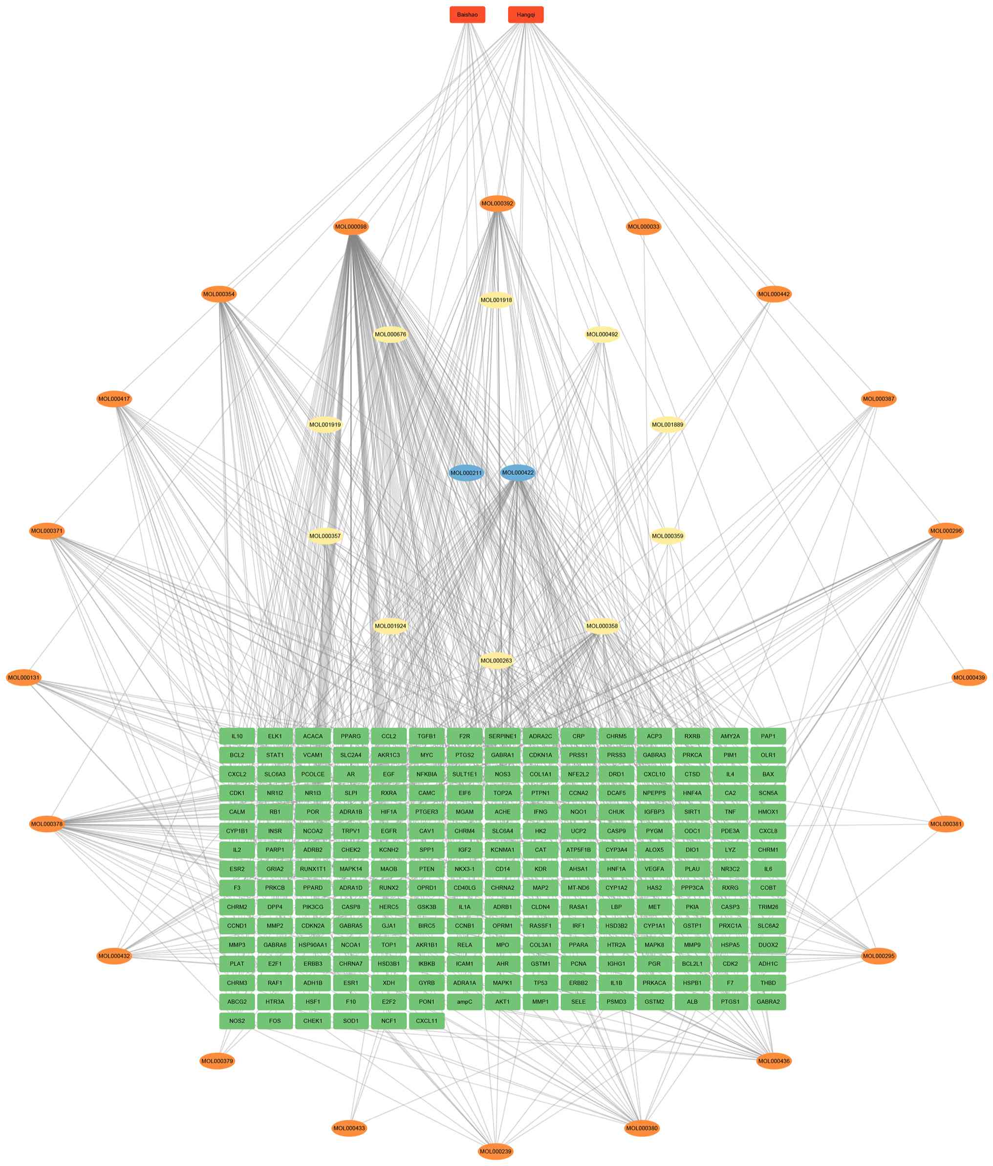
bioactive ingredients and their respective target genes. In the
diagram, the orange rectangular nodes at the top denote the two
constituent herbs of BSHQ, ovals represent the active ingredients
and the green rectangular nodes in the inner layer symbolize the
target genes. This layout provides a clear and comprehensive
overview of the interactions among these herbal ingredients and
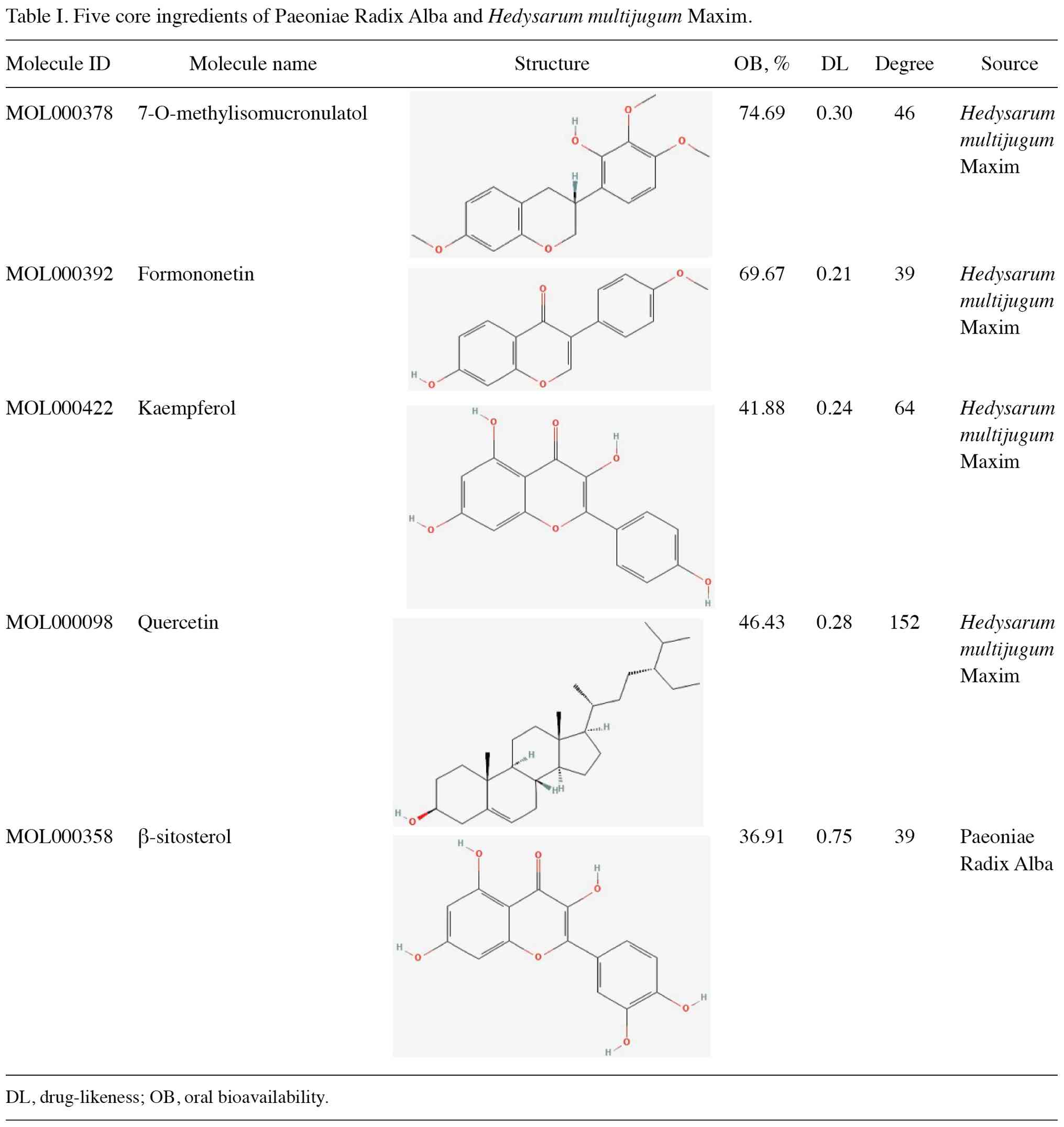
their targets (Fig. 1). Subsequent
analysis within NetworkAnalyst (embedded in Cytoscape) was carried
out to identify the top five active ingredients with the highest
degree of connectivity in the network. The ingredients with the
highest connectivity were identified as quercetin, kaempferol,
7-O-methylisomucronulatol, formononetin and β-sitosterol (Table I).
GEO dataset analysis
The GEO datasets GSE7305, GSE11691 and GSE58178 were
retrieved from the GEO database and subsequently merged for further
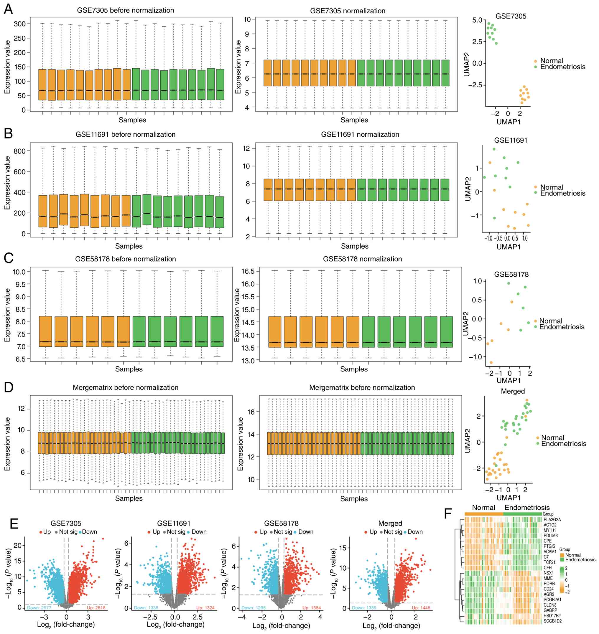
analysis. After normalization, boxplots of the processed data
confirmed that the medians across all samples were comparable,
indicating consistency in preprocessing steps (Fig. 2A-D). PCA was performed to visualize
the global transcriptomic distribution of the normalized samples; a
clear tendency toward separation was identified between the normal
and endometriosis groups, supporting the presence of transcriptomic
differences between the two tissue types (Fig. 2A-D). DEGs were identified using the
criteria |logFC| >0.3 and P<0.05. The results of differential
gene expression were visualized using a volcano plot (Fig. 2E).
In GSE7305, 2,977 genes were downregulated and 2,818
genes were upregulated. In GSE11691, there were 1,336 downregulated
genes and 1,324 upregulated genes. In GSE58178, 1,295 genes were
downregulated and 1,384 genes were upregulated. In the merged
datasets, 1,389 genes were downregulated and 1,445 genes were
upregulated (Fig. 2E). To further
illustrate these findings, a heatmap was generated to display the
top 10 most upregulated and downregulated genes from the merged
dataset (Fig. 2F).
WGCNA
The gene expression profiles from 25 endometriosis
samples and 25 normal endometrial samples (merged from GSE7305,
GSE11691 and GSE58178 datasets) were used to construct
co-expression modules using the WGCNA algorithm. Sample clustering
was performed using the flashClust package. When the
soft-thresholding power was set to 8, the scale-free topology fit
index reached 0.85, optimizing the average connectivity of the
network (Fig. 3A). Hierarchical
clustering was performed to organize genes into co-expression
modules, and the gene dendrogram with module color assignments is
shown in Fig. 3E. Applying the
dynamic tree cut algorithm, four distinct gene co-expression
modules were identified. The eigengene adjacency heatmap was
generated to visualize the correlation relationships among
different modules (Fig. 3B).
Subsequent analysis of the relationships between module eigengenes
and clinical traits revealed that the turquoise module exhibited
the strongest correlation with endometriosis, comprising 698 genes
(Table SII; Fig. 3C). Furthermore, a strong positive
correlation was observed between the module eigengenes and the
genes within the turquoise module (Fig. 3D).
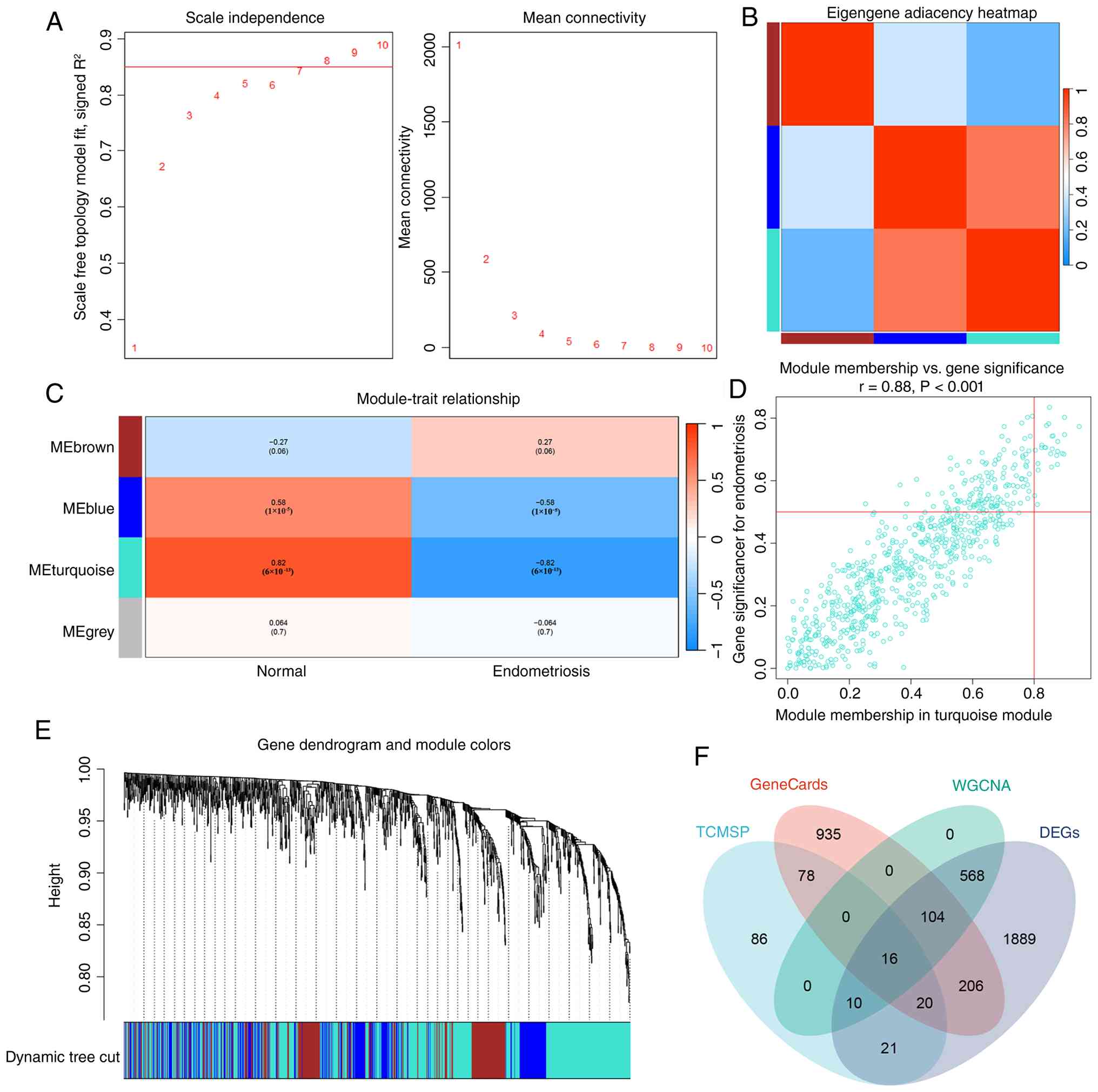 | Figure 3WGCNA. (A) Scale independence and
mean connectivity analysis for the soft thresholding power in
WGCNA, identifying the optimal power for network construction. (B)
Eigengene adjacency heatmap illustrates the relationships between
different modules in the WGCNA. The intensity of the color reflects
the strength of correlation (from 0 to 1) between module
eigengenes. (C) Module-trait relationships, with the correlation
values between different modules (MEbrown, MEblue, MEturquoise and
MEgrey) and normal/endometriosis traits. The most significant
module for endometriosis was MEturquoise. (D) Scatter plot showing
the relationship between module membership and gene significance
for the turquoise module in endometriosis samples. A positive
correlation was observed (r=0.88; P<0.001). (E) Gene dendrogram
from hierarchical clustering of genes, colored by module
membership. Modules were identified by dynamic tree cut, with color
coding for different modules. (F) Venn diagram showing the overlap
of genes from TCMSP, WGCNA, DEGs and GeneCards databases. A total
of 16 common genes were identified as BSHQ-related genes. BSHQ,
Paeoniae Radix Alba and Hedysarum multijugum Maxim; DEGs,
differentially expressed genes; TCMSP, Traditional Chinese Medicine
Systems Pharmacology; WGCNA, weighted gene co-expression network
analysis. |
Identification of BSHQ-related
genes
A total of 1,359 endometriosis-related genes were
retrieved from the GeneCards database (with a relevance score
>1). The effective ingredients of BSHQ, as analyzed through
TCMSP, along with endometriosis-related genes from GeneCards, the
turquoise module genes from WGCNA and DEGs from the merged
datasets, were intersected, resulting in the identification of 16
BSHQ-related genes (Fig. 3F).
These 16 genes were as follows: Cyclin dependent kinase inhibitor
1A (CDKN1A), claudin 4 (CLDN4), cytochrome P450 family 1 subfamily
B member 1 (CYP1B1), estrogen receptor 1 (ESR1), Fos proto-oncogene
(FOS), heme oxygenase 1 (HMOX1), heat shock protein family A member
5 (HSPA5), interleukin (IL)6, MET proto-oncogene (MET), matrix
metallopeptidase 9 (MMP9), prostaglandin E receptor 3 (PTGER3),
selectin E (SELE), SERPINE1, secretory leukocyte peptidase
inhibitor (SLPI), tumor protein p53 (TP53) and vascular cell
adhesion molecule 1 (VCAM1).
To explore the relationships between these 16 genes,
the present study first performed chromosomal localization analysis
(Fig. 4A). Following this, an
enrichment analysis of the 16 genes was conducted. The top three
most enriched items in each of the GO BP, CC and MF categories, and
KEGG pathways were identified. For GO BP terms, the top three
enriched items were ‘cellular response to oxidative stress’,
‘cellular response to chemical stress’ and ‘response to reactive
oxygen species’. For GO CC terms, the top three were ‘caveola’,
‘plasma membrane raft’ and ‘collagen-containing extracellular
matrix’. For GO MF terms, the top three were ‘RNA polymerase II
general transcription initiation factor binding’, ‘oxidoreductase
activity, acting on paired donors, with incorporation or reduction
of molecular oxygen, reduced flavin or flavoprotein as one donor,
and incorporation of one atom of oxygen’ and ‘core promoter
sequence-specific DNA binding’. For KEGG pathways, the top three
enriched items were ‘lipid and atherosclerosis’, ‘fluid shear
stress and atherosclerosis’ and ‘endocrine resistance’ (Fig. 4B; Table SIII).
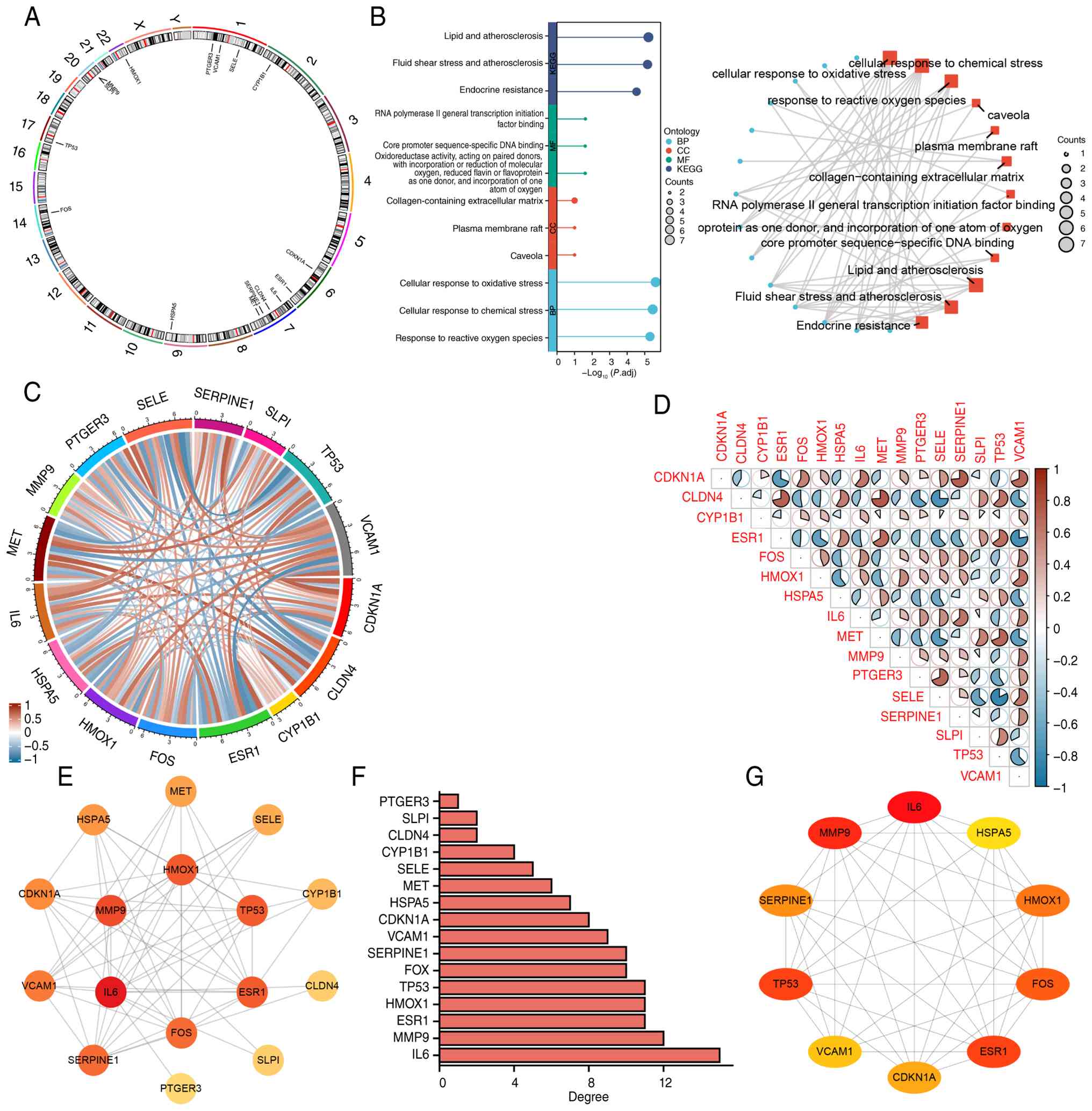 | Figure 4Network and enrichment analyses of
BSHQ-related genes. (A) Circular plot showing the chromosomal
locations of the BSHQ-related genes. Each chromosome is annotated
with its chromosome number, and the significant BSHQ-related genes
are marked with official gene symbol labels at their corresponding
genomic positions. (B) GO and KEGG pathway enrichment analyses for
the BSHQ-related genes. (C) Chord plots showing the relationships
between the expression levels of the 16 BSHQ-related genes. The
lines connecting genes indicate the strength and direction of their
correlation, with red lines representing positive correlations and
blue lines representing negative correlations. (D) Correlation plot
illustrating the relationships between the expression levels of the
16 BSHQ-related genes, with the strength of the correlation
indicated by the intensity of the color. (E) Protein-protein
interaction network showing the interactions between the 16 key
genes identified in the present study. Nodes represent proteins and
edges between them indicate functional associations. The color
intensity of the nodes reflects the relative importance of each
gene, with darker nodes indicating higher importance. (F) Degree
distribution plot showing the number of connections (degree) for
each gene. (G) Network analysis of the top 10 BSHQ-related genes
which were identified using the maximal clique centrality
algorithm. BP, biological processes; BSHQ, Paeoniae Radix Alba and
Hedysarum multijugum Maxim; CC, cellular components; GO,
Gene Ontology; KEGG, Kyoto Encyclopedia of Genes and Genomes; MF,
molecular functions. |
To further investigate the relationships between the
expression levels of these 16 BSHQ-related genes, chord and
correlation plots were constructed (Fig. 4C and D). In the correlation plot, the color
intensities or color scale were used to indicate the strength of
the correlation, with blue and red representing negative and
positive correlations, respectively. Additionally, using the STRING
database, a PPI network for the 16 BSHQ-related genes was
constructed (Fig. 4E). The darker
colors in the network indicate a higher influence on endometriosis.
Fig. 4F displays the ranking of
all 16 genes by degree. The top 10 hub genes (CDKN1A, ESR1, FOS,
HMOX1, HSPA5, IL6, MMP9, SERPINE1, TP53 and VCAM) were identified
using the MCC algorithm in the cytoHubba plug-in (Fig. 4G).
scRNA-seq analysis
The present study analyzed scRNA-seq data from
endometriosis (GSE213216), applying rigorous raw data
preprocessing. After normalizing the data and performing PCA to
reduce dimensionality, the present study used the UMAP method to
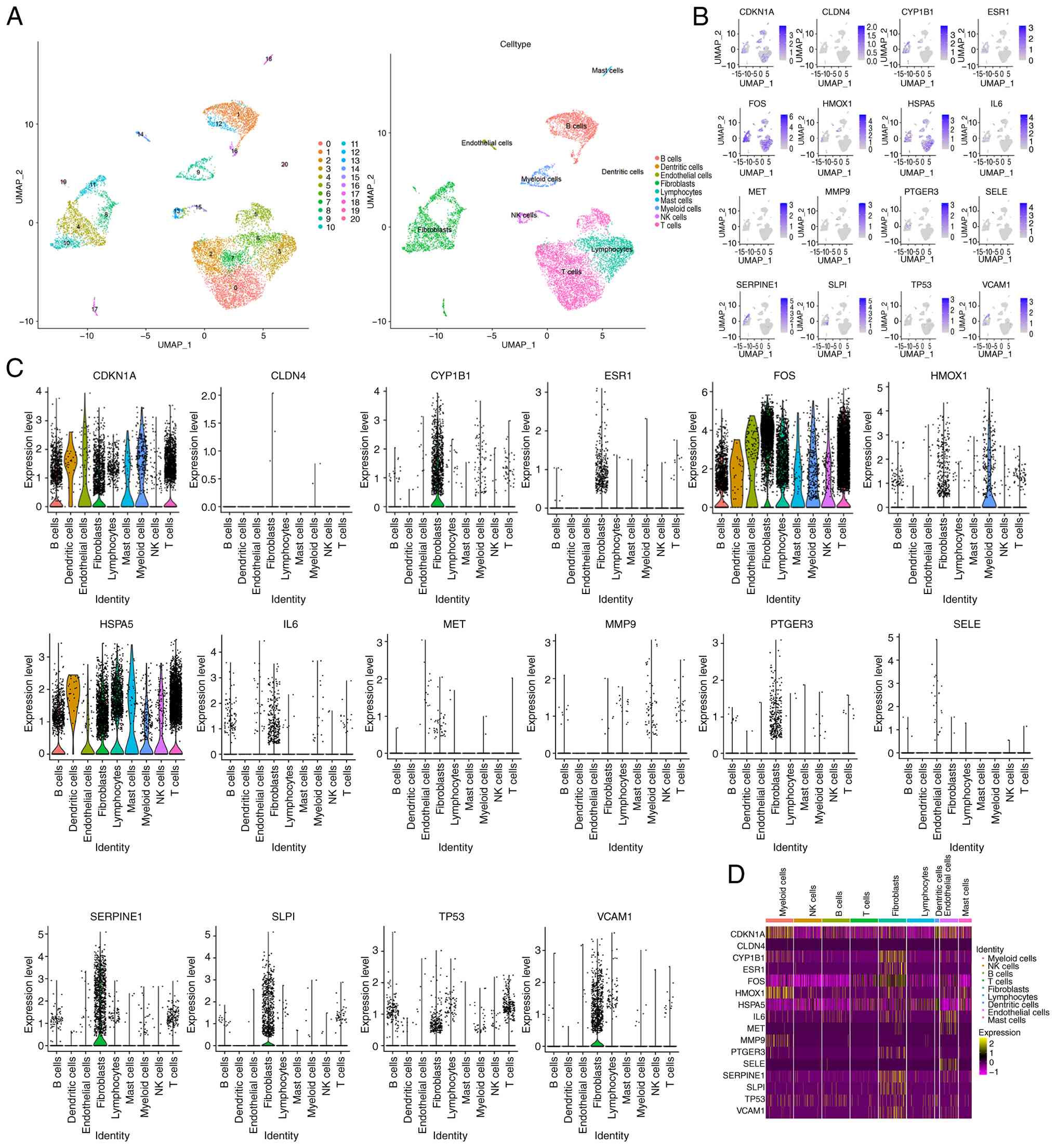
visualize all cells, which were subsequently grouped into 20
distinct clusters. Finally, the expression of the 16 BSHQ-related
genes across different cell types was visualized. The analysis
revealed distinct clusters of immune cell types based on their gene
expression patterns. UMAP clustering analysis identified separate
clusters corresponding to different cell types. Notably, clear
separation was observed for B cells, dendritic cells, endothelial
cells, fibroblasts, lymphocytes, mast cells, myeloid cells, natural
killer (NK) cells and T cells, with clear delineation between these
cell populations (Fig. 5A). These
clusters were defined by their unique transcriptional profiles, as
indicated by the different colors representing the various cell
types.
Next, the present study examined the expression of
the 16 BSHQ-related genes, CDKN1A, CLDN4, CYP1B1, ESR1, FOS, HMOX1,
HSPA5, IL6, MET, MMP9, PTGER3, SELE, SERPINE1, SLPI, TP53 and
VCAM1. The feature plots (Fig. 5B)
and violin plots (Fig. 5C)
illustrated the differential expression of these selected marker
genes across the various immune cell types. Several of these genes,
such as CDKN1A, FOS and HSPA5, were highly expressed in nearly all
cell types, whereas others, including CLDN4, MET, MMP9 and SELE,
exhibited low expression levels across almost all cell types. This
highlights the diversity in gene expression profiles within the
immune cell populations. Notably, SERPINE1 was highly expressed
specifically in fibroblasts.
Finally, a heatmap of gene expression across all
cell types was generated, revealing distinct expression patterns
for each gene (Fig. 5D). The
heatmap confirmed the tissue- and cell-type-specific expression of
the selected genes, offering valuable insights into the regulatory
mechanisms that govern immune cell functions.
Screening hub genes by machine
learning
To identify key biomarkers for endometriosis, the
present study applied a combination of LASSO regression and SVM-RFE
algorithms for feature selection. The LASSO analysis was first
conducted to identify the most predictive features, as illustrated
in Fig. 6A and B, which shows the relationship between
the fraction of deviance explained and the coefficients of each
feature. As the model was regularized, the coefficients for each
feature were progressively shrunk towards zero, with the optimal
features emerging when the deviance explained by each feature
plateaued. The SVM-RFE analysis, shown in Fig. 6C, was used to rank the features
based on their importance. To refine the list of candidate
biomarkers, the present study intersected the results from both the
LASSO and SVM-RFE analyses. The Venn diagram (Fig. 6D) revealed three overlapping genes,
which were selected as the most significant biomarkers for
endometriosis: PTGER3, SERPINE1 and VCAM1.
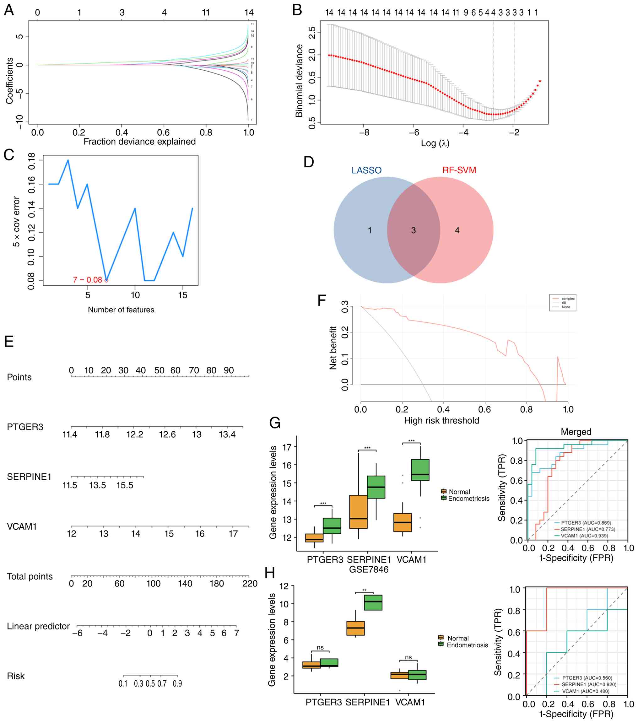 | Figure 6Machine learning analysis and
identification of hub genes. (A) LASSO regression coefficient plot
showing the selection of key genes based on their coefficients. The
plot illustrates the shrinkage of coefficients as the
regularization parameter increases. (B) LASSO cross-validation plot
determining the optimal log(λ) value for the model. The red dot
represents the selected λ with the minimal mean cross-validation
error. (C) Cross-validation error plot for feature selection. (D)
Venn diagram showing the overlap of genes selected by LASSO and
SVM-RFE. Three hub genes were identified by both methods. (E)
Nomogram for predicting risk based on the expression levels of the
hub genes. The nomogram shows how to calculate the total score and
the associated risk for each patient. (F) DCA for evaluating the
clinical utility of the nomogram. The DCA plot assesses the net
benefit of applying the nomogram at various risk thresholds,
providing insights into its potential clinical application across
different decision-making scenarios. (G) ROC curve analysis for the
merged dataset, showing the predictive power of PTGER3, SERPINE1
and VCAM1 with AUC values. The boxplots display the distribution of
expression of these three genes in normal and endometriosis
samples. (H) ROC curve analysis for the GSE746 dataset used as a
validation set, showing the predictive performance of PTGER3,
SERPINE1 and VCAM1. The boxplots display the expression of these
genes in both normal and endometriosis samples.
nsP>0.05, **P<0.01,
***P<0.001. AUC, area under the curve; DCA, decision
curve analysis; FPR, false positive rate; LASSO, least absolute
shrinkage and selection operator; PTGER3, prostaglandin E receptor
3; ROC, receiver operating characteristic; SERPINE1, serpin family
E member 1; SVM-RFE, support vector machine-recursive feature
elimination; TPR, true positive rate; VCAM1, vascular cell adhesion
molecule 1. |
Development and validation of a
nomogram for endometriosis risk prediction
To further assess the diagnostic potential of the
identified hub genes (PTGER3, SERPINE1 and VCAM1), the present
study developed a nomogram incorporating these biomarkers to
predict the risk of endometriosis. The nomogram (Fig. 6E) visually represents the weighted
contributions of each gene to the overall risk score. The
individual scores for each biomarker were calculated based on their
expression levels, with PTGER3, SERPINE1 and VCAM1 each
contributing specific points corresponding to their respective
expression levels. The total score, which is the sum of the
individual gene scores, was then used to compute the linear
predictor. The nomogram revealed a linear predictor ranging from -6
to 7, with higher scores associated with an increased predicted
risk of endometriosis. The total points, ranging from 0 to 220, can
be used to stratify patients based on their likelihood of
developing endometriosis. A risk score between 0.1 and 0.9 was
observed across the merged GEO dataset cohort, making this a
reliable tool for risk stratification in clinical practice.
Moreover, the clinical utility of the nomogram was
validated through DCA (Fig. 6F),
which evaluated the net benefit of applying the nomogram at
different risk thresholds. The DCA curve demonstrated that using
the nomogram to predict endometriosis risk provided a notable net
benefit over traditional strategies (such as treating all or no
patients), particularly at higher risk thresholds. This indicated
that the nomogram, incorporating PTGER3, SERPINE1 and VCAM1, may
represent a valuable tool for clinical decision-making and
personalized risk assessment in endometriosis.
Diagnostic performance and validation
of key biomarkers for endometriosis
To assess the diagnostic performance of the
identified biomarkers, the present study generated ROC curves. In
the merged datasets, PTGER3 achieved an AUC of 0.869, SERPINE1 had
an AUC of 0.773 and VCAM1 showed an AUC of 0.939 (Fig. 6G), indicating that the three genes
exhibited strong diagnostic potential for endometriosis. Boxplots
showed that the expression levels of all three genes were
significantly different between normal and endometriosis samples
(P<0.05).
Finally, the present study validated the performance
of these biomarkers in an independent validation cohort (GSE7846).
The ROC curve analysis in the validation set (Fig. 6H) revealed that SERPINE1 maintained
high diagnostic value with an AUC of 0.920, whereas PTGER3 and
VCAM1 exhibited reduced AUC values of 0.560 and 0.480,
respectively. Expression analysis further confirmed the significant
differential expression of SERPINE1, whereas PTGER3 and VCAM1 did
not show significant differences in the validation set. Therefore,
the SERPINE1 gene was selected as the next research object.
Molecular docking
Based on the aforementioned network pharmacology
analysis of BSHQ active ingredients and their target genes,
quercetin was identified as a common BSHQ-derived bioactive ligand
that interacts with the proteins encoded by PTGER3, SERPINE1 and
VCAM1. The molecular structure file of quercetin was retrieved from
the TCMSP database and subjected to docking preparation. Molecular
docking of the three proteins with the core active ingredient
quercetin was conducted using AutoDock Vina, and the most optimal
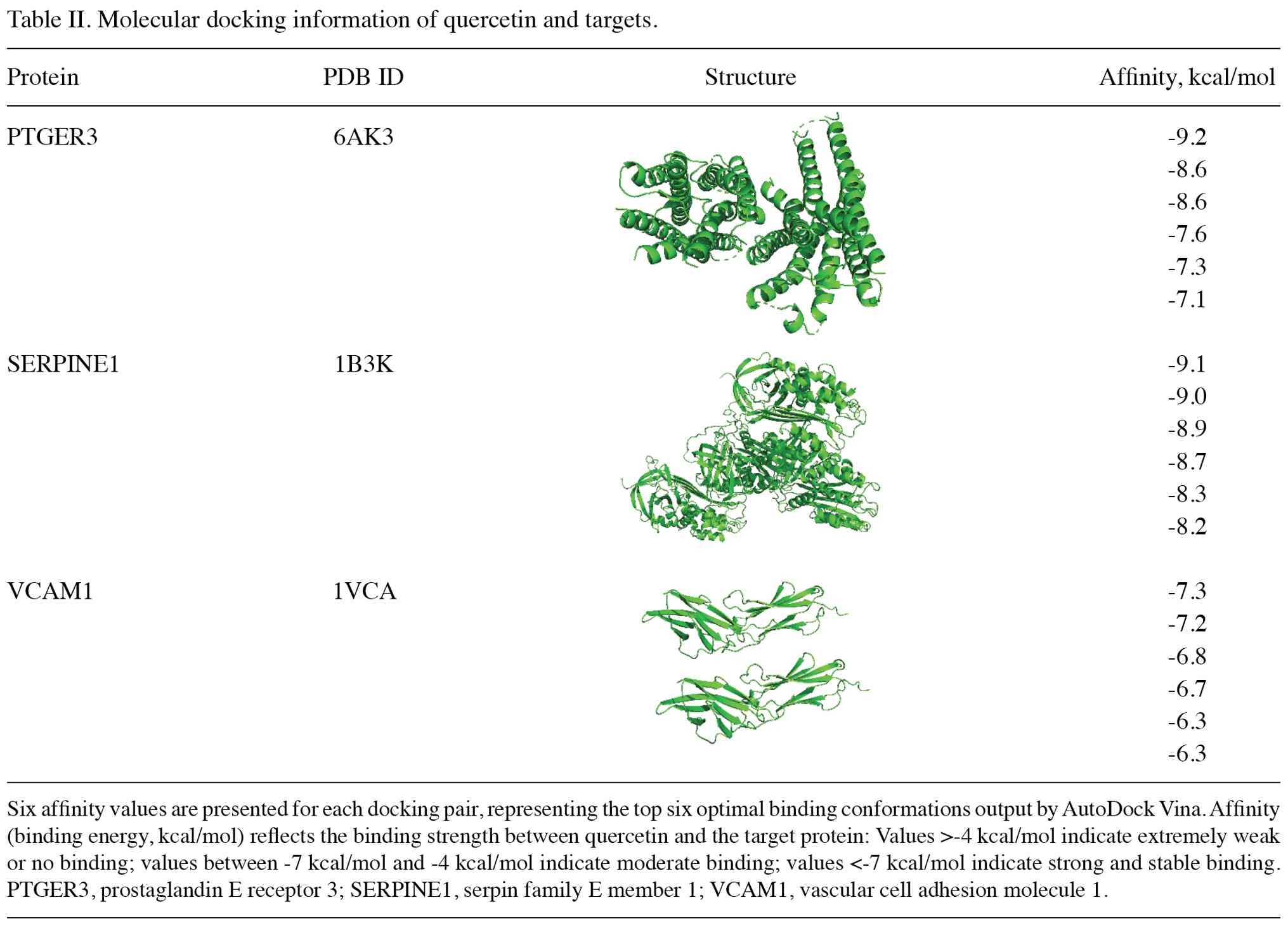
binding poses were selected. The resulting protein-ligand docking
structures showed that the binding free energies of all the optimal
poses were <-7 kcal/mol (Table
II). Molecular interaction analysis of the docking poses was
performed using PLIP and visualization was carried out with
PyMOL.
In the binding pocket of PTGER3, the amino acids
Q339, Q103, T107 and T106 form hydrophobic interactions with
quercetin. Additionally, amino acids F140 and T136 form hydrogen
bonds with quercetin (Fig. 7A). In
the binding pocket of SERPINE1, the amino acids V32, V284, E283,
T214, Y220 and R30 participate in hydrophobic interactions with
quercetin, whereas N31, E283, D285 and G218 form hydrogen bonds
with quercetin (Fig. 7B). In the
binding pocket of VCAM1, the amino acids P120 and Y119 interact
hydrophobically with quercetin and R123, D122, A144 and I88 form
hydrogen bonds with quercetin (Fig.
7C).
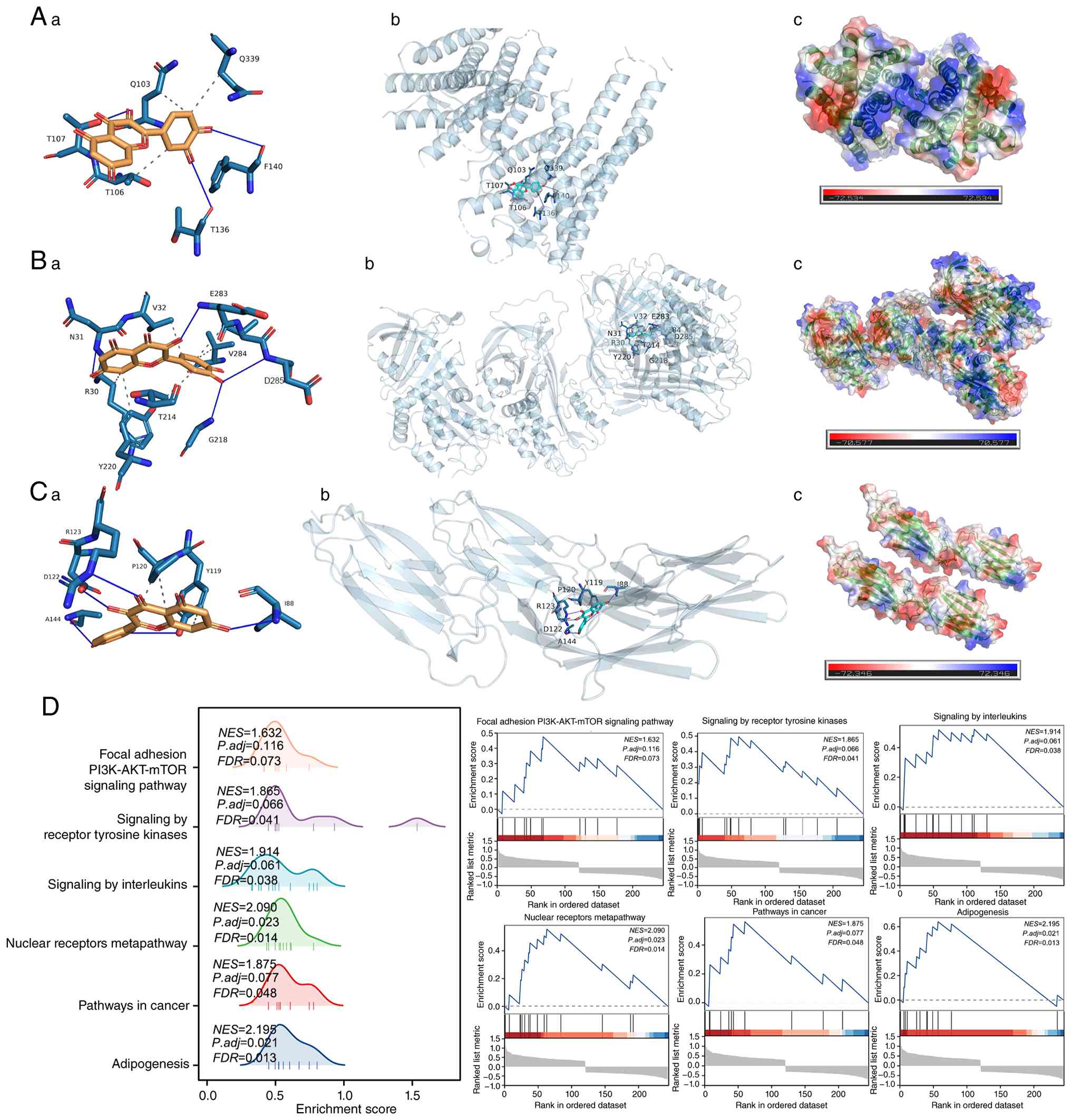 | Figure 7Molecular docking and GSEA. (Aa) 2D
interaction diagram showing the binding interactions between PTGER3
and quercetin. (Ab) 3D structure of the PTGER3-quercetin complex,
highlighting the binding pocket. (Ac) Electrostatic surface
potential map of the PTGER3-quercetin complex, showing the
distribution of charge (red for negative and blue for positive).
(Ba) 2D interaction diagram showing the binding interactions
between SERPINE1 and quercetin. (Bb) 3D structure of the
SERPINE1-quercetin complex, highlighting the binding pocket. (Bc)
Electrostatic surface potential map of the SERPINE1-quercetin
complex, showing the distribution of charge (red for negative and
blue for positive). (Cc) 2D interaction diagram showing the binding
interactions between VCAM1 and quercetin. (Cb)3D structure of the
VCAM1-quercetin complex, highlighting the binding pocket. (Cc)
Electrostatic surface potential map of the VCAM1-quercetin complex,
showing the distribution of charge (red for negative and blue for
positive). (D) GSEA showing the NES for various pathways. GSEA,
Gene Set Enrichment Analysis; NES, normalized enrichment scores;
PTGER3, prostaglandin E receptor 3; SERPINE1, serpin family E
member 1; VCAM1, vascular cell adhesion molecule 1. |
GSEA
Subsequently, based on the median expression level
of the SERPINE1 gene, the present study classified the
endometriosis samples from the merged dataset into two groups: The
SERPINE1 low-expression group and the SERPINE1 high-expression
group. DEGs (|logFC| >0.3 and P<0.05) between the two groups
were identified, followed by GSEA to further investigate the
biological pathways associated with these genes. GSEA was performed
to identify the significantly enriched biological pathways within
the dataset. The analysis revealed several prominent pathways with
varying levels of enrichment, as indicated by their normalized
enrichment scores and corresponding statistical parameters. The
enriched biological pathways included: ‘Focal adhesion
PI3K-AKT-mTOR signaling pathway’, ‘Signaling by receptor tyrosine
kinases’, ‘Signaling by interleukins’, ‘Nuclear receptors
meta-pathway’, ‘Pathways in cancer’ and ‘Adipogenesis’ (Fig. 7D).
Effect of quercetin on proliferation
in endometriosis cells
To evaluate the effects of quercetin on the
proliferative capacity of the endometriosis cell lines VK2/E6E7 and
12Z, a IC50 assay was performed.
The dose-response curve was constructed by plotting
the relationship between the drug concentration and the percentage
of cell proliferation. Through curve fitting, the IC50
values for each cell line were calculated. The results demonstrated
a dose-dependent decrease in cell proliferation as the drug
concentration increased. For the 12Z cell line, the calculated
IC50 value was found to be 41.49 µM, whereas for the
VK2/E6E7 cell line, the IC50 value was 9.518 µM
(Fig. 8A).
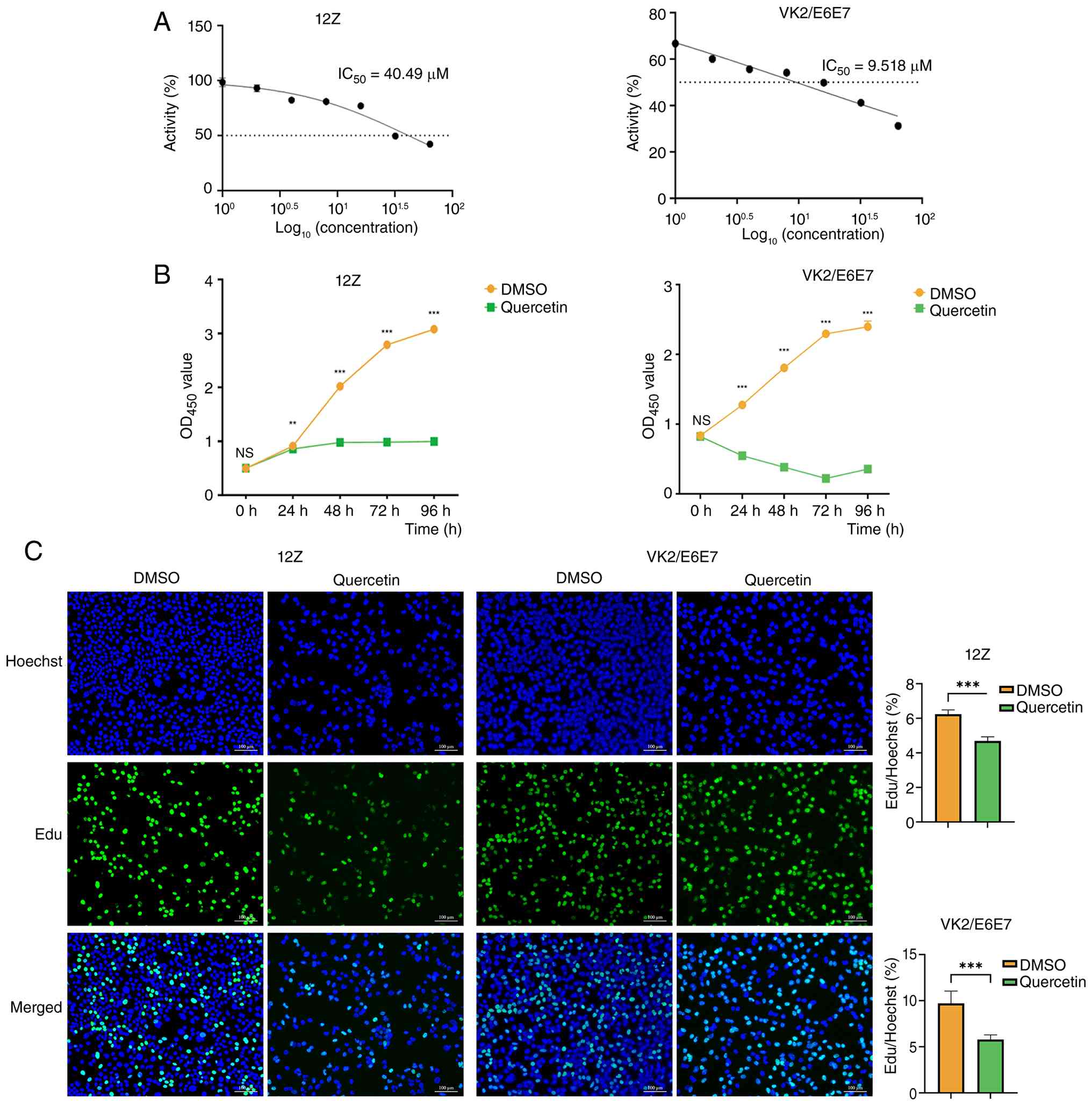 | Figure 8Effect of quercetin on the
proliferation of endometriosis cells. (A) IC50 values
for quercetin in 12Z and VK2/E6E7 cells were determined, with the
dose-response curves showing a dose-dependent decrease in cell
viability. The IC50 value for 12Z was 41.49 µM, while
for VK2/E6E7 it was 9.518 µM. (B) Cell Counting Kit-8 assay results
demonstrated a significant reduction in cell proliferation
following quercetin treatment in both cell lines over 96 h. (C) EdU
fluorescence staining revealed a significant decrease in the
percentage of EdU-positive cells following quercetin treatment,
with a marked reduction in both 12Z and VK2/E6E7 cells compared
with the DMSO control group, all fluorescence micrographs in this
panel were acquired at x10 magnification. NSP>0.05,
**P<0.01, ***P<0.001. DMSO, dimethyl
sulfoxide; EdU, 5-ethynyl-2'-deoxyuridine; IC50,
half-maximal inhibitory concentration; OD, optical density. |
To further investigate the effects of quercetin on
the proliferative capacity of the endometriosis cell lines VK2/E6E7
and 12Z, the present study employed both CCK-8 and EdU assays. The
CCK-8 assay results revealed a significant reduction in cell
proliferation following quercetin treatment, as evidenced by the
substantial decrease in OD450 values for both cell lines.
Specifically, for the 12Z cell line, the cell proliferation rate
was significantly diminished at various time points, with a notable
reduction observed at 24 h (t=3.553; P<0.01), 48 h (t=34.59,
P<0.001), 72 h (t=42.95, P<0.001) and 96 h (t=45.88,
P<0.001). Similarly, the VK2/E6E7 cell line demonstrated a
pronounced decrease in cell proliferation across the same time
intervals, with statistical significance observed at 24 h (t=44.60;
P<0.001), 48 h (t=58.21; P<0.001), 72 h (t=126.2; P<0.001)
and 96 h (t=24.73; P<0.001) (Fig.
8B). These findings indicated that quercetin exerts a potent
inhibitory effect on the proliferation of both VK2/E6E7 and 12Z
cells, with the effect becoming more pronounced over time.
To further evaluate the effects of quercetin on cell
proliferation, the present study conducted an EdU fluorescence
staining assay using both the 12Z and VK2/E6E7 cell lines. The
results demonstrated a significant reduction in the EdU-positive
cell rate after quercetin treatment, indicating a marked inhibition
of cell proliferation (Fig. 8C).
In the 12Z cell line, the EdU-positive cell rate was 6.234±0.249%
in the DMSO control group, whereas it decreased to 4.692±0.241% in
the quercetin-treated group, with a significant difference between
the two groups (t=20.40; P<0.001). Similarly, for the VK2/E6E7
cell line, the EdU-positive cell rate in the DMSO control group was
9.708±1.337% and after treatment with quercetin, it decreased to
5.791±0.504%, with statistical significance observed (t=13.15;
P<0.001). These findings confirmed that quercetin significantly
inhibits the proliferative capacity of both 12Z and VK2/E6E7 cells,
as evidenced by the reduced EdU incorporation and the decreased
number of proliferating cells.
Effect of quercetin on SERPINE1
expression in endometriosis cells
To investigate the effect of quercetin on SERPINE1
expression in 12Z and VK2/E6E7 cells, both RT-qPCR and WB were
conducted at 24 and 48 h after treatment. The results of the
RT-qPCR experiments demonstrated a significant reduction in
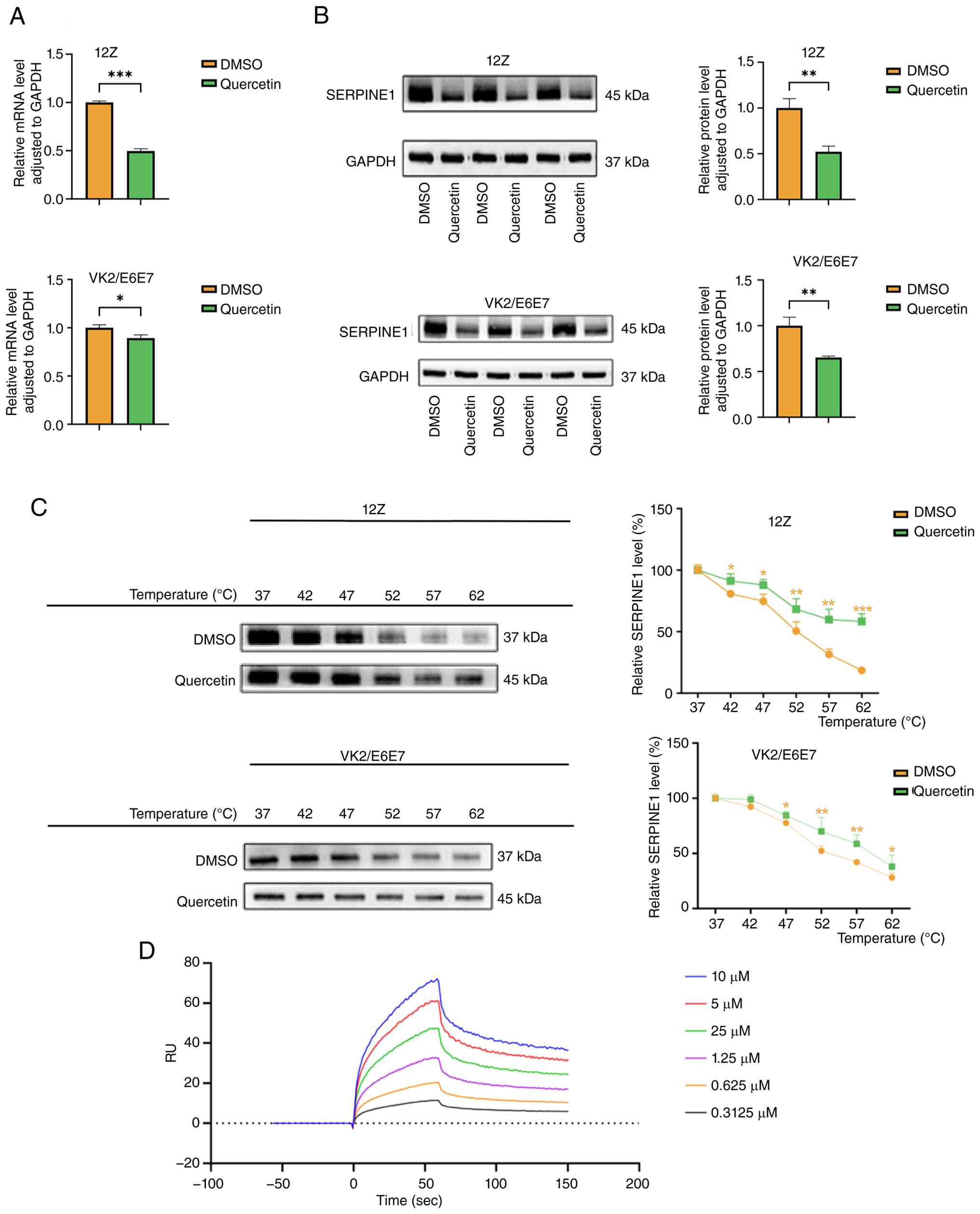
SERPINE1 mRNA expression levels following quercetin treatment in
both cell lines (Fig. 9A). In the
12Z cell line, SERPINE1 mRNA expression decreased from 1.000±0.015
in the DMSO control group to 0.497±0.023 in the quercetin-treated
group, with a highly significant difference (t=31.35; P<0.001).
Similarly, in the VK2/E6E7 cell line, the mRNA expression of
SERPINE1 decreased from 1.000±0.031 in the DMSO control group to
0.894±0.033 in the quercetin-treated group, with a statistically
significant reduction (t=4.089; P=0.01).
WB further confirmed these findings at the protein
level (Fig. 9B). In the 12Z cell
line, quercetin treatment led to a substantial reduction in
SERPINE1 protein expression, which decreased from 1.000±0.102 in
the DMSO control group to 0.522±0.061 in the quercetin-treated
group, with a significant difference (t=6.955; P=0.002). Similarly,
in VK2/E6E7 cells, SERPINE1 protein expression decreased from
1.000±0.093 in the DMSO control group to 0.653±0.015 in the
quercetin-treated group, with a statistically significant reduction
(t=6.401; P<0.01). These results collectively suggested that
quercetin treatment may significantly inhibit both the mRNA and
protein levels of SERPINE1 in 12Z and VK2/E6E7 cell lines,
indicating its potential role in modulating the expression of key
genes involved in the pathogenesis of endometriosis.
Evaluation of the interaction of
quercetin with SERPINE1
To evaluate the effect of quercetin on the stability
of cellular targets, the present study conducted a CETSA to assess
the interaction between quercetin and SERPINE1 (Fig. 9C). The results showed that SERPINE1
protein levels decreased progressively with increasing temperature.
However, this temperature-induced degradation was markedly
mitigated in the quercetin-treated groups, suggesting that
quercetin may effectively stabilize SERPINE1 by protecting it from
temperature-dependent degradation.
Based on the findings from both molecular docking
and CETSA, the present study preliminarily confirmed that quercetin
can bind to SERPINE1. To further validate this interaction, the
present study performed SPR experiments. The SPR sensorgram curves
in Fig. 9D recorded real-time
response units across serially diluted quercetin concentrations
(0.3125-10 µM). Fitting these response curves yielded a KD of
2.03x10⁻⁶ M for the interaction between quercetin and
SERPINE1, indicating a strong affinity between the two (Fig. 9D). Consistent with this binding
effect, quercetin treatment suppressed SERPINE1 mRNA and protein
levels, thus reducing its expression. These results collectively
supported the conclusion that quercetin interacts with SERPINE1,
potentially influencing its stability and function in endometriosis
cells.
SERPINE1 overexpression rescues cell
proliferation inhibition induced by quercetin
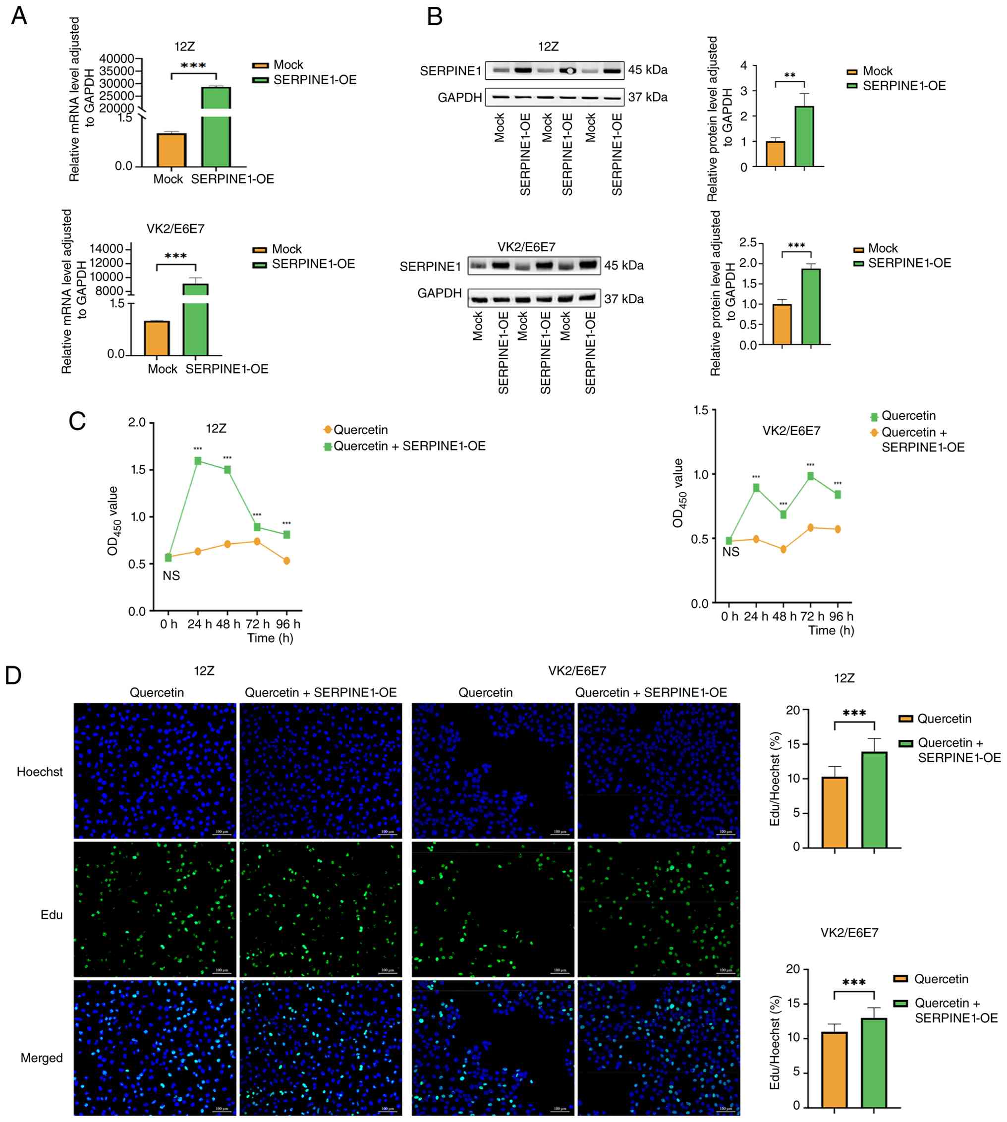
To assess the overexpression efficiency of SERPINE1
in 12Z and VK2/E6E7 cells, the present study transfected both cell
lines with a SERPINE1 overexpression plasmid, and performed RT-qPCR
and WB at 24 and 48 h post-transfection, respectively. The RT-qPCR
results demonstrated a significant increase in SERPINE1 mRNA
expression in the overexpression groups for both cell lines
(Fig. 10A). In the 12Z cell line,
the SERPINE1 mRNA expression increased from 1.003±0.095 in the DMSO
control group to 2,8658.739±779.729 in the SERPINE1 overexpression
group, with a highly significant difference (t=63.66; P<0.001).
Similarly, in the VK2/E6E7 cell line, SERPINE1 mRNA expression
increased from 1.000±0.019 in the DMSO control group to
9,115.191±1479.679 in the overexpression group, also showing a
statistically significant increase (t=10.67; P<0.001).
WB further confirmed the successful overexpression
of SERPINE1 at the protein level (Fig. 10B). In the 12Z cell line, SERPINE1
protein expression increased from 1.000±0.139 in the DMSO control
group to 2.399±0.489 in the SERPINE1 overexpression group, with a
significant difference (t=4.769; P=0.009). Similarly, in VK2/E6E7
cells, the SERPINE1 protein expression increased from 1.000±0.119
in the DMSO control group to 1.880±0.118 in the overexpression
group, with a highly significant difference (t=9.126; P<0.001).
These results indicated that transfection with the SERPINE1
overexpression plasmid effectively enhanced both mRNA and protein
levels of SERPINE1 in 12Z and VK2/E6E7 cell lines, confirming the
successful overexpression of SERPINE1 in these models.
To evaluate the effect of SERPINE1 overexpression on
cell proliferation in the presence of quercetin, the present study
transfected 12Z and VK2/E6E7 cells with a SERPINE1 overexpression
plasmid and treated them with quercetin. Cell proliferation was
assessed using the CCK-8 assay at multiple time points: 0, 24, 48,
72 and 96 h. The results from the CCK-8 assay indicated a
significant increase in the OD450 values in the SERPINE1
overexpression + quercetin treatment groups compared with in the
quercetin-only treatment groups, suggesting enhanced cell
proliferation (Fig. 10C). In the
12Z cell line, the OD450 values at 24 h (t=63.28; P<0.001), 48 h
(t=26.05; P<0.001), 72 h (t=6.011; P<0.001) and 96 h
(t=13.03; P<0.001) were all significantly higher in the SERPINE1
overexpression group treated with quercetin. Similarly, in the
VK2/E6E7 cell line, significant increases in cell proliferation
were observed at all time points, with OD450 values at 24 h
(t=65.96; P<0.001), 48 h (t=70.81; P<0.001), 72 h (t=37.70;
P<0.001) and 96 h (t=32.22; P<0.001) showing a marked
elevation in the SERPINE1 overexpression + quercetin treatment
group compared with the quercetin-only treatment.
To further confirm these findings, EdU fluorescence
staining was performed to assess cell proliferation. The results
revealed a significant increase in the percentage of EdU-positive
cells in the SERPINE1 overexpression + quercetin treatment groups
compared with the quercetin-only treatment groups (Fig. 10D). In the 12Z cell line, the
percentage of EdU-positive cells was 10.315±1.458% in the
quercetin-only treatment group and increased to 13.952±1.882% in
the SERPINE1 overexpression + quercetin treatment group, with a
significant difference (t=7.325; P<0.001). Similarly, in the
VK2/E6E7 cell line, the percentage of EdU-positive cells was
11.010±1.111% in the quercetin-only group and increased to
13.002±1.466% in the SERPINE1 overexpression + quercetin treatment
group, also showing a significant increase (t=5.080; P<0.001).
These findings collectively suggested that quercetin suppresses
cell proliferation, while SERPINE1 overexpression reverses this
inhibitory effect and restores cellular proliferative capacity in
both 12Z and VK2/E6E7 endometriosis cells.
The results suggested that quercetin alone may
suppress the proliferation of endometriosis cells and reduce
endogenous SERPINE1 expression, whereas SERPINE1 overexpression
counteracts this inhibitory effect. Specifically, SERPINE1
overexpression markedly increased cell proliferation in both 12Z
and VK2/E6E7 cell lines in the presence of quercetin, as evidenced
by the elevated OD450 values and increased EdU-positive cell rates
at all time points.
Discussion
The present study identified 32 bioactive
ingredients from BSHQ, with 12 from Paeoniae Radix Alba and 22 from
Hedysarum multijugum Maxim. These ingredients demonstrated
the notable pharmacological potential of these herbs in treating
endometriosis. A total of 231 target genes were identified,
highlighting their therapeutic value. The analysis revealed the
interactions between bioactive ingredients and target genes,
shedding light on the molecular mechanisms involved. Notably,
quercetin, kaempferol and other compounds such as
7-O-methylisomucronulatol showed the highest connectivity,
suggesting their critical role in modulating biological pathways
related to endometriosis. The anti-inflammatory and antioxidant
properties of quercetin may markedly contribute to the therapeutic
effects of these herbs, modulating inflammatory responses, immune
activity and tissue remodeling in endometriosis (23,24).
A comprehensive bioinformatics analysis was
performed to identify 16 key genes associated with endometriosis,
derived from herbal ingredients in BSHQ. The GO and KEGG pathway
analyses provided crucial insights into the roles of these genes in
endometriosis. These genes were involved in immune response, cell
proliferation, inflammation and cell migration, all of which are
fundamental to disease development. The GO analysis revealed that
genes such as IL6, SERPINE1 and MMP9 were deeply involved in immune
regulation and inflammation, pivotal factors in the progression of
the disease (25-28).
Meanwhile, genes such as CDKN1A, ESR1 and FOS may influence cell
proliferation and migration, facilitating tissue invasion and
growth in endometriosis. The KEGG analysis identified pathways
related to oxidative stress, lipid metabolism and endocrine
resistance. Genes such as HMOX1 and SERPINE1 serve key roles in
oxidative stress responses, which contribute to the chronic
inflammation seen in endometriosis (29). Additionally, VCAM1 is involved in
immune cell adhesion and persistent inflammation, whereas ESR1 and
PTGER3 are implicated in endocrine resistance, suggesting that
targeting these pathways may improve the effectiveness of hormonal
therapies for endometriosis (30,31).
For an improved understanding of the interactions of
the 16 BSHQ-related genes in endometriosis, the present study
constructed a PPI network and performed correlation analysis. The
PPI network, built using the STRING database, identified key hub
genes, with CDKN1A, ESR1, FOS, HMOX1, HSPA5, IL6, MMP9, SERPINE1,
TP53 and VCAM1 emerging as the top 10 hub genes. These genes,
characterized by high connectivity, may have critical roles in
regulating key cellular processes, such as cell cycle progression,
inflammation and tissue remodeling, and could serve as key
regulators in the development of endometriosis.
The application of scRNA-seq has markedly enhanced
the understanding of cellular diversity in endometriosis. The
present study analyzed scRNA-seq data from endometriotic tissue and
identified 20 distinct cellular clusters using UMAP clustering,
providing clear insights into various immune cell types, including
B cells, dendritic cells, fibroblasts, myeloid cells, NK cells and
T cells. This highlights the immune complexity and heterogeneity
within endometriotic tissue. The present study further examined the
expression patterns of 16 BSHQ-related genes, which are involved in
processes such as cell cycle regulation, inflammation and immune
responses. Genes such as CDKN1A, FOS and HSPA5 were widely
expressed across immune cells, indicating their involvement in
proliferation, stress response and apoptosis. By contrast, genes
such as CLDN4, MET, MMP9 and SELE exhibited more selective roles.
Notably, SERPINE1 was highly expressed in fibroblasts, suggesting
its critical role in tissue remodeling and the chronic inflammatory
environment of endometriosis. Fibroblasts influence ECM formation
and immune responses, potentially driving disease progression in
endometriotic lesions (32). These
findings underscored the immune landscape of endometriosis and
suggested that genes such as SERPINE1 could serve as potential
biomarkers for diagnosis or monitoring. Targeting the pathways
regulated by these genes may offer new therapeutic strategies for
modulating immune responses and tissue remodeling in
endometriosis.
In the present study, PTGER3, SERPINE1 and VCAM1
were identified as key biomarkers for endometriosis using LASSO
regression and SVM-RFE algorithms. The ROC curve analysis revealed
that PTGER3 and SERPINE1 possess strong diagnostic potential,
effectively distinguishing between endometriosis and normal tissue.
These genes are integral to the pathogenesis of the disease,
underscoring their value as both diagnostic and therapeutic
targets. Incorporating these biomarkers into a nomogram for risk
prediction enhanced their clinical utility. This nomogram assigned
weighted scores to each biomarker based on their expression levels,
markedly improving the personalized diagnosis of endometriosis. By
providing a quantitative risk score, the model allows for more
accurate patient stratification, facilitating early intervention
and tailored treatment approaches.
As a common bioactive flavonoid extracted from BSHQ,
quercetin exerts multifaceted regulatory effects on pathological
signaling pathways linked to endometriosis. Consistent with
previous research, it exerts prominent anti-inflammatory activity:
BSHQ has previously been shown to suppress macrophage activation,
lower reactive oxygen species (ROS) levels and reduce the secretion
of pro-inflammatory cytokines, including TNF-α and IL-1β, thereby
modulating the inflammatory microenvironment of endometriotic
lesions (33,34). Prior reports have also confirmed
that quercetin restrains endometriotic cell proliferation by
inhibiting ERK1/2, p38 MAPK and AKT cascades to trigger cell cycle
arrest and apoptosis (35). It
also suppresses MMP2 and MMP9, enzymes critical for cell invasion
and migration, further hindering disease progression (36). Moreover, quercetin serves a vital
role in oxidative stress regulation by scavenging ROS and promoting
the expression of antioxidant enzymes such as HMOX1(24). Its ability to modulate hormonal
resistance, particularly through the regulation of estrogen
signaling, offers therapeutic advantages, especially when used in
conjunction with hormonal therapies (37). Furthermore, quercetin interacts
with key biomarkers, including SERPINE1, ESR1 and MMP9, potentially
influencing the progression of endometriosis (38).
The present study identified SERPINE1 as a key
target gene of quercetin and a central hub gene in BSHQ-related
pathways in endometriosis. Located at 7q22.1, SERPINE1 encodes
plasminogen activator inhibitor-1, which regulates fibrinolysis by
inhibiting tissue-type and urokinase-type plasminogen activators.
This activity is essential for ECM remodeling, cell migration and
tissue repair. Elevated SERPINE1 expression has been linked to
cancer progression, particularly in gastric cancer, where it
regulates epithelial-mesenchymal transition (39). Additionally, SERPINE1 has a pivotal
role in wound healing, immune responses and fibrosis, establishing
it as a critical factor in chronic inflammatory diseases (40,41).
SERPINE1 serves a critical role in the inflammatory
microenvironment of endometriotic lesions. Reduced SERPINE1
expression in endometrial stromal cells enhances the production of
inflammatory cytokines through Toll-like receptor signaling,
contributing to the inflammation observed in endometriosis
(26). This inflammation
accelerates disease progression by altering cytokine production. As
a protease inhibitor, SERPINE1 regulates ECM remodeling, which is
essential for tissue invasion and lesion establishment. In
endometriotic fibroblasts, SERPINE1 is abnormally upregulated, and
this dysregulated upregulation leads to persistent ECM accumulation
and lesion expansion (42). While
SERPINE1 loss in stromal cells induces inflammation, its
overexpression in fibroblasts promotes ECM deposition and lesion
growth. Elevated SERPINE1 levels have been linked to endometrial
cancer risk, and this cell-type-specific abnormal expression
pattern makes it a promising biomarker for endometriosis (43). Additionally, SERPINE1 affects
immune cell recruitment by altering cytokine secretion and ECM
remodeling, facilitating immune cell infiltration and tissue
invasion (44). Targeting SERPINE1
could offer therapeutic benefits by modulating inflammation and ECM
remodeling, reducing lesion progression and alleviating symptoms
(45). Its effect on hormonal
regulation may also contribute to infertility in endometriosis
(46). Environmental and oxidative
stress factors further modulate SERPINE1 expression, exacerbating
the chronic inflammatory state in endometriosis (47).
The present study highlighted the ability of
quercetin to inhibit proliferation in endometriosis cells. CCK-8
and EdU assays both demonstrated significant reductions in cell
proliferation following quercetin treatment. These findings
suggested that quercetin suppresses cell proliferation in a dose-
and time-dependent manner. The EdU assay further confirmed a
decrease in DNA synthesis, indicating that quercetin effectively
inhibits cell cycle progression, positioning it as a promising
anti-proliferative agent for treating endometriosis.
The molecular analyses in the present study provided
valuable insights into the interactions between quercetin and key
biomarkers associated with endometriosis, specifically PTGER3,
SERPINE1 and VCAM1. These findings underscored the potential of
quercetin to modulate inflammation and tissue remodeling, both of
which are critical processes in the disease. The analysis revealed
that quercetin binds effectively to PTGER3, SERPINE1 and VCAM1,
demonstrating strong binding affinities. These interactions suggest
that quercetin may influence the prostaglandin E2 pathway via
PTGER3, regulate inflammation and tissue remodeling through
SERPINE1, and reduce immune cell infiltration by modulating VCAM1.
Additionally, quercetin stabilizes SERPINE1 under stress
conditions, protecting it from degradation at the protein level;
simultaneously, quercetin suppresses abnormally high SERPINE1
transcription in endometriotic fibroblasts to restrict excessive
ECM deposition and lesion growth. These findings suggest that
quercetin can effectively modulate the inflammatory
microenvironment, which is a key characteristic of endometriosis,
positioning it as a promising therapeutic agent for the condition.
By interacting with SERPINE1, quercetin further demonstrates its
potential in treating endometriosis.
In the present study, the effects of quercetin on
the proliferation of endometriosis cells were comprehensively
assessed, revealing key insights into its potential therapeutic
effects. The effect of quercetin on cell proliferation was evident
across various cell lines, with both the IC50 and
proliferation assays demonstrating that quercetin markedly
inhibited the proliferation of the 12Z and VK2/E6E7 endometriosis
cell lines. This anti-proliferative effect was observed to be dose-
and time-dependent, with greater reductions in cell proliferation
and DNA synthesis as the treatment duration extended. These
findings align with previous studies on the anticancer properties
of quercetin, highlighting its role in modulating cell cycle
progression and survival pathways, which are crucial for the growth
and persistence of endometriotic lesions (35,48).
Furthermore, the ability of quercetin to inhibit DNA synthesis, as
evidenced by the EdU assay, suggested that it could act as an
effective agent to target the proliferative aspect of
endometriosis.
An additional critical aspect explored in the
present study was the influence of quercetin on the expression of
SERPINE1, a protein involved in inflammation, ECM remodeling and
cell migration, which are key contributors to endometriosis
pathology. The results demonstrated that quercetin effectively
downregulated both the mRNA and protein levels of SERPINE1 in 12Z
and VK2/E6E7 cell lines. This finding is significant as SERPINE1
has been implicated in the regulation of chronic inflammation and
tissue remodeling in endometriosis. The reduction in SERPINE1
expression following quercetin treatment suggested that quercetin
may modulate the inflammatory microenvironment that characterizes
endometriotic lesions. By decreasing SERPINE1 levels, quercetin
could potentially alleviate some of the inflammation and ECM
remodeling that exacerbate the disease. Moreover, this
downregulation of SERPINE1 may further underscore the potential of
quercetin to modulate key pathways involved in disease
progression.
To further investigate the molecular interaction
between quercetin and SERPINE1, the present study employed advanced
techniques such as CETSA and SPR. These assays provided strong
evidence that quercetin binds directly to SERPINE1, stabilizing the
protein and preventing its degradation under stress conditions. The
results from CETSA indicated that quercetin effectively stabilizes
SERPINE1, protecting it from temperature-induced degradation.
Normally, an increase in temperature leads to the denaturation of
proteins, but in the presence of quercetin, the degradation of
SERPINE1 was markedly reduced. This suggests that quercetin may
bind directly to SERPINE1, preventing its destabilization under
stressful conditions. The stabilizing effect observed in the CETSA
experiment provides strong evidence that quercetin can interact
with SERPINE1 in a manner that potentially influences its
stability, which may be critical for maintaining proper cellular
function in endometriotic lesions.
To further substantiate these findings, SPR
analysis was performed, revealing a strong binding affinity between
quercetin and SERPINE1. The KD value obtained from the SPR analysis
indicated that quercetin interacts with SERPINE1 with high
specificity and affinity, suggesting a potentially robust and
biologically relevant interaction. This strong binding affinity
highlights the potential of quercetin to influence the function of
SERPINE1 within endometriotic tissue. The results from both CETSA
and SPR experiments collectively support the hypothesis that
quercetin interacts with SERPINE1; it may stabilize SERPINE1
protein to maintain basal stability at post-translational levels,
while inhibiting its aberrantly high expression to block
pathological activity; rescue experiments confirmed SERPINE1
overexpression reversed the therapeutic effects of quercetin on the
disease process.
The interaction between quercetin and SERPINE1 is
of particular importance in the context of endometriosis, as
SERPINE1 regulates ECM remodeling, which is central to the
formation and growth of endometriotic lesions. The ability of
quercetin to stabilize SERPINE1 may help modulate these processes
by fine-tuning the uPA/tPA-plasmin-MMP proteolytic cascade that
governs ECM turnover, potentially preventing excessive tissue
invasion and fibrosis (49,50).
Additionally, SERPINE1 is involved in immune cell recruitment and
inflammatory response, both of which are key drivers of
endometriosis pathology. By stabilizing SERPINE1, quercetin may
help regulate these processes by preserving the homeostatic
immunomodulatory function of SERPINE1 and restraining Toll-like
receptor-mediated overproduction of inflammatory cytokines,
contributing to a reduction in the chronic inflammation and tissue
remodeling that exacerbate the condition (26,51).
The present study has several limitations that
should be acknowledged. First, the bioinformatics analyses were
based on publicly available datasets, and although multiple
datasets were integrated and an independent validation cohort was
used, potential heterogeneity among platforms and sample sources
may still have influenced the results. Second, the functional
experiments were performed primarily in vitro using 12Z and
VK2/E6E7 cell lines, which cannot fully recapitulate the complex
immune, hormonal and microenvironmental features of endometriosis
in vivo. Third, although SERPINE1 was identified and
validated as a key target of quercetin, the potential contributions
of other candidate genes and signaling pathways identified in the
present study were not thoroughly investigated. In addition, the
direct therapeutic efficacy, pharmacokinetics and long-term safety
of quercetin were not evaluated in animal models or clinical
settings. Therefore, further studies using primary cells, animal
models and clinical samples with standardized collection protocols
and strict inclusion/exclusion criteria are required to validate
the present findings and to further clarify the therapeutic
potential and molecular mechanisms of quercetin in
endometriosis.
In conclusion, the present study provided valuable
insights into the therapeutic potential of quercetin in treating
endometriosis, demonstrating its ability to inhibit cell
proliferation and regulate key biomarkers such as SERPINE1. These
results provide a solid basis for future studies, emphasizing the
importance of clinical trials to evaluate the long-term
effectiveness and safety of quercetin in treating
endometriosis.
Supplementary Material
Consensus coding sequence region of
serpin family E member 1 (NM_000602.5).
Map of the SERPINE1 overexpression
plasmid. The SERPINE1 coding sequence was cloned into the
pcDNA3.1(+) vector to construct the overexpression plasmid (total
length: 6,589 bp). The plasmid carries an AmpR for bacterial
selection, and drives SERPINE1 expression under the control of the
CMV promoter. Common restriction enzyme cutting sites are annotated
on the map. AmpR, ampicillin resistance gene; CMV, cytomegalovirus;
SERPINE1, serpin family E member 1.
Active ingredients of Paeoniae Radix
Alba and Hedysarum multijugum Maxim.
Turquoise module genes of WGCNA.
GO and KEGG analyses for the 16
Paeoniae Radix Alba and Hedysarum Multijugum Maxim-related
genes.
Acknowledgements
Not applicable.
Funding
Funding: No funding was received.
Availability of data and materials
The data generated in the present study may be
requested from the corresponding author.
Authors' contributions
XG, DR, XL and YX were responsible for designing
the study. XG, DR, XC, RW, XL and YX were responsible for writing
the manuscript, extracting and analyzing data, interpreting the
results and experiments. XG, DR XC confirm the authenticity of all
the raw data. PZ, LW, MW, JZ, PL and DZ contributed to experiments
and reviewing the article. All authors read and approved the final
manuscript.
Ethics approval and consent to
participate
Not applicable.
Patient consent for publication
Not applicable.
Competing interests
The authors declare that they have no competing
interests.
References
|
1
|
Kwiatkowski P, Gładysz K, Szydłowska J,
Żuchnik O, Król O, Kuczyńska B, Czelej M, Kłos A, Gieroba K and
Szydłowski M: Endometriosis - Pathogenesis, diagnosis and
treatment. J Educ Health Sport. 13:302–308. 2023.
|
|
2
|
Bokor A, Meuleman C and D'Hooghe T:
Clinical Aspects of Endometriosis. In: Reproductive Endocrinology
and Infertility: Integrating Modern Clinical and Laboratory
Practice. Carrell D and Peterson C (eds). Springer New York, New
York, NY, pp191-207, 2010.
|
|
3
|
Chamie LP, Ribeiro DMRF, Tiferes DA,
Macedo Neto AC and Serafini PC: Atypical sites of deeply
infiltrative endometriosis: Clinical characteristics and imaging
findings. Radiographics. 38:309–328. 2018.PubMed/NCBI View Article : Google Scholar
|
|
4
|
Kim H and Cho S: Diagnosis and treatment
of endometriosis. Korean Med Assoc. 62:513–518. 2019.
|
|
5
|
Giudice LC: Advances in approaches to
diagnose endometriosis. Glob Reprod Health. 9(e0074)2024.
|
|
6
|
Andrade APP, Oliveira CVBL, Araújo LBF,
Donald MED, Portugal FFD, Costa MS, Andrade CP, Chianca BL and
Bacha E: Management of Endometriosis: An Integrative Review of
National and International Guidelines. Adv Soc Sci Res J.
10:328–342. 2023.
|
|
7
|
Luna Russo MA, Chalif JN and Falcone T:
Clinical management of endometriosis. Minerva Ginecol. 72:106–118.
2020.
|
|
8
|
Chauhan S, More A, Chauhan V and Kathane
A: Endometriosis: A review of clinical diagnosis, treatment, and
pathogenesis. Cureus. 14(e28864)2022.PubMed/NCBI View Article : Google Scholar
|
|
9
|
De Morais HB, De Sousa LMLL, Santos AMI,
Ribeiro VEA and Carvalho LMB: Negative impacts of endometriosis on
the quality of life of affected women: an integrative literature
review. Brazilian MedStud 5: No 8, 2021.
|
|
10
|
Yang Y, Yuan L, Wang K, Lu D, Meng F, Xu
D, Li W and Nan Y: The role and mechanism of paeoniae radix alba in
tumor therapy. Molecules. 29(1424)2024.PubMed/NCBI View Article : Google Scholar
|
|
11
|
Zhang S, Yuan Z, Wu H, Li W, Li L and
Huang H: Network pharmacology-based strategy reveals the effects of
hedysarum multijugum maxim.-radix salviae compound on oxidative
capacity and cardiomyocyte apoptosis in rats with diabetic
cardiomyopathy. Biomed Res Int. 2020(8260703)2020.PubMed/NCBI View Article : Google Scholar
|
|
12
|
Gao X, Ren C, Li L, Zhao H, Liu K, Zhuang
M, Lv X, Zhi X, Jiang H, Chen Q, et al: Pharmacological action of
Hedysarum polysaccharides: A review. Front Pharmacol.
14(1119224)2023.PubMed/NCBI View Article : Google Scholar
|
|
13
|
Yang K, Zeng L, Ge A, Shi Y, Zhu X, Liu W
and Ge J: The effect of hedysarum multijugum maxim.-chuanxiong
rhizoma compound on ischemic stroke: a research based on network
and experimental pharmacology. Oxid Med Cell Longev.
2020(6072380)2020.PubMed/NCBI View Article : Google Scholar
|
|
14
|
Su KH, Su SY, Ko CY, Cheng YC, Huang SS
and Chao J: Ethnopharmacological survey of traditional Chinese
medicine pharmacy prescriptions for Dysmenorrhea. Front Pharmacol.
12(746777)2021.PubMed/NCBI View Article : Google Scholar
|
|
15
|
Zhao M, Zhao P, Wang C, Ren D, Song Y and
Lu X: Bioinformatics analysis of hypoxia-related mechanisms in
endometriosis: DDR2 as a potential diagnostic and therapeutic
biomarker. Biomed Res Int. 2025(4765791)2025.PubMed/NCBI View Article : Google Scholar
|
|
16
|
Hever A, Roth RB, Hevezi P, Marin ME,
Acosta JA, Acosta H, Rojas J, Herrera R, Grigoriadis D, White E, et
al: Human endometriosis is associated with plasma cells and
overexpression of B lymphocyte stimulator. Proc Natl Acad Sci USA.
104:12451–12456. 2007.PubMed/NCBI View Article : Google Scholar
|
|
17
|
Hull ML, Escareno CR, Godsland JM, Doig
JR, Johnson CM, Phillips SC, Smith SK, Tavaré S, Print CG and
Charnock-Jones DS: Endometrial-peritoneal interactions during
endometriotic lesion establishment. Am J Pathol. 173:700–715.
2008.PubMed/NCBI View Article : Google Scholar
|
|
18
|
Monsivais D, Dyson MT, Yin P, Coon JS,
Navarro A, Feng G, Malpani SS, Ono M, Ercan CM, Wei JJ, et al: ERβ-
and prostaglandin E2-regulated pathways integrate cell
proliferation via Ras-like and estrogen-regulated growth inhibitor
in endometriosis. Mol Endocrinol. 28:1304–1315. 2014.PubMed/NCBI View Article : Google Scholar
|
|
19
|
Sha G, Wu D, Zhang L, Chen X, Lei M, Sun
H, Lin S and Lang J: Differentially expressed genes in human
endometrial endothelial cells derived from eutopic endometrium of
patients with endometriosis compared with those from patients
without endometriosis. Hum Reprod. 22:3159–3169. 2007.PubMed/NCBI View Article : Google Scholar
|
|
20
|
Fonseca MAS, Haro M, Wright KN, Lin X,
Abbasi F, Sun J, Hernandez L, Orr NL, Hong J, Choi-Kuaea Y, et al:
Single-cell transcriptomic analysis of endometriosis. Nat Genet.
55:255–267. 2023.PubMed/NCBI View Article : Google Scholar
|
|
21
|
Vickers AJ and Elkin EB: Decision curve
analysis: A novel method for evaluating prediction models. Med
Decis Making. 26:565–574. 2006.PubMed/NCBI View Article : Google Scholar
|
|
22
|
Livak KJ and Schmittgen TD: Analysis of
relative gene expression data using real-time quantitative PCR and
the 2(-Delta Delta C(T)) method. Methods. 25:402–408.
2001.PubMed/NCBI View Article : Google Scholar
|
|
23
|
Bastin A, Teimouri M, Faramarz S, Shabani
M, Doustimotlagh AH and Sadeghi A: In vitro and molecular docking
analysis of quercetin as an anti-inflammatory and antioxidant. Curr
Pharm Des. 29:883–891. 2023.PubMed/NCBI View Article : Google Scholar
|
|
24
|
Luo M, Tian R, Yang Z, Peng YY and Lu N:
Quercetin suppressed NADPH oxidase-derived oxidative stress via
heme oxygenase-1 induction in macrophages. Arch Biochem Biophys.
671:69–76. 2019.PubMed/NCBI View Article : Google Scholar
|
|
25
|
Kitawaki J, Kiyomizu M, Obayashi H, Ohta
M, Ishihara H, Hasegawa G, Nakamura N, Yoshikawa T and Honjo H:
Synergistic effect of interleukin-6 promoter (IL6-634C/G) and
intercellular adhesion molecule-1 (ICAM-1 469K/E) gene
polymorphisms on the risk of endometriosis in Japanese women. Am J
Reprod Immunol. 56:267–274. 2006.PubMed/NCBI View Article : Google Scholar
|
|
26
|
Kusama K, Satoyoshi A, Azumi M, Yoshie M,
Kojima J, Mizuno Y, Ono M, Nishi H, Kajihara T and Tamura K:
Toll-like receptor signaling pathway triggered by inhibition of
serpin A1 stimulates production of inflammatory cytokines by
endometrial stromal cells. Front Endocrinol (Lausanne).
13(966455)2022.PubMed/NCBI View Article : Google Scholar
|
|
27
|
Miyashita M, Koga K, Izumi G, Sue F,
Makabe T, Taguchi A, Nagai M, Urata Y, Takamura M, Harada M, et al:
Effects of 1,25-dihydroxy vitamin D3 on endometriosis. J Clin
Endocrinol Metab. 101:2371–2379. 2016.PubMed/NCBI View Article : Google Scholar
|
|
28
|
Vargas E, Aghajanova L, Gemzell-Danielsson
K, Altmäe S and Esteban FJ: Cross-disorder analysis of
endometriosis and its comorbid diseases reveals shared genes and
molecular pathways and proposes putative biomarkers of
endometriosis. Reprod Biomed Online. 40:305–318. 2020.PubMed/NCBI View Article : Google Scholar
|
|
29
|
Milewski L, Sciezynska A, Poninska J,
Soszyńska M, Barcz E, Roszkowski PI, Kamiński P, Włodarski P,
Płoski R and Malejczyk J: Endometriosis is associated with
functional polymorphism in the promoter of heme oxygenase 1 (HMOX1)
gene. Cells. 10(695)2021.PubMed/NCBI View Article : Google Scholar
|
|
30
|
Kuessel L, Wenzl R, Proestling K,
Balendran S, Pateisky P, Yotova 1st, Yerlikaya G, Streubel B and
Husslein H: Soluble VCAM-1/soluble ICAM-1 ratio is a promising
biomarker for diagnosing endometriosis. Hum Reprod. 32:770–779.
2017.PubMed/NCBI View Article : Google Scholar
|
|
31
|
Santulli P, Borghese B, Noël JC, Fayt I,
Anaf V, de Ziegler D, Batteux F, Vaiman D and Chapron C: Hormonal
therapy deregulates prostaglandin-endoperoxidase synthase 2 (PTGS2)
expression in endometriotic tissues. J Clin Endocrinol Metab.
99:881–890. 2014.PubMed/NCBI View Article : Google Scholar
|
|
32
|
Ma J, Zhang L, Zhan H, Mo Y, Ren Z, Shao A
and Lin J: Single-cell transcriptomic analysis of endometriosis
provides insights into fibroblast fates and immune cell
heterogeneity. Cell Biosci. 11(125)2021.PubMed/NCBI View Article : Google Scholar
|
|
33
|
Peng J, Yang Z, Li H, Hao B, Cui D, Shang
R, Lv Y, Liu Y, Pu W, Zhang H, et al: Quercetin reprograms
immunometabolism of macrophages via the SIRT1/PGC-1α signaling
pathway to ameliorate lipopolysaccharide-induced oxidative damage.
Int J Mol Sci. 24(5542)2023.PubMed/NCBI View Article : Google Scholar
|
|
34
|
Yao S, Sang H, Song G, Yang N, Liu Q,
Zhang Y, Jiao P, Zong C and Qin S: Quercetin protects macrophages
from oxidized low-density lipoprotein-induced apoptosis by
inhibiting the endoplasmic reticulum stress-C/EBP homologous
protein pathway. Exp Biol Med (Maywood). 237:822–831.
2012.PubMed/NCBI View Article : Google Scholar
|
|
35
|
Park S, Lim W, Bazer FW, Whang KY and Song
G: Quercetin inhibits proliferation of endometriosis regulating
cyclin D1 and its target microRNAs in vitro and in vivo. J Nutr
Biochem. 63:87–100. 2019.PubMed/NCBI View Article : Google Scholar
|
|
36
|
Xu W, Song Y, Li K, Zhang B and Zhu X:
Quercetin inhibits adenomyosis by attenuating cell proliferation,
migration and invasion of ectopic endometrial stromal cells. Drug
Des Devel Ther. 14:3815–3826. 2020.PubMed/NCBI View Article : Google Scholar
|
|
37
|
Chaichian S, Nikfar B, Arbabi Bidgoli S
and Moazzami B: The role of quercetin for the treatment of
endometriosis and endometrial cancer: A comprehensive review. Curr
Med Chem. 32:74–86. 2025.PubMed/NCBI View Article : Google Scholar
|
|
38
|
Tri Wulandari E, Soetrisno Purwanto B,
Reviono Ertanto Y and Sunowo J: A molecular docking insight:
Phyllanthus niruri L. constituents targeting MMP-9 for angiogenesis
inhibition and IL-1beta for antiinflammatory action in
endometriosis therapy. Plant Sci Today, 2024.
|
|
39
|
Yang JD, Ma L and Zhu Z: SERPINE1 as a
cancer-promoting gene in gastric adenocarcinoma: Facilitates tumour
cell proliferation, migration, and invasion by regulating EMT. J
Chemother. 31:408–418. 2019.PubMed/NCBI View Article : Google Scholar
|
|
40
|
Simone TM and Higgins PJ: Inhibition of
SERPINE1 function attenuates wound closure in response to tissue
injury: A role for PAI-1 in re-epithelialization and granulation
tissue formation. J Dev Biol. 3:11–24. 2015.
|
|
41
|
Chen TY, Zhou M, Lin MQ, Liang ST, Yan Y,
Wang SM, Fang CS, Li D and Ruan Y: Research progress on the
SERPINE1 protein and chronic inflammatory diseases of the upper
respiratory tract: A literature review. Int Arch Allergy Immunol.
182:1097–1102. 2021.PubMed/NCBI View Article : Google Scholar
|
|
42
|
Wilson MR, Reske JJ, Holladay J, Neupane
S, Ngo J, Cuthrell N, Wegener M, Rhodes M, Adams M, Sheridan R, et
al: ARID1A mutations promote P300-dependent endometrial invasion
through super-enhancer hyperacetylation. Cell Rep.
33(108366)2020.PubMed/NCBI View Article : Google Scholar
|
|
43
|
Trabert B, Eldridge RC, Pfeiffer RM,
Shiels MS, Kemp TJ, Guillemette C, Hartge P, Sherman ME, Brinton
LA, Black A, et al: Prediagnostic circulating inflammation markers
and endometrial cancer risk in the prostate, lung, colorectal and
ovarian cancer (PLCO) screening trial. Int J Cancer. 140:600–610.
2017.PubMed/NCBI View Article : Google Scholar
|
|
44
|
Hirota Y, Osuga Y, Hirata T, Yoshino O,
Koga K, Harada M, Morimoto C, Nose E, Yano T, Tsutsumi O and
Taketani Y: Possible involvement of thrombin/protease-activated
receptor 1 system in the pathogenesis of endometriosis. J Clin
Endocrinol Metab. 90:3673–3679. 2005.PubMed/NCBI View Article : Google Scholar
|
|
45
|
Cakmak H, Seval-Celik Y, Arlier S,
Guzeloglu-Kayisli O, Schatz F, Arici A and Kayisli UA: p38
mitogen-activated protein kinase is involved in the pathogenesis of
endometriosis by modulating inflammation, but not cell survival.
Reprod Sci. 25:587–597. 2018.PubMed/NCBI View Article : Google Scholar
|
|
46
|
Wu MH, Hsiao KY and Tsai SJ: Endometriosis
and possible inflammation markers. Gynecol Minim Invasive Ther.
4:61–67. 2015.
|
|
47
|
Saguyod SJU, Kelley AS, Velarde MC and
Simmen RC: Diet and endometriosis-revisiting the linkages to
inflammation. Journal of Endometriosis and Pelvic Pain Disorders,
2018. 10:51–58. 2018.
|
|
48
|
Zhang L, Mohankumar K, Martin G, Mariyam
F, Park Y, Han SJ and Safe S: Flavonoids quercetin and kaempferol
are NR4A1 antagonists and suppress endometriosis in female mice.
Endocrinology. 164(bqad133)2023.PubMed/NCBI View Article : Google Scholar
|
|
49
|
Vassalli JD, Sappino AP and Belin D: The
plasminogen activator/plasmin system. J Clin Invest. 88:1067–1072.
1991.PubMed/NCBI View Article : Google Scholar
|
|
50
|
Senthilkumar K, Arunkumar R, Elumalai P,
Sharmila G, Gunadharini DN, Banudevi S, Krishnamoorthy G, Benson CS
and Arunakaran J: Quercetin inhibits invasion, migration and
signalling molecules involved in cell survival and proliferation of
prostate cancer cell line (PC-3). Cell Biochem Funct. 29:87–95.
2011.PubMed/NCBI View Article : Google Scholar
|
|
51
|
Jiang M, Lv Z, Huang Y, Cheng Z, Meng Z,
Yang T, Yan Q, Lin M, Zhan K and Zhao G: Quercetin alleviates
lipopolysaccharide-induced inflammatory response in bovine mammary
epithelial cells by suppressing TLR4/NF-κB signaling pathway. Front
Vet Sci. 9(915726)2022.PubMed/NCBI View Article : Google Scholar
|