Introduction
Endometrial carcinosarcoma (ECS) is a rare
high-grade malignant tumor of the uterus, which exhibits both
epithelial carcinoma and mesenchymal sarcoma components. The fifth
edition of the World Health Organization (WHO) classification
categorizes ECS as a subtype of endometrial carcinoma, which
accounts for ~5% of all uterine malignant tumors (1,2). ECS
has a high degree of malignancy and a 5-year survival rate of
33-39% in the United States (3).
After the molecular classification of endometrial cancer was
conducted in 2013, molecular pathological studies found that 7-12%
of endometrial cancer cases have mutations in the DNA polymerase ε
(POLE) gene. These tumors exhibit an ultra-high mutational
phenotype and demonstrate a markedly better prognosis compared with
the other three TCGA molecular subtypes of endometrial carcinoma
(4-6).
Furthermore, the ratio of microsatellite stability to TP53 mutation
has been reported to be 1.9:1 in POLE-mutant endometrial cancer
(7). In the absence of molecular
typing, POLE-mutant endometrial cancer with good prognosis may be
misinterpreted as invasive endometrial cancer, which has an
unfavorable prognosis.
To the best of our knowledge, POLE-mutant ECS has
rarely been reported (8-11).
In 2025, Toboni et al (10)
reported that among 2,235 cases of ECS, 48 cases (48/2,235; 2.1%)
were POLE-mutant. The prognoses of the patients with POLE-mutant
ECS were considerably improved compared with those of the patients
with the TP53-abnormal (TP53-mutant molecular subtype) ECS (median
overall survival, 74.8 vs. 21.9 months, respectively). Therefore,
correctly screening POLE-mutant ECS by molecular typing is
important for effective treatment. The present study reports a case
of POLE-mutant ECS diagnosed via molecular detection, and discusses
its diagnosis and treatment strategy based on previous studies. The
current findings may serve as a reference for clinicians and
pathologists.
Case report
Clinical presentation
A female patient (age, 61 years), whose menopause
occurred prior to admission, was admitted to The Second People's
Hospital of Xiaoshan District (Hangzhou, China) in February 2025,
after experiencing yellow vaginal discharge for >2 months. A
gynecological examination revealed that the vulva was intact. The
patient had a history of childbirth; their vagina appeared smooth,
and their uterus was in an anterior position, of normal size,
medium texture, non-tender and with acceptable mobility. No obvious
mass or tenderness was detected in the bilateral ovarian and
fallopian tube regions. A human papillomavirus test of cervical
cell samples showed positive results for subtype 52, and the
cervical liquid-based cytology examination (Shijiazhuang New
Century Engineering Co., Ltd.) showed no intraepithelial lesions or
malignant cells. Gynecological B-ultrasound examination showed that
the uterus was clear, with a slightly stronger heterogeneous echo
mass of 4.3x2.9x4.1 cm in size in the uterine cavity,
low-resistance blood flow signals and no prominent endometrium.
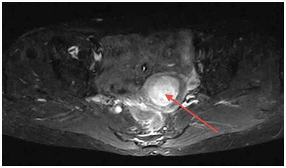
Pelvic magnetic resonance imaging, plain scan and functional
imaging showed abnormal signals in the uterine cavity measuring
4.2x3.1 cm; the boundary between the lesion and the muscular layer
was well-defined and a malignant tumor was considered (Fig. 1). No abnormal signal shadow was
found in the cervix, bilateral ovarian and fallopian tube regions,
and uterorectal fossa. The bladder and pelvic wall had a normal
structure, and the lymph nodes in the pelvic cavity were not
enlarged. The patient had a body mass index of 22.7
kg/m2 (within the normal healthy range for Chinese
adults). The patient had two sons and one daughter; obstetric
history was recorded using the TPAL system (3-0-0-3:
term-preterm-abortion-living children). Notably, the patient had
undergone surgical repair of a perforated gastric ulcer >10
years ago and had lived with chronic hepatitis B for >50 years
without complications. Abdominal B-ultrasound examination showed no
prominent masses in the liver, kidney or retroperitoneal space, and
no tumors were detected using head and chest computed tomography.
Serological test results showed that carcinoembryonic antigen (CEA;
reference range, 0-5 ng/ml), cancer antigen (CA)199 (reference
range, 0-37 U/ml), CA125 (reference range, 0-30.2 U/ml),
α-fetoprotein (AFP: reference range, 0-8.1 ng/ml) and squamous cell
carcinoma-associated antigen (SCC-Ag; reference range, 0-1.5 ng/ml)
levels were within normal limits. Diagnostic curettage was
performed 3 days after admission. The obtained intrauterine tissue
specimens were fixed in 10% neutral-buffered formalin at room
temperature (25˚C) for 24 h, embedded in paraffin, sectioned and
stained with haematoxylin-eosin (H&E). Histopathological
examination of curettage material yielded a diagnosis of poorly
differentiated adenocarcinoma of the uterine cavity. The patient
underwent laparoscopic hysterectomy, bilateral ovariectomy and
salpingectomy, para-aortic lymph node resection and pelvic lymph
node dissection under general anesthesia. The surgical size of the
uterus was 6.5x5.5x5.5 cm. The cervix was atrophic with a smooth
surface, the length of the cervical canal was 2 cm and the
endometrial thickness was 0.1 cm. A 4.0x4.0x2.0 cm exogenic
gray-yellow soft mass was located near the uterine horn on the
right side of the uterus, with a gray-yellow, fish-meat-like cut
surface and a gelatinous area infiltrating the superficial muscle
layer. No notable abnormalities were detected in the bilateral
ovaries and fallopian tubes.
Resected uterine tumor tissue was fixed with 10%
neutral formalin at room temperature (25˚C) for 24 h, embedded in
paraffin, cut into sections (3 µm) and stained with H&E.
H&E staining was performed at room temperature (25˚C):
Hematoxylin for 5 min and 1% aqueous eosin for 1 min. Slide
observation was carried out using a bright-field light microscope.
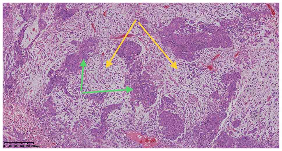
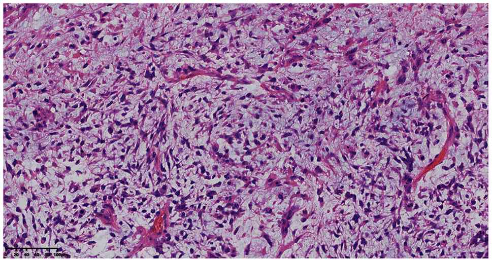
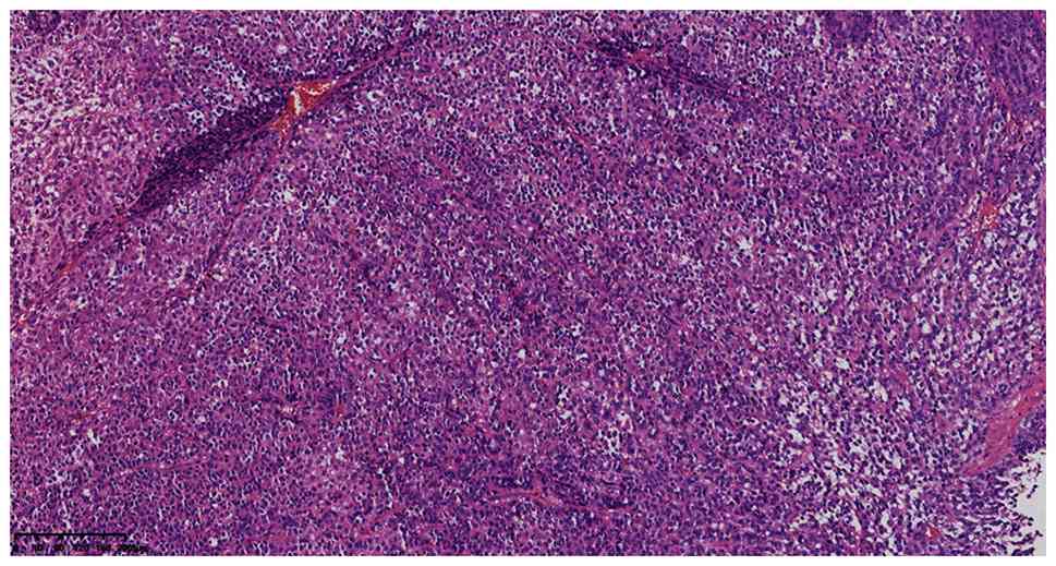
Microscopic examination showed that the tumor consisted of
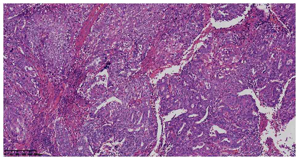
intermingled carcinoma and sarcoma components (Fig. 2). The epithelial components
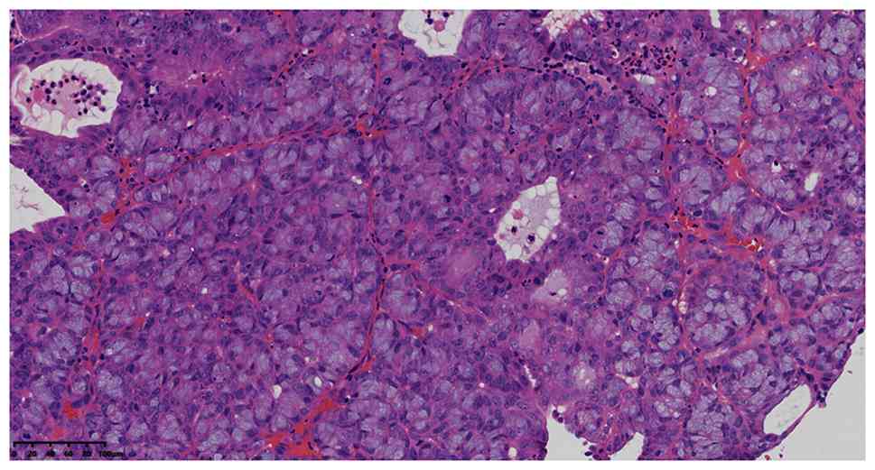
included endometrioid carcinoma (FIGO grade 3, 10%) (Fig. 3), mucinous adenocarcinoma (FIGO
grade 3, 30%) (Fig. 4), and a
high-grade undifferentiated sarcoma characterized by spindle and
small round cells (60%) (Figs. 5
and 6) (1). The tumor invaded the myometrium
(<1/2) without lymphovascular space invasion (LVSI).
Immunohistochemistry (IHC) analysis was performed
using an EnVision IHC kit (polymer method; cat. no. KIT-0014;
Fuzhou Maixin Biotechnology Development Co., Ltd.) using primary
antibodies (incubated for 50 min at 25˚C) purchased from Beijing
Zhongshan Jinqiao Biological Co., Ltd. and Fuzhou Maixin
Biotechnology Development Co., Ltd., to target the following
proteins (pre-diluted working solutions unless otherwise
indicated): Cytokeratin (CK)7 (1:100; cat. no. 21022407), Cam5.2
(working fluid; cat. no. 19121791), estrogen receptor (ER) (1:200;
cat. no. 6072353), progesterone receptor (PR) (1:400; cat. no.
9071722), vimentin (Vim) (1:100; cat. no. 21031351), paired box
gene 8 (Pax-8) (1:100; cat. no. 21012350), p16 (working fluid; cat.
no. 2107210673d), TP53 (p53) (1:100; cat. no. 20082125), SMARCA4
(BRG1) (working fluid; cat. no. 21045528), MutL homolog 1 (MLH1)
(1:100; cat. no. 2109290789d), MutS Homolog 2 (MSH2) (1:200; cat.
no. 2104220836C2), MutS Homolog 6 (MSH6) (1:200; cat. no.
2101280831C1), Mismatch repair endonuclease PMS2 (PMS2) (1:100;
cat. no. 2109150775d), NapsinA (1:100; cat. no. 20091108), CerbB-2
(working fluid; cat. no. H13317) and Ki-67 proliferation index
(Ki-67) (1:200; cat. no. 20091812). IHC sections were observed
under a light microscope without software analysis.
The results were as follows: CK7 (carcinoma, +;
sarcoma, -) (Fig. 7A), Cam5.2
(carcinoma, +; sarcoma, -) (Fig.
7B), estrogen receptor (carcinoma, 10% +; sarcoma, -) (Fig. 7C), progesterone receptor
(carcinoma, 1% +; sarcoma, -) (Fig.
7D), vimentin (carcinoma, weak +; sarcoma, +) (Fig. 7E), paired box gene 8 (carcinoma, +;
sarcoma, partial +) (Fig. 7F), P16
(+) (Fig. 7G), TP53 (mucinous
adenocarcinoma and small round cell sarcoma showed aberrant
immunohistochemical overexpression ; Figs. 7H and I), SMARCA4 (+) (Fig. 8A), MLH1 (+) (Fig. 8B), MSH2 (+) (Fig. 8C), MSH6 (+) (Fig. 8D), PMS2 (+) (Fig. 8E), NapsinA (-) (Fig. 8F), CerbB-2 (1+) (Fig. 8G), desmin (-) (Fig. 8H) and Ki-67 proliferation index
(80%) (Fig. 8I) (Table I). Consequently, the primary
pathological diagnosis was identified as ECS (TP53-abnormal type),
which invaded less than one-half of the myometrium. LVSI was not
detected and all 39 resected lymph nodes were negative for
metastatic tumor cells. The tumor was classified as FIGO 2023 stage
IIC endometrial carcinoma (1).
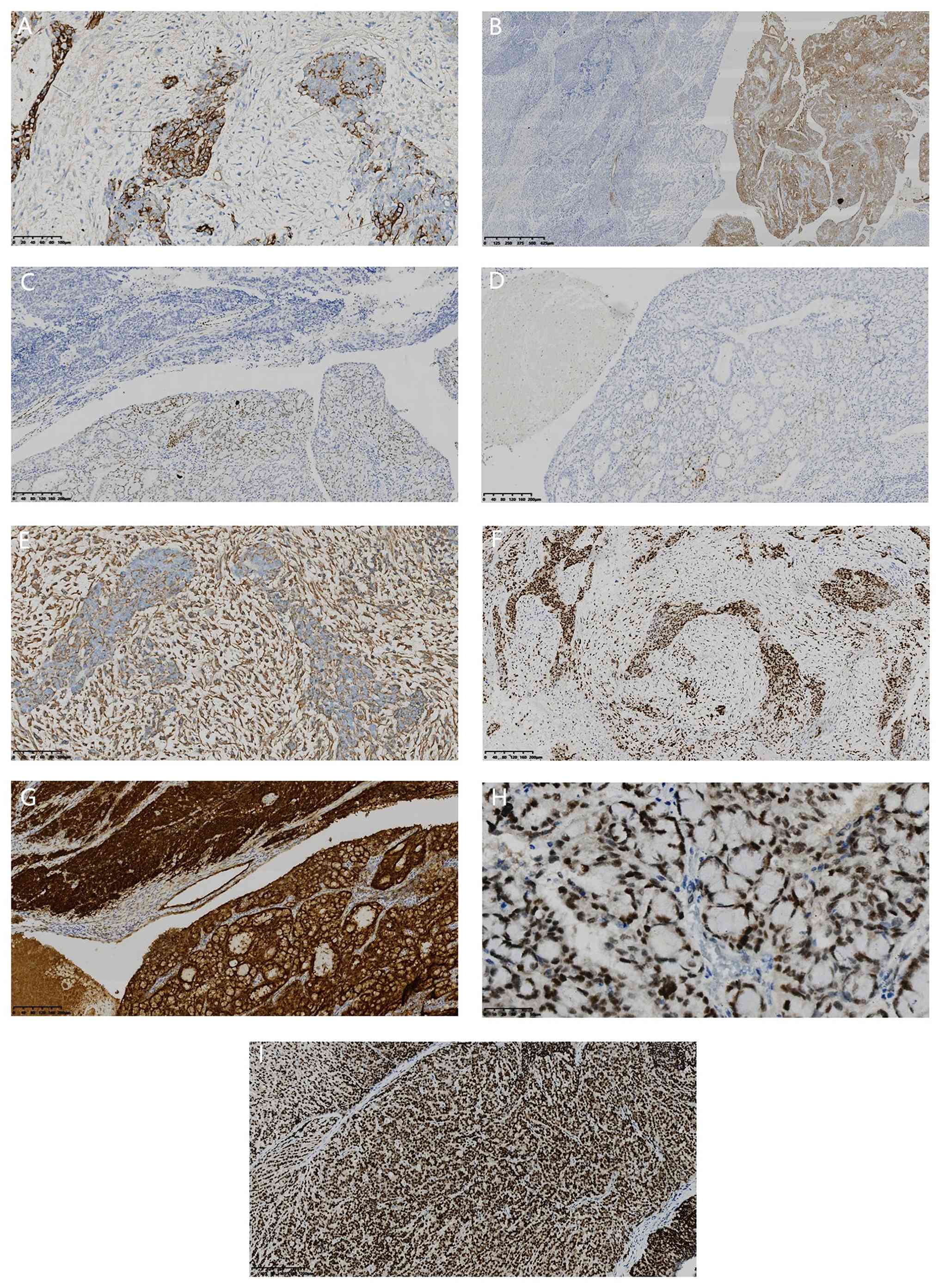 | Figure 7Immunohistochemical staining. (A)
Cytokeratin 7 expression (carcinoma, + (arrow); sarcoma, -)
(magnification, x200; scale bar, 100 µm). (B) Cam5.2 expression
(carcinoma, +; sarcoma, -) (magnification, x32; scale bar, 625 µm).
(C) Estrogen receptor expression (carcinoma, 10% +; sarcoma, -)
(magnification, x100; scale bar, 200 µm). (D) Progesterone receptor
expression (carcinoma, 1% +; sarcoma, -) (magnification, x100;
scale bar, 200 µm). (E) Vimentin expression (carcinoma, weak +;
sarcoma, +) (magnification, x200; scale bar, 100 µm). (F) Paired
box gene 8 expression (carcinoma, +; sarcoma, partial +)
(magnification, x100; scale bar, 200 µm). (G) p16 expression (+)
(magnification, x100; scale bar, 200 µm). (H) p53 aberrant
immunohistochemical overexpression in mucinous adenocarcinoma
(magnification, x400; scale bar, 50 µm). (I) p53 aberrant
immunohistochemical overexpression in small round cell sarcoma
(magnification, x100; scale bar, 200 µm). |
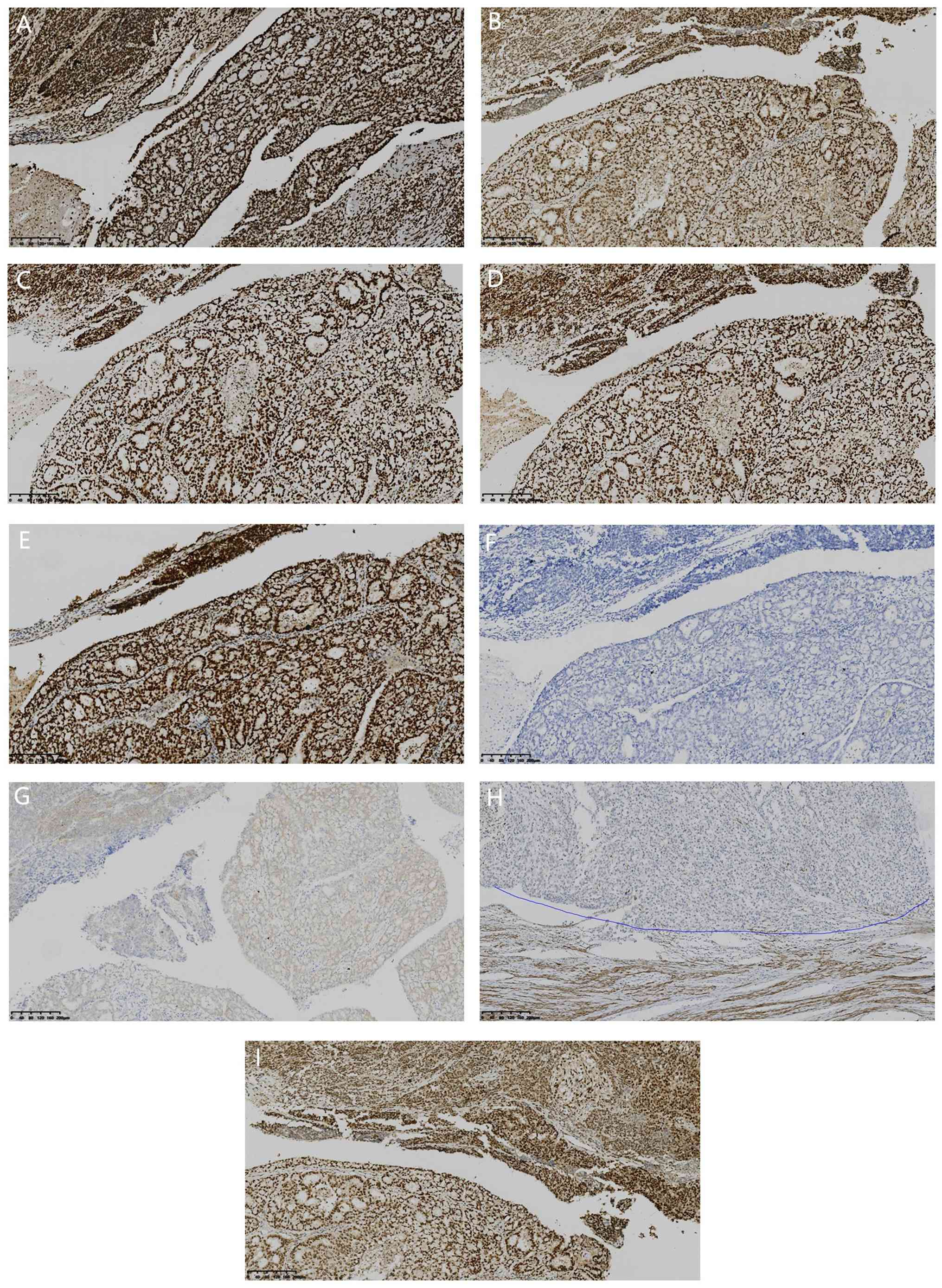 | Figure 8Immunohistochemical staining. (A)
SMARCA4 expression (+) (magnification, x100; scale bar, 200 µm).
(B) MutL homolog 1 expression (+) (magnification, x100; scale bar,
200 µm). (C) MutS Homolog 2 expression (+) (magnification, x100;
scale bar, 200 µm). (D) MutS Homolog 6 expression (+)
(magnification, x100; scale bar, 200 µm). (E) Mismatch repair
endonuclease PMS2 expression (+) (magnification, x100; scale bar,
200 µm). (F) NapsinA expression (-) (magnification, x100; scale
bar, 200 µm). (G) CerbB-2 (1+) expression (magnification, x100;
scale bar, 200 µm). (H) Desmin expression (-) (above the blue line.
The tumor tissue was negative, while the smooth muscle tissue of
the internal control was positive.) (magnification, x100; scale
bar, 200 µm). (I) Ki-67 proliferation index of 80% (magnification,
x100; scale bar, 200 µm). |
 | Table IImmunohistochemical staining results
of endometrial carcinosarcoma. |
Table I
Immunohistochemical staining results
of endometrial carcinosarcoma.
| Tumor name | ER | PR | Pax8 | CK7 | Cam5.2 | Vim | Desmin | P16 | SMARCA4 | TP53 | NapsinA | CerbB-2 | MSH2 | MLH1 | PMS2 | MSH6 |
|---|
| Carcinoma | 10%P | 1%P | P | P | P | WP | N | P | P | MAO | N | 1+ | P | P | P | P |
| Sarcoma | N | N | PP | N | N | P | N | P | P | SRCSO | N | 1+ | P | P | P | P |
Molecular testing methods
Molecular testing was performed by the Molecular
Laboratory of Hangzhou Adicon Medical Laboratory Center Co., Ltd.
(Zhejiang, China) using next-generation sequencing (NGS) technology
on the GeneMind SURFSeq 5000 sequencing system (ref. genome,
GRCh37/hg19). This SURFSeq 5000 system is used as described in the
sequencing details below.
Sample preparation reagents and
quality control methods
Details of the reagents used for sample preparation
are described below. Specifically, for genomic DNA extraction from
formalin-fixed paraffin-embedded (FFPE) samples, the FastPure FFPE
DNA Isolation Kit (cat. no. DC105-01; Vazyme Biotech Co., Ltd.) was
used. DNA concentration was quantified using the Qubit fluorometer
(Thermo Fisher Scientific, Inc.). DNA integrity and fragment size
distribution were assessed using the Fragment Analyzer system
(Agilent Technologies, Inc.). Library preparation was conducted
using the Adicon LDT Solid Tumor Mini panel (Twist Bioscience). For
library construction, the required reagents consisted of enzymes
for DNA fragmentation and end repair (fragmentation enzyme, T4 DNA
polymerase; Klenow fragment, T4 polynucleotide kinase), enzymes for
A-tailing and adapter ligation [Klenow fragment (3'-5' exo-) and T4
DNA ligase], dual-indexed sequencing adapters (with unique
molecular identifiers), high-fidelity DNA polymerase, a dNTP mix
(dATP, dTTP, dCTP, dGTP) and a primer mix (for library
amplification). Hybridization was performed using the Twist Fas
Hybridization Reagents Kit (Twist Bioscience). For hybridization,
the core reagents included biotinylated DNA capture probes (120-150
bp), Human Cot-1 DNA, universal blocking oligos (serving as
blocking agents), a high-salt hybridization buffer containing
formamide, low-stringency wash buffer (2X SSC; 0.1% SDS),
high-stringency wash buffer (0.1X SSC; 0.1% SDS),
streptavidin-coated magnetic beads and elution buffer (0.1 M NaOH
or Tris-HCl). In addition, quality control reagents, including
fragment analyzer reagents, were used in the process to ensure the
reliability of the experiment. The SURFSeq 5000 Sequencing Kit V2.0
(FCH 300 cycles) (cat. no. S000369; GeneMind Biosciences Co., Ltd.)
was used for sequencing.
Sequencing details. i) Sequencing type:
Paired-end NGS with a read length of 2x150 bp; ii) sequencing
platform: SURFSeq 5000 (GeneMind Biosciences); iii) final library
loading concentration and quantification: The final library was
loaded at a concentration of 1 nM and quantified by the Qubit assay
(Thermo Fisher Scientific).
Bioinformatic analysis. Raw sequencing reads
were trimmed to remove adapters and low-quality bases using
Cutadapt (v4.1, https://github.com/marcelm/cutadapt). Clean reads were
aligned to the human reference genome GRCh37/hg19 (UCSC Genome
Browser; https://hgdownload.soe.ucsc.edu/goldenPath/hg19/)
with BWA-MEM (v0.7.17; https://bio-bwa.sourceforge.net/). PCR duplicates were
marked by Picard (v2.27.5; Broad Institute; https://github.com/broadinstitute/picard). Base
quality recalibration and somatic variant calling were performed
using GATK (v4.2.6.1; Broad Institute; https://gatk.broadinstitute.org/). Detected variants
were functionally annotated with ANNOVAR (v20210201; https://annovar.openbioinformatics.org/).
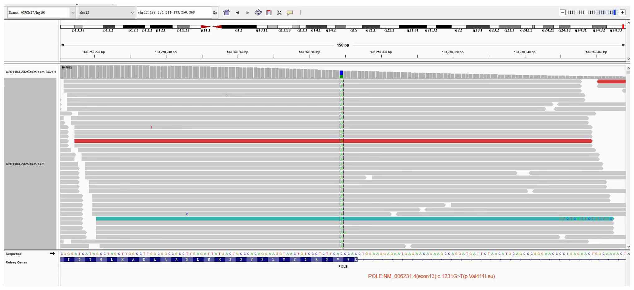
Molecular analysis was performed using NGS and
revealed a c.1231G>T (p.Val411Leu) mutation (variant allelic
frequency was 45.23%) in exon 13 of the POLE gene (Fig. 9). The TP53 gene harbored three
mutations: c.809T>G (p.Phe270Cys) in exon 8, c.637C>T
(p.Arg213Ter) in exon 5 and c.413C>T (p.Ala138Val) in exon 6,
with variant allelic frequencies of 17.63, 22.70 and 24.93%,
respectively. The final pathological diagnosis was ECS with a POLE
gene mutation, FIGO 2023 stage IA, POLE-mutant subtype (1).
Treatment and follow-up
After the surgery, the patient was referred to the
Binjiang Branch of The Second Affiliated Hospital of Zhejiang
University School of Medicine (Hangzhou, China) for consultation.
After a comprehensive assessment, the doctors decided not to
administer adjuvant therapy. The tumor was FIGO 2023 stage IA,
POLE-mutant endometrial carcinosarcoma with less than one-half
myometrial invasion and negative lymph nodes. POLE-mutant
endometrial carcinoma is associated with an excellent prognosis,
therefore, adjuvant therapy was withheld after multidisciplinary
discussion, given the low risk of recurrence and the potential
toxicities of adjuvant treatment. No recurrence or metastasis was
observed during the 18-month follow-up period. The last follow-up
was performed in September 2026.
Discussion
ECS, also known as malignant mixed Müllerian tumor,
exhibits bidirectional differentiation and includes components of
both epithelial carcinoma and mesenchymal sarcoma, displaying
notable histological heterogeneity (1). It is a rare malignant tumor of the
female reproductive system and accounts for ~5% of uterine
malignancies among women with uterine cancer in Western populations
(2). The incidence of ECS ranges
from 2-5 cases per 100,000 women, and postmenopausal women (median
age, 65 years) are at a greater risk. The 5-year overall survival
rate is <40% (3), which is
markedly lower than the survival rate of >80% typical of common
endometrial cancer. Moreover, the mortality rate of ECS is the
highest among uterine malignant tumors. However, by applying
molecular typing in endometrial cancer, some studies have revealed
that the prognosis for patients with a POLE mutation is improved
compared with the prognosis for those with a TP53-abnormal
(9,10). Additionally, unnecessary pain and
costs related to excessive treatment can be reduced. The present
study presented a case of POLE-mutant ECS, and reviewed the
relevant literature to provide novel insights into the diagnosis
and treatment of these tumors within the endometrium.
ECS is composed of epithelial carcinoma (such as
endometrioid carcinoma and serous carcinoma) and sarcomatous
components, which are classified into homologous subtypes
(including endometrial stromal sarcoma, leiomyosarcoma and
undifferentiated sarcoma) or heterologous subtypes (including
rhabdomyosarcoma, osteosarcoma and chondrosarcoma) (1). The cancer cells are irregularly
arranged and characterized by large hyperchromatic nuclei and
visible mitotic figures. Most sarcoma cells are spindle or
polygonal, with notable atypia and active mitotic figures, and
necrosis is common. Tumor cells often invade blood vessels,
lymphatic vessels or surrounding tissues, and may undergo distant
metastasis (for example, to the lung and liver) (2).
In a previous study, an immunohistochemical analysis
demonstrated that the carcinomatous component expresses CK and
epithelial membrane antigen, while the sarcomatous component
expresses vimentin and other mesenchymal markers (CD10, smooth
muscle actin, desmin and myogenin). Specific markers of
heterologous components [such as MyoD1(+)/S-100(+)] have also been
detected and TP53 is aberrantly expressed in ~50% of cases
(abnormal type) (2). In the
present study, histological examination of tissue samples from the
patient revealed that they had high-grade endometrioid carcinoma,
mucinous adenocarcinoma and undifferentiated sarcoma.
Immunohistochemical staining revealed the carcinoma component had
positive expression of epithelial markers and the sarcomatous
component had vimentin positivity. TP53 showed aberrant
immunohistochemical overexpression. The microsatellite stability
marker was not lost, as demonstrated by intact expression of MMR
proteins on immunohistochemistry. Based on these findings, a
pathological diagnosis of ECS was confirmed (TP53-abnormal
type).
A previous study revealed that POLE-mutant
endometrial cancer generally exhibits microsatellite stability
(65%) and TP53 gene mutations in ~35% of cases (7). In the absence of molecular typing,
POLE-mutant endometrial cancer with good prognosis may be
misdiagnosed and treated as invasive endometrial cancer, which has
an unfavorable prognosis; this may lead to unnecessary
overtreatment and thus harm the patient. The development of
molecular subtyping and its application in endometrial cancer led
the 2020 5th edition of the WHO classification of tumors of the
female reproductive system to recommend using a modified version of
The Cancer Genome Atlas (TCGA) classification. The classification
scheme also proposed a diagnostic algorithm based on sequencing of
the POLE gene, mismatch repair (MMR) protein immunohistochemistry
and TP53 immunohistochemistry. Subtyping follows a hierarchical
algorithm in sequence: POLE-mutant, MMR-deficient (MMRd),
non-specific molecular profile (NSMP) and TP53-abnormal subtypes
(1). Endometrial carcinomas
harboring both pathogenic POLE exonuclease domain mutations
and TP53 aberrations are classified as the POLE-mutant
subtype (1). Researchers have also
performed molecular subtyping of endometrial cancer via biopsy
before surgery; this strategy provides early prognostic value for
guiding personalized treatment (12). Endometrial cancer after surgery,
including carcinosarcoma, is also suitable for the modified TCGA
classification (1).
In the current patient, TP53 showed aberrant
immunohistochemical overexpression and the MMR proteins MLH1, MSH2,
MSH6 and PMS2 were positively expressed, as determined by
immunohistochemistry. NGS confirmed the presence of the
c.1231G>T (p.Val411 Leu) mutation in exon 13 of the POLE gene
and three distinct TP53 mutations, which agreed with the
diagnosis of ECS with POLE mutation. Based on the findings, the
original stage IIc was updated to stage I Am-POLE mut (FIGO 2023)
(13).
As reported in previous studies, POLE mutations
occur in 7-12% of endometrioid carcinoma cases (4,5) and
2.7% of ECS cases (10). Nero
et al (14) reported that
of 61 (61/596; 10.2%) patients with endometrial carcinoma with POLE
hotspot mutations, 27 (44.3%) and 34 (55.7%) patients had low- and
high-grade cancer, respectively; 20 (32.8%) patients had LVSI; and
16 patients had co-mutations [MMRd, 9, 56.3%; TP53-abnormal, 4,
25.0%; MMRd plus TP53-abnormal, 3, 18.8%]. Furthermore, FIGO 2023
staging revealed that 57 cases (93.4%) were stage IA with
POLE-mutant subtype, and four cases (6.6%) were stage III-IV. The
median tumor mutation burden (TMB) was 150.8 mut/Mb (14). Notably, in endometrial carcinoma
(including ECS), pathogenic POLE exonuclease-domain
mutations mainly occur in six exons (exons 9-14), and the mismatch
base cannot be recognized and repaired; this leads to an abnormal
increase in the number of genomic mutations and causes
tumorigenesis (4). This site is
located in the exonuclease domain, and endometrial carcinoma
carrying mutations in the exonuclease domain of POLE usually has a
TMB exceeding 100 mut/Mb (15). In
the context of a combined TP53 mutation (which ordinarily confers
the poor prognostic characteristic of the TP53-abnormal subtype),
the POLE mutation may still dominate the biological behavior. Nero
et al (14) and Bogani
et al (16) reported that
four and 20 patients with endometrial cancer (25.0 and 27.8%,
respectively) with both POLE and TP53 mutations had a better
prognosis than patients with TP53-abnormal subtype lacking POLE
mutations (14,16). Therefore, the present case was
consistent with the WHO classification of POLE mutation.
For prognosis and treatment, comprehensive staging
surgery is conducted in the early stages, while cytoreductive
surgery is performed in later stages. Surgical pathological staging
and adjuvant therapy, including systemic therapy and radiotherapy,
are based on postoperative pathology. Patients for whom resection
will not be satisfactory may be reassessed for surgery following
chemotherapy or radiotherapy (17). While POLE-mutant ECS has
demonstrated favorable outcomes in limited clinical series
(8-10),
substantial long-term prognostic data are preliminary and
continuously evolving. As this subtype is rare [accounting for 2.7%
of all ECS cases (10)] and
single-case reports have limitations (for example, small overall
sample size, absence of TMB data and individual variability in
biological behavior), there is a risk of overgeneralization of
prognostic conclusions from isolated cases. Further validation is
required through larger-scale cohorts with extended follow-up to
confirm the durability of favorable outcomes and to determine the
effect of concurrent genetic alterations (for example, TP53
mutations) on long-term prognosis. Molecular typing should be
performed to guide future treatment decisions. If stage I and II
patients have POLE mutations, they can be closely followed up and
observed regularly with full communication without radiotherapy and
chemotherapy (8,18). Hammer et al (9) reported on 11 patients with
POLE-mutant ECS, with mean and median follow-up times of 24.3 and
14.1 months, respectively (range, 1.4-61.1 months); no disease
recurrence or death was detected in 10 patients, whereas one
patient with a subclonal POLE mutation died 1.4 months after
surgery. Toboni et al (10)
reported that the median overall survival of 48 patients was 74.8
months, which was notably longer than that of other subtypes.
Bhangoo et al (11)
reported a case of a stage III chemotherapy-ineffective relapse
patient who was found to have a POLE mutation by molecular
detection; the condition improved after 12 months of follow-up
after immunotherapy; however, as this is a case report, more data
are needed to confirm these findings.
In terms of advantages, the present case of
POLE-mutant ECS is rare and has a unique pathological component of
mucinous adenocarcinoma. After a comprehensive analysis, further
treatment was not required, which increases the reference value of
clinical diagnosis and treatment of ECS with POLE mutations. In
terms of limitations of the present study, only a single case
report was provided with a short follow-up period (18 months) and
no TMB data, which restricts the generalizability of the prognostic
observations. Therefore, long-term follow-up of this patient and
data on more clinical cases are necessary to validate the
prognostic significance of POLE mutations in ECS.
In summary, the present diagnosis and treatment
experience combined with the literature review suggests that POLE
mutations are rare in ECS and may be associated with a relatively
favorable short-term prognosis; however, long-term outcomes need to
be confirmed in larger-scale studies. For morphologically
high-grade ECS, molecular subtyping should be routinely performed,
particularly to determine the status of POLE mutations. When a
POLE-mutant subtype is diagnosed, adjuvant therapy can be
administered individually based on a comprehensive evaluation of
tumor stage to reduce the risk of overtreatment.
Acknowledgements
Not applicable.
Funding
Funding: No funding was received.
Availability of data and materials
The data generated in the present study may be found
in the Genome Sequence Archive for Humans under accession number
HRA014729 or at the following URL: https://ngdc.cncb.ac.cn/gsa-human/browse/HRA014729.
Authors' contributions
YZ was responsible for the conceptual framework
design and initial manuscript drafting of this case report. HG
participated in the conceptual design, methodological design, data
collation, formal analysis and implementation of the study. BH was
responsible for project management, result validation, initial
manuscript writing, and manuscript revision and review. BH and YZ
confirm the authenticity of all the raw data. BH and YZ confirm the
accuracy and integrity of all raw clinical data of this study and
are responsible for the validity of the original data. All authors
have read and approved the final manuscript.
Ethics approval and consent to
participate
Not applicable.
Patient consent for publication
The patient provided written informed consent for
the case study to be published.
Competing interests
The authors declare that they have no competing
interests.
References
|
1
|
WHO Classification of Tumours Editorial
Board. WHO classification of tumours. Female genital tumours (M).
IARC Press, Sthed, Lyon, 2020.
|
|
2
|
Bogani G, Ray-Coquard I, Concin N, Ngoi
NYL, Morice P, Caruso G, Enomoto T, Takehara K, Denys H, Lorusso D,
et al: Endometrial carcinosarcoma. Int J Gynecol Cancer.
33:147–174. 2023.PubMed/NCBI View Article : Google Scholar
|
|
3
|
Siegel RL, Miller KD and Jemal A: Cancer
statistics, 2020. CA Cancer J Clin. 70:7–30. 2020.PubMed/NCBI View Article : Google Scholar
|
|
4
|
Cancer Genome Atlas Research Network.
Kandoth C, Schultz N, Cherniack AD, Akbani R, Liu Y, Shen H,
Robertson AG, Pashtan I, Shen R, et al: Integrated genomic
characterization of endometrial carcinoma. Nature. 497:67–73.
2013.PubMed/NCBI View Article : Google Scholar
|
|
5
|
Yao X, Feng M and Wang W: The clinical and
pathological characteristics of POLE-mutated endometrial cancer: A
comprehensive review. Cancer Manag Res. 16:117–125. 2024.PubMed/NCBI View Article : Google Scholar
|
|
6
|
Restaino S, Paglietti C, Arcieri M,
Biasioli A, Della Martina M, Mariuzzi L, Andreetta C, Titone F,
Bogani G, Raimondo D, et al: Management of patients diagnosed with
endometrial cancer: Comparison of guidelines. Cancers (Basel).
15(1091)2023.PubMed/NCBI View Article : Google Scholar
|
|
7
|
Hussein YR, Weigelt B, Levine DA,
Schoolmeester JK, Dao LN, Balzer BL, Liles G, Karlan B, Köbel M,
Lee CH and Soslow RA: Clinicopathological analysis of endometrial
carcinomas harboring somatic POLE exonuclease domain mutations. Mod
Pathol. 28:505–514. 2015.PubMed/NCBI View Article : Google Scholar
|
|
8
|
Ebner C, Frosch A, Leitner K, Soucek R,
Marth C and Zeimet AG: An uncommon case of POLE mutated uterine
carcinosarcoma-complemented by a review of literature. Gynecol
Oncol Rep. 54(101442)2024.PubMed/NCBI View Article : Google Scholar
|
|
9
|
Hammer PM, Momeni-Boroujeni A, Kolin DL,
Kingsley L, Folkins A, Geisick RLP, Ho C, Suarez CJ and Howitt BE:
POLE-mutated uterine carcinosarcomas: A clinicopathologic and
molecular study of 11 cases. Mod Pathol. 38(100676)2025.PubMed/NCBI View Article : Google Scholar
|
|
10
|
Toboni MD, Dinkins K, Wu S, Mattox T,
Oberley MJ, Thaker PH, Herzog TJ, Powell MA and Jones N: Not all
uterine carcinosarcomas are created equal: Survival outcomes
according to molecular characterization of uterine carcinosarcoma.
Gynecol Oncol. 193:89–97. 2025.PubMed/NCBI View Article : Google Scholar
|
|
11
|
Bhangoo MS, Boasberg P, Mehta P, Elvin JA,
Ali SM, Wu W and Klempner SJ: Tumor mutational burden guides
therapy in a treatment refractory POLE-mutant uterine
carcinosarcoma. Oncologist. 23:518–523. 2018.PubMed/NCBI View Article : Google Scholar
|
|
12
|
Restaino S, Poli A, Arcieri M, Mariuzzi L,
Orsaria M, Tulisso A, Pellecchia G, Paparcura F, Petrillo M, Bogani
G, et al: Molecular classification of endometrial carcinoma on
endometrial biopsy: An early prognostic value to guide personalized
treatment. Int J Gynecol Cancer. 34:1211–1216. 2024.PubMed/NCBI View Article : Google Scholar
|
|
13
|
Occhiali T, Poli A, Arcieri M, Driul L,
Ditto A, Bogani G, Mariani A, Scambia G, Restaino S and Vizzielli
G: The exciting journey of progress: Exploring FIGO 2023 staging
for endometrial cancer at a leading ESGO institution. Eur J Surg
Oncol. 51(109695)2025.PubMed/NCBI View Article : Google Scholar
|
|
14
|
Nero C, Trozzi R, Persiani F, Rossi S,
Mastrantoni L, Duranti S, Camarda F, Marino I, Giacò L, Pasciuto T,
et al: POLE mutations in endometrial carcinoma: Clinical and
genomic landscape from a large prospective single-center cohort.
Cancer. 131(e35731)2025.PubMed/NCBI View Article : Google Scholar
|
|
15
|
León-Castillo A, Britton H, McConechy MK,
McAlpine JN, Nout R, Kommoss S, Brucker SY, Carlson JW, Epstein E,
Rau TT, et al: Interpretation of somatic POLE mutations in
endometrial carcinoma. J Pathol. 250:323–335. 2020.PubMed/NCBI View Article : Google Scholar
|
|
16
|
Bogani G, Betella I, Multinu F, Casarin J,
GhezzI F, Sorbi F, VizziellI G, Petrillo M, Cianci S, Berretta R,
et al: Characteristics and outcomes of surgically staged multiple
classifier endometrial cancer. Eur J Surg Oncol.
50(107269)2024.PubMed/NCBI View Article : Google Scholar
|
|
17
|
NCCN Clinical Practice Guidelines in
Oncology (NCCN Guidelines). Uterine Νeoplasms, version 2, 2024.
|
|
18
|
Concin N, Matias-Guiu X, Vergote I, Cibula
D, Mirza MR, Marnitz S, Ledermann J, Bosse T, Chargari C, Fagotti
A, et al: ESGO/ESTRO/ESP guidelines for the management of patients
with endometrial carcinoma. Int J Gynecol Cancer. 31:12–39.
2021.PubMed/NCBI View Article : Google Scholar
|