Introduction
Gene amplification is a genetic process where the
copy number of certain genes increases, without a proportional
increase of the genome. Gene amplification is not only a hallmark
of genetic alterations in tumor cells but is also well described
and studied since decades as physiological mechanism in amphibians
and flies. For example, during the development of Drosophila
melanogaster and Xenopus laevis, specific DNA regions
are amplified within defined developmental time windows (1).
There is growing evidence that gene amplification
plays a physiological role during differentiation of stem and
progenitor cells. Previously we described gene amplification during
differentiation of mesenchymal stem cells towards osteoblasts and
adipocytes (2). Interestingly,
gene amplifications that arise during stem cell differentiation
often overlap with those found in the corresponding tumor types.
For example, the genes CDK4 and MDM2 were shown to
undergo co-amplification during the differentiation of mesenchymal
stem cells (MSCs) into adipocytes, mirroring the high-level
amplifications characteristic of liposarcoma (3). Similarly, several studies on
chondrosarcoma have reported MYC gene amplification on
chromosome 8, with array-CGH analyses demonstrating that MYC
amplification occurs more frequently in higher-grade and
dedifferentiated chondrosarcomas (4,5). In
addition, genomic profiling of chondrosarcoma revealed several
amplified chromosome regions including regions on chromosomes 5,
12, 19 and 22(6). Notably,
amplification of CDK4 without accompanying MDM2
co-amplification has also been documented in at least one
chondrosarcoma case, further underscoring the heterogeneity and
potential clinical relevance of these genetic events (7).
Mesenchymal stem cells (MSCs) possess significant
potential for therapeutic applications in tissue regeneration. One
of the most ambitious investigations is probably regeneration of
articular cartilage since many health restrictions are based on
cartilage destruction (8-10).
Understanding both the regenerative capacity and the potential
risks caused by genetic changes that are associated with directing
hMSCs toward a chondrogenic lineage is therefore essential. Here we
set out to investigate whether gene amplifications are detectable
during chondrogenic differentiation of human mesenchymal stem cells
(hMSCs).
Materials and methods
Cell culture and differentiation
Primary hMSCs from bone marrow were purchased form
PromoCell and were cultured until they formed spheres in U-bottom
96-well plates or were near confluent on Zeiss FrameSlides PET
(polyethylene terephthalate) membrane for laser microdissection.
Chondrocyte differentiation was induced in conditions using
chondrogenic differentiation medium (PromoCell) (11) for 5-7 days.
A detailed characterization of hMSCs is provided in
the supplier's certificates of analysis for Lot numbers 488Z010
(female donor, 79 years) and 479Z025 (male donor, 80 years), both
of which were used in this study. As indicated in these
certificates, surface marker analysis at passage 3 showed that 100%
of cells were positive for CD73, CD90 and CD105, while 99-100% were
negative for CD34 CD45. The certificates also document
differentiation potential toward adipogenic, osteogenic, and
chondrogenic lineages. All experiments were performed using cells
at passage 3.
RNA-seq
RNA was isolated using miRNAeasy Kit (Qiagen) and
used for next generation sequencing. Sequencing was performed on an
DNBSEQ-G400RS instrument by the NGS Sequencing Facility of Saarland
University using the 100bp paired end sequencing strategy.
Paired-end fastq files were processed using the mRNA module of
SnakePipes 3.0.0(12). The reads
were aligned using STAR using default settings provided by
snakepipes (13) against
GRCh38(14) in order to generate
bam and bai files. Quality control checks were carried out using
multiQC (15).
Toluidine blue staining of
proteoglycan during differentiation
HMSCs were cultivated and differentiated as
described above. For toluidine blue staining cells were washed once
with PBSx1 for 10 min and either fixed with 4% PFA
(paraformaldehyde) for 5 min or with ice cold methanol for 10 min.
Toluidine staining solution (10% Toluidine blue, 1% Borax) was
applied for 15 min, and excess staining was removed by three PBSx1
washing steps (16).
Differentiation was checked with a bright-field microscope at x20
magnification.
Laser microdissection of
differentially toluidine-blue stained areas
For laser microdissection hMSCs were differentiated
for 5 days towards chondrocytes and stained with toluidine blue for
proteoglycan detection of differentiating cells. In total 20 areas
(for each staining subpopulation: grey, light blue, blue and dark
blue) of 0.76 mm2 were laser microdissected, using the
PALM® MicroBeam Microdissection System (Carl Zeiss AG).
These experiments were done in three biological replicates. DNA
extraction was performed using the QIAamp DNA Micro Kit (Qiagen)
according to the manufacturer's instructions. The final eluate was
concentrated to a total volume of approximately 12 µl and DNA
concentration was determined using Qubit™ dsDNA HS Assay Kit
(Thermo Fisher Scientific).
qPCR analysis
TaqMan Copy Number Assays for genes CDK4
(Hs00957586_cn), MDM2 (Hs00181272_cn), AGAP2
(Hs01425078_cn), CPT1B (Hs01741692cn), MYC
(Hs02602824_cn), SHANK3 (Hs04081743_cn), and TRIB1
(Hs00836770_cn) were performed following manufacturer's
instructions (Applied Biosystems®). RNaseP TaqMan
Copy number reference assay was used for relative quantitation of
copy number of target genes. DNA from not differentiated controls
of hMSCs cultivated in DMEM was used as control standard for normal
diploid copy number. TaqMan assays were run in four technical
replicates and results were analyzed using StepOne™ Software v2.0
and CopyCaller™ software (Applied Biosystems® by Life
Technologies Corporation) for differentiation in U-bottom 96-well
plates. TaqMan Copy Number Assays for genes CDK4
(Hs00957586_cn), AGAP2 (Hs01425078_cn), CPT1B
(Hs01741692cn), SHANK3 (Hs04081743_cn), and TRIB1
(Hs00836770_cn) were performed with 250 pg DNA per reaction. TaqMan
assays were run in two technical replicates and results were
analyzed using StepOne™ Software v2.0 and CopyCaller™ software for
laser microdissected areas. For analysis of this low amount of DNA
input the Cq-threshold was set to exclude values above 35.
Statistical analysis
Copy number values extracted from CopyCaller™ were
analyzed for each gene separately. One-way ANOVA (alpha level 0.05,
n=6) was used to test the null hypothesis that staining color
(CTRL, grey, light blue, blue, dark blue) and copy number are
independent for the genes AGAP2, CDK4, CPT1B,
SHANK3 and TRIB1. We performed Tukey's HSD post hoc
test (alpha=0.05) to find pairs of staining area that are
significantly different in terms of copy number for each of the
genes mentioned. Calculations were performed using R version 4.3.3,
specifically the aov function (https://search.r-project.org/R/refmans/stats/html/aov.html)
with the formula Copy_number ~ Staining followed by the Tukey HSD
function for post hoc comparisons. P<0.05 was considered to
indicate a statistically significant difference.
Results
HMSCs were differentiated in U-bottom 96-wells and
differentiation was confirmed by toluidine blue staining and
RNA-seq analysis of chondrogenic differentiation specific genes
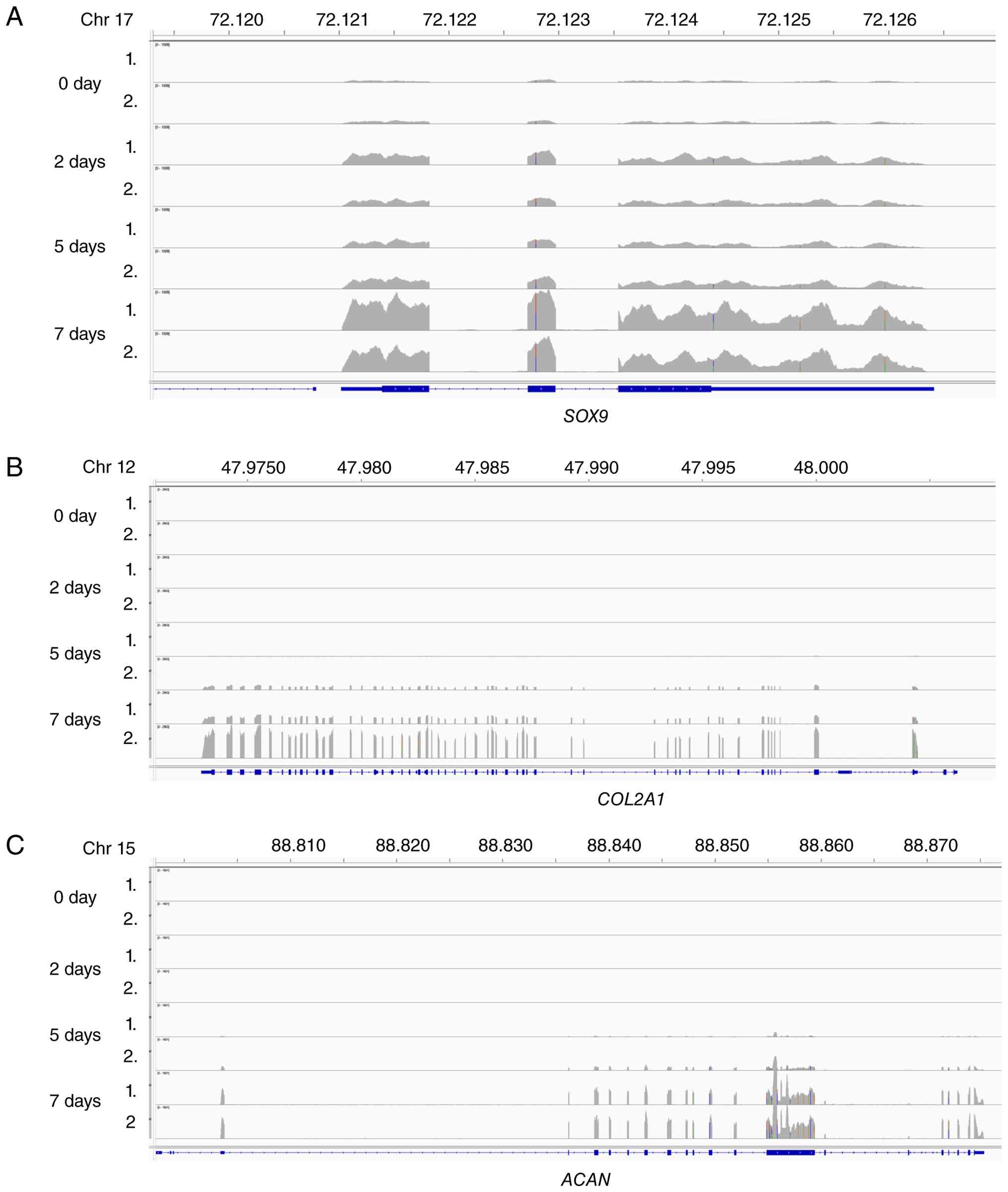
SOX9, COL2A1 and ACAN (17,18).
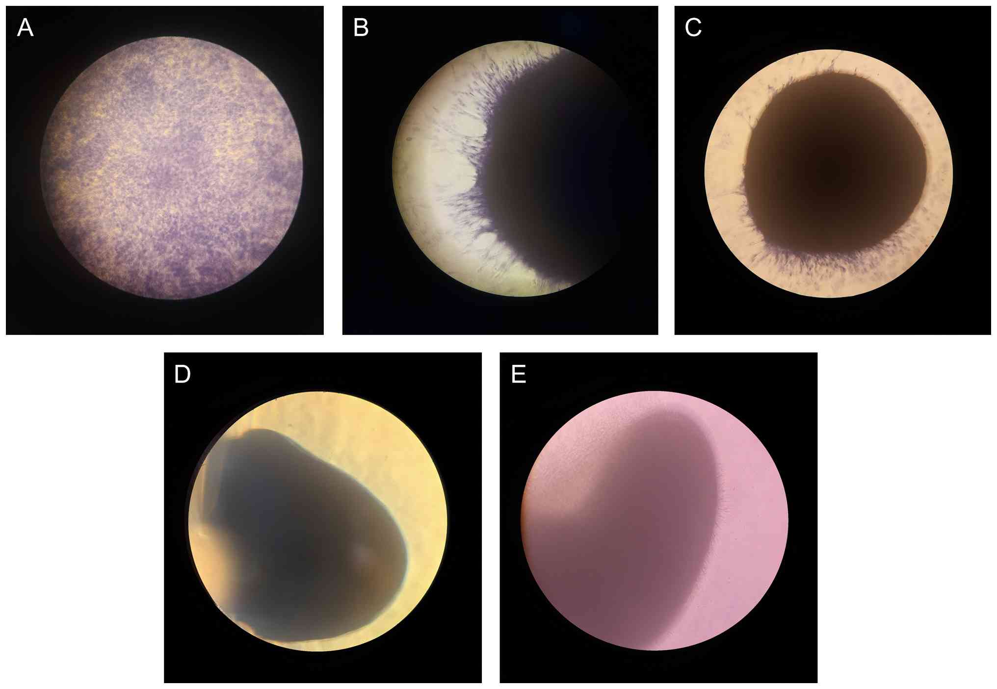
Already at 5 days post differentiation induction cells revealed
three-dimensional cell growth as spheres and high intensity of
proteoglycan specific toluidine blue staining (Fig. 1). Expression analysis of two
independent biological replicates revealed an increase of
SOX9 expression after 2 days and a further increase after 7
days of differentiation and an increase of COL2A1 and
ACAN expression after 5 and 7 days of differentiation
(Fig. 2). To detect gene
amplifications, we analyzed bulk DNA from hMSCs differentiated
toward chondrocytes for 1, 2, 5, and 7 days, as well as from
undifferentiated hMSCs serving as controls. Amplification was
assessed by quantitative PCR (TaqMan) in four replicates, and data
were evaluated using the CopyCaller software (Applied
Biosystems).
For our investigation we selected genes previously
found to be amplified during osteoblast and adipocyte
differentiation and genes that were known to be amplified in tumors
derived from chondrocytes including genes on chromosome 12,
chromosome 22 and chromosome 8 (2,6).
Copy number was determined for genes AGAP2, CDK4 and
MDM2 localized on chromosome 12, CPT1B and
SHANK3 localized on chromosome 22 and TRIB1 and
MYC localized on chromosome 8. As shown in Fig. 3 after 1 day of differentiation none
of the investigated genes revealed a copy number increase. After 2
days of differentiation CDK4, CPT1B and SHANK
3 revealed a 60% copy number increase, AGAP2 and
TRIB1 exhibited a 20% copy number increase; MDM2 and
MYC remained unchanged (Fig.
3). After 5 days of differentiation AGAP2 and
MDM2 displayed increases, whereas CDK4, CPT1B
and SHANK3, which were highest at 2 days, decreased and
MYC still remained unchanged. After 7 days of
differentiation CDK4, CPT1B, SHANK3, and
TRIB1 decreased further, AGAP2 increased, and no
further copy number change was detected for MDM2. MYC
showed no change throughout the 7-day differentiation period.
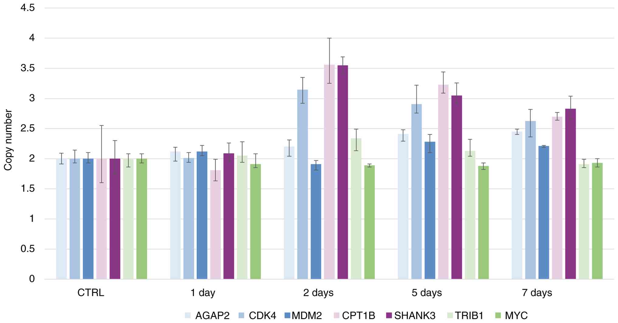 | Figure 3Descriptive amplification analysis of
AGAP2, CDK4, MDM2, CPT1B,
SHANK3, TRIB1 and MYC using qPCR during
chondrogenic differentiation. Amplification of AGAP2,
CDK4, MDM2, CPT1B, SHANK3, TRIB1
and MYC was analyzed by qPCR using TaqMan copy number assays
and RNAseP was used as reference TaqMan assay. DNA from
undifferentiated hMSCs (CTRL) served as standard for normal diploid
copy number with copy number values indicated on the y-axis and
normal diploid copy number is referred as 2. Copy numbers are shown
as mean from four technical replicates from one experiment (3
pooled spheroid samples) with vertical lines indicating the range.
Chondrogenic-induced hMSC cells were analyzed at four time points
after differentiation induction (1, 2, 5 and 7 days). CPT1B
and SHANK3 revealed the highest copy numbers of all
investigations after 2 days of differentiation and a decrease until
7 days. CDK4 revealed also a high copy number after 2 days
of differentiation induction (green) and a decrease until 7 days.
TRIB1 revealed a copy number increase only 2 days after
differentiation induction. AGAP2 and MDM2 revealed a
copy number increase only after 5 and 7 days. MYC revealed
no copy number increase at any time point after differentiation
induction. qPCR, quantitative PCR. |
Previous published investigations on gene
amplification during differentiation revealed a high heterogeneity
of gene amplification between single cells. During osteoblast
differentiation for example, cells with high level amplification
were detected next to cells with normal copy number and during
neural differentiation composition and copy number of co-amplified
genes varied between neighboring cells (2,19).
One possible reason for the differences in gene copy numbers is
differences in the degree of differentiation upon differentiation
induction. After five days upon differentiation induction, we
performed laser microdissection on hMSCs after toluidine blue
staining for detection of proteoglycan production that is
indicative for chondrocyte differentiation (Fig. 4A-D). Toluidine blue stain revealed
heterogenous blue intensities with areas of grey (Fig. 4A), light blue (Fig. 4B), blue (Fig. 4C) and dark blue (Fig. 4D) staining as result of different
amounts of produced proteoglycans indicating different stages of
differentiation.
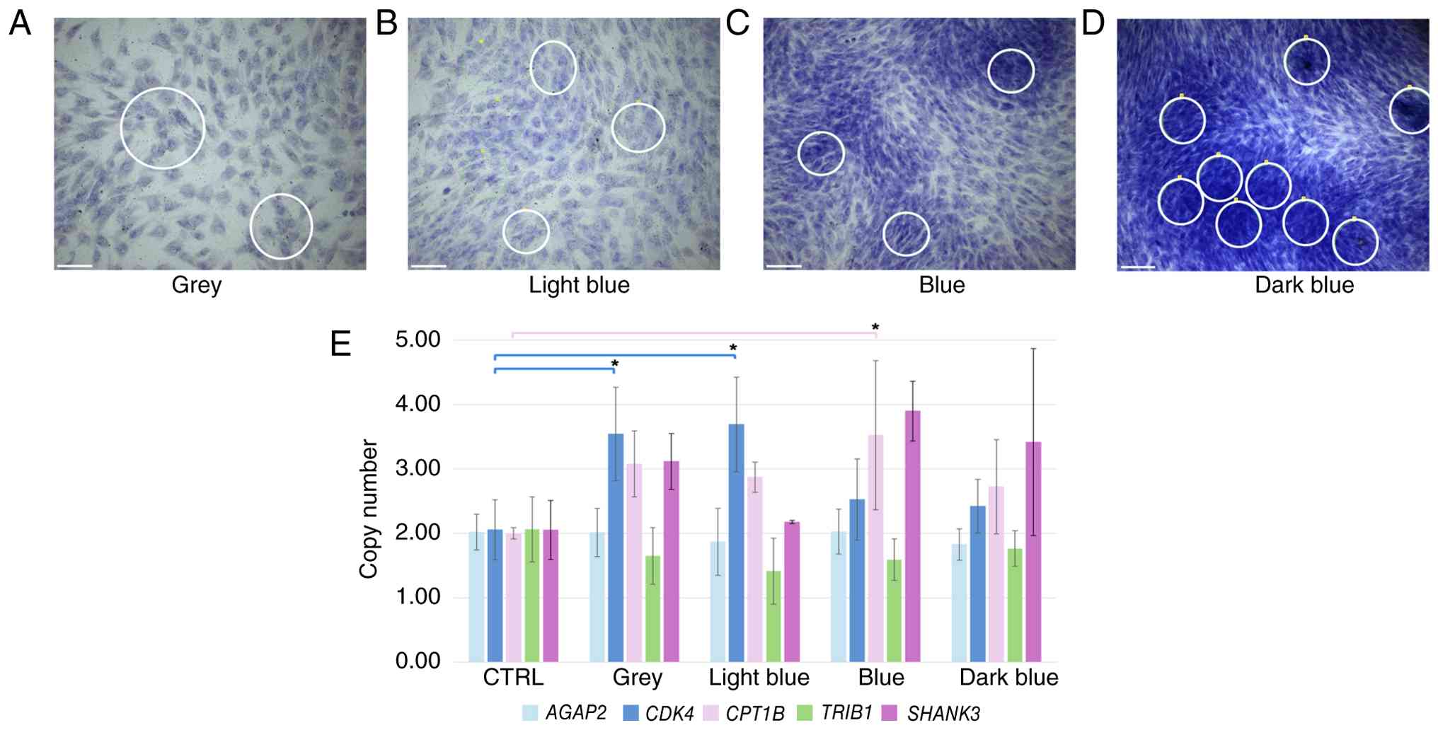 | Figure 4Amplification analysis of
AGAP2, CDK4, CPT1B, SHANK3 and
TRIB1 after laser microdissection. hMSCs were differentiated
towards chondrocytes for 5 days on PET frame slides for laser
microdissection. Cells were seeded at high density, and slide was
stained with toluidine blue for proteoglycan detection during
differentiation. Different areas of staining intensity could be
detected and were defined as areas with (A) grey cells, (B) light
blue cells, (C) blue cells and (D) dark blue cells, all indicating
stages during the differentiation towards chondrocytes. In total
for each area type twice 20 areas were laser microdissected and
used for DNA isolation. Experiments were done in three biological
replicates. Examples of laser microdissected areas are circled in
white and scale bars represent 150 µm. (E) Results of amplification
analysis of our quantitative PCR using TaqMan copy number assays
and RNAseP as reference are shown as diagram with
AGAP2, CDK4 and CPT1B SHANK3 and TRIB1.
DNA from undifferentiated hMSCs (CTRL) served as standard for
normal diploid copy number with copy number values indicated on the
y-axis and normal diploid copy number is referred as 2. Copy
numbers are presented as means of two technical replicates across
three biological replicates (N=6), with error bars representing
standard deviation. *P<0.05. CDK4 revealed the
highest copy number in areas with grey and light blue cells
indicating gene amplification at the beginning of differentiation.
CPT1B revealed high copy numbers in grey, light blue and
blue cells indicating gene amplification during differentiation and
SHANK3 revealed the highest copy numbers in areas with blues
cells indicating gene amplifications during later differentiation
progression. AGAP2 and TRIB1 revealed no copy number
increase. In areas with dark blues cells no significant
amplification was detectable indicating a reduction of gene
amplification towards final differentiation. hMSC, human
mesenchymal stem cell. |
In total we laser microdissected 20 areas for each
blue intensity as shown in Fig.
4A-D. Although the laser microdissected regions were comparable
in size the amount of isolated DNA varied between 1.8 ng for grey
and light blue areas and 6 ng for blue and dark blue areas. Copy
analysis was done by TaqMan qPCR with 250 pg laser microdissected
DNA for each experiment. Analyses were performed using three
biological replicates, each with two technical replicates, and
undifferentiated hMSC DNA served as the control. Copy number
variations for AGAP2, CDK4, CPT1B,
TRIB1, and SHANK3 are shown in Fig. 4E, and corresponding calculated
P-values were summarized in Tables
I and II. A significantly
increased CDK4 copy number was observed in cells from grey
and light blue regions, corresponding to absent or low proteoglycan
content. CPT1B copy number was significantly elevated in
blue regions, indicative of proteoglycan deposition. In addition,
an increase in SHANK3 copy number was detected in blue
regions, reflecting higher proteoglycan levels. Interestingly, dark
blue areas with highest proteoglycan levels revealed no significant
copy number increase for the investigated genes. AGAP2
showed no copy number increase in the laser microdissected probes
for all staining intensities. TaqMan analysis of MDM2
revealed no results for technical reasons. Unfortunately, the very
low amount of DNA from microdissected areas is a limitation of this
study since chondrocyte marker analysis was not possible.
 | Table IOne-way ANOVA results for
AGAP2, CDK4, CPT1B, SHANK3 and
TRIB1 copy number values grouped by laser-microdissected
areas with different toluidine blue staining intensities. |
Table I
One-way ANOVA results for
AGAP2, CDK4, CPT1B, SHANK3 and
TRIB1 copy number values grouped by laser-microdissected
areas with different toluidine blue staining intensities.
| Gene | Term | Degrees of
freedom | Sum of squares | Mean of
squares | F-value | Pr (>F) |
|---|
| AGAP2 | Staining | 4 | 0.2175 | 0.0544 | 0.3395 | 0.8487 |
| | Residuals | 25 | 4.0042 | 0.1602 | | |
| CDK4 | Staining | 4 | 11.6078 | 2.9019 | 6.6149 | 0.0011a |
| | Residuals | 23 | 10.0901 | 0.4387 | | |
| TRIB1 | Staining | 4 | 1.3868 | 0.3467 | 1.5625 | 0.2164 |
| | Residuals | 24 | 5.3256 | 0.2219 | | |
| SHANK3 | Staining | 4 | 11.0040 | 2.7510 | 2.7278 | 0.0689 |
| | Residuals | 15 | 15.1273 | 1.0085 | | |
| CPT1B | Staining | 4 | 7.3789 | 1.8447 | 3.0215 | 0.0409a |
| | Residuals | 21 | 12.8213 | 0.6105 | | |
| Table IITukey's HSD post hoc test for
staining pairs with significant P-values (adj P<0.05). |
Table II
Tukey's HSD post hoc test for
staining pairs with significant P-values (adj P<0.05).
| Gene | Comparison | Mean
difference | CI lower 95 | CI upper 95 | Adj. P-value |
|---|
| CDK4 | Light blue vs.
CTRL | 1.6367 | 0.5063 | 2.7671 | 0.0024 |
| CDK4 | Grey vs. CTRL | 1.4867 | 0.2228 | 2.7505 | 0.0158 |
| CPT1B | Blue vs. CTRL | 1.5217 | 0.1778 | 2.8656 | 0.0215 |
| CDK4 | Dark blue vs. light
blue | -1.2700 | -2.4004 | -0.1396 | 0.0225 |
| CDK4 | Blue vs. light
blue | -1.1650 | -2.2954 | -0.0346 | 0.0412 |
Discussion
Gene amplification has long been associated with
tumorigenesis and multidrug resistance, although its physiological
role was first recognized in Drosophila melanogaster during
eggshell development (20,21). Here, we identified gene
amplifications in hMSCs during chondrogenic differentiation.
Previous fluorescence-in-situ-hybridization (FISH) analyses
of hMSCs differentiating into adipocytes and osteoblasts revealed
heterogenous populations, with amplified and non-amplified cells
coexisting (2). However, FISH
analysis proved challenging for chondrocyte differentiation due to
the three-dimensional growth pattern and proteoglycan production,
which interfere with FISH. Here, we investigated gene amplification
during the first seven days of chondrocyte differentiation using
qPCR and Taq Man assays targeting genes previously shown to be
amplified during osteoblast and adipocyte differentiation, as well
as genes that were known to be amplified in chondrosarcoma
(2,6).
DNA sequence amplification, as defined by Bostock CJ
in 1986, refers to any event that increases a gene's copy number
per haploid genome above the level characteristic for that organism
(1). In accordance with a
criterion defined in our recent study (22), qPCR-derived copy number values
exceeding 2.3 were considered indicative of gene amplification. To
distinguish gene amplification from whole-chromosome duplication,
we included genes located on the same chromosomes (AGAP2,
CDK4, and MDM2 on chromosome 12; CPT1B and
SHANK3 on chromosome 22; and TRIB1 and MYC on
chromosome 8).
During chondrocyte differentiation, qPCR analysis
revealed amplification of several investigated genes at days 2 and
5, followed by a decline by day 7. To assess stage-specific
amplification, we performed laser microdissection of toluidine
blue-stained areas after five days of differentiation, when
proteoglycan deposition was evident. Cells were grown on PET
membrane slides, enabling precise isolation of regions with varying
staining intensities.
Amplification patterns differed across
differentiation stages: CDK4 showed the highest copy number
in regions lacking and low proteoglycans, indicating early
activation, whereas SHANK3 peaked in areas with strong
proteoglycan staining, suggesting stage-specific amplification.
Regions with the most intense staining displayed no significant
amplifications for all tested genes, consistent with cessation of
amplification during terminal differentiation. Although
AGAP2 increased in bulk DNA at days 5 and 7, it was
undetectable in microdissected samples, and MDM2 yielded no
results, likely due to technical limitations of microdissection and
DNA isolation. A possible reason for failure of MDM2 qPCR
amplification could be the very low amount of DNA per reaction (250
pg) and degradation effects during laser microdissection and DNA
isolation. In this respect we would like to mention that gene
amplification analysis using microdissected samples is a starting
point to address gene amplifications in a heterogenous cell
population but has limitations because of low DNA yield and
degradation during laser exposure. Future studies need to be
expanded to increase DNA yield and to have the possibility to test
more genes on gene amplification including chondrocyte marker genes
and additional genome regions. Furthermore, although CPT1B
and SHANK3 are both located on chromosome 22, their distinct
amplification patterns across microdissected regions argues against
whole-chromosome duplication and instead support gene-specific
amplification events.
Chondrosarcoma development is closely linked to the
dysregulation of normal chondrogenic differentiation pathways.
Chondrosarcoma is increasingly viewed as a disease in which
molecular alterations accumulate along the differentiation
trajectory of chondrogenic cells rather than arising independently
of cartilage development. During early differentiation mutations
alter proliferation and differentiation of chondrocytes and very
likely support malignant transformation. Those early mutations were
supposed to alter the IHH/PTHrP and IDH1/IDH2 pathways (23). Late-stage mutations were suggested
to involve cell cycle regulating pathways including p53 and RB
(23). As early as 1980 Kreicbergs
et al (24) found that many
highly malignant and metastasizing chondrosarcoma were hyperploid.
In this respect it is interesting that we found CDK4
amplifications predominantly during early differentiation stages
and no significant gene amplifications during the latest
differentiation stage. So early differentiation associated
mutations can lead to unscheduled CDK4 amplification with
known growth advantages whereas late differentiation mtations
especially TP53 mutations probably can disturb proper cease
of gene amplification and lead to genome instability.
In addition, for normal chondrocyte differentiation
of mesenchymal stem cells, a precise timing of induction and
silencing of SOX9 expression is necessary (25). Here we found an increasing
SOX9 expression during differentiation with highest SOX9
expression after 7 days. Posttranscriptional regulation of
SOX9 expression through altered miR-145 expression was
described as epigenetic regulation of SOX9 in human
chondrosarcoma (26). We can
speculate if this regulation occurs late during differentiation
maybe a loss of SOX9 gene function thereby leads to
additional genome instability, polyploidy and further promotes
tumorigenesis.
Despite tumor development chondrocyte
differentiation may also affect Osteoarthritis (OA) and
extracellular matrix (ECM) dysregulation. OA is mainly a disease of
extracellular matrix (ECM) imbalance (27). Therefore, any gene affecting
chondrocyte proliferation and metabolism of ECM proteins, could
potentially contribute to disease susceptibility or
progression.
Chondrocytes were the main producer of ECM proteins.
During OA progression chondrocytes secrete inflammatory cytokines
that were involved in disruption of cartilage homeostasis and
cartilage degradation (28).
Several studies report an association between OA and fibrosis
(29). During normal chondrocyte
differentiation CDK4 amplification likely necessary for
regulating the balance between proliferation and differentiation.
Since CDK4 gene amplification is no more detectable in late
differentiation stages and as mentioned above for chondrosarcoma
development mutations can lead to growth advantage of cells with
unscheduled CDK4 amplification resulting in proliferation of
chondrocytes and imbalances of ECM proteins and inflammatory
cytokines.
CPT1B is a rate-limiting enzyme for
mitochondrial fatty-acid β-oxidation and was recently shown to be
highly expressed in myoblasts of OA patients (30). During normal differentiation
CPT1B amplification is probably necessary for an enhanced
metabolism and for OA development the involvement is still unknown.
SHANK3 is known as a postsynaptic scaffold protein in
neurons and an involvement in normal cartilage development is
unknown. We can only speculate that during normal chondrocyte
differentiation SHANK3 is necessary for cytoskeletal
organization or cell-matrix communication but a functional role in
OA development remains unclear.
In conclusion, we observed in both bulk and
microdissected analyses that gene amplification is an early,
transient process during chondrogenic differentiation, diminishing
as cells mature-likely to preserve genomic stability. Since this
study has limitations, further orthogonal validation methods
including single cell analysis were needed. Future studies should
explore mechanisms that regulate amplification cessation and their
implications for regenerative therapies. Nevertheless, the current
work provides valuable insights into advantages and capabilities
including risks of these cells.
Acknowledgements
Not applicable.
Funding
Funding: This work was supported by the Universität des
Saarlandes (grant no. 2025-Fischer) to UF and the Deutsche
Forschungsgemeinschaft (DFG; grant no. 447452855 INST 256/541-1) to
EM.
Availability of data and materials
The data generated in the present study may be found
in the SRA database under BioProject accession number PRJNA1504250
or at the following URL: https://www.ncbi.nlm.nih.gov/sra/?term=PRJNA1504250.
Authors' contributions
PES performed the investigation, and reviewed and
edited the manuscript; MC performed the investigation, and reviewed
and edited the manuscript; SR performed formal analysis, including
statistical analysis and sequencing analysis, and reviewed and
edited the manuscript; AK was involved in conceptualization,
acquired funding, and reviewed and edited the manuscript; EM was
involved in conceptualization, acquired funding, wrote the original
draft, and reviewed and edited the manuscript; UF was involved in
conceptualization, performed the investigation, wrote the original
draft, and reviewed and edited the manuscript. SR and AK confirm
the authenticity of all the raw data. All authors read and approved
the final manuscript.
Ethics approval and consent to
participate
The in vitro use of primary human cells from
PromoCell does not require ethics approval from the institutional
ethics committee; this does not violate the ethical, legal or
political standards or principles within Germany. PromoCell's donor
acquisition, documents and cell collection process has been
assessed and evaluated by the ethical commission of the doctor's
medical association of Baden-Württemberg (ethic approval nos. #
219-04 and # F-2020-157).
Patient consent for publication
Not applicable.
Competing interests
The authors declare that they have no competing
interests.
References
|
1
|
Bostock CJ: Mechanisms of DNA sequence
amplification and their evolutionary consequences. Philos Trans R
Soc Lond B Biol Sci. 312:261–273. 1986.PubMed/NCBI View Article : Google Scholar
|
|
2
|
Altmayer NC, Galata V, Warschburger N,
Keller A, Meese E and Fischer U: Gene amplification in mesenchymal
stem cells and during differentiation towards adipocytes or
osteoblasts. Oncotarget. 9:1803–1812. 2017.PubMed/NCBI View Article : Google Scholar
|
|
3
|
Thway K: What's new in adipocytic
neoplasia? Histopathology. 80:76–97. 2022.PubMed/NCBI View Article : Google Scholar
|
|
4
|
Barrios C, Castresana JS, Ruiz J and
Kreicbergs A: Amplification of c-myc oncogene and absence of
c-Ha-ras point mutation in human bone sarcoma. J Orthop Res.
11:556–563. 1993.PubMed/NCBI View Article : Google Scholar
|
|
5
|
Morrison C, Radmacher M, Mohammed N,
Suster D, Auer H, Jones S, Riggenbach J, Kelbick N, Bos G and
Mayerson J: MYC amplification and polysomy 8 in chondrosarcoma:
Array comparative genomic hybridization, fluorescent in situ
hybridization, and association with outcome. J Clin Oncol.
23:9369–9376. 2005.PubMed/NCBI View Article : Google Scholar
|
|
6
|
Hallor KH, Staaf J, Bovée JVMG, Hogendoorn
PCW, Cleton-Jansen AM, Knuutila S, Savola S, Niini T, Brosjö O,
Bauer HCF, et al: Genomic profiling of chondrosarcoma: Chromosomal
patterns in central and peripheral tumors. Clin Cancer Res.
15:2685–2694. 2009.PubMed/NCBI View Article : Google Scholar
|
|
7
|
Kanoe H, Nakayama T, Murakami H, Hosaka T,
Yamamoto H, Nakashima Y, Tsuboyama T, Nakamura T, Sasaki MS and
Toguchida J: Amplification of the CDK4 gene in sarcomas: Tumor
specificity and relationship with the RB gene mutation. Anticancer
Res. 18:2317–2321. 1998.PubMed/NCBI
|
|
8
|
Prieto-Alhambra D, Judge A, Javaid MK,
Cooper C, Diez-Perez A and Arden NK: Incidence and risk factors for
clinically diagnosed knee, hip and hand osteoarthritis: Influences
of age, gender and osteoarthritis affecting other joints. Ann Rheum
Dis. 73:1659–1664. 2014.PubMed/NCBI View Article : Google Scholar
|
|
9
|
Widjaja G, Jalil AT, Budi HS, Abdelbasset
WK, Efendi S, Suksatan W, Rita RS, Satria AP, Aravindhan S, Saleh
MM, et al: Mesenchymal stromal/stem cells and their exosomes
application in the treatment of intervertebral disc disease: A
promising frontier. Int Immunopharmacol. 105(108537)2022.PubMed/NCBI View Article : Google Scholar
|
|
10
|
Cucchiarini M, Henrionnet C, Mainard D,
Pinzano A and Madry H: New trends in articular cartilage repair. J
Exp Orthop. 2(8)2015.PubMed/NCBI View Article : Google Scholar
|
|
11
|
Gonzalez AO, Azees PAA, Chen JP,
Marinkovic M, Cao B, Liang T, Hu P, Yeh CK, Dean DD, Bai Y and Chen
XD: Aging adipose-derived mesenchymal stem cells, cultured on a
native young extracellular matrix, are protected from senescence
and apoptosis along with increased expression of HLA-DR and CD74
associated with PI3K signaling. Aging Cell.
24(e70165)2025.PubMed/NCBI View Article : Google Scholar
|
|
12
|
Bhardwaj V, Heyne S, Sikora K, Rabbani L,
Rauer M, Kilpert F, Richter AS, Ryan DP and Manke T: snakePipes:
Facilitating flexible, scalable and integrative epigenomic
analysis. Bioinformatics. 35:4757–4759. 2019.PubMed/NCBI View Article : Google Scholar
|
|
13
|
Dobin A, Davis CA, Schlesinger F, Drenkow
J, Zaleski C, Jha S, Batut P, Chaisson M and Gingeras TR: STAR:
Ultrafast universal RNA-seq aligner. Bioinformatics. 29:15–21.
2013.PubMed/NCBI View Article : Google Scholar
|
|
14
|
Frankish A, Diekhans M, Jungreis I,
Lagarde J, Loveland JE, Mudge JM, Sisu C, Wright JC, Armstrong J,
Barnes I, et al: Gencode 2021. Nucleic Acids Res. 49:D916–D923.
2021.PubMed/NCBI View Article : Google Scholar
|
|
15
|
Ewels PA, Peltzer A, Fillinger S, Patel H,
Alneberg J, Wilm A, Garcia MU, Tommaso PD and Nahnsen S: The
nf-core framework for community-curated bioinformatics pipelines.
Nat Biotechnol. 38:276–278. 2020.PubMed/NCBI View Article : Google Scholar
|
|
16
|
Fortier LA, Mohammed HO, Lust G and Nixon
AJ: Insulin-like growth factor-I enhances cell-based repair of
articular cartilage. J Bone Joint Surg Br. 84:276–288.
2002.PubMed/NCBI View Article : Google Scholar
|
|
17
|
Lebedis I, Pachaleva J, Bernotiene E,
Bironaite D, Ragauskas T, Kvedaras G, Terbetas G and Uzieliene I:
Effect of HIF-1alpha inhibitor LW6 on chondrogenic properties of
mesenchymal stromal cells and chondrocytes in cell sheets under
physioxia. J Biol Eng. 20(30)2026.PubMed/NCBI View Article : Google Scholar
|
|
18
|
Zuncheddu D, Bella ED, Petta D, Bärtschi
C, Häckel S, Deml MC, Stoddart MJ, Grad S and Basoli V: Effect of
glucose depletion and fructose administration during chondrogenic
commitment in human bone marrow-derived stem cells. Stem Cell Res
Ther. 13(533)2022.PubMed/NCBI View Article : Google Scholar
|
|
19
|
Fischer U, Backes C, Raslan A, Keller A,
Meier C and Meese E: Gene amplification during differentiation of
mammalian neural stem cells in vitro and in vivo. Oncotarget.
6:7023–7039. 2015.PubMed/NCBI View Article : Google Scholar
|
|
20
|
Matsui A, Ihara T, Suda H, Mikami H and
Semba K: Gene amplification: Mechanisms and involvement in cancer.
Biomol Concepts. 4:567–582. 2013.PubMed/NCBI View Article : Google Scholar
|
|
21
|
Claycomb JM, Benasutti M, Bosco G, Fenger
DD and Orr-Weaver TL: Gene amplification as a developmental
strategy: Isolation of two developmental amplicons in Drosophila.
Dev Cell. 6:145–155. 2004.PubMed/NCBI View Article : Google Scholar
|
|
22
|
Fischer U, Kim E, Keller A and Meese E:
Specific amplifications and copy number decreases during human
neural stem cells differentiation towards astrocytes, neurons and
oligodendrocytes. Oncotarget. 8:25872–25884. 2017.PubMed/NCBI View Article : Google Scholar
|
|
23
|
Samuel AM, Costa J and Lindskog DM:
Genetic alterations in chondrosarcomas-keys to targeted therapies?
Cell Oncol (Dordr). 37:95–105. 2014.PubMed/NCBI View Article : Google Scholar
|
|
24
|
Kreicbergs A, Zetterberg A and Soderberg
G: The prognostic significance of nuclear DNA content in
chondrosarcoma. Anal Quant Cytol. 2:272–279. 1980.PubMed/NCBI
|
|
25
|
Stockl S, Lindner G, Li S, Schuster P,
Haferkamp S, Wagner F, Prodinger PM, Multhoff G, Boxberg M,
Hillmann A, et al: SOX9 knockout induces polyploidy and changes
sensitivity to tumor treatment strategies in a chondrosarcoma cell
line. Int J Mol Sci. 21(20)2020.PubMed/NCBI View Article : Google Scholar
|
|
26
|
Mak IW, Singh S, Turcotte R and Ghert M:
The epigenetic regulation of SOX9 by miR-145 in human
chondrosarcoma. J Cell Biochem. 116:37–44. 2015.PubMed/NCBI View Article : Google Scholar
|
|
27
|
Vincent TL, McClurg O and Troeberg L: The
extracellular matrix of articular cartilage controls the
bioavailability of pericellular matrix-bound growth factors to
drive tissue homeostasis and repair. Int J Mol Sci.
23(6003)2022.PubMed/NCBI View Article : Google Scholar
|
|
28
|
Akkiraju H and Nohe A: Role of
chondrocytes in cartilage formation, progression of osteoarthritis
and cartilage regeneration. J Dev Biol. 3:177–192. 2015.PubMed/NCBI View Article : Google Scholar
|
|
29
|
Usher KM, Zhu S, Mavropalias G, Carrino
JA, Zhao J and Xu J: Pathological mechanisms and therapeutic
outlooks for arthrofibrosis. Bone Res. 7(9)2019.PubMed/NCBI View Article : Google Scholar
|
|
30
|
Greggi C, Montanaro M, Scioli MG, Puzzuoli
M, Grillo SG, Scimeca M, Mauriello A, Orlandi A, Gasbarra E,
Iundusi R, et al: Modulation of carnitine palmitoyl transferase 1b
expression and activity in muscle pathophysiology in osteoarthritis
and osteoporosis. Biomolecules. 14(10)2024.PubMed/NCBI View Article : Google Scholar
|