Introduction
Pulmonary hypertension (PH) is a fatal
cardiopulmonary vascular disorder characterized by progressive
elevation of pulmonary arterial pressure, driven by a combination
of pulmonary vasoconstriction, vascular remodeling and inflammation
(1). These pathological changes
collectively increase pulmonary vascular resistance, leading to
progressive right ventricular (RV) pressure overload and eventual
heart failure. PH is estimated to affect ≥1% of the global
population, with its prevalence approaching 10% in individuals aged
>65 years (2-4).
Despite advances in vasodilator therapies, including prostacyclin
analogues, endothelin receptor antagonists and phosphodiesterase-5
inhibitors, the long-term outcomes of PH remain poor, with 5-year
survival rates of 50-60% (5).
RV dysfunction is increasingly recognized as the
primary determinant of survival in patients with PH, surpassing the
prognostic importance of pulmonary vascular pathology alone
(6-9).
The right ventricle is anatomically and functionally distinct from
the left ventricle; it is thinner-walled, has a more complex fiber
orientation, and possesses limited coronary perfusion reserve and
metabolic capacity, rendering it particularly vulnerable to
sustained pressure overload (10).
Chronic pressure overload triggers a cascade of RV changes,
including concentric hypertrophy, metabolic reprogramming toward
glycolysis, increased expression of apoptosis-related proteins,
such as Bax, interstitial fibrosis and capillary rarefaction,
ultimately progressing to chamber dilation, systolic dysfunction
and clinical RV failure (11,12).
These findings highlight the urgent need for therapeutic strategies
that directly target RV protection, rather than focusing
exclusively on pulmonary vasodilation.
The PI3K/AKT signaling pathway is a central
regulator of cell survival, proliferation, metabolism and apoptosis
in the cardiovascular system (13,14).
Upon activation by growth factors, cytokines or mechanical stimuli,
PI3K phosphorylates its downstream effector AKT, which in turn
modulates the expression and activity of numerous target proteins
involved in mitochondrial integrity, protein synthesis and redox
balance. In cardiomyocytes, PI3K/AKT signaling promotes survival by
upregulating anti-apoptotic proteins, such as Bcl-2, and
suppressing pro-apoptotic mediators, such as Bax (15-17).
However, the role of this pathway in PH-induced RV remodeling
remains poorly characterized. While previous studies have focused
on the pulmonary vasculature (18,19),
whether PI3K/AKT signaling serves as a compensatory protective
mechanism in the pressure-overloaded RV, and whether its modulation
could attenuate RV dysfunction, remains largely unexplored.
The present study aimed to characterize molecular
changes associated with RV remodeling in a murine model of
hypobaric hypoxia-induced PH. Following transcriptomic
identification of PI3K/AKT pathway enrichment, its potential
involvement in apoptosis-associated changes was further evaluated
using in vitro inhibition experiments.
Materials and methods
Animals and PH model
Male C57BL/6J mice (age, 8 weeks; weight, 20-25 g)
were purchased from Beijing Huafukang Biotechnology Co., Ltd.
[license no. SCXK (Beijing) 2024-0003]. A total of 16 mice were
used in the present study. After 1 week of acclimation, the mice
were randomly assigned to the normoxic control (n=8, housed under
normal atmospheric conditions with 21% O2) or hypobaric
hypoxia-induced PH (n=8) groups. PH was induced by continuous
exposure to a hypobaric chamber simulating 5,500 m altitude (10%
O2) for 28 days (19).
Soda lime and anhydrous calcium chloride were used to absorb
CO2 and moisture in the enclosures of PH-group mice. All
animal procedures were performed in accordance with the Animal
Research: Reporting of In Vivo Experiments guidelines
(20) and approved by the Animal
Care and Welfare Committee of Beijing Institute of Heart, Lung and
Blood Vessel Diseases and Beijing Anzhen Hospital, Capital Medical
University (Beijing, China; approval no. 2025205x).
Mice were housed in standard polycarbonate cages
(maximum, 4 mice/cage) under a 12-h light/dark cycle, at 22±2˚C and
55±5% humidity, with ad libitum access to filtered water and
a standard commercial rodent maintenance diet (Beijing Huafukang
Biotechnology Co., Ltd.). Animal bedding was enriched with nesting
material. For echocardiography, mice were anesthetized with
isoflurane (2% induction; 1-1.5% maintenance). For terminal tissue
collection, mice were deeply anesthetized with tribromoethanol (250
mg/kg; intraperitoneal) and euthanized by cervical dislocation.
Humane endpoints were predefined (≥20% weight loss, severe
respiratory distress or immobility), but no early euthanasia was
required. All animals were euthanized at the end of the
experiment.
Cell culture and treatments
H9C2 cells (cat. no. CRL-1446; American Type Culture
Collection) were cultured in DMEM/F-12 (cat. no. D6572; Beijing
Solarbio Science & Technology Co., Ltd.) with 10% fetal bovine
serum (cat. no. 10099141C; Thermo Fisher Scientific, Inc.) and 1%
penicillin/streptomycin (cat. no. P1400; Beijing Solarbio Science
& Technology Co., Ltd.) at 37˚C in 5% CO2. Prior to
stimulation, cells were serum-starved for 12 h. Cells were
subsequently incubated with 1 µM Ang II (cat. no. P1085; Selleck
Chemicals) for 24 h at 37˚C to induce apoptosis. Ang II was
selected as it is a well-established pro-apoptotic stimulus in
cardiomyocytes and has been shown to regulate PI3K/AKT signaling
(21). In selected experiments,
cells were pretreated with 10 µM LY294002 (cat. no. S1105; Selleck
Chemicals) 30 min before Ang II exposure.
Echocardiography
Transthoracic echocardiography was performed using a
VINNO 6 system [VINNO Technology (Suzhou) Co., Ltd.] with a 40 MHz
transducer at baseline and after 28 days of exposure to either
normoxic (control) or hypobaric hypoxic conditions. For this
procedure, mice were anesthetized with 2% isoflurane, which was
maintained at a concentration of 1-1.5%. Left ventricular
dimensions, volumes (including end-diastolic and end-systolic
volumes), stroke volume, ejection fraction and fractional
shortening were measured and averaged over three cycles.
Tissue collection
On the final day of the experimental protocol, mice
were fasted overnight (~12 h) with free access to water prior to
tissue collection. After echocardiography, mice were deeply
anesthetized with tribromoethanol (250 mg/kg; intraperitoneal) and
euthanized by cervical dislocation. Following euthanasia, the
hearts and lungs of mice were excised following intratracheal
perfusion with cold saline. Tissues were fixed in 4%
paraformaldehyde at 4˚C for 24 h or snap-frozen in liquid nitrogen
(-196˚C) for subsequent molecular analysis.
RNA sequencing and analysis
Total RNA was extracted from RV tissue using
TRIzol® reagent (cat. no. 15596026; Invitrogen; Thermo
Fisher Scientific, Inc.), and RNA integrity was confirmed using an
Agilent 2100 Bioanalyzer (Agilent Technologies, Inc.) with an RNA
integrity number ≥7.0. Strand-specific libraries were prepared
using the NEBNext® Ultra™ II Directional RNA Library
Prep Kit for Illumina® (cat. no. E7760; New England
Biolabs, Inc.) and sequenced on an Illumina NovaSeq 6000 platform
(Illumina, Inc.; paired-end; 150 bp). The final library loading
concentration was 1.8 pM, as measured by quantitative PCR (qPCR)
using the KAPA Library Quantification Kit (cat. no. KK4824; Roche
Diagnostics). Raw sequencing reads were quality-controlled with
FastQC (v0.11.9; https://www.bioinformatics.babraham.ac.uk/projects/fastqc/),
aligned to the mouse reference genome GRCm38 (https://www.ensembl.org/Mus_musculus/Info/Index) using
HISAT2 (v2.2.1; https://daehwankimlab.github.io/hisat2/), and
quantified with featureCounts (v2.0.1; https://subread.sourceforge.net/), which is an R
package included in the Subread software suite. Differentially
expressed genes were identified using DESeq2 (v1.38.3; https://bioconductor.org/packages/DESeq2/) with
thresholds of |log2 fold change (FC)| ≥1 and false
discovery rate (FDR) <0.05. Further analyses using Gene Ontology
(GO), Kyoto Encyclopedia of Genes and Genomes (KEGG) and Gene Set
Enrichment Analysis (GSEA) were performed using the clusterProfiler
R package (v4.6.0; https://bioconductor.org/packages/clusterProfiler/).
All R packages were run on R (version 4.3.0; R Foundation for
Statistical Computing).
Western blotting
Proteins were extracted from RV tissue samples and
H9C2 cells using radio-immunoprecipitation assay buffer (cat. no.
R0010; Beijing Solarbio Science & Technology Co., Ltd.) and
quantified by bicinchoninic acid assay (cat. no. PC0020; Beijing
Solarbio Science & Technology Co., Ltd.). Equal amounts of
protein (20 µg) were separated by sodium dodecyl
sulfate-polyacrylamide gel electrophoresis on 12% gels and
transferred to PVDF membranes (cat. no. HVLP09050; MilliporeSigma).
After blocking with 5% bovine serum albumin (cat. no. SW3015;
Beijing Solarbio Science & Technology Co., Ltd.) in
Tris-buffered saline-0.1% Tween-20 at room temperature for 1 h, the
membranes were incubated overnight at 4˚C with primary antibodies
against Bcl-2 (1:1,000; cat. no. 68103-1-Ig; Proteintech Group,
Inc.), Bax (1:1,000; cat. no. 60267-1-Ig; Proteintech Group, Inc.)
and GAPDH (1:10,000; cat. no. 60004-1-Ig; Proteintech Group, Inc.).
The membranes were subsequently incubated with HRP-conjugated
secondary antibodies (1:5,000; cat. no. ZB-2305; OriGene
Technologies, Inc.) at room temperature for 1 h. Bands were
visualized using an enhanced chemiluminescence kit (cat. no. G2020;
Wuhan Servicebio Technology Co., Ltd.) and semi-quantified using
ImageJ software (version 1.53; National Institutes of Health).
Reverse transcription-qPCR
(RT-qPCR)
Total RNA was extracted from RV tissues using TRIzol
reagent and was reverse-transcribed into cDNA using the
PrimeScript™ RT Reagent Kit (cat. no. RR037A; Takara Biotechnology
Co., Ltd.) according to the manufacturer's protocol, with the
following thermal profile: 37˚C for 15 min, followed by 85˚C for 5
sec. The resulting cDNA was amplified using SYBR Green Master Mix
(cat. no. 9109-036-820; Takara Biotechnology Co., Ltd.) on a
LightCycler® 96 System (Roche Diagnostics). The
thermocycling protocol was as follows: Initial denaturation at 95˚C
for 30 sec, followed by 40 cycles at 95˚C for 5 sec and 60˚C for 30
sec, with a final melt-curve analysis to verify amplification
specificity. The following primer sequences were used: Bax, forward
5'-AAACTGGTGCTCAAGGCCC-3', reverse 5'-GGTCCCGAAGTAGGAGAGGA-3';
Bcl-2, forward 5'-GACTGAGTACCTGAACCGGC-3', reverse
5'-AGTTCCACAAAGGCATCCCAG-3'; and Gapdh, forward
5'-ACCCTTAAGAGGGATGCTGC-3', reverse 5'-CCCAATACGGCCAAATCCGT-3'.
Gapdh served as the internal control. Relative gene expression was
calculated using the 2-ΔΔCq method (22). All reactions were performed in
triplicate.
Histology and fibrosis
quantification
Paraffin-embedded myocardial sections (4 µm) were
fixed with 4% paraformaldehyde at 4˚C for 24 h prior to embedding.
Sections were then stained with Masson's trichrome at room
temperature for 60 min according to the manufacturer's protocol
(cat. no. G1340; Solarbio Science & Technology Co., Ltd.).
Collagen-positive areas were quantified in five random fields per
section using Image-Pro Plus software (version 6.0; Media
Cybernetics, Inc.) and expressed as a percentage of the total area.
Images were captured using a light microscope (BX53; Olympus
Corporation).
Immunofluorescence
The aforementioned paraffin-embedded myocardial
sections underwent immunofluorescence staining. After dewaxing in
xylene I, II and III (10 min each) and rehydration through a graded
alcohol series (100, 95, 90, 80 and 70% ethanol; 5 min each),
antigen retrieval was performed by heating the sections in citrate
buffer (10 mM, pH 6.0) at 95˚C for 20 min, followed by cooling to
room temperature. After washing with PBS, the sections were
permeabilized with 0.25% Triton X-100 in PBS for 10 min at room
temperature and then blocked with 5% normal goat serum (cat. no.
SL038; Beijing Solarbio Science & Technology Co., Ltd.) in PBS
at room temperature for 1 h. Subsequently, the myocardial sections
were incubated with primary antibodies against Ki-67 (cat. no.
66555-6-Ig; Proteintech Group, Inc.), Bax (cat. no. 60267-1-Ig;
Proteintech Group, Inc.) and Bcl-2 (cat. no. 68103-1-Ig;
Proteintech Group, Inc.) overnight at 4˚C. After washing with PBS,
the sections were incubated with Multi-rAb®
CoraLite® Plus 488-conjugated secondary antibody (cat.
no. RGAM002; Proteintech Group, Inc.) and Multi-rAb CoraLite Plus
594-conjugated secondary antibody (cat. no. RGAM004; Proteintech
Group, Inc.) at room temperature for 1 h. Nuclei were
counterstained with DAPI (cat. no. ab104139; Abcam) at room
temperature for 5 min. Images were captured under a fluorescence
microscope (BX53; Olympus Corporation), and positively stained cell
ratios were quantified using ImageJ software (version 1.53).
Statistical analysis
Data are presented as the mean ± SD. Comparisons
between two groups were made using unpaired Student's t-tests and
comparisons among multiple groups were made using one-way ANOVA
with Tukey's post hoc test. Analyses were performed with GraphPad
Prism 9.0 (Dotmatics). P<0.05 was considered to indicate a
statistically significant difference.
Results
Echocardiography reveals impaired
cardiac function in PH model mice
After 28 days of hypoxia, echocardiography (Fig. 1A) demonstrated preserved
interventricular septal thickness at diastole (Fig. 1B), left ventricular posterior wall
thickness at diastole (Fig. 1D),
left ventricular internal diameter at diastole (Fig. 1F), left ventricular end-diastolic
volume (Fig. 1H) and left
ventricular diastolic mass (Fig.
1M) in PH mice compared with in the control group. However, PH
model mice showed significant reductions in interventricular septal
thickness at systole (Fig. 1C),
left ventricular posterior wall thickness at systole (Fig. 1E), fractional shortening (Fig. 1J), stroke volume (Fig. 1K) and left ventricular ejection
fraction (Fig. 1L) compared with
those in the control group. These changes were accompanied by
significant increases in left ventricular internal diameter at
systole (Fig. 1G) and left
ventricular end-systolic volume (Fig.
1I) in PH model mice compared with control mice. These findings
indicated that PH-induced RV failure was associated with secondary
left ventricular systolic dysfunction.
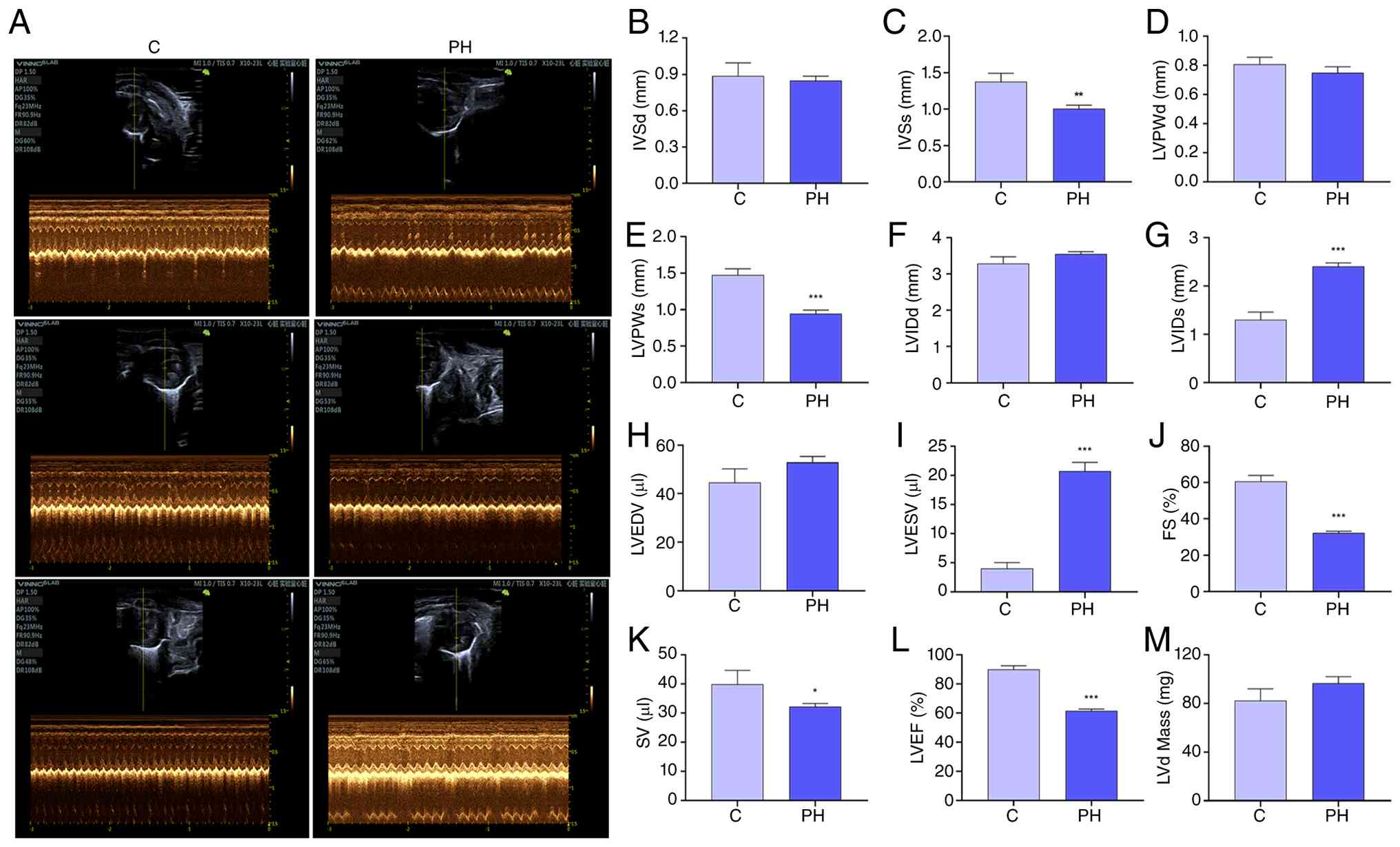 | Figure 1Echocardiographic assessment of
cardiac function in PH model mice. (A) Representative M-mode
echocardiographic images of the left ventricle from normoxic C mice
and PH model mice after 28 days of hypobaric hypoxia exposure.
Parameters were measured by echocardiography, including (B) IVSd,
(C) IVSs, (D) LVPWd, (E) LVPWs, (F) LVIDd, (G) LVIDs, (H) LVEDV,
(I) LVESV, (J) FS, (K) SV, (L) LVEF and (M) LVd mass. Data are
presented as mean ± SD (n=8 per group). *P<0.05,
**P<0.01 and ***P<0.001 vs. C group. C,
control; PH, pulmonary hypertension; IVSd, interventricular septal
thickness at diastole; IVSs, interventricular septal thickness at
systole; LVPWd, left ventricular posterior wall thickness at
diastole; LVPWs, left ventricular posterior wall thickness at
systole; LVIDd, left ventricular internal diameter at diastole;
LVIDs, left ventricular internal diameter at systole; LVEDV, left
ventricular end-diastolic volume; LVESV, left ventricular
end-systolic volume; FS, fractional shortening; SV, stroke volume;
LVEF, left ventricular ejection fraction; LVd, left ventricular
diastolic. |
Transcriptomic profiling identifies
enrichment of PI3K/AKT signaling-related genes in PH model
mice
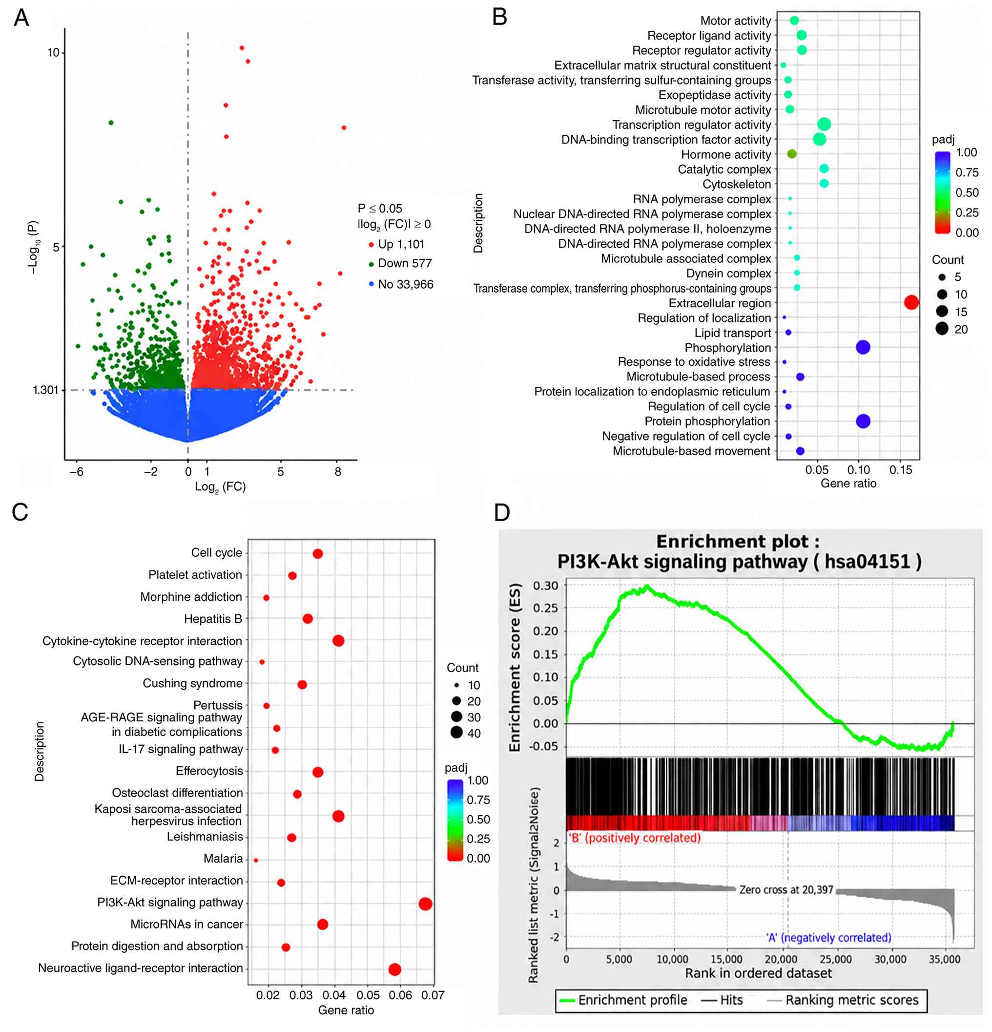
RNA sequencing revealed 1,101 significantly
upregulated genes and 577 significantly downregulated genes in PH
model mice compared with in the control group (Fig. 2A). KEGG enrichment analysis
identified significant enrichment of the ‘PI3K-AKT signaling
pathway’ (FDR<0.01; Fig. 2C).
GO enrichment analysis identified significantly enriched terms,
including ‘extracellular matrix structural constituent’,
‘DNA-binding transcription factor activity’, ‘response to oxidative
stress’ and ‘regulation of cell cycle’ (Fig. 2B), indicating that PH-induced RV
remodeling involves alterations in extracellular matrix
organization, transcriptional regulation, oxidative stress
responses and cell cycle control. KEGG enrichment analysis
identified significant enrichment of the ‘PI3K-Akt signaling
pathway’ (FDR<0.01; Fig. 2C).
GSEA further supported enrichment of the PI3K/AKT signaling pathway
in PH model mice (normalized enrichment score, 2.14; P<0.001;
Fig. 2D).
PH induces changes in
apoptosis-associated protein expression and RV fibrosis
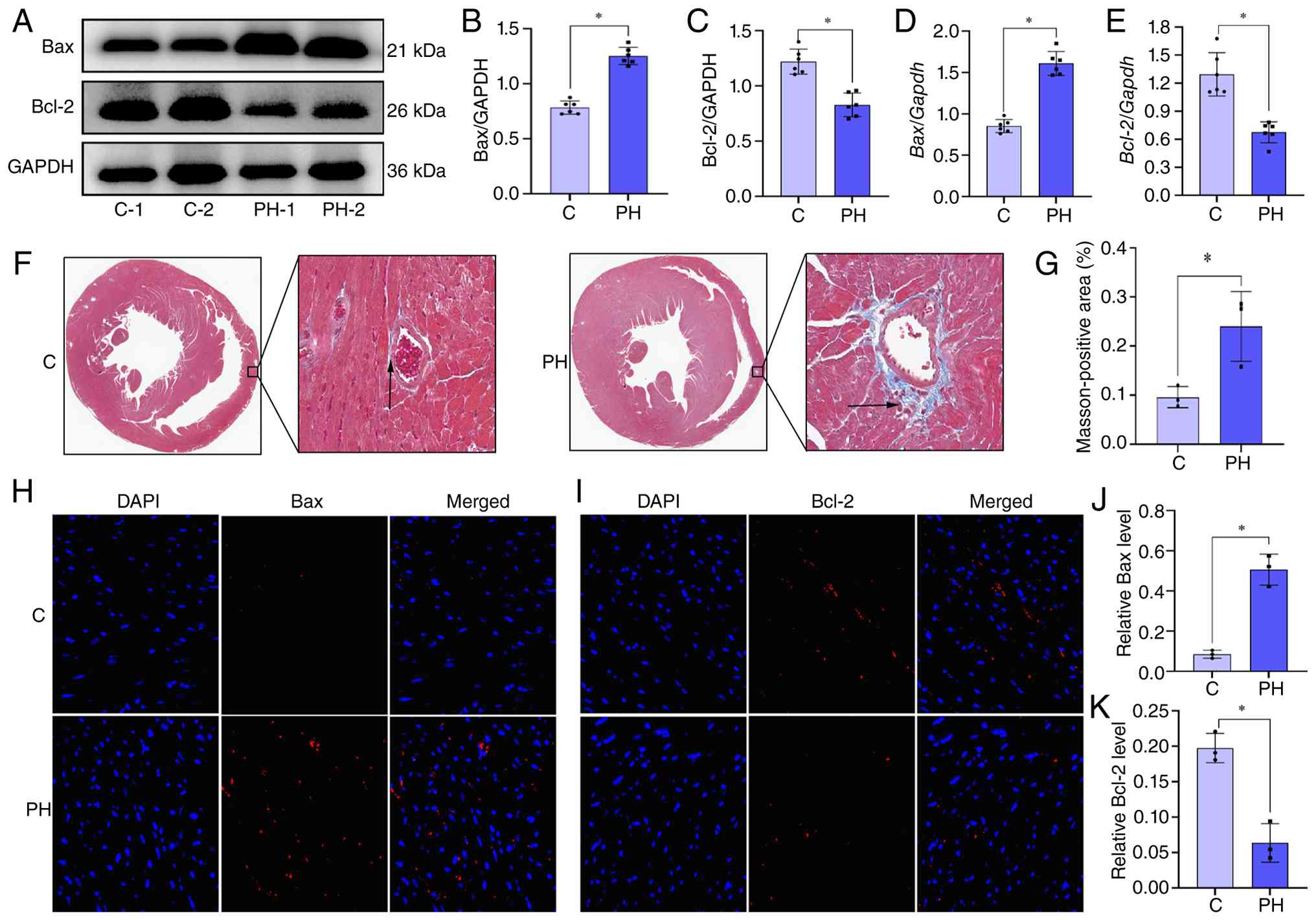
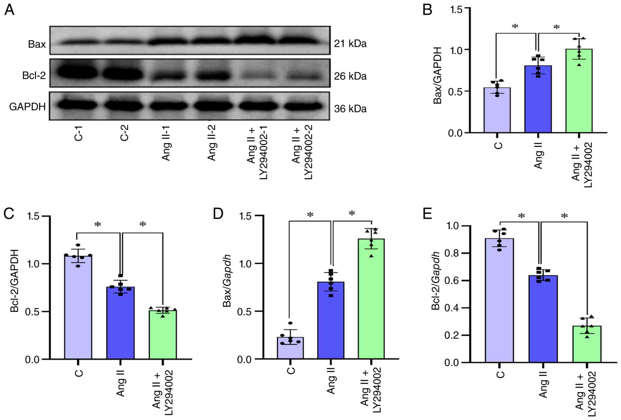
Western blotting and RT-qPCR showed significantly
increased Bax expression and significantly decreased Bcl-2
expression at the mRNA and protein levels in PH model mice compared
with those in the control mice, indicating a pro-apoptotic shift in
apoptosis-associated signaling (Fig.
3A-E). Immunofluorescence staining supported significantly
enhanced Bax and reduced Bcl-2 levels in PH model mice compared
with in control mice (Fig. 3H-K).
Furthermore, Masson's trichrome staining revealed significantly
increased collagen deposition in PH model mice compared with in
control mice, which is consistent with RV fibrosis (Fig. 3F and G).
PH is associated with a small increase
in Ki-67-positive cardiomyocytes
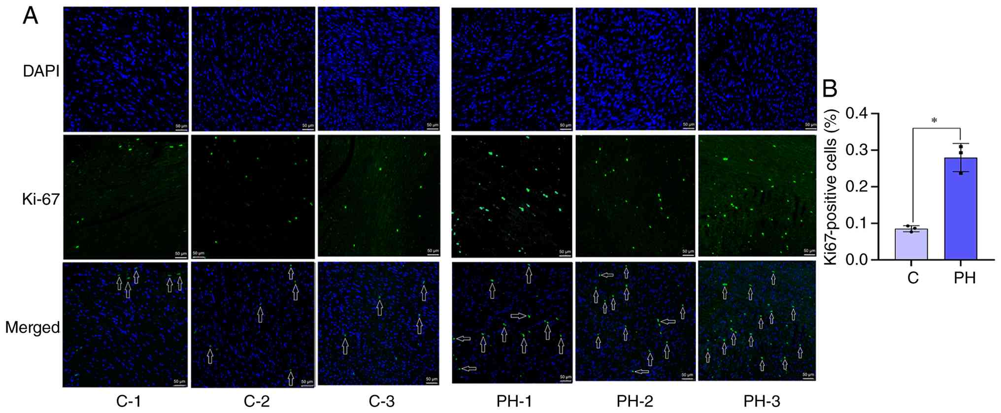
Immunofluorescence analysis showed a small but
statistically significant increase in Ki-67-positive cardiomyocytes
in RV tissues from PH model mice compared with controls (Fig. 4A and B). However, the proportion of
Ki-67-positive cardiomyocytes remained very low (~0.4%). As Ki-67
positivity indicates cell-cycle activity but does not demonstrate
completion of cell division (18,19),
these findings indicate only limited cardiomyocyte cell-cycle
activity in the PH right ventricle, and the biological significance
of this low-frequency response remains uncertain.
PI3K inhibition alters Ang II-induced
apoptosis-associated protein expression in H9C2 cells)
In H9C2 cells, treatment with Ang II resulted in
significant Bax upregulation and Bcl-2 downregulation at both the
protein and mRNA levels (Fig.
5A-E), indicating a pro-apoptotic shift in apoptosis-associated
protein expression. LY294002, a selective PI3K inhibitor that
blocks PI3K/AKT signaling, further upregulated Bax and
downregulated Bcl-2 expression in Ang II-treated cells compared
with Ang II alone (Fig. 5A-E).
These findings support a protective role for endogenous PI3K/AKT
signaling against pro-apoptotic protein changes in
cardiomyocytes.
Discussion
The present study investigated the role of PI3K/AKT
signaling in RV remodeling induced by PH. The findings demonstrated
that PH model mice developed RV dysfunction with changes in
apoptosis-associated protein expression (increased Bax and
decreased Bcl-2) and fibrosis, which was accompanied by
transcriptomic enrichment of the PI3K/AKT pathway. Inhibition of
PI3K/AKT signaling with LY294002 further altered
apoptosis-associated protein expression (further upregulated Bax
and downregulated Bcl-2) in Ang II-treated H9C2 cells. In addition,
Ki-67 staining in PH model mouse RV tissues indicated a limited
proliferative response, which was considered insufficient to
counterbalance the pro-apoptotic changes observed. Together, these
findings suggested that PI3K/AKT pathway enrichment may represent a
compensatory response, but this response was insufficient to
prevent RV dysfunction.
The PI3K/AKT pathway is a central regulator of cell
survival, apoptosis and metabolism in the cardiovascular system
(20,21). Previous studies have demonstrated
that activation of this pathway promotes the expression of
anti-apoptotic proteins, such as Bcl-2, and inhibits pro-apoptotic
mediators, such as Bax, thereby enhancing cardiomyocyte survival
under stress (22-24).
Consistent with these reports, the present study observed that the
inhibition of PI3K/AKT signaling with LY294002 further altered
apoptosis-associated protein expression (further upregulated Bax
and downregulated Bcl-2) in Ang II-treated H9C2 cells. Notably,
despite transcriptomic enrichment of the PI3K/AKT pathway in PH
model mice, pro-apoptotic Bax expression remained elevated and
anti-apoptotic Bcl-2 expression was reduced compared with controls,
indicating that compensatory PI3K/AKT signaling was unable to fully
counteract the pro-apoptotic shift under sustained pathological
stress. This paradox highlights the complexity of signaling
regulation in RV remodeling.
In addition to changes in apoptosis-associated
protein expression, the present study observed a small but
significant increase in Ki-67-positive cardiomyocytes in PH model
mice compared with in the controls.Ki-67 is a marker associated
with active phases of the cell cycle and is generally absent in
quiescent cells (25). Therefore,
the presence of Ki-67-positive cardiomyocytes suggests cell-cycle
activity in a small subset of cardiomyocytes; however, Ki-67
staining alone cannot determine whether these cells progressed
through the cell cycle or completed cytokinesis. Moreover,
Ki-67-positive cardiomyocytes accounted for only ~0.4% of the cells
examined. Accordingly, these findings should be interpreted as
evidence of limited cell-cycle activity rather than definitive
cardiomyocyte proliferation or regeneration. Previous studies have
reported that cardiomyocytes under pathological conditions can
re-express cell cycle markers (26-28).
However, such activity rarely translates into effective
regeneration (26). The findings
of the present study supported this notion, indicating that
proliferative attempts in RV tissues were insufficient to
compensate for ongoing pro-apoptotic changes, ultimately leading to
cardiac functional decline.
Clinically, RV failure remains the leading cause of
mortality in patients with PH (29), and current therapies predominantly
target pulmonary vasculature (30,31).
The data of the present study suggested that modulation of PI3K/AKT
signaling could help to preserve cardiomyocyte survival and delay
RV dysfunction, as supported by the following observations: i)
Transcriptomic enrichment of the PI3K/AKT pathway in PH model mice;
ii) the pro-apoptotic shift indicated by increased Bax and
decreased Bcl-2 expression in PH model mice; and iii) further
exacerbation of these apoptosis-associated protein changes by
LY294002 in Ang II-treated H9C2 cells. However, excessive
activation of this pathway may promote maladaptive remodeling
through crosstalk with mTOR, autophagy- or fibrosis-related
pathways (32). Thus, therapeutic
strategies for PH using the PI3K/AKT pathway must emphasize precise
signaling regulation rather than indiscriminate pathway
activation.
From a translational perspective, the findings of
the present study suggested that targeting the PI3K/AKT signaling
pathway may offer a promising adjunctive strategy for preventing RV
dysfunction in PH. Currently, approved PH therapies primarily
target pulmonary vasodilation and exhibit limited direct effects on
myocardial survival pathways (33,34).
The observation that transcriptomic enrichment of the PI3K/AKT
pathway was associated with a pro-apoptotic shift in PH model mice
(increased Bax and decreased Bcl-2 expression), and that inhibition
of this pathway with LY294002 further exacerbated these changes in
Ang II-treated H9C2 cells, implies that endogenous PI3K/AKT
signaling may provide partial protection against pro-apoptotic
protein changes. Therefore, augmenting this pathway, either through
pharmacological activators or gene therapy approaches, could
potentially delay or attenuate RV failure in patients with PH,
particularly in those at high risk for RV decompensation (16,35,36).
However, given the pleiotropic nature of PI3K/AKT signaling and its
involvement in cell proliferation, fibrosis and metabolic
regulation (17,27,37),
indiscriminate activation carries the risk of promoting maladaptive
remodeling or oncogenic effects. Therefore, future clinical
strategies should prioritize precise, context-dependent modulation
of this pathway, possibly through isoform-specific inhibitors or
activators, or via combination with existing PH therapies to
achieve synergistic benefits. Additionally, the development of
biomarkers reflecting PI3K/AKT pathway activity in RV tissues could
facilitate patient stratification and personalized therapeutic
approaches. Although these possibilities are encouraging, notable
challenges remain, including the requirement for cardiac-specific
delivery systems and rigorous preclinical safety evaluation.
Therefore, the findings of the present study provide a rationale
for further translational studies to explore these
opportunities.
It is important to acknowledge that the experiments
in the present study were conducted exclusively in male mice, which
may limit the generalizability of findings to female populations.
Notably, PH exhibits well-recognized sex disparities, with higher
prevalence but potentially improved survival outcomes in female
patients compared with male patients (28,38).
The underlying mechanisms of these differences are complex and have
been shown to involve sex hormones, particularly estrogen, sex
chromosomes and differential RV adaptation (39-42).
In the context of PI3K/AKT signaling, estrogen has been shown to
activate this pathway in various cell types, raising the
possibility that the compensatory activation observed in male mice
in the present study might differ in magnitude or functional
consequence to PI3K/AKT activation in females (43-45).
Given that RV failure remains the leading cause of mortality in
patients with PH regardless of sex (46,47),
the findings of the present study derived from male PH model mice
still provide valuable insights into RV dysfunction. Future studies
incorporating both male and female animals, as well as
investigations into the effects of estrogen and other sex-specific
factors on PI3K/AKT signaling in the right ventricle, will be
important for determining whether the observations of the present
study are universally applicable or sex-dependent. Furthermore,
although this limitation warrants consideration, the use of male
mice in the present study aligns with common practice in numerous
preclinical cardiovascular studies, in which male animals are used
to minimize variability associated with the estrous cycle. Notably,
the results of the present study established a clear mechanistic
foundation that may be extended to female models in subsequent
investigations. Several additional experimental limitations should
be acknowledged. Primarily, the present study focused on apoptosis
and did not explore other forms of programmed cell death, such as
pyroptosis or ferroptosis, which may also contribute to RV
remodeling. Furthermore, downstream effectors and crosstalk with
other signaling cascades were not investigated in detail.
Additionally, the conclusions of the present study were drawn from
animal and cell models only, and validation in human PH samples is
required to establish clinical relevance. Furthermore, although
Ki-67 staining indicated limited cell-cycle activity in a small
subset of cardiomyocytes, Ki-67 positivity alone cannot determine
whether these cells completed the cell cycle or cytokinesis;
therefore, definitive conclusions regarding cardiomyocyte
proliferation or regeneration cannot be drawn from the present
data.
In summary, the present study demonstrated that
transcriptomic enrichment of the PI3K/AKT pathway was observed in
the right ventricle during PH. Although this pathway enrichment was
accompanied by a pro-apoptotic shift (increased Bax and decreased
Bcl-2 expression) and inhibition of PI3K/AKT signaling with
LY294002 further exacerbated these changes in vitro, this
compensatory response appeared insufficient to prevent progressive
cell loss and functional deterioration. Targeted and precise
modulation of PI3K/AKT signaling may therefore represent a
promising therapeutic strategy for preventing RV failure in
patients with PH.
Acknowledgements
Not applicable.
Funding
Funding: The present study was supported by the Capital's Funds
for Health Improvement and Research (grant no. CFH-2024-1-2062) and
the Beijing Natural Science Foundation - Beijing
Economic-Technological Development Area Innovation Joint Fund
(grant no. L258027).
Availability of data and materials
The RNA sequencing data generated in the present
study may be found in the Genome Sequence Archive repository under
accession number CRA033550 or at the following URL: https://ngdc.cncb.ac.cn/gsa/browse/CRA033550. The
other data generated in the present study may be requested from the
corresponding author.
Authors' contributions
QW and GL were responsible for the conceptualization
and design of the study. QZ wrote the original draft of the
manuscript. HZ was responsible for reviewing, editing and
critically revising the manuscript for important intellectual
content. JS was responsible for data curation, including the
collection, organization and verification of experimental datasets
derived from echocardiography, molecular assays and histological
analyses; checking the completeness and consistency of data
records; and preparing the curated datasets for subsequent
statistical analysis and figure preparation. YD contributed to the
acquisition of resources and materials essential for the study, and
participated in the experimental design discussions. GX performed
the bioinformatics analysis and software implementation for the
RNA-seq data processing and analysis. WD contributed to the
visualization and presentation of the results, including the
preparation of figures. All authors participated in the
interpretation of the data, reviewed the manuscript, and read and
approved the final manuscript. All authors agree to be accountable
for all aspects of the work and to ensure that questions related to
the accuracy or integrity of any part of the work are appropriately
investigated and resolved. QW and GL confirm the authenticity of
all the raw data.
Ethics approval and consent to
participate
All animal experiments were authorized by the Animal
Care and Welfare Committee of Beijing Institute of Heart, Lung and
Blood Vessel Diseases and Beijing Anzhen Hospital, Capital Medical
University (approval no. 2025205x).
Patient consent for publication
Not applicable.
Competing interests
The authors declare that they have no competing
interests.
References
|
1
|
Liu HT, Zhang S, Liu Y, Ma J, Chen W, Yin
T, Li TB, Liang B and Tao L: Knockdown of HSP110 attenuates
hypoxia-induced pulmonary hypertension in mice through suppression
of YAP/TAZ-TEAD4 pathway. Respir Res. 23(209)2022.PubMed/NCBI View Article : Google Scholar
|
|
2
|
Huang S, Qiu J, Wang A, Ma Y, Wang P, Ding
D, Qiu L, Li S, Liu M, Zhang J, et al: Burden of pulmonary arterial
hypertension in Asia from 1990 to 2021: Findings from Global burden
of disease study 2021. Chin Med J (Engl). 138:1324–1333.
2025.PubMed/NCBI View Article : Google Scholar
|
|
3
|
Mocumbi A, Humbert M, Saxena A, Jing ZC,
Sliwa K, Thienemann F, Archer SL and Stewart S: Pulmonary
hypertension. Nat Rev Dis Primers. 10(1)2024.PubMed/NCBI View Article : Google Scholar
|
|
4
|
Huang H, Hu C, Zhang R, Xu H, Cao M and Fu
Y: Global burden of pulmonary arterial hypertension and associated
heart failure: Global burden of disease 2021 analysis. JACC Heart
Fail. 13(102385)2025.PubMed/NCBI View Article : Google Scholar
|
|
5
|
Farber HW, Miller DP, Poms AD, Badesch DB,
Frost AE, Muros-Le Rouzic E, Romero AJ, Benton WW, Elliott CG,
McGoon MD and Benza RL: Five-year outcomes of patients enrolled in
the REVEAL registry. Chest. 148:1043–1054. 2015.PubMed/NCBI View Article : Google Scholar
|
|
6
|
Deng Y, Wu W, Guo S, Chen Y, Liu C, Gao X
and Wei B: Altered mTOR and Beclin-1 mediated autophagic activation
during right ventricular remodeling in monocrotaline-induced
pulmonary hypertension. Respir Res. 18(53)2017.PubMed/NCBI View Article : Google Scholar
|
|
7
|
Boucherat O, Yokokawa T, Krishna V,
Kalyana-Sundaram S, Martineau S, Breuils-Bonnet S, Azhar N, Bonilla
F, Gutstein D, Potus F, et al: Identification of LTBP-2 as a plasma
biomarker for right ventricular dysfunction in human pulmonary
arterial hypertension. Nat Cardiovasc Res. 1:748–760.
2022.PubMed/NCBI View Article : Google Scholar
|
|
8
|
Amaki M, Nakatani S, Kanzaki H, Kyotani S,
Nakanishi N, Shigemasa C, Hisatome I and Kitakaze M: Usefulness of
three-dimensional echocardiography in assessing right ventricular
function in patients with primary pulmonary hypertension. Hypertens
Res. 32:419–422. 2009.PubMed/NCBI View Article : Google Scholar
|
|
9
|
Vonk-Noordegraaf A, Haddad F, Chin KM,
Forfia PR, Kawut SM, Lumens J, Naeije R, Newman J, Oudiz RJ,
Provencher S, et al: Right heart adaptation to pulmonary arterial
hypertension: Physiology and pathobiology. J Am Coll Cardiol. 62
(25 Suppl):D22–D33. 2013.PubMed/NCBI View Article : Google Scholar
|
|
10
|
Weber KT and Brilla CG: Pathological
hypertrophy and cardiac interstitium. Fibrosis and
renin-angiotensin-aldosterone system. Circulation. 83:1849–1865.
1991.PubMed/NCBI View Article : Google Scholar
|
|
11
|
Lazzeroni D, Rimoldi O and Camici PG: From
left ventricular hypertrophy to dysfunction and failure. Circ J.
80:555–564. 2016.PubMed/NCBI View Article : Google Scholar
|
|
12
|
Xiao F, Li HL, Wang JR, Wu HM, Cai YQ,
Wang JW, Che H, Li G, Xia ZY, Xie YB and Wang S: Research progress
on programmed cell death of cardiomyocytes in pressure-overload
hypertrophic cardiomyopathy. Apoptosis. 30:2191–2224.
2025.PubMed/NCBI View Article : Google Scholar
|
|
13
|
Xin G, Xu-Yong L, Shan H, Gang W, Zhen C,
Ji-Jun L, Ping Y and Man-Hua C: SH2B1 protects cardiomyocytes from
ischemia/reperfusion injury via the activation of the PI3K/AKT
pathway. Int Immunopharmacol. 83(105910)2020.PubMed/NCBI View Article : Google Scholar
|
|
14
|
Meco M, Giustiniano E, Nisi F, Zulli P and
Agosteo E: MAPK, PI3K/Akt Pathways, and GSK-3β Activity in severe
acute heart failure in intensive care patients: An updated review.
J Cardiovasc Dev Dis. 12(266)2025.PubMed/NCBI View Article : Google Scholar
|
|
15
|
Cheng Y, Shen A, Wu X, Shen Z, Chen X, Li
J, Liu L, Lin X, Wu M, Chen Y, et al: Qingda granule attenuates
angiotensin II-induced cardiac hypertrophy and apoptosis and
modulates the PI3K/AKT pathway. Biomed Pharmacother.
133(111022)2021.PubMed/NCBI View Article : Google Scholar
|
|
16
|
Chen L, Chen LJ, Shen HW, Hsu C, Zeng JH,
Li JH, Liu JL, Yang JZ, Liu Y, Li XW, et al: Inhibition of HIF-2α
expression in cardiomyocytes attenuates PCB126-induced
cardiotoxicity associated with decreased apoptosis through the
PI3K/Akt and p53 signaling pathways. Ecotoxicol Environ Saf.
286(117185)2024.PubMed/NCBI View Article : Google Scholar
|
|
17
|
Zhao Q, Zhao X, Zhang X, Cong R, Chen X,
Dong D and Sun M: Mechanisms of aerobic exercise on
doxorubicin-induced cardiomyocyte apoptosis: Based on AMPK/PI3K/AKT
signalling pathway. Life Sci. 376(123731)2025.PubMed/NCBI View Article : Google Scholar
|
|
18
|
Goncharova EA, Ammit AJ, Irani C, Carroll
RG, Eszterhas AJ, Panettieri RA and Krymskaya VP: PI3K is required
for proliferation and migration of human pulmonary vascular smooth
muscle cells. Am J Physiol Lung Cell Mol Physiol. 283:L354–L363.
2002.PubMed/NCBI View Article : Google Scholar
|
|
19
|
Garat CV, Crossno JT Jr, Sullivan TM,
Reusch JE and Klemm DJ: Inhibition of phosphatidylinositol
3-kinase/Akt signaling attenuates hypoxia-induced pulmonary artery
remodeling and suppresses CREB depletion in arterial smooth muscle
cells. J Cardiovasc Pharmacol. 62:539–548. 2013.PubMed/NCBI View Article : Google Scholar
|
|
20
|
Kilkenny C, Browne W, Cuthill IC, Emerson
M and Altman DG: Animal research: Reporting in vivo experiments:
The ARRIVE guidelines. Br J Pharmacol. 160:1577–1579.
2010.PubMed/NCBI View Article : Google Scholar
|
|
21
|
Wang Y, Fan Y, Song Y, Han X, Fu M, Wang
J, Cui X, Cao J, Chen L, Hu K, et al: Angiotensin II induces
apoptosis of cardiac microvascular endothelial cells via regulating
PTP1B/PI3K/Akt pathway. In Vitro Cell Dev Biol Anim. 55:801–811.
2019.PubMed/NCBI View Article : Google Scholar
|
|
22
|
Livak KJ and Schmittgen TD: Analysis of
relative gene expression data using real-time quantitative PCR and
the 2(-Delta Delta C(T)) Method. Methods. 25:402–408.
2001.PubMed/NCBI View Article : Google Scholar
|
|
23
|
Qin W, Cao L and Massey IY: Role of
PI3K/Akt signaling pathway in cardiac fibrosis. Mol Cell Biochem.
476:4045–4059. 2021.PubMed/NCBI View Article : Google Scholar
|
|
24
|
Xiao F, Li HL, Yang B, Che H, Xu F, Li G,
Zhou CH and Wang S: Disulfidptosis: A new type of cell death.
Apoptosis. 29:1309–1329. 2024.PubMed/NCBI View Article : Google Scholar
|
|
25
|
Scholzen T and Gerdes J: The Ki-67
protein: From the known and the unknown. J Cell Physiol.
182:311–22. 2000.PubMed/NCBI View Article : Google Scholar
|
|
26
|
Kar A, Gupta S, Matilal A and Sarkar S:
Tissue engineering with targeted delivery of nanotized S-nitrosyl
mutant of NEMO ameliorates myocardial infarction. Nanomedicine
(Lond). 20:1085–1099. 2025.PubMed/NCBI View Article : Google Scholar
|
|
27
|
Han Z, Zhang X, Liu C, Lu M, Wang J, Nie Y
and Zhang H: Analysis of long noncoding RNAs expression profiles in
the human cardiac fibroblasts with cardiac fibrosis. Biochem
Biophys Res Commun. 660:73–81. 2023.PubMed/NCBI View Article : Google Scholar
|
|
28
|
Franco V, Ryan JJ and McLaughlin VV:
Pulmonary hypertension in women. Heart Fail Clin. 15:137–145.
2019.PubMed/NCBI View Article : Google Scholar
|
|
29
|
Yogeswaran A, Mamazhakypov A, Schermuly RT
and Weiß A: Right ventricular failure in pulmonary hypertension:
Recent insights from experimental models. Herz. 48:285–290.
2023.PubMed/NCBI View Article : Google Scholar
|
|
30
|
Namazi M, Eftekhar SP, Mosaed R,
Shiralizadeh Dini S and Hazrati E: Pulmonary hypertension and right
ventricle: A pathophysiological insight. Clin Med Insights Cardiol.
18(11795468241274744)2024.PubMed/NCBI View Article : Google Scholar
|
|
31
|
Waddingham MT, Tsuchimochi H, Sonobe T,
Sequeira V, Nayeem MJ, Shirai M, Pearson JT and Ogo T: The
selective serotonin reuptake inhibitor paroxetine improves right
ventricular systolic function in experimental pulmonary
hypertension. J Mol Cell Cardiol Plus. 8(100072)2024.PubMed/NCBI View Article : Google Scholar
|
|
32
|
Ma ZG, Yuan YP, Fan D, Zhang X, Teng T,
Song P, Kong CY, Hu C, Wei WY and Tang QZ: IRX2 regulates
angiotensin II-induced cardiac fibrosis by transcriptionally
activating EGR1 in male mice. Nat Commun. 14(4967)2023.PubMed/NCBI View Article : Google Scholar
|
|
33
|
Mandell E, Kinsella JP and Abman SH:
Persistent pulmonary hypertension of the newborn. Pediatr Pulmonol.
56:661–669. 2021.PubMed/NCBI View Article : Google Scholar
|
|
34
|
Mukherjee D and Konduri GG: Pediatric
pulmonary hypertension: Definitions, mechanisms, diagnosis, and
treatment. Compr Physiol. 11:2135–2190. 2021.PubMed/NCBI View Article : Google Scholar
|
|
35
|
Hou C, Chen J, Zhao Y, Niu Y, Lin S, Chen
S, Zong Y, Sun X, Xie L and Xiao T: The emerging role of fatty acid
synthase in hypoxia-induced pulmonary hypertensive mouse energy
metabolism. Oxid Med Cell Longev. 2021(9990794)2021.PubMed/NCBI View Article : Google Scholar
|
|
36
|
Hou B, Su S, Ji L, Huayu M, Yang N, Yang
Z, Li Z and Lu D: Luteolin alleviates right ventricular hypertrophy
in high altitude pulmonary hypertension rats by regulating
PI3K/AKT/mTOR signalling pathway. Eur J Pharmacol.
1003(177898)2025.PubMed/NCBI View Article : Google Scholar
|
|
37
|
Noorabadi P, Shahabi Rabori V, Jamali S,
Jafari N and Saberiyan M: An overview on cardiac regeneration
revolution: Exploring the promise of stem cell therapies. Mol Biol
Rep. 52(511)2025.PubMed/NCBI View Article : Google Scholar
|
|
38
|
Park E and Safdar Z: Pulmonary
hypertension in women. Methodist Debakey Cardiovasc J. 20:70–80.
2024.PubMed/NCBI View Article : Google Scholar
|
|
39
|
Wiese CB, Avetisyan R and Reue K: The
impact of chromosomal sex on cardiometabolic health and disease.
Trends Endocrinol Metab. 34:652–665. 2023.PubMed/NCBI View Article : Google Scholar
|
|
40
|
Dignam JP, Sharma S, Stasinopoulos I and
MacLean MR: Pulmonary arterial hypertension: Sex matters. Br J
Pharmacol. 181:938–966. 2024.PubMed/NCBI View Article : Google Scholar
|
|
41
|
Reue K and Wiese CB: Illuminating the
mechanisms underlying sex differences in cardiovascular disease.
Circ Res. 130:1747–1762. 2022.PubMed/NCBI View Article : Google Scholar
|
|
42
|
Ventetuolo CE and Sherman-Roe AE: Sex
differences in pulmonary (arterial) hypertension: does it matter?
Curr Opin Pulm Med. 31:411–428. 2025.PubMed/NCBI View Article : Google Scholar
|
|
43
|
Savineau JP, Marthan R and Dumas de la
Roque E: Role of DHEA in cardiovascular diseases. Biochem
Pharmacol. 85:718–726. 2013.PubMed/NCBI View Article : Google Scholar
|
|
44
|
Dumas de la Roque E, Savineau JP and
Bonnet S: Dehydroepiandrosterone: A new treatment for vascular
remodeling diseases including pulmonary arterial hypertension.
Pharmacol Ther. 126:186–199. 2010.PubMed/NCBI View Article : Google Scholar
|
|
45
|
Yuan P, Wu WH, Gao L, Zheng ZQ, Liu D, Mei
HY, Zhang ZL and Jing ZC: Oestradiol ameliorates monocrotaline
pulmonary hypertension via NO, prostacyclin and endothelin-1
pathways. Eur Respir J. 41:1116–1125. 2013.PubMed/NCBI View Article : Google Scholar
|
|
46
|
Al-Qazazi R, Lima PDA, Prisco SZ, Potus F,
Dasgupta A, Chen KH, Tian L, Bentley RET, Mewburn J, Martin AY, et
al: Macrophage- NLRP3 activation promotes right ventricle failure
in pulmonary arterial hypertension. Am J Respir Crit Care Med.
206:608–624. 2022.PubMed/NCBI View Article : Google Scholar
|
|
47
|
Latimer K, Layne1 M and Payne M: Pulmonary
hypertension. Am Fam Physician. 110:183–191. 2024.PubMed/NCBI
|