Introduction
Malignant pleural mesothelioma (MPM) is a highly
aggressive tumor with a poor survival rate that arises from the
surface cells of the pleura. It is a rare tumor; however, MPM has
become a very serious public health concern in Japan. A newspaper
article, published in June 2005, reported that five residents who
had lived near a now-closed asbestos cement pipe plant in
Amagasaki, Japan, developed pleural mesothelioma (1). The industrial use of asbestos has
been banned in Japan since 2006, but the incidence of MPM is
expected to continue increasing for the next few decades due to the
past usage of asbestos (2).
MPM has therapeutic and diagnostic challenges.
Surgical resection, often combined with radiotherapy or adjuvant
chemotherapy, is indicated for the treatment of MPM in the earlier
stage. There is a small population of patients who achieve
prolonged disease-free survival. Yet the majority of cases are
already progressive at the time of diagnosis, and these patients
exhibit an extremely poor prognosis (3). Systemic chemotherapy or radiotherapy
to date has not had an impact on patient survival for advanced
cases. Thus, it is quite important to diagnosis MPM at an early
stage. Most MPM cases demonstrate pleural effusion at the time of
diagnosis, but cytological diagnosis with pleural effusion is
usually difficult and has limited utility. To obtain a definite
diagnosis, a thoracoscopic or percutaneous biopsy should be
performed to obtain adequate specimens for pathological and
immunohistochemical analyses. Yet, even with these procedures, it
is sometimes difficult to differentiate MPM from other pleural
diseases including benign asbestos pleurisy (BAP), tuberculosis
pleurisy (TP), or pleural metastasis of lung cancer (LC). Several
investigators have sought to improve the differential diagnosis of
pleural effusion by measuring tumor markers. Shi et al
reported the usefulness of measuring the pleural carcinoembryonic
antigen for the diagnosis of malignant pleural effusion (4). Similar findings were reported
regarding cytokeratin 19 fragment 21-1 and carbohydrate antigen
(CA) 125, CA15-3 and CA19-9 (5).
Aoe et al previously reported that the concentration of
receptor-binding cancer antigen expressed on Siso cells (RCAS1) was
higher in malignant pleural effusion than in non-malignant effusion
(6), but the usefulness of these
markers has not yet been fully established in clinical practice. A
useful molecular marker for the differential diagnosis of these
diseases is therefore urgently needed.
Mesothelin is a 40-kDa cell surface glycosylated
phosphatidylinositol (GPI)-anchored glycoprotein which has putative
functions in cell-to-cell adhesion (7). Mesothelin is expressed on normal
mesothelial cells (8); however, it
is highly overexpressed in cancers such as MPM (9,10),
pulmonary carcinomas (11–14) and other neoplasms (15,16).
Soluble mesothelin-related protein (SMRP) is recognized as a
cleaved fragment of membrane-bound mesothelin (17). Robinson and colleagues reported
that serum SMRP levels were elevated in MPM when compared with
healthy asbestos-exposed and non-exposed subjects, and with other
pulmonary diseases including LC (18). Similar results were reported by
Cristaudo et al (19) and
Schneider et al (20) who
demonstrated that SMRP blood concentrations were significantly
higher in MPM than in LC cases. These findings suggest the
usefulness of serum SMRP as a diagnostic or screening marker of
MPM.
The SMRP value in pleural fluid was evaluated by
Scherpereel et al (21) and
Pass et al (22). Both
research groups reported that the pleural SMRP value was higher
than that in serum, and the level was higher in MPM than in other
pulmonary diseases. Therefore, the aim of the present study was to
investigate the SMRP level in pleural fluid in Japanese patients
with MPM. For this purpose, SMRP concentrations in pleural fluid
from Japanese patients with MPM were examined and compared with
those of patients with BAP, TP or LC. Correlations between SMRP and
asbestos exposure were also examined.
Materials and methods
Materials
Pleural fluid was collected from patients with MPM.
For these cases, pathological diagnosis of MPM was confirmed based
on standard H&E staining and positive immunohistochemical
reactivity to mesothelial markers such as calretinin, Wilms’ tumor
1, or thrombomodulin, and negative reactivity to carcinoembryonic
antigen. The clinical stage of MPM was determined according to the
International Mesothelioma Interest Group (IMIG) criteria (23) and was based on staging procedures
including computed tomographic (CT) scans of the chest and abdomen,
magnetic resonance images of the brain and Technetium-99m
hydroxymethylene diphosphonate bone scans. Survival data of the
patients with MPM were determined from the day of diagnosis to the
day of death or last follow-up. Pleural fluid was also collected
from patients with LC, BAP, TP and with chronic heart failure (CHF)
as controls. LC was diagnosed in cases where lung cancer cells were
detected in the pleural effuion. Histological subtypes of LC were
based on the World Health Organization (WHO) classification
(24). The clinical stage of the
disease was assessed using the International Staging System
(25). TP was diagnosed in cases
in which Mycobacterium tuberculosis was detected in the
pleural fluid. TP was also diagnosed in cases with higher
concentrations of adenosine deaminase (>50 IU/l) and when
lymphocyte dominancy was shown in the fluid. CHF was diagnosed in
cases which demonstrated transudate fluid with known cardiac
diseases. The diagnosis of BAP was determined by exclusion of other
specific causes in patients with past asbestos exposure, in which
malignant diseases were ruled out with thoracoscopy. Informed
consent was provided by all patients, and the study was conducted
with approval of the appropriate institutional review boards.
SMRP measurement
SMRP was measured using the chemiluminescent enzyme
immunoassay (CLEIA) (Fujirebio Diagnostics, Malvern, PA, USA) based
on the 2-step sandwich method. In brief, 20 μl of sample was mixed
with 180 μl of sample diluents, then 20 μl of the diluted sample
was incubated with 250 μl of anti-SMRP antibody-coated ferrite
particles at 37°C for 10 min. After washing, 250 μl of anti-SMRP
antibodies coupled with alkaline phosphate was added and incubated
at 37°C for 10 min. After a washing step, 200 μl of substrate
[3-(2′-spiroadamantane)-4-methoxy-4-(3″-phosphoryloxy)
phenyl-1,2-dioxetane disodium salt; AMPPD] solution was added,
followed by incubation at 37°C for 5 min. Luminescence at a
wavelength of 477 nm was measured, and the SMRP concentration of
each sample was calculated with the standard curve method.
Asbestos body burden
Quantification of asbestos bodies was performed
using the protocol modified by Kohyama and Suzuki (26). In brief, portions of
paraffin-embedded normal lung tissue (1–2 g) obtained from surgery
or autopsy were deparaffinized with xylene, then microcut. These
were digested with solution containing 5–20% sodium hypochlorite
and KOH for 6 h at 60°C. Following digestion, samples were pelleted
and resuspended in distilled water. Samples were then mixed well
and filtered through a cellulose ester membranous filter which was
dehydrated and cut in half. Pieces of the filter were mounted on
microscope slides and dried with acetone vapor. Asbestos bodies
were then counted, and the asbestos bodies per (wet weight) gram of
lung were calculated.
Statistical analyses
Comparisons between groups were performed using the
Kruskal-Wallis test and non-parametric analysis using the
Mann-Whitney U test. Areas under receiver operating curves (ROC)
were calculated using standard techniques. Survival data were
determined from the day of diagnosis to the day of death or last
follow-up and analyzed based on the Kaplan-Meyer method.
Correlations between pleural SMRP values and asbestos body or
patient survival were calculated based on Pearson’s correlation
coefficient (PCI). Statistical calculations were performed with
SPSS Statistical Package version 11.0 (SPSS Inc., Chicago, IL,
USA).
Results
Patient characteristics
Between January 2004 and July 2007, pleural fluids
were collected from 23 patients with MPM, 38 with LC, 26 with BAP,
5 with TP and 4 with CHF at the Okayama Rosai Hospital. Of the 23
cases (median age 64 years; range 47–89; male/female 21/2)
diagnosed with MPM, there were 15 epithelioid, 2 biphasic, 4
sarcomatoid and 2 unknown pathological subtypes. According to the
IMIG staging system, there were 3 cases in stage I, 2 in stage II,
9 in stage III, 6 in stage IV and 3 unknown. Of the 38 cases
(median age 69.5 years; range 46–91; male/female 29/9) diagnosed
with LC, there were 24 patients with adenocarcinoma, 4 with
small-cell carcinoma, 3 with squamous cell carcinoma and 7
undetermined pathological subtypes. The characteristics of the
patients are summarized in Table
I.
 | Table I.Patient characteristics. |
Table I.
Patient characteristics.
| MPM | PMLC | BAP | TP | CHF |
|---|
| No. | 23 | 38 | 26 | 5 | 4 |
| Age (years) | | | | | |
| Median (range) | 64 (47–89) | 70 (48–90) | 75.5 (58–88) | 82 (68–88) | 74 (68–82) |
| Gender | | | | | |
| Male/Female | 21/2 | 28/10 | 26/0 | 5/0 | 3/1 |
| Asbestos exposure
period (years) | | | | | |
| Median (range) | 33 (5–51) | - | 30 (3–46) | - | |
| Histology | | | | | |
| Epithelioid | 15 | - | - | - | |
| Biphasic | 2 | - | - | - | |
| Sarcomatoid | 4 | - | - | - | |
| Unknown | 2 | - | - | - | |
| Adenocarcinoma | | 24 | | | |
| Squamous cell
carcinoma | | 3 | | | |
| Small-cell
carcinoma | | 4 | | | |
| Not determined | | 7 | | | |
| Stage | | | | | |
| I | 3 | - | - | - | |
| II | 2 | - | - | - | |
| III | 9 | - | - | - | |
| IV | 6 | - | - | - | |
| Unknown | 3 | - | - | - | |
SMRP value in MPM
According to the clinical stage and pathological
subtypes of MPM, a trend was noted in which the SMRP value was
higher in advanced stages (III and IV, n=16; median 13.8, range
2.85–82.8 nmol/l) compared with the value in early stages (I and
II, n=5; median 7.9, range 2.5–33.9 nmol/l), and higher in
epithelioid type (n=13; median 15.4, range 2.2–82.8 nmol/l) than in
sarcomatoid (n=4; median 13.8, range 2.85–10.45 nmol/l), though
there were no significant differences (P=0.158 and 0.389,
respectively).
SMRP and asbestos exposure
Occupational asbestos exposure was revealed in 21
patients with MPM. We examined the duration of asbestos exposure
and the SMRP value in the pleural fluid, but no correlation was
shown (PCI, −0.069). Quantification of asbestos bodies was
performed in 17 cases of MPM. The median number of bodies was 2,180
(239–526,000) per gram of dried lung. We examined the correlation
between the SMRP value in pleural fluid and the number of asbestos
bodies, but no correlation was found (PCI, −0.156). Survival data
was available in 22 cases. No correlation was found between the
SMRP value and survival (PCI, −0.179). We compared the survival of
two groups, those with a lower concentration of SMRP (≤8.0 nmol/l)
and those with a higher concentration, but no statistical
difference was demonstrated (data not shown).
SMRP value for dif ferential
diagnosis
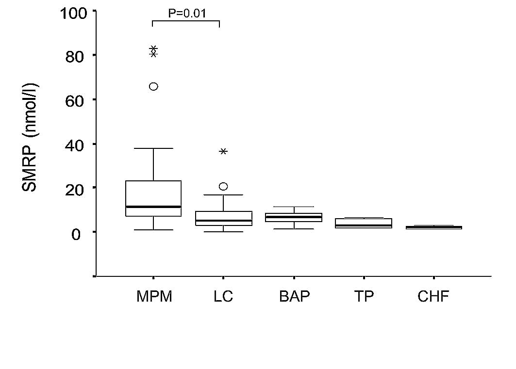
The median concentration of SMRP in MPM, LC, BAP, TP
and CHF were 11.5 (range 0.9–82.8), 5.2 (0.05–36.4), 6.65
(1.45–11.25), 3.20 (1.65–6.5) and 2.03 (1.35–2.8) nmol/l,
respectively. The SMRP concentration was significantly higher in
MPM than in the other diseases (P=0.001, Kruskal-Wallis test,
Fig. 1). The area under the ROC
curve (AUC) values of the MPM diagnosis was 0.75 [95% confidence
interval (CI), 0.615–0.884] for the differential diagnosis from the
other groups. Based on the cut-off value of 8 nmol/l, the
sensitivity and specificity for diagnosis of MPM were 70.0 and
68.4%, respectively. The SMRP concentration in MPM was
significantly higher than that in LC (P=0.004, Mann-Whitney U
test). The AUC for the differential diagnosis of MPM and LC was
0.724 (95% CI, 0.583–0.866). Based on the cut-off value of 8
nmol/l, the sensitivity and specificity for diagnosis of MPM were
69.6 and 68.4%, respectively. The SMRP concentration in MPM was
significantly higher than in BAP (P=0.004, Mann-Whitney U test).
The AUC value for the differential diagnosis of MPM and BAP was
0.74 (95% CI, 0.586–0.894). Based on the cut-off value of 8 nmol/l,
the sensitivity and specificity for diagnosis of MPM were 69.6 and
69.2%, respectively.
Discussion
In this study, we first examined the SMRP value in
pleural fluid from patients with MPM. SMRP was higher in the
epithelioid subtype than in the sarcomatoid, and higher in advanced
stages (III and IV) than in early stages (I and II), though the
differences were not statistically significant. These findings
collaborate a previous study by Scherpereel et al (21). They examined the SMRP values, both
in serum and pleural fluid, and reported that SMRP both in serum
and pleural fluid was higher in the epithelioid subtype and in
advanced diseases of MPM. The differences in our study were not
statistically significant, probably due to the small number of
samples, but our results reflect a similar trend in MPM in Japan.
In addition, we examined the correlation between pleural SMRP and
overall survival of patients with MPM, but no correlation was
found. The role of serum SMRP as a prognostic marker was examined
by Cristaudo et al. In their study, a high SMRP level in
serum was an independent negative prognostic factor in patients
with MPM (19). The present study
is the first report to examine the role of pleural SMRP as a
prognostic factor, but these results should be interpreted
carefully because of the small number of cases. Further studies are
warranted to clarify the role of pleural SMRP as a prognosis
predictive marker.
We next examined the usefulness of pleural SMRP as a
diagnostic marker of MPM. We compared the SMRP value in the pleural
fluid of MPM to that of LC, BAP, TP and CHF. The SMRP value in MPM
was significantly higher than in the other diseases. Similar
findings were also reported by Scherpereel et al (21). They reported that the serum or
pleural fluid SMRP level was significantly higher in patients with
MPM than in subjects with benign pleural lesions related to
asbestos exposure (BPLAE) or in LC. In their report, BPLAE was
defined based on the definition by the American Thoracic Society
(27), which corresponds with BPE
in our study. In our study, subjects with TP and CHF were also
included as controls. TP is the single most frequent cause of death
by an infectious agent and is also a major cause of pleural
effusion (28). Several molecular
markers in pleural effusion have been examined as diagnostic
markers of TP (29), but the
differential diagnosis is still often problematic in clinical
practice. Our results revealed, for the first time, the usefulness
of pleural SMRP to distinguish MPM and TP.
We also analyzed the correlations between the SMRP
concentration and asbestos exposure. We determined the number of
asbestos bodies in the lungs of patients with MPM. The duration of
occupational asbestos exposure was determined through patient
interview. As a result, no correlation was revealed between SMRP
values and the duration of asbestos exposure or asbestos bodies in
the lung. These findings indicate that elevation of SMRP in the
pleural effusion of MPM is not influenced by asbestos, but is one
of the cancer-specific events. The mechanisms of accumulation of
SMRP in pleural fluid have not as yet been established. SMRP is
reported as a proteolytically cleaved fragment of membrane-bound
mesothelin (17). The release of
SMRP could also be due to a frameshift mutation of the protein
(21). Further studies are
warranted to examine the mechanisms involved in the elevation of
SMRP in MPM.
In conclusion, we examined the SMRP concentration in
pleural fluid from patients with MPM, LC, BAP, TP and CHF and
demonstrated that the SMRP value in MPM was significantly higher
than that in the other diseases. These results indicate the
usefulness of pleural SMRP as a diagnostic marker of MPM.
Acknowledgements
This research is a part of the
research and development, and dissemination projects related to the
13 fields of occupational injuries and illnesses of the Japan
Labour Health and Welfare Organization. This research is supported
by the Program for the Promotion of Fundamental Studies in Health
Science of the National Institute of Biomedical Innovation of
Japan.
References
|
1.
|
Ohshima H: Five cases with mesothelioma
living near a now-defunct asbestos cement plant in Amagasaki city.
Mainichi Newspaper (in Japanese). 1June 29–2005.
|
|
2.
|
Robinson BW and Lake RA: Advances in
malignant mesothelioma. N Engl J Med. 353:1591–1603. 2005.
View Article : Google Scholar : PubMed/NCBI
|
|
3.
|
Ray M and Kindler HL: Malignant pleural
mesothelioma: an update on biomarkers and treatment. Chest.
136:888–896. 2009. View Article : Google Scholar : PubMed/NCBI
|
|
4.
|
Shi HZ, Liang QL, Jiang J, Qin XJ and Yang
HB: Diagnostic value of carcinoembryonic antigen in malignant
pleural effusion: a meta-analysis. Respirology. 13:518–527. 2008.
View Article : Google Scholar : PubMed/NCBI
|
|
5.
|
Liang QL, Shi HZ, Qin XJ, Liang XD, Jiang
J and Yang HB: Diagnostic accuracy of tumour markers for malignant
pleural effusion: a meta-analysis. Thorax. 63:35–41. 2008.
View Article : Google Scholar : PubMed/NCBI
|
|
6.
|
Aoe K, Hiraki A, Maeda T, et al: Soluble
receptor-binding cancer antigen expressed on SiSo cells in pleural
fluid: a potential diagnostic marker for malignant pleural
effusion. Chest. 126:1195–1197. 2004. View Article : Google Scholar
|
|
7.
|
Rump A, Morikawa Y, Tanaka M, et al:
Binding of ovarian cancer antigen CA125/MUC16 to mesothelin
mediates cell adhesion. J Biol Chem. 279:9190–9198. 2004.
View Article : Google Scholar : PubMed/NCBI
|
|
8.
|
Chang K, Pai LH, Pass H, et al: Monoclonal
antibody K1 reacts with epithelial mesothelioma but not with lung
adenocarcinoma. Am J Surg Pathol. 16:259–268. 1992. View Article : Google Scholar : PubMed/NCBI
|
|
9.
|
Chang K and Pastan I: Molecular cloning of
mesothelin, a differentiation antigen present on mesothelium,
mesotheliomas and ovarian cancers. Proc Natl Acad Sci USA.
93:136–140. 1996. View Article : Google Scholar : PubMed/NCBI
|
|
10.
|
Ordonez NG: Value of mesothelin
immunostaining in the diagnosis of mesothelioma. Mod Pathol.
16:192–197. 2003. View Article : Google Scholar : PubMed/NCBI
|
|
11.
|
Ordonez NG: The immunohistochemical
diagnosis of mesothelioma: a comparative study of epithelioid
mesothelioma and lung adenocarcinoma. Am J Surg Pathol.
27:1031–1051. 2003. View Article : Google Scholar : PubMed/NCBI
|
|
12.
|
Scholler N, Fu N, Yang Y, et al: Soluble
member(s) of the mesothelin/megakaryocyte potentiating factor
family are detectable in sera from patients with ovarian carcinoma.
Proc Natl Acad Sci USA. 96:11531–11536. 1999. View Article : Google Scholar
|
|
13.
|
Frierson HF Jr, Moskaluk CA, Powell SM, et
al: Large-scale molecular and tissue microarray analysis of
mesothelin expression in common human carcinomas. Hum Pathol.
34:605–609. 2003. View Article : Google Scholar : PubMed/NCBI
|
|
14.
|
Bhattacharjee A, Richards WG, Staunton J,
et al: Classification of human lung carcinomas by mRNA expression
profiling reveals distinct adenocarcinoma subclasses. Proc Natl
Acad Sci USA. 98:13790–13795. 2001. View Article : Google Scholar : PubMed/NCBI
|
|
15.
|
Hornick JL, Lauwers GY and Odze RD:
Immunohistochemistry can help distinguish metastatic pancreatic
adenocarcinomas from bile duct adenomas and hamartomas of the
liver. Am J Surg Pathol. 29:381–389. 2005. View Article : Google Scholar : PubMed/NCBI
|
|
16.
|
Watanabe H, Okada G, Ohtsubo K, et al:
Expression of mesothelin mRNA in pure pancreatic juice from
patients with pancreatic carcinoma, intraductal papillary mucinous
neoplasm of the pancreas and chronic pancreatitis. Pancreas.
30:349–354. 2005. View Article : Google Scholar
|
|
17.
|
Hassan R, Bera T and Pastan I: Mesothelin:
a new target for immunotherapy. Clin Cancer Res. 10:3937–3942.
2004. View Article : Google Scholar : PubMed/NCBI
|
|
18.
|
Robinson BW, Creaney J, Lake R, et al:
Mesothelin-family proteins and diagnosis of mesothelioma. Lancet.
362:1612–1616. 2003. View Article : Google Scholar : PubMed/NCBI
|
|
19.
|
Cristaudo A, Foddis R, Vivaldi A, et al:
Clinical significance of serum mesothelin in patients with
mesothelioma and lung cancer. Clin Cancer Res. 13:5076–5081. 2007.
View Article : Google Scholar : PubMed/NCBI
|
|
20.
|
Schneider J, Hoffmann H, Dienemann H,
Herth FJ, Meister M and Muley T: Diagnostic and prognostic value of
soluble mesothelin-related proteins in patients with malignant
pleural mesothelioma in comparison with benign asbestosis and lung
cancer. J Thorac Oncol. 3:1317–1324. 2008. View Article : Google Scholar : PubMed/NCBI
|
|
21.
|
Scherpereel A, Grigoriu B, Conti M, et al:
Soluble mesothelin-related peptides in the diagnosis of malignant
pleural mesothelioma. Am J Respir Crit Care Med. 173:1155–1160.
2006. View Article : Google Scholar : PubMed/NCBI
|
|
22.
|
Pass HI, Wali A, Tang N, et al: Soluble
mesothelin-related peptide level elevation in mesothelioma serum
and pleural effusions. Ann Thorac Surg. 85:265–272. 2008.
View Article : Google Scholar : PubMed/NCBI
|
|
23.
|
Rusch VW: A proposed new international TNM
staging system for malignant pleural mesothelioma from the
International Mesothelioma Interest Group. Lung Cancer. 14:1–12.
1996. View Article : Google Scholar
|
|
24.
|
Histological Typing of Lung and Pleural
Tumors. World Health Organization; Geneva: 1999
|
|
25.
|
Mountain CF: Revisions in the
International System for staging lung cancer. Chest. 111:1710–1717.
1997. View Article : Google Scholar : PubMed/NCBI
|
|
26.
|
Kohyama N and Suzuki Y: Analysis of
asbestos fibers in lung parenchyma, pleural plaques and
mesothelioma tissues of North American insulation workers. Ann NY
Acad Sci. 643:27–52. 1991. View Article : Google Scholar : PubMed/NCBI
|
|
27.
|
Diagnosis and initial management of
nonmalignant diseases related to asbestos. Am J Respir Crit Care
Med. 170:691–715. 2004. View Article : Google Scholar : PubMed/NCBI
|
|
28.
|
Raviglione MC and Luelmo F: Update on the
global epidemiology of tuberculosis. Curr Issues Public Health.
2:192–197. 1996.PubMed/NCBI
|
|
29.
|
Hiraki A, Aoe K, Eda R, et al: Comparison
of six biological markers for the diagnosis of tuberculous
pleuritis. Chest. 125:987–989. 2004. View Article : Google Scholar : PubMed/NCBI
|