Introduction
Conventional treatment options for the triple-negative breast cancer (TNBC) subtype include anthracycline/taxol/platinum-based conventional cytotoxic therapy or phosphoinositol-3-kinase (PI3K), AKT, molecular target of rapamycin (mTOR) and poly(ADP-ribose) polymerase (PARP) selective small molecule based targeted therapy (1-3). These long-term treatment options are frequently associated with systemic toxicity and acquired drug resistance, leading to the progression of therapy-resistant disease (4). At present, no drug is yet available for the secondary prevention of TNBC, at least to the best of our knowledge. These therapeutic limitations emphasize the need for the identification of non-toxic efficacious agents as testable alternatives against TNBC.
Natural products, including dietary and medicinal phytochemicals, botanicals and nutritional herbs have documented preventative/therapeutic/efficacy in complementary and alternative medicine against breast cancer subtypes (5,6). These naturally occurring compounds may represent effective dietary supplements for the prevention/therapy of organ site cancers. Several mechanistically distinct Chinese nutritional herbs have been demonstrated to exert growth inhibitory effects in cellular models for the estrogen receptor (ER)-α+/progesterone receptor (PR)+/human epidermal growth factor receptor-2 (HER2)- (Luminal A) and ER-α-/PR-/HER-2- (triple-negative) molecular subtypes of breast cancer (7-10). Published evidence from a comparative study on isogenic MCF-7 cellular phenotypes with modulated ER-α function demonstrated that the Chinese nutritional herb, Psoralea corylifolia (PC), effectively inhibited growth in the phenotypes with functional or non-functional ER-α (7).
The aim of the present cell culture study was to evaluate the growth inhibitory effects of PC in a cellular model for TNBC and to identify possible mechanistic leads for the efficacy of this nutritional herb. These in vitro data may provide a scientific basis for subsequent in vivo studies on triple-negative tumors. The MDA-MB-231 cellular model of TNBC was selected as the experimental system. The TNBC breast cancer subtype is notable for the compromised tumor suppressive function of the RB gene (11). Thus, the experimental downregulation of the aberrant function of RB may identify potential molecular targets for the efficacy of PC.
Materials and methods
Experimental model
The MDA-MB-231 cell line is isolated from the pleural effusion of human metastatic breast carcinoma. The carcinoma cells lack the expression levels of ER-α, PR and amplified HER-2, and represent an experimental model for TNBC (12,13). This cell line was obtained from the American Type Culture Collection (ATCC), and the cells were maintained in RPMI-1640 medium with L-glutamine and 5% fetal bovine serum (Life Technologies; Thermo Fisher Scientific, Inc.) following the protocol a recommended by ATCC.
Test agent
A non-fractionated aqueous extract of PC was prepared by boiling 20 g of seeds of PC provided by the author GYCW in 200 ml of deionized distilled water followed by sequential boiling and centrifugation to obtain a concentrated extract at a concentration of 20 g extract in 20 ml deionized distilled water (7,8,10). This stock solution was subsequently diluted in the culture medium to obtain a concentration of 1 mg extract/1 ml. This diluted stock solution was subsequently used on the cell cultures at the µg/ml concentration range.
Analysis of dose response effect of PC
The dose response effect of PC was determined by examining cell viability using the Cell Titre Glo assay (Promega Corporation) as recommended in the protocol provided by the manufacturer. This assay measures relative luminescent units (RLUs) as a quantitative end point. Briefly, 5.0x10³ cells were seeded in 96-well plates and treated with PC at a 1-100 µg/ml concentration range for 6 days at 37˚C in a CO2 incubator. The medium containing PC was replenished every 72 h. Untreated cultures maintained in the culture medium represented the control. Cell viability was measured on day 7 using a Fluoroskan plate reader (Thermo Fisher Scientific, Inc.), and was expressed as RLUs. The data obtained from the dose response experiment were extrapolated to identify the minimally effective (IC25), half maximum (IC50) and maximally cytostatic (IC90) inhibitory concentrations for the cells treated with PC.
Anchorage-independent (AI) growth assay
This assay was performed following the optimized protocol. Briefly, MDA-MB-231 cell suspensions, at a density of 5x10⁵ per ml prepared in 0.33% agar with a concentration range of 1-80 µg/ml PC. The untreated cell suspension represented the control. These cell suspensions were overlaid on a basement matrix of 0.6% agar. The cultures were incubated at 37˚C in a CO2 incubator for 21 days. The AI colonies were fixed in 4% phosphate-buffered formaldehyde (cat. no. 100494, Sigma-Aldrich; Merck KGaA, stained with 0.005% crystal violet (cat. no. C-6158, Sigma-Aldrich; Merck KGaA), overnight at room temperature. The colony counts were determined at x10 magnification. The data were expressed as the AI colony number. These data were extrapolated to identify the IC25, IC50 and IC90 inhibitory concentrations.
Cell cycle progression assay
For the cell cycle analysis, 5x10⁵ cells were seeded in T-25 flasks and treated for 24 h after seeding with 3, 6 and 12 µg/ml of PC for 48 h. The cells were harvested by trypsinization, pelleted at 500 x g, and washed twice with cold PBS. The cells were then fixed with cold 70% ethanol, washed with cold PBS, and stained with 50 µg/ml propidium iodide in PBS (Sigma-Aldrich; Merck KGaA), followed by the addition of 10 µg/ml ribonuclease (Sigma-Aldrich; Merck KGaA), and incubation at 37˚C for 20 min and a 4-h incubation of the mixture at 4˚C in the dark. Cell cycle progression was monitored following an optimized protocol that included the fluorescence-assisted sorting of PI-stained cells, with the use of a 488 nm excitation filter and a 520 nm band pass filter, and gating of the sorted cells on a forward versus side scatter. The DNA content was analyzed using a BD FACScan Flow Cytometer (BD Biosciences) and analyzed by FCS Express software version 306 (De Novo Software). The flow cytometry-based analyses determined the distribution of individual cell populations in the G1 (quiescent), S, G2/M (proliferative) and sub G0 (apoptotic) phases of the cell cycle. These primary data were expressed as the G1: S + G2/M ratio and the percentage sub G0 population to assess altered cell cycle progression and cellular apoptosis, respectively.
Western blot analysis
Cells were seeded in 10-cm dishes at 70% confluence 1 day prior to treatment. Cells were treated with 2, 4 and 6 µg/ml of PC and the cultures were incubated for 48 h in a CO2 incubator at 37˚C. Cells were harvested and immediately lysed with RIPA buffer containing protease inhibitors (Sigma-Aldrich; Merck KGaA), and centrifuged at 4˚C for 15 min at 10,000 x g. The protein content of the lysate was quantified using the Bradford method, and an equal quantity of protein was separated by 10% SDS-PAGE and transferred onto a nitrocellulose membrane (Bio-Rad Laboratories) and was blocked at 4˚C overnight with 5% non-fat dry milk followed by incubation with primary and secondary antibodies (Table I). The chemiluminescent signal was developed with ECL-plus reagent (Bio-Rad, Laboratories, Inc.) and detected by autoradiography. The antibody for β-actin was used as the reference protein for western blot analysis. The signal intensity in the western blots was quantified as arbitrary scanning units (ASU) using Molecular Imager GS800 and Quantity One software (Bio-Rad Laboratories, Inc.)
 |
Table I
Source of antibodies.
|
Table I
Source of antibodies.
| Antibody |
Vendor |
Catalogue no. |
Dilution |
| Primary |
|
|
|
| β-actin C4 |
Santa Cruz Biotechnology, Inc. |
sc-47778 |
1:200 |
| Cyclin D1 |
Santa Cruz Biotechnology, Inc. |
sc-246 |
1:200 |
| RB |
Santa Cruz Biotechnology, Inc. |
sc-74562 |
1:100 |
| CDK4 |
Cell Signaling Technology, Inc. |
12790 |
1:1,000 |
| CDK6 |
Cell Signaling Technology, Inc. |
7961 |
1:200 |
| p-RB Ser780 |
Cell Signaling Technology, Inc. |
3590 |
1:100 |
| Secondary |
|
|
|
| Anti-rabbit IgG-HRP |
Santa Cruz Biotechnology, Inc. |
sc-2357 |
1:1,000 |
Caspase assay
Caspase-3/7 activity in the MDA-MB-231 cells was measured using a Caspase-Glo assay kit (Promega Corporation). Briefly, the cells treated with PC were homogenized by sonication in homogenization buffer (25 mmol/l HEPES, pH 7.5, 5 mmol/l MgCl2, and 1 mmol/l EGTA) and protease inhibitors (all from Sigma-Aldrich; Merck KGaA). The homogenate was centrifuged at 6,500 x g at 4˚C for 15 min and the supernatant was used for experiments. To 10 µl of the supernatant, equal volume of the assay reagent was added, and the mixture was incubated at room temperature for 2 h. The luminescence was measured using a luminometer (Thermo Fisher Scientific, Inc.). The data were expressed as RLUs.
Statistical analysis
The data were obtained from replicate experiments for the dose response, AI growth, cell cycle progression and caspase-3/7 activity assays and are expressed as the mean ± SD, n=3 per treatment group. The comparisons of statistically significant differences between the common control and multiple treatment groups were analyzed using one-way analysis of variance with Dunnett's multiple comparison test as a post-hoc with a threshold of α=0.05. P<0.05 was considered to indicate a statistically significant difference.
Results
Growth inhibitory effect of PC
The data on the growth inhibitory effects of PC are presented in Table II. These data exhibited a dose-dependent inhibitory response in MDA-MB-231 cells. As indicated by the RLU values, treatment with 6, 12 and 24 µg/ml PC for 7 days resulted in a decrease of 67.7% (P=0.020), in a decrease of 72.3% (P=0.014) and in a decrease of 84.8% (P=0.001), respectively, relative to the untreated control. These data identified the IC25 as 2.4±0.3 µg/ml, the IC50 as 4.4±1.0 µg/ml and the IC90 as 25.5±1.8 µg/ml, respectively, relative to the untreated control.
|
Table II
Growth inhibitory effects of PC on MDA-MB-231 cells.
|
Table II
Growth inhibitory effects of PC on MDA-MB-231 cells.
| Treatment |
Concentration (µg/ml) |
Relative luminescent unitsa |
P-value |
Inhibition (% control) |
| Control |
- |
0.65±0.02 |
|
- |
| PC |
3 |
0.45±0.01 |
|
-30.8 |
| |
6 |
0.21±0.01 |
0.020 |
-67.7 |
| |
12 |
0.18±0.02 |
0.014 |
-72.3 |
| |
24 |
0.10±0.01 |
0.001 |
-84.8 |
Effect of PC on AI colony formation
The data on inhibition of AI colony formation by PC are presented in Tables III and SI. A 21-day treatment with PC identified the IC25 as 5 µg (P=0.050), the IC50 as 10 µg (P=0.037) and the IC90 as 80 µg (P=0.014), respectively, relative to the untreated control.
|
Table III
Inhibition of anchorage-independent growth of MDA-MB-231 cells by PC.
|
Table III
Inhibition of anchorage-independent growth of MDA-MB-231 cells by PC.
| Treatment |
Concentration (µg/ml) |
AI colony numbera |
IC |
P-value |
| Control |
- |
745±51 |
- |
- |
| PC |
1 |
641±59 |
|
|
| |
5 |
559±56 |
IC25 |
0.050 |
| |
10 |
373±48 |
IC50 |
0.037 |
| |
20 |
307±74 |
|
|
| |
40 |
230±58 |
|
|
| |
60 |
123±11 |
|
|
| |
80 |
69±5 |
IC90 |
0.014 |
Effect of PC on cell cycle progression
The effect of PC on cell cycle progression is presented in Tables IV, SII and SIII. The data expressed as the G1: S + G2/M ratio demonstrated that treatment with PC at 6 µg/ml exhibited a 2.5-fold increase (P=0.020) and treatment with PC at 12 µg/ml exhibited a 3.5-fold (P=0.014) increase respectively, relative to the untreated control.
|
Table IV
Effect of PC on cell cycle progression in MDA-MB-231 cells.
|
Table IV
Effect of PC on cell cycle progression in MDA-MB-231 cells.
| Treatment |
Concentration (µg/ml) |
G1: S + G2/Ma |
P-value |
Fold of control |
| Control |
- |
0.6±0.1 |
|
- |
| PC |
3 |
0.9±0.2 |
|
|
| |
6 |
2.1±0.3 |
0.020 |
2.5x |
| |
12 |
2.7±0.3 |
0.014 |
3.5x |
Effect of PC on RB signaling
The data presented in Fig. 1 are the results of the analyses of the effects of PC on the RB signaling. The proteins, cyclin D1, CDK4, CDK6 and p-RB, involved in this pathway exhibited a dose-dependent decrease, relative to the untreated control.
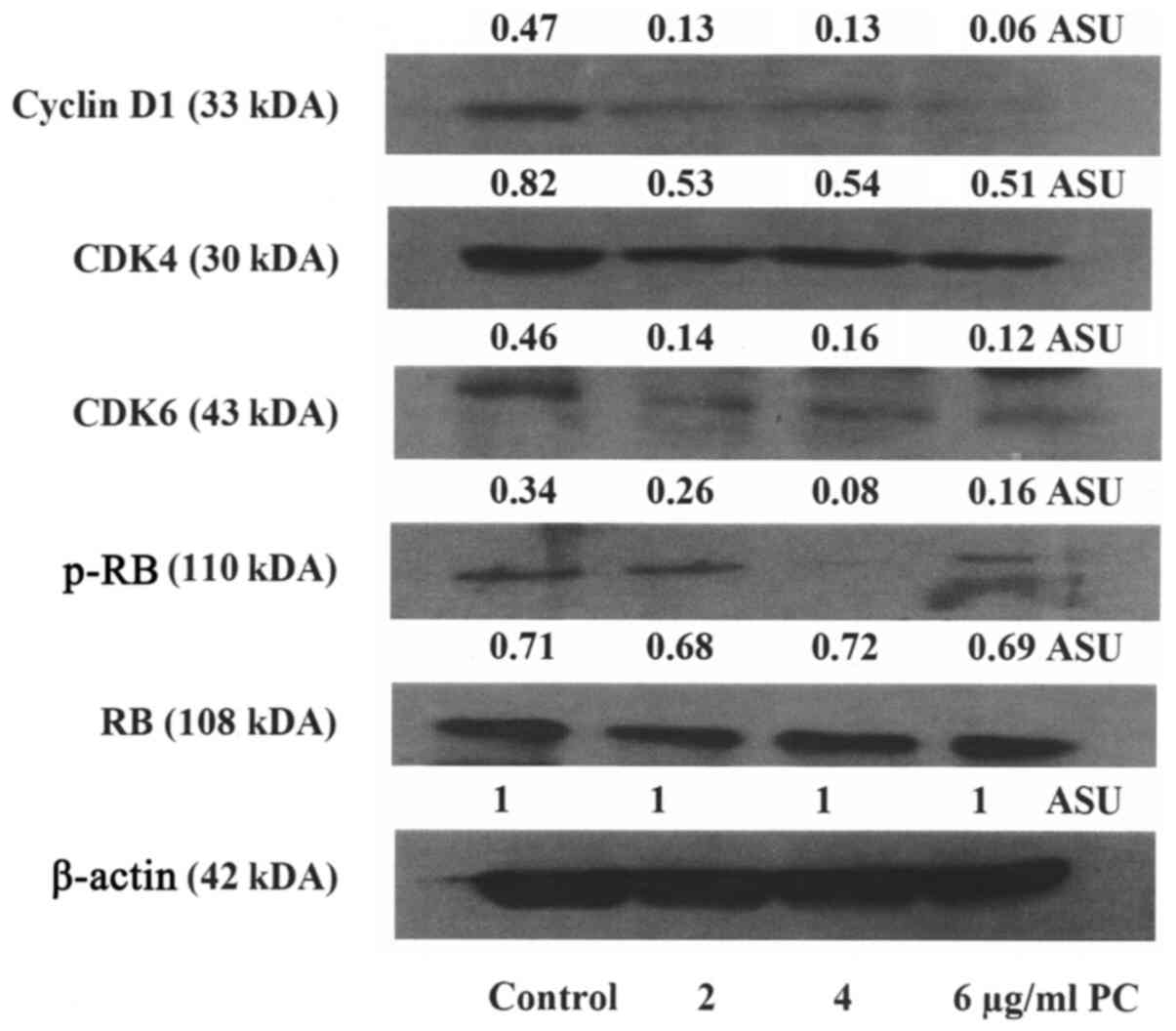 |
Figure 1
Western blot analysis of select proteins of the RB pathway. The expression levels of cyclin D1, CDK4, CDK6 and p-RB were inhibited in response to treatment with PC. PC, Psoralea corylifolia; CDK4, cyclin dependent kinase 4; CDK6, cyclin dependent kinase 6, p-RB, phosphorylated retinoblastoma; RB, retinoblastoma; ASU; arbitrary scanning unit.
|
Effect of PC on cellular apoptosis
The data presented in Tables V and SIV are the results of the analyses of the effect of PC on cellular apoptosis by quantifying the status of cells in sub G0 (apoptotic) phase of the cell cycle and the pro-apoptotic caspase-3/7 activity. Treatment with PC resulted in a dose-dependent increase in the sub G0 phase, as well as an increase in the caspase activity, relative to the untreated control.
|
Table V
Effect of PC on the apoptosis of MDA-MB-231 cells.
|
Table V
Effect of PC on the apoptosis of MDA-MB-231 cells.
| Treatment |
Concentration (µg/ml) |
Sub G0 (%)a |
P-value |
Caspase-3/7 activity (RLU)a |
P-value |
| Control |
- |
5.0±1.2 |
|
0.5±0.1 |
|
| PC |
3 |
18.0±1.4 |
0.020 |
6.5±1.3 |
0.014 |
| |
6 |
20.0±2.7 |
0.020 |
50.8±3.1 |
0.005 |
| |
12 |
60.0±5.0 |
0.014 |
75.0±5.0 |
0.001 |
Discussion
Conventional treatment options for TNBC are frequently associated with systemic toxicity and acquired drug resistance leading to progression of therapy resistant disease. Moreover, there are no drugs available for secondary prevention of TNBC. These considerations emphasize a need to identify relatively non-toxic efficacious naturally occurring testable alternatives. The nutritional herb, PC, has documented anti-oxidative, chemo-protective and anti-inflammatory activities. This herb is used in the traditional Chinese medicine and in Indian Ayurvedic medicine for general health management, as a treatment for leukoderma and psoriasis, and also as a palliative for breast cancer patients. PC is commonly used in a dose range of 10 to 15 g in herbal formulations in combination with additional herbs (14).
In the present study, treatment of the MDA-MB-231 cells with PC at a low concentration range of 3 to 24 µg/ml led to a dose-dependent reduction in viable cell number, identifying an effective dose range of 3 to 12 µg/ml, and a maximum cytostatic dose of 24 µg/ml.
Experiments using the anchorage-independent colony formation assay revealed a dose-dependent reduction in the number of AI colonies in response to treatment with PC. Published evidence on the present TNBC model has demonstrated quantifiable increase in AI colony number (9,10). By contrast, the triple-negative non-tumorigenic 184-B5 breast epithelial cells are notable for the absence of AI colony formation (15). The selectivity of AI growth in the carcinoma derived cells suggests that the AI colony formation represents a sensitive and specific in vitro surrogate end point biomarker for in vivo tumorigenic potential. Thus, the inhibition of AI colonies in response to treatment with PC suggests that PC may reduce the risk of developing breast cancer.
In the present study, experiments to examine the effects of PC on cell cycle progression revealed an increased G1: S + G2/M ratio due to G1 phase arrest. At the mechanistic level, the inhibition of cell cycle progression was associated with reduced expression levels of cyclin D1, CDK4, CDK6 and p-RB. TNBC is notable for defective RB signaling pathway, and cyclin D1 is involved in the CDK4/CDK6- pRB pathway (16). Cyclin D1 functions as a G1 specific cyclin for G1 to S phase transition during cell cycle progression, and represents a potential therapeutic target (17). Pharmacological inhibitors of CDK4 and CDK6 function during G1 to S phase transition (18). Thus, the data demonstrating that PC induced G1 arrest, and inhibited CDK4 and CDK6 expression, suggest that PC may function as a natural CDK inhibitor. Collectively, these data suggest that PC may downregulate defective RB signaling and thereby, provide plausible mechanistic leads for the anti-proliferative effects of PC in the present experimental system.
Experiments to examine the effect of PC on cellular apoptosis revealed that treatment with PC at the concentration range of 3 to 12 µg/ml induced an increase in the subG0 (apoptotic) phase of the cell cycle, and a corresponding increase in the pro-apoptotic caspase-3/7 activity. Collectively, these data provide a mechanistic lead for pro-apoptotic effects of PC and suggest possible involvement of the intrinsic pathway of cellular apoptosis.
Traditional Chinese medicine involves boiling the herbal formulation to prepare an aqueous extract for patient consumption. To simulate this clinical scenario, a non-fractionated aqueous extract of PC was used in the present study. The specific role of individual bioactive components in the herbal formulation is not well-defined. It is conceivable that the bioactive agents may act in a synergistic/additive manner, leading to growth inhibitory efficacy. The phytochemical analysis of PC has demonstrated the presence of coumarins, flavonoids and meroterpenes (19). The phytochemical isobavachalcone present in PC has been documented to increase ER-α expression, CD44 expression and affects paclitaxel resistance in MCF-7 breast cancer cells (20). In colon carcinoma-derived HCT-116 and SW480 cells, this phytochemical has documented efficacy via the AKT/GSK-3β/β-catenin axis (21), and has also been shown to inhibit cyclin D1 and CDK-4 in HCT116, SW480, LoVo and HT29 colon carcinoma-derived cellular models (22). The anti-proliferative and pro-apoptotic effects of PC extract in the MCF-7 model have been shown to be due to the upregulation of PARP, reactive oxygen species (ROS), pro-apoptotic caspase-7, caspase-9 and BCl-2 associated protein X (BAX), respectively (23). Psoralen, another putative bioactive agent present in PC, has been shown to induce G1 arrest and reverse the expression of multi-drug resistance (MDR) genes in an adriamycin-resistant MCF-7/ADR model (24). Taken together, these mechanistic data provide a proof of concept for involvement of select cell cycle regulatory and apoptosis specific proteins in the growth inhibitory effects of PC.
At the present time, no drugs are available for the secondary prevention of TNBC, at least to the best of our knowledge. Since PC has demonstrated efficacy against TNBC and is a non-toxic nutritional herb, it carries great promise for effective long-term secondary prevention for this type of breast cancer.
In conclusion, the present study provides plausible mechanistic leads for the efficacy of PC as a testable alternative against TNBC. Furthermore, these in vitro data on a cellular model of TNBC represent a scientific rationale for subsequent in vivo investigations on TNBC tumors. Overall, the study outcome validates the present experimental approach to identify and prioritize efficacious natural products as potential dietary supplements for prevention/therapy of breast cancer.
Supplementary Material
AI growth assay.
Cell cycle progression assay.
G1: S + G2/M ratio.
Apoptosis and caspase assay.
Acknowledgements
Not applicable.
Funding
Major funding for the present study was provided by philanthropic contributions to the American Foundation of Chinese Medicine from the Randall and Barbara Smith Foundation, and the Sophie Stenbeck Family Foundation.
Availability of data and materials
The datasets used and/or analyzed during the current study are available from the corresponding author on reasonable request.
Authors' contributions
NTT conceived the study design, formulated the experimental protocols and prepared the manuscript. HBN conducted the experiments, organized and analyzed the data and participated in the preparation of the manuscript. GYCW selected the nutritional herb for the study and contributed to the data interpretation and preparation of the manuscript.
Ethics approval and consent to participate
Not applicable.
Patient consent for publication
Not applicable.
Competing interests
The authors declare that they have no competing interests.
References
|
1
|
Dinh P, Sotiriou C and Piccart MJ: The evolution of treatment strategies: Aiming at the target. Breast. 16 (Suppl 2):S10–S16. 2007.PubMed/NCBI View Article : Google Scholar
|
|
2
|
Anders CK, Winer EP, Ford JM, Dent R, Silver DP, Sledge GW and Carey LA: Poly(ADP-Ribose) polymerase inhibition: ‘targeted’ therapy for triple-negative breast cancer. Clin Cancer Res. 16:4702–4710. 2010.PubMed/NCBI View Article : Google Scholar
|
|
3
|
Ibrahim YH, García-García C, Serra V, He L, Torres-Lockhart K, Prat A, Anton P, Cozar P, Guzmán M, Grueso J, et al: PI3K inhibition impairs BRCA1/2 expression and sensitizes BRCA-proficient triple-negative breast cancer to PARP inhibition. Cancer Discov. 2:1036–1047. 2012.PubMed/NCBI View Article : Google Scholar
|
|
4
|
Dean M, Fojo T and Bates S: Tumour stem cells and drug resistance. Nat Rev Cancer. 5:275–284. 2005.PubMed/NCBI View Article : Google Scholar
|
|
5
|
Neergheen VS, Bahorun T, Taylor EW, Jen LS and Aruoma OI: Targeting specific cell signaling transduction pathways by dietary and medicinal phytochemicals in cancer chemo-prevention. Toxicology. 278:229–241. 2010.PubMed/NCBI View Article : Google Scholar
|
|
6
|
Ye L, Jia Y, Ji KE, Sanders AJ, Xue K, Ji J, Mason MD and Jiang WG: Traditional Chinese medicine in the prevention and treatment of cancer and cancer metastasis. Oncol Lett. 10:1240–1250. 2015.PubMed/NCBI View Article : Google Scholar
|
|
7
|
Telang N, Li G, Katdare M, Sepkovic D, Bradlow L and Wong G: Inhibitory effects of Chinese nutritional herbs in isogenic breast carcinoma cells with modulated estrogen receptor function. Oncol Lett. 12:3949–3957. 2016.PubMed/NCBI View Article : Google Scholar
|
|
8
|
Telang NT, Li G, Katdare M, Sepkovic DW, Bradlow HL and Wong GYC: The nutritional herb Epimedium grandiflorum inhibits the growth in a model for the Luminal A molecular subtype of breast cancer. Oncol Lett. 13:2477–2482. 2017.PubMed/NCBI View Article : Google Scholar
|
|
9
|
Telang N, Nair HB and Wong GYC: Efficacy of Dipsacus asperoides (DA) in a model for triple negative breast cancer. Cancer Res. 77 (Suppl 4):Abstract nr P4-13-04. 2017.
|
|
10
|
Telang NT, Nair HB and Wong GYC: Growth inhibitory efficacy of Cornus officinalis in a cell culture model for triple-negative breast cancer. Oncol Lett. 17:5261–5266. 2019.PubMed/NCBI View Article : Google Scholar
|
|
11
|
Bosco EE and Knudsen ES: RB in breast cancer: At the crossroads of tumorigenesis and treatment. Cell Cycle. 6:667–671. 2007.PubMed/NCBI View Article : Google Scholar
|
|
12
|
Neve RM, Chin K, Fridlyand J, Yeh J, Baehner FL, Fevr T, Clark L, Bayani N, Coppe J-P, Tong F, et al: A collection of breast cancer cell lines for the study of functionally distinct cancer subtypes. Cancer Cell. 10:515–527. 2006.PubMed/NCBI View Article : Google Scholar
|
|
13
|
Subik K, Lee JF, Baxter L, Strzepek T, Costello D, Crowley P, Xing L, Hung MC, Bonfiglio T, Hicks DG, et al: The expression patterns of ER, PR, HER-2, CK5/6, EGFR, Ki 67, and AR by immune-histochemical analysis in breast cancer cell lines. Breast Cancer (Auckl). 4:35–41. 2010.PubMed/NCBIErratum in: Breast Cancer (Auckl) 12: 1178223418806626, 2018.
|
|
14
|
Khushboo PS, Jadhav VM, Kadam VJ and Sathe NS: Psoralea corylifolia Linn. - ‘Kushtanashini’. Pharmacogn Rev. 4:69–76. 2010.PubMed/NCBI View Article : Google Scholar
|
|
15
|
Telang N: Anti-proliferative and pro-apoptotic effects of rosemary and constituent terpenoids in a model for the HER-2-enriched molecular subtype of clinical breast cancer. Oncol Lett. 16:5489–5497. 2018.PubMed/NCBI View Article : Google Scholar
|
|
16
|
Burkhart DL and Sage J: Cellular mechanisms of tumour suppression by the retinoblastoma gene. Nat Rev Cancer. 8:671–682. 2008.PubMed/NCBI View Article : Google Scholar
|
|
17
|
Musgrove EA, Caldon CE, Barraclough J, Stone A and Sutherland RL: Cyclin D as a therapeutic target in cancer. Nat Rev Cancer. 11:558–572. 2011.PubMed/NCBI View Article : Google Scholar
|
|
18
|
Sherr CJ and Roberts JM: CDK inhibitors: Positive and negative regulators of G1-phase progression. Genes Dev. 13:1501–1512. 1999.PubMed/NCBI View Article : Google Scholar
|
|
19
|
Zhang X, Zhao W, Wang Y, Lu J and Chen X: The chemical constituents and bioactivities of Psoralia corylifolia Linn.: A Review. Am J Chin Med. 44:35–60. 2016.PubMed/NCBI View Article : Google Scholar
|
|
20
|
Shi J, Chen Y, Chen W, Tang C, Zhang H, Chen Y, Yang X, Xu Z, Wei J and Chen J: Isobavachalcone sensitizes cells to E2-induced paclitaxel resistance by down-regulating CD44 expression in ER+ breast cancer cells. J Cell Mol Med. 22:5220–5230. 2018.PubMed/NCBI View Article : Google Scholar
|
|
21
|
Li Y, Qin X, Li P, Zhang H, Lin T, Miao Z and Ma S: Isobavachalcone isolated from Psoralia corylifolia inhibits cell proliferation and induces apoptosis via inhibiting AKT/GSK-3β/β-catenin pathway. Drug Des Devel Ther. 13:1449–1460. 2019.PubMed/NCBI View Article : Google Scholar
|
|
22
|
Park GH, Sung JH, Song HM and Jeong JB: Anti-cancer activity of Psoralea fructus through the downregulation of cyclin D1 and CDK4 in human colorectal cancer cells. BMC Complement Altern Med. 16(373)2016.PubMed/NCBI View Article : Google Scholar
|
|
23
|
Rajan V, Tripathi J, Variyar P and Pandey BN: Mechanism of cytotoxicity by Psoralea corylifolia extract in human breast carcinoma cells. J Environ Pathol Toxicol Oncol. 33:265–277. 2014.PubMed/NCBI View Article : Google Scholar
|
|
24
|
Wang X, Cheng K, Han Y, Zhang G, Dong J, Cui Y and Yang Z: Effect of Psoralen as an anti-tumor agent in human breast cancer MCF-7/ADR cells. Biol Pharm Bull. 39:815–822. 2016.PubMed/NCBI View Article : Google Scholar
|


