Introduction
While highly active antiretroviral therapy (HAART)
reduces the morbidity and mortality of patients infected with the
human immunodeficiency virus (HIV) (1), prior studies report adverse drug
reactions that may alter the course of the antiviral therapy.
Various cutaneous and mucosal lesions may result from the use of
reverse transcriptase inhibitors (RTIs), as well as protease
inhibitors (2). For example,
adverse skin reactions including rash, urticaria, erythema
multiforme, toxic epidermolysis or Stevens-Johnson syndrome (SJS)
have been associated with HAART (3). Similarly, mucosal epithelial lesions
in the oral cavity include epithelial desquamation, exfoliative
cheilitis, cracks, ulceration and fissure formation (4). These cutaneous and mucosal lesions
may result from the use of RTIs, such as azidovudine (AZT),
didanosine and efavirenz (EFV), as well as protease inhibitors
(2). EFV is one of the commonly
used drugs in HAART and is the first medication approved for
once-daily dosing (5). Despite
its antiviral efficacy at a therapeutic dose, EFV has been linked
to skin lesions (3,6,7).
In addition, EFV can sometimes cause severe hepatitis, central
nervous system (CNS) complications, renal failure and pulmonary
complications (8–11). Although these adverse effects may
be viewed as hypersensitivity, direct phenotypic and genetic
effects of RTIs at the cellular level have not been
investigated.
Epithelial tissue regeneration can be hampered by
several cell death pathways, including apoptosis, terminal
differentiation, cellular senescence and autophagy. Genotoxic
signals trigger premature senescence or terminal differentiation in
human keratinocytes (12).
Oncogene-induced senescence (OIS) is a well-characterized
epigenetic phenomenon to halt cellular transformation (13). A recent report showed that mitotic
arrest in OIS is mediated by autophagy, a metabolic program leading
to catabolic processing of self proteins and organelles (14). Autophagic cell death is
characterized by numerous autophagosomes and degradation of
cytosolic proteins, whereas the nucleus remains intact until the
late stage of cell death (15).
This process is orchestrated by multiple protein factors, most
notably the autophagy-essential proteins (ATGs), which may exceed
30 different proteins (16).
Autophagy can be identified by degradation and lipidation of light
chain 3 (LC3), which becomes incorporated into the membrane of
autophagosomes and autophagolysosomes until it is degraded
(17). Autophagy can be
suppressed by chemical inhibitors of PI3K, such as 3-methylalanine
(3-MA), and induced by rapamycin (18).
In the present study, we investigated the cellular
effects of EFV on normal human keratinocytes (NHKs) and the
underlying molecular mechanisms. Cultured NHKs exposed to EFV
exhibited notable loss of cell viability accompanied by elongated
morphological alterations. Assessment of the cell death pathway
revealed lack of apoptotic responses but induction of autophagy in
NHKs exposed to EFV. Sublethal dose of EFV rapidly induced
degradation of p53. p53 degradation by EFV occurred together with a
reduced mTOR level and activation of ERK phosphorylation,
indicating that EFV triggers the canonical autophagic pathway in
NHKs. The EFV-treated cells demonstrated premature terminal
differentiation, while this effect was attenuated in cells treated
with 3-MA. These data indicate that EFV limits epithelial
regeneration by triggering autophagy, in part, through
proteasome-dependent degradation of p53.
Materials and methods
Cells and cell culture
NHKs and normal human fibroblasts (NHFs) were
prepared from discarded skin or mucosal tissues according to the
methods described elsewhere (19). The discarded tissues were utilized
to establish the primary cultures as guided by the UCLA Medical
Institutional Review Board (MIRB). Human small intestine epithelial
cells (CCL-241) were purchased from the American Type Culture
Collection (ATCC, Manassas, VA, USA) and cultured in Hybri-Care
Medium (ATCC) with 10% FBS and 30 ng/ml human EGF (Sigma, St.
Louis, MO, USA). HaCaT cells represent a spontaneously immortalized
human keratinocyte cell line (20), and OFK6/T cells are oral
keratinocytes immortalized with the hTERT gene (21). These cells were maintained in
Keratinocyte Growth Medium (Lonza, Walkersville, MD, USA).
Peripheral blood mononuclear cells (PBMCs) and Jurket cells were
obtained from Dr Anahid Jewett (UCLA School of Dentistry, Los
Angeles, CA) and cultured in RPMI-1640 (Invitrogen) supplemented
with 10% FBS. Cell viability was determined by MTT assay after a
48-h exposure to EFV according to the manufacturer's guidelines
(ATCC). Replication kinetics was determined by calculating the
population doubling (PD) levels according to the methods described
elsewhere (19).
Organotypic reconstructs were established using NHKs
(22). EFV (10 μM) was added to
the culture medium at the time of airlifting for 7 days until
harvested by fixing in 10% buffered formalin. Subsequently, H&E
staining was performed on thick (6 μm) sagittal sections.
Consecutive sections of the same specimens were used for
immunohistochemistry (IHC) for proliferating cell nuclear antigen
(PCNA) and K1.
Assay of apoptotic cell death
NHKs seeded on 96-well plates were exposed to 10 μM
EFV for 72 h. DNA fragmentation was determined by staining the
cells and TUNEL assay using the In Situ Cell Death detection
kit (Roche, South San Francisco, CA, USA). Apoptosis was also
detected by western blotting for caspase-3 and poly-ADP-ribose
polymerase (PARP) using the cells exposed to EFV and Jurkat cells
exposed to ionizing radiation (IR).
Western blotting
Whole cell extracts (50 μg) were separated on 4–20%
SDS-PAGE gel and transferred onto Immobilon protein membranes
(Millipore, Billerica, MA, USA). The membranes were incubated
successively with the primary and the secondary antibodies. Signals
were detected using ECL Western blotting detection reagents
(Amersham Pharmacia Biotech, Piscataway, NJ, USA).
Reverse transcription and real-time
PCR
Total RNA was isolated from cultured cells using the
RNeasy Mini kit (Qiagen, Valencia, CA, USA). cDNA was synthesized
from 5 μg RNA using the Superscript first-strand synthesis system
(Invitrogen). We used 1 μl cDNA for qPCR amplification using
SYBR-Green I Master Mix (Roche). The primer sequences were obtained
from the Universal Probe Library (Roche). PCR amplification was
performed on LightCycler 480 (Roche). Second derivative Cq value
determination method was used to compare fold-differences according
to the manufacturer's instructions (Roche).
Indirect in situ immunostaining
NHKs were exposed to 10 µM EFV for 48 h, fixed in
3.7% formaldehyde for 15 min, and permeabilized in 0.25% Triton
X-100 in PBS for 10 min. Mouse monoclonal anti-p53 or α-tubulin and
Alex Fluor® 488 goat anti-mouse IgG (Invitrogen) were
used as primary and secondary antibodies, respectively. Cells were
then counterstained with Hoechst 33342 (3.3 μg/ml). Images were
obtained using a Nikon fluorescence microscope. NHKs were treated
with 10 μM EFV in the absence or presence of 5 mM 3-methylalanine
(3-MA) for 48 h. Indirect immunoperoxidase staining (IPS) was
performed for involucrin as described previously (23).
Results
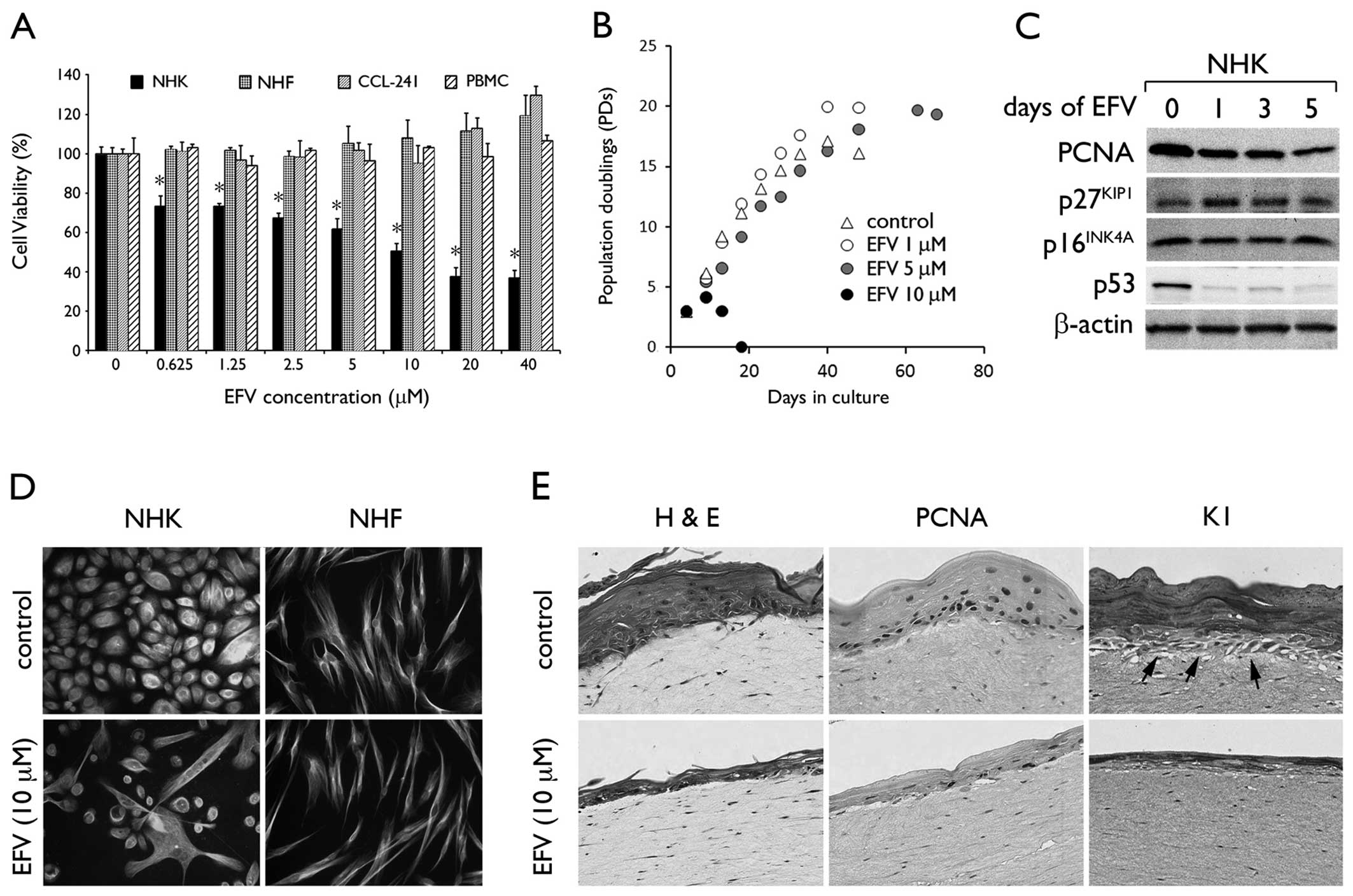
EFV inhibits cell proliferation of NHKs
in a cell type-specific manner
To determine the effects of EFV on NHKs, we
performed a cell viability assay in cultures exposed to EFV at
varying doses from 0 to 40 μM. After 48 h, we found a
dose-dependent reduction in cell viability with the IC50
at ~10 μM. As a negative control, we included 0.1%
dimethylsulfoxide (DMSO). In contrast, EFV exposure conferred no
cytotoxic effects on other cell types, including NHFs, PBMCs, or
CCL-241 cells (Fig. 1A). We
determined the long-term effects of EFV by serial subculture of
primary NHKs in the presence of EFV (Fig. 1B). At 10 μM EFV, NHKs completely
lost their viability within 10–15 days of exposure, while EFV at 1
and 5 μM allowed the cells to undergo continued proliferation.
Notably, the cells exhibited extended lifespan at these lower
doses, completing PD 20 and 19 at 1 and 5 μM EFV, respectively,
while the untreated cells senescenced after PD 17. We performed
western blot analysis for the proteins involved in cell cycle
regulation, such as p53, p16INK4A, p27KIP1
and PCNA in the NHKs following exposure to 10 μM EFV (Fig. 1C). After 1 day of EFV treatment,
an increase in p27KIP1 expression and a drastic loss of
p53 were noted, while the level of p16INK4A did not
change. PCNA expression was reduced by EFV, reflecting reduced cell
proliferation. Acute exposure to 10 μM EFV led to marked changes in
cellular morphology characteristic of terminally differentiated
keratinocytes, while NHFs remained unchanged (Fig. 1D). We exposed the 3 dimensional
(3D) organotypic culture of NHKs to EFV at 10 μM for 7 days and
found epithelial atrophy, with reduced proliferating and
undifferentiated basal cell content, following exposure to EFV
(Fig. 1E). However, NHFs in the
dermal-equivalent layer remained viable and unaltered following EFV
exposure. These results demonstrate the cytotoxic and growth
inhibitory effects of EFV in a cell type- and dose-dependent
manner.
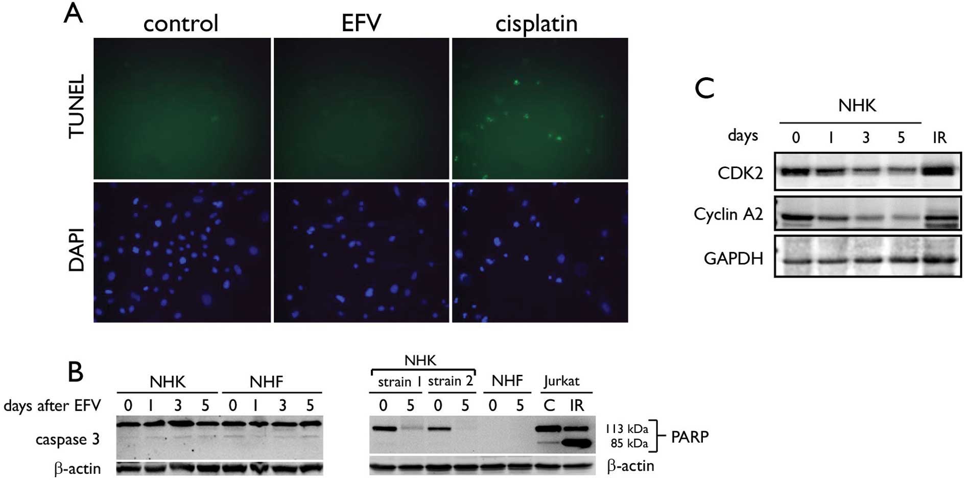
EFV-induced cell death in NHKs involves
autophagy but not apoptosis
We explored the hypothesis that apoptosis is induced
by EFV in NHKs. We performed terminal deoxynucleotidyl
transferase-mediated dUTP nick end-labeling (TUNEL) assay to check
for DNA fragmentation in NHKs treated with 10 μM EFV for 3 days. As
a comparison, we included NHKs exposed to 10 μM cisplatin, which
induces apoptosis by causing DNA damage (24). Approximately 60% of the culture
showed positive TUNEL staining after cisplatin treatment, while
positive TUNEL staining was not detected in the control untreated
cells and those exposed to EFV (Fig.
2A). EFV treatment did not cause TUNEL-positive staining in the
NHKs. Western blotting was performed for detection of cleaved
caspase-3 and PARP, both of which are markers of apoptotic events
(25). EFV treatment did not
cause caspase-3 activation or cleavage of PARP (Fig. 2B). NHKs treated with EFV showed no
change in the level of caspase-3, but almost complete loss of
full-length PARP. However, this was not consistent with the
apoptotic response as evinced in Jurkat cells exposed to 5 Gy IR,
which showed accumulation of the cleaved PARP, reflecting
activation of caspase-3. Notably, EFV treatment led to a notable
reduction in the levels of CDK2 and cyclin A2 (Fig. 2C), which are required for entry
into mitosis (26), suggesting
potential arrest of the cell cycle in the S phase. Hence, the cell
death noted in NHKs following exposure to EFV did not involve
apoptosis.
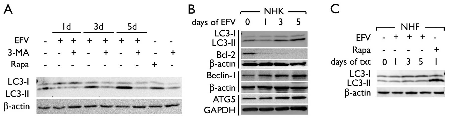
Next, we examined the occurrence of autophagy, known
as an alternative pathway of programmed cell death (27). We assessed the levels of LC3-I and
LC3-II, which represent the parent form and the cleaved form,
respectively. Following 3 days of EFV treatment, we noted an
increased level of LC3-II, while this was abolished by co-treatment
with 3-MA (Fig. 3A). As a
control, NHKs were exposed to rapamycin, which induces the level of
LC3-II. EFV treatment almost completely abolished the level of
Bcl-2 by day 1. There was a time-dependent increase in the levels
of Beclin-1 and ATG5, and this paralleled the increase in LC3-II in
the NHKs following treatment with EFV (Fig. 3B). The LC3-II level did not change
in the NHFs exposed to EFV, whereas rapamycin led to a notable
induction in LC3-II (Fig. 3C).
These data suggest that EFV induces autophagy in NHKs but not in
NHFs, and this may explain the cell type-specific cytotoxicity of
EFV.
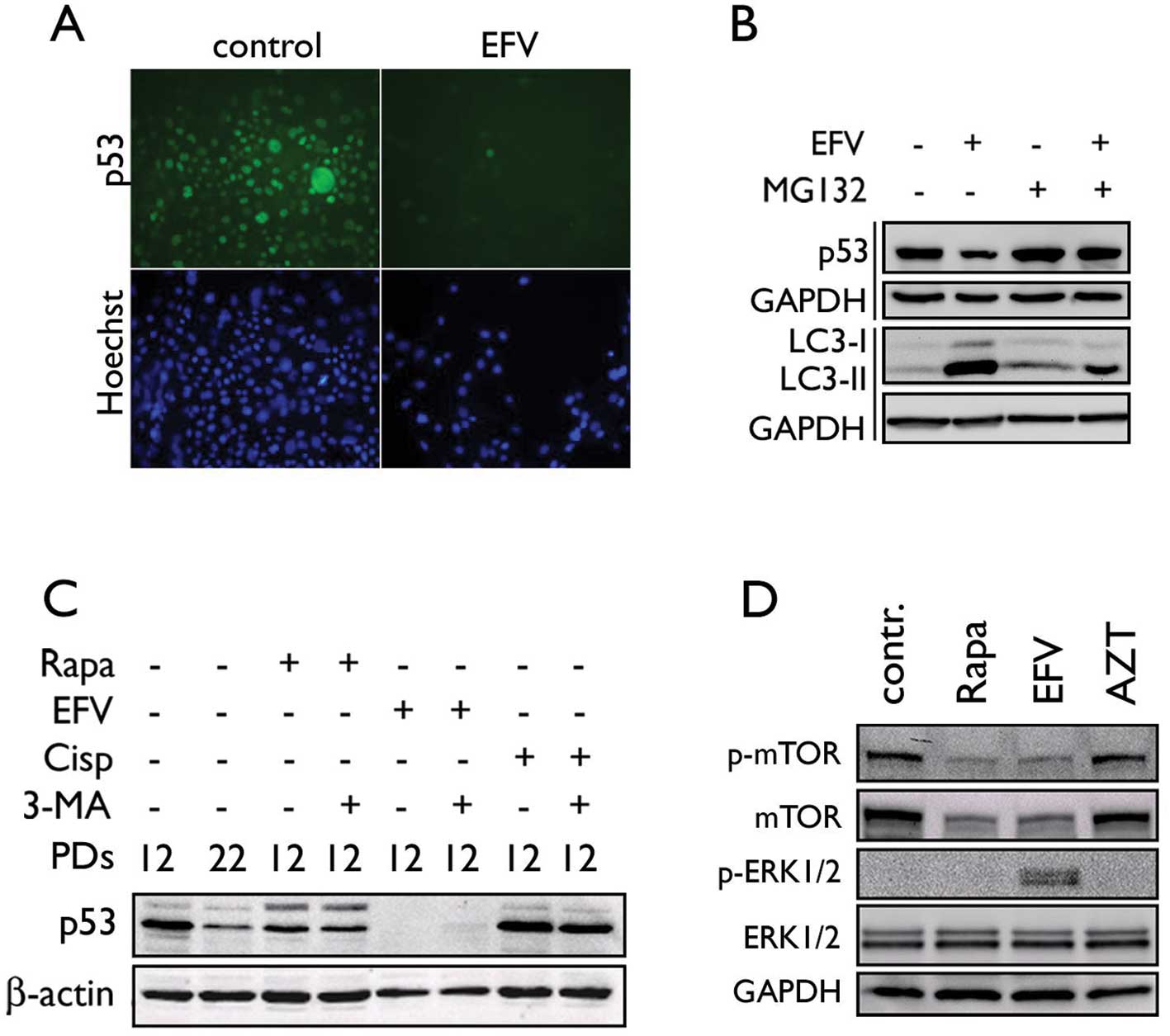
EFV stimulates proteosome-dependent
degradation of p53, loss of mTOR, and activation of ERK1/2 in
NHKs
Previous studies have demonstrated that cytoplasmic
p53 plays a causative role in autophagy (28). Western blotting showed that EFV
treatment led to a rapid reduction in p53 in NHKs (Fig. 4B). This was confirmed by
immunofluorescence staining of p53, which showed loss of nuclear
and cytoplasmic p53 in the EFV-treated cells (Fig. 4A). Since p53 undergoes
ubiquitin-dependent proteasomal degradation, we exposed the cells
to EFV in the presence of MG132, a proteasome inhibitor. EFV
treatment led to a reduction in the p53 level and a strong increase
in LC3-II accumulation, reflecting induction of autophagy (Fig. 4B). MG132 blocked the EFV-mediated
p53 degradation and notably reduced the level of LC3-II in the
cells exposed to EFV. On the contrary, co-treatment of 3-MA, which
suppressed EFV-mediated autophagy in NHKs, did not inhibit p53
degradation in the cells exposed to EFV (Fig. 4C), suggesting that p53 degradation
does not result from EFV-mediated autophagy. The EFV-treated NHKs
showed reduced phosphorylation of mTOR (Ser2448), similar to the
cells treated with rapamycin (Fig.
4D). This occurred together with a reduced protein level of
mTOR. In contrast, the azidothymidine (AZT)-treated cells exhibited
no changes in mTOR phosphorylation. EFV treatment also led to
phosphorylation of ERK1/2 (Thr202 and Tyr204) while other
treatments showed no effect. The above data suggest that
EFV-mediated autophagy in NHKs occurs with loss of p53 and mTOR,
and activation of the ERK pathway.
Autophagy induced by EFV is linked with
terminal differentiation of NHKs
Prior to cell death, the EFV-treated cells exhibited
morphological changes resembling terminal differentiation (Fig. 1D). This was confirmed by western
blotting for the markers of keratinocyte differentiation, i.e.
involucrin, filaggrin and loricrin. The levels of these markers
increased in NHKs exposed to EFV in a time-dependent manner
(Fig. 5A). After 5 days of EFV
exposure, involucrin expression was increased to a level similar to
calcium treatment (data not shown), which triggers terminal
differentiation (19).
EFV-induced keratinocyte differentiation occurred with loss of the
Notch1 protein level after exposure to EFV. We confirmed the
induction of the genes involved in keratinocyte differentiation by
qPCR. All tested genes, LCE3E, LCE3D, SPRR2A, keratin 1, loricrin,
involucrin and filaggrin, were progressively induced at the mRNA
expression level by EFV treatment (Fig. 5B). In situ immunoperoxidase
staining revealed the increased intensity of involucrin staining
and lack of cell proliferation in NHKs exposed to EFV (Fig. 5C). However, when the cells were
co-treated with EFV and 3-MA, there was a reduction in involucrin
staining and partial recovery of replicating and surviving cells.
Western blotting revealed that EFV strongly induced the involucrin
level beyond that of senescent NHKs at late-passage (PD 22), and
3-MA treatment notably reduced the involucrin protein level in the
EFV-treated cells (Fig. 5D).
Notably, rapamycin or AZT did not trigger keratinocyte
differentiation. Thus, EFV-mediated autophagy in the NHKs was
uniquely linked with aberrant keratinocyte differentiation, which
is partially responsible for the cytotoxic effects of the drug.
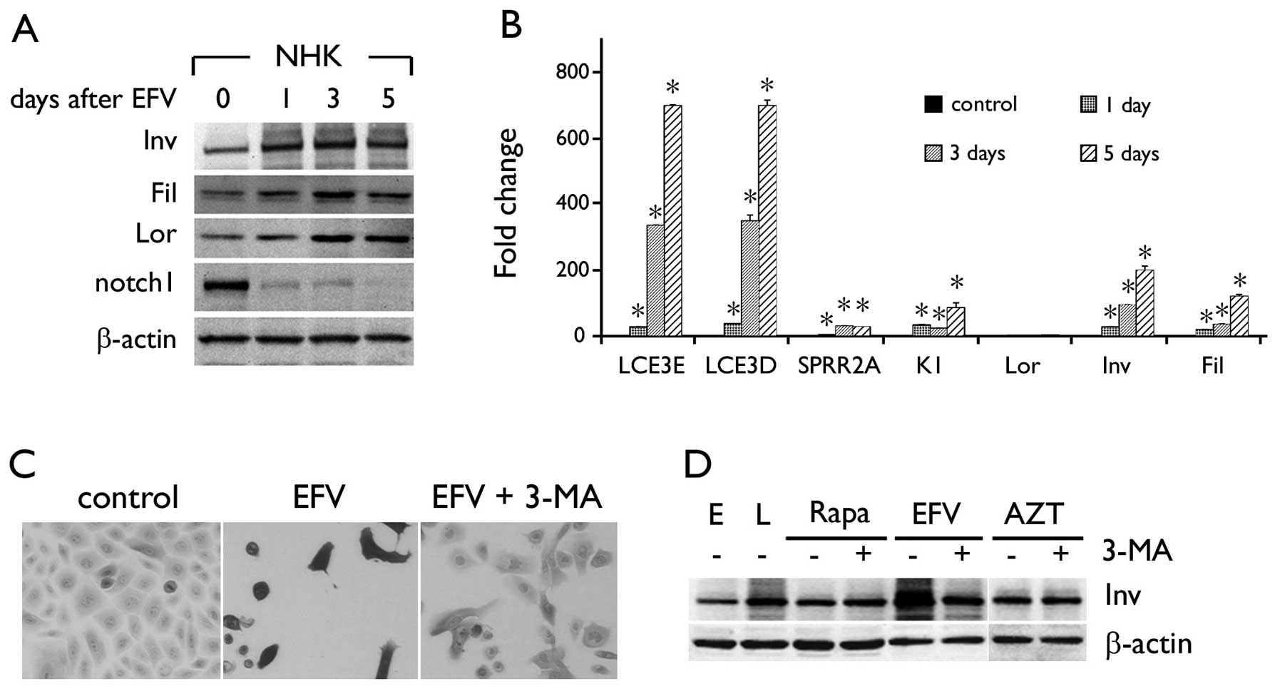 | Figure 5EFV induces terminal differentiation
in NHKs. (A) Western blotting was performed with EFV-treated NHKs
to detect various proteins as indicated. (B) mRNA expression levels
for differentiation-associated genes were determined by qPCR. Error
bars, SD. *P<0.05 against control. (C) NHKs were
exposed to 10 μM EFV for 48 h with or without 3-MA (5 mM) and
stained for involucrin by IPS. Original magnification, ×100. (D)
Involucrin expression was determined in NHKs exposed to rapamycin
(Rapa), EFV or AZT in the absence or presence of 3-MA. E,
proliferating cells at PD 12; L, senescent cells at PD 22 were
included as controls. β-actin served as a loading control. Inv,
involucrin; Fil, filaggrin; Lor, loricrin, K1, keratin 1. |
Discussion
Exposure of NHKs to EFV led to autophagic cell death
associated with terminal differentiation. This was demonstrated by
rapid loss of cell proliferation and viability that accompanied
induction of LC3-II and the markers of keratinocyte terminal
differentiation in cells exposed to EFV. Organotypic culture study
showed that EFV treatment eliminates the undifferentiated and
proliferating cells in the epithelium. This leads to lack of
epithelial regeneration and causes atrophy. Therefore, aberrant
differentiation of keratinocytes caused by EFV would, at least in
part, be responsible for cutaneous adverse effects noted in
patients. These phenotypic responses occurred uniquely in NHKs but
not in other cell types, i.e. fibroblasts, intestinal cells or
leukocytes. The 3D tissue reconstruction model revealed epithelial
atrophy and the lack of basal cell proliferation and tissue
regeneration upon exposure to EFV, while NHFs in the dermal
equivalent layer remained unchanged. This cell type-specificity was
paralleled by induction of autophagy in NHKs but not in NHFs,
indicating that the cytotoxicity of EFV is linked with autophagy.
On the contrary, induction of apoptosis was not noted in
EFV-treated cells. We also tested several other genotoxic agents,
such as actinomycin D, dexamethasone, etoposide and
methyl-nitro-nitrosoguanine (data not shown). Apoptotic signaling
from these strong genotoxic agents was extremely weak or absent in
NHKs. Likewise, we recently reported that IR failed to trigger
apoptosis in NHKs, while it strongly induced apoptosis in Jurkat
cells (29). Thus, apoptosis was
poorly induced in these cells.
EFV treatment increased the protein level of
Beclin-1, ATG5 and p27KIP1, and drastically reduced p53
and Bcl-2. The increased p27KIP1 level may have resulted
from autophagy through the AMP-activated protein kinase pathway
(30). Loss of Bcl-2 may allow
Beclin-1 binding to hVps34, a class III PI3K (31), thereby promoting autophagy in the
cells treated with EFV. EFV also led to marked reduction in cyclin
A2, which is required for mitotic entry (26), suggesting possible S phase arrest
in the treated cells. Loss of cyclin A2 may have contributed to the
growth inhibitory effects of EFV on NHKs. Autophagy occurs along
with proteosome-dependent degradation of p53; specific inhibition
of p53 is sufficient to trigger autophagic cell death (28). p53 degradation in the EFV-treated
NHKs occurred quite rapidly in a proteosome-dependent manner, and
blockage of the p53 degradation notably reduced the autophagic
response to EFV. p53 degradation did not occur in NHFs, which
showed no phenotypic changes to EFV (data not shown). We previously
showed that the levels of PCNA, involucrin, p16INK4A and
p53 do not change in exponentially replicating cells until the
cells approach replicative senescence (32,33). In the present study, the
inhibitory effect of EFV was noted primarily on p53, while that of
p16INK4A was not evident (Fig. 1C). In addition, we used rapidly
proliferating cells for the experiments. Thus, the possibility that
the loss of p53 occurred through replicative senescence during EFV
exposure is very remote. These data suggest that p53 degradation in
NHKs may have a causative role in autophagy in response to EFV.
Autophagy is often linked with a variety of other
cellular processes, such as senescence, ER stress, or
differentiation. A recent study showed that autophagy mediates the
mitotic arrest during OIS and demonstrated the interdependence
between the two processes (14).
Our data showed that autophagy was linked to terminal
differentiation in NHKs as a mechanism of cell death. The
EFV-treated NHKs demonstrated elongated morphology in culture and
strongly expressed the markers of keratinocyte differentiation.
These phenotypic changes including cell death were partially
inhibited by 3-MA, an inhibitor of the PI3K pathway (18). Rapamycin also induced autophagy in
NHKs but did not trigger differentiation, suggesting that the
mechanisms causing autophagy were different between rapamycin and
EFV. Keratinocyte differentiation is regulated by the notch
signaling pathway (34). However,
EFV-induced differentiation occurred without notch induction.
Rather, the notch protein level rapidly decreased in cells exposed
to EFV, presumably due to p53 degradation since p53 is required for
notch expression (34).
Although autophagy is a method of programmed cell
death and can function as a tumor-suppressive mechanism, it may
also lead to cell survival under stressful conditions. Beclin-1 was
found to be deleted in large portions (50–75%) of various types of
human cancers, including breast and ovarian (35,36). Introduction of beclin-1 into a
cancer cell line led to autophagy and loss of cell proliferation
and in vivo tumorigenicity (37). A subsequent study showed that
monoallelic deletion of beclin-1 in a mouse model led to increased
tumorigenesis associated with reduced autophagy (38). These studies suggest the tumor
suppressive effects of autophagy. In contrast, autophagy does play
a role in maintaining the viability of highly proliferative cancer
cells, particularly in the center of the tumor mass where the cells
are under severe metabolic stress. A study by Degenhardt et
al(39) demonstrated
induction of autophagy in regions of metabolic stress to mitigate
the ischemic cell death. Furthermore, autophagy was found to be
induced in leukemic cells undergoing anti-cancer therapy with a
histone deacetylase inhibitor, such as suberoylanilide hydroxamic
acid (SAHA), as a protective and survival mechanism (40). Chemical and genetic disruption of
autophagy led to enhanced anticancer efficacy of SAHA in the
present study. Therefore, autophagy may indeed be a mechanism by
which cancer cells gain resistance to chemotherapeutic agents or
protect cancer cells from metabolic stress.
In HIV+ patients undergoing long-term
therapeutic exposure to EFV, the occurrence of autophagy and its
contribution to tumorigenesis need to be investigated. The mean
plasma concentration of EFV in HIV+ patients under the
antiretroviral regimen was found to be 8.7 μM for those who
responded to therapy (41).
Another study showed the mean plasma concentration to be 6.9 μM
with a wide range from 0.4 to 48 μM, and suggested an effective
therapeutic plasma concentration of EFV at 3–13 μM (42). Thus, 10 μM EFV used in the present
study was within the clinically relevant concentration at which EFV
exhibits an antiviral effect. It is possible that EFV-induced
autophagy has contrasting effects on cells at different
concentrations. As shown in Fig.
1D, EFV caused cell death at 10 μM but allowed enhanced cell
proliferation at 1 and 5 μM, extending the replicative lifespan of
cells. We also found that chronic exposure of immortalized oral
keratinocytes harboring the human papillomavirus (HPV) genome to 5
μM EFV interfered with terminal differentiation and led to a
transformed phenotype (data not shown). Although EFV triggered
terminal differentiation and autophagic cell death in NHKs at 10
μM, it may have a tumor-promoting effect at a lower concentration
by protecting aberrant cells from metabolic stress and suppressing
the cell death pathway. These possibilities warrant further
investigation.
Acknowledgements
This study was supported in part by
the grants (DE18295 and DE18959) from the National Institute of
Dental and Craniofacial Research (NIDCR) and the Jack Weichman
Endowed Fund. We thank NIH AIDS Research and the Reference Reagents
Program for providing efavirenz.
References
|
1
|
Kalkut G: Antiretroviral therapy: an
update for the non-AIDS specialist. Curr Opin Oncol. 17:479–484.
2005. View Article : Google Scholar : PubMed/NCBI
|
|
2
|
Borrás-Blasco J, Navarro-Ruiz A, Borrás C
and Casterá E: Adverse cutaneous reactions associated with the
newest antiretroviral drugs in patients with human immunodeficiency
virus infection. J Antimicrob Chemother. 62:879–888.
2008.PubMed/NCBI
|
|
3
|
Borrás-Blasco J, Belda A, Rosique-Robles
JD, Casterá MD and Abad FJ: Burning mouth syndrome due to efavirenz
therapy. Ann Pharmacother. 40:1471–1472. 2006.PubMed/NCBI
|
|
4
|
Casariego Z and Ben G: Oral manifestations
of HIV infection in Argentina: a study of 1889 cases. Med Oral.
3:271–276. 1998.
|
|
5
|
Porche DJ: Efavirenz. J Assoc Nurses AIDS
Care. 11:95–98. 2000. View Article : Google Scholar
|
|
6
|
Dona C, Soriano V, Barreiro P,
Jiménez-Náchera I and González-Lahoz J: Toxicity associated to
efavirenz in HIV-infected persons enrolled in an expanded access
program. Med Clin. 115:337–338. 2000.PubMed/NCBI
|
|
7
|
Yoshimoto E, Konishi M, Takahashi K,
Murakawa K, Maeda K, Mikasa K and Yamashina Y: The first case of
efavirenz-induced photosensitivity in a Japanese patient with HIV
infection. Intern Med. 43:630–631. 2004. View Article : Google Scholar : PubMed/NCBI
|
|
8
|
Behrens GM, Stoll M and Schmidt RE:
Pulmonary hypersensitivity reaction induced by efavirenz. Lancet.
357:1503–1504. 2001. View Article : Google Scholar : PubMed/NCBI
|
|
9
|
Angel-Moreno-Maroto A, Suárez-Castellano
L, Hernández-Cabrera M and Pérez-Arellano JL: Severe
efavirenz-induced hypersensitivity syndrome (not-DRESS) with acute
renal failure. J Infect. 52:e39–e40. 2006. View Article : Google Scholar
|
|
10
|
Punwani K, Suedkamp S, Nguyen D and Song
JC: Update on the CNS adverse effects of Sustiva (efavirenz). AIDS
Alert. 22:32–34. 2007.PubMed/NCBI
|
|
11
|
Brück S, Witte S, Brust J, Schuster D,
Mosthaf F, Procaccianti M, Rump JA, Klinker H, Petzold D and
Hartmann M: Hepatotoxicity in patients prescribed efavirenz or
nevirapine. Eur J Med Res. 13:343–348. 2008.
|
|
12
|
Bertrand-Vallery V, Boilan E, Ninane N,
Demazy C, Friguet B, Toussaint O, Poumay Y and Debacq-Chainiaux F:
Repeated exposures to UVB induce differentiation rather than
senescence of human keratinocytes lacking p16(INK-4A).
Biogerontology. 11:167–181. 2010. View Article : Google Scholar : PubMed/NCBI
|
|
13
|
Agger K, Cloos PA, Rudkjaer L, Williams K,
Andersen G, Christensen J and Helin K: The H3K27me3 demethylase
JMJD3 contributes to the activation of the INK4A-ARF locus in
response to oncogene- and stress-induced senescence. Genes Dev.
23:1171–1176. 2009. View Article : Google Scholar : PubMed/NCBI
|
|
14
|
Young AR and Narita M, Ferreira M,
Kirschner K, Sadaie M, Darot JF, Tavaré S, Arakawa S, Shimizu S,
Watt FM and Narita M: Autophagy mediates the mitotic senescence
transition. Genes Dev. 23:798–803. 2009. View Article : Google Scholar : PubMed/NCBI
|
|
15
|
Klionsky DJ: Autophagy: from phenomenology
to molecular understanding in less than a decade. Nat Rev Mol Cell
Biol. 8:931–937. 2007. View
Article : Google Scholar : PubMed/NCBI
|
|
16
|
Liang C and Jung JU: Autophagy genes as
tumor suppressors. Curr Opin Cell Biol. 22:226–233. 2010.
View Article : Google Scholar : PubMed/NCBI
|
|
17
|
Kabeya Y, Mizushima N, Ueno T, Yamamoto A,
Kirisako T, Noda T, Kominami E, Ohsumi Y and Yoshimori T: LC3, a
mammalian homologue of yeast Apg8p, is localized in autophagosome
membranes after processing. EMBO J. 19:5720–5728. 2000. View Article : Google Scholar : PubMed/NCBI
|
|
18
|
Wu YT, Tan HL, Shui G, Bauvy C, Huang Q,
Wenk MR, Ong CN, Codogno P and Shen HM: Dual role of
3-methyladenine in modulation of autophagy via different temporal
patterns of inhibition on class I and III phosphoinositide
3-kinase. J Biol Chem. 285:10850–10861. 2010. View Article : Google Scholar : PubMed/NCBI
|
|
19
|
Kang MK, Bibb C, Baluda MA, Rey O and Park
NH: In vitro replication and differentiation of normal human oral
keratinocytes. Exp Cell Res. 258:288–297. 2000. View Article : Google Scholar : PubMed/NCBI
|
|
20
|
Boukamp P, Petrussevska RT, Breitkreutz D,
Hornung J, Markham A and Fusenig NE: Normal keratinization in a
spontaneously immortalized aneuploid human keratinocyte cell line.
J Cell Biol. 106:761–771. 1988. View Article : Google Scholar : PubMed/NCBI
|
|
21
|
Dickson MA, Hahn WC, Ino Y, Ronfard V, Wu
JY, Weinberg RA, Louis DN, Li FP and Rheinwald JG: Human
keratinocytes that express hTERT and also bypass a
p16(INK4a)-enforced mechanism that limits life span become immortal
yet retain normal growth and differentiation characteristics. Mol
Cell Biol. 20:1436–1447. 2000. View Article : Google Scholar
|
|
22
|
Dongari-Bagtzoglou A and Kashleva H:
Development of a highly reproducible three-dimensional organotypic
model of the oral mucosa. Nat Protoc. 1:2012–2018. 2006. View Article : Google Scholar : PubMed/NCBI
|
|
23
|
Kang MK, Kim RH, Kim SJ, Yip FK, Shin KH,
Dimri GP, Christensen R, Han T and Park NH: Elevated Bmi-1
expression is associated with dysplastic cell transformation during
oral carcinogenesis and is required for cancer cell replication and
survival. Br J Cancer. 96:126–133. 2007. View Article : Google Scholar : PubMed/NCBI
|
|
24
|
Kim Y, McBride J, Zhang R, Zhou X and Wong
DT: p12 (CDK2-AP1) mediates DNA damage responses induced by
cisplatin. Oncogene. 24:407–418. 2005. View Article : Google Scholar : PubMed/NCBI
|
|
25
|
Kurokawa M and Kornbluth S: Caspases and
kinases in a death grip. Cell. 138:838–854. 2009. View Article : Google Scholar : PubMed/NCBI
|
|
26
|
Gong D and Ferrell J Jr: The roles of
cyclin A2, B1, and B2 in early and late mitotic events. Mol Biol
Cell. 21:3149–3161. 2010. View Article : Google Scholar : PubMed/NCBI
|
|
27
|
Bursch W, Ellinger A, Gerner C, Fröhwein U
and Schulte-Hermann R: Programmed cell death (PCD). Apoptosis,
autophagic PCD, or others? Ann NY Acad Sci. 926:1–12. 2000.
View Article : Google Scholar : PubMed/NCBI
|
|
28
|
Tasdemir E, Maiuri MC, Galluzzi L, Vitale
I, Djavaheri-Mergny M, D'Amelio M, Criollo A, Morselli E, Zhu C,
Harper F, Nannmark U, Samara C, Pinton P, Vicencio JM, Carnuccio R,
Moll UM, Madeo F, Paterlini-Brechot P, Rizzuto R, Szabadkai G,
Pierron G, Blomgren K, Tavernarakis N, Codogno P, Cecconi F and
Kroemer G: Regulation of autophagy by cytoplasmic p53. Nat Cell
Biol. 10:676–687. 2008. View
Article : Google Scholar : PubMed/NCBI
|
|
29
|
Dong Q, Oh JE, Chen W, Kim R, Kim RH, Shin
KH, McBride WH, Park NH and Kang MK: Radioprotective effects of
Bmi-1 involve epigenetic silencing of oxidase genes and enhanced
DNA repair in normal human keratinocytes. J Invest Dermatol.
131:1216–1225. 2011. View Article : Google Scholar : PubMed/NCBI
|
|
30
|
Liang J, Shao SH, Xu ZX, Hennessy B, Ding
Z, Larrea M, Kondo S, Dumont DJ, Gutterman JU, Walker CL,
Slingerland JM and Mills GB: The energy sensing LKB1-AMPK pathway
regulates p27(kip1) phosphorylation mediating the decision to enter
autophagy or apoptosis. Nat Cell Biol. 9:218–224. 2007. View Article : Google Scholar : PubMed/NCBI
|
|
31
|
Pattingre S, Tassa A, Qu X, Garuti R,
Liang XH, Liang XH, Mizushima N, Packer M, Schneider MD and Levine
B: Bcl-2 antiapoptotic proteins inhibit Beclin 1-dependent
autophagy. Cell. 122:927–939. 2005. View Article : Google Scholar : PubMed/NCBI
|
|
32
|
Kang MK, Guo W and Park NH: Replicative
senescence of normal human oral keratinocytes is associated with
the loss of telomerase activity without shortening of telomeres.
Cell Growth Differ. 9:85–95. 1998.PubMed/NCBI
|
|
33
|
Kang MK, Kameta A, Shin KH, Baluda MA and
Park NH: Senescence occurs with hTERT repression and limited
telomere shortening in human oral keratinocytes cultured with
feeder cells. J Cell Physiol. 199:364–370. 2004. View Article : Google Scholar : PubMed/NCBI
|
|
34
|
Yugawa T, Narisawa-Saito M, Yoshimatsu Y,
Haga K, Ohno S, Egawa N, Fujita M and Kiyono T: DeltaNp63alpha
repression of the Notch1 gene supports the proliferative capacity
of normal human keratinocytes and cervical cancer cells. Cancer
Res. 70:4034–4044. 2010. View Article : Google Scholar : PubMed/NCBI
|
|
35
|
Futreal PA, Söderkvist P, Marks JR,
Iglehart JD, Cochran C, Barrett JC and Wiseman RW: Detection of
frequent allelic loss on proximal chromosome 17q in sporadic breast
carcinoma using microsatellite length polymorphisms. Cancer Res.
52:2624–2627. 1992.
|
|
36
|
Cliby W, Ritland S, Hartmann L, Dodson M,
Halling KC, Keeney G, Podratz KC and Jenkins RB: Human epithelial
ovarian cancer allelotype. Cancer Res. 53(Suppl 10): S2393–S2398.
1993.
|
|
37
|
Liang XH, Jackson S, Seaman M, Brown K,
Kempkes B, Hibshoosh H and Levine B: Induction of autophagy and
inhibition of tumorigenesis by beclin 1. Nature. 402:672–676. 1999.
View Article : Google Scholar : PubMed/NCBI
|
|
38
|
Qu X, Yu J, Bhagat G, Furuya N, Hibshoosh
H, Troxel A, Rosen J, Eskelinen EL, Mizushima N, Ohsumi Y,
Cattoretti G and Levine B: Promotion of tumorigenesis by
heterozygous disruption of the beclin 1 autophagy gene. J Clin
Invest. 112:1809–1820. 2003. View
Article : Google Scholar : PubMed/NCBI
|
|
39
|
Degenhardt K, Mathew R, Beaudoin B, Bray
K, Anderson D, Chen G, Mukherjee C, Shi Y, Gélinas C, Fan Y, Nelson
DA, Jin S and White E: Autophagy promotes tumor cell survival and
restricts necrosis, inflammation, and tumorigenesis. Cancer Cell.
10:51–64. 2006. View Article : Google Scholar : PubMed/NCBI
|
|
40
|
Carew JS, Nawrocki ST, Kahue CN, Zhang H,
Yang C, Chung L, Houghton JA, Huang P, Giles FJ and Cleveland JL:
Targeting autophagy augments the anticancer activity of the histone
deacetylase inhibitor SAHA to overcome Bcr-Abl-mediated drug
resistance. Blood. 110:313–322. 2007. View Article : Google Scholar : PubMed/NCBI
|
|
41
|
Ståhle L, Moberg L, Svensson JO and
Sönnerborg A: Efavirenz plasma concentrations in HIV-infected
patients: inter- and intraindividual variability and clinical
effects. Ther Drug Monit. 26:267–270. 2004.PubMed/NCBI
|
|
42
|
Marzolini C, Telenti A, Decosterd LA,
Greub G, Biollaz J and Buclin T: Efavirenz plasma levels can
predict treatment failure and central nervous sytem side effects in
HIV-1-infected patients. AIDS. 15:71–75. 2001. View Article : Google Scholar : PubMed/NCBI
|