Introduction
Wound healing comprises several phases, such as
inflammation, granulation, re-epithelization and tissue remodeling
(1). The wound healing processes,
include cell migration, simultaneous stimulation of new blood
vessel formation, the proliferation of cells, such as keratinocytes
and fibroblasts, and the production of basement membrane zones and
connective tissues. A number of different cell types are recruited
to the wounded skin areas during the healing process (2). After an injury to the skin has
occurred, inflammatory cells are attracted to the site of infection
to remove the damaged tissues, and then fibroblasts and
keratinocytes migrate to the wound lesion and proliferate in order
to generate new tissue. Fibroblasts, the most common connective
tissue cells, are the predominant cell type in the dermis and play
a critical role in wound healing. Fibroblasts produce
collagen-based extracellular matrix (ECM) and help reapproximate
wound edges through their contractile properties. The remodeling
phase, which is a period of consolidation and strengthening,
promotes a change in the composition of the ECM of fibers and cells
(3).
A variety of herbs and plants have been
traditionally used for the treatment of various diseases and wounds
in folk medicine in many developing countries (4–6).
The choice of herbal products for the treatment of wounds and other
ailments depends on the tradition and the plant species grown in
different regions of the world. In recent years, many efforts have
been made worldwide to discover agents from natural resources that
can promote wound healing, reduce the cost of treatment, and save
patients from other severe complications (7–9).
Stewartia koreana [S. koreana; (Theaceae)], a
deciduous tree, is native to Korea, and its tree leaves have been
used as a traditional oriental medicine for the prevention and
treatment of various diseases. Previous studies have demonstrated
that extracts of S. koreana exhibit various biological
activities, such as anti-inflammatory activity, the inhibition of
osteoclast differentiation and angiogenic activity (10–12).
In the present study, we analyzed the effects of
S. koreana extract (SKE) and its isolated compounds on the
proliferation and migration of normal human dermal fibroblasts
(NHDFs) and evaluated its wound healing activity in mice. We found
that SKE stimulated the proliferation and migration of fibroblast
cells, and, among the isolated compounds, hyperin was the most
effective in stimulating the proliferation and migration of NHDFs.
Moreover, we demonstrated that SKE stimulated wound healing in the
skin of animals. Our results suggest that SKE is very effective in
wound healing, and that hyperin may contribute to the strong wound
healing activity of SKE by stimulating the proliferation and
migration of normal dermal fibroblasts.
Materials and methods
Materials and cell culture
Methanol extracts of S. koreana were prepared
as previously described (10).
Briefly, S. koreana leaves were dried and refluxed twice
with 80% methanol. The extract was then filtered and centrifuged at
800 × g for 10 min. Methanol in the extract was removed under
reduced pressure by rotary evaporation and the water residue was
removed by lyophilization. The percentage yield was calculated
based on the weight of the dried plant material. The dried powder
was dissolved in aliquots of DMSO and diluted to the desired
concentrations.
NHDFs were cultured in FGM-2 (Clonetics, Lonza,
Walkersville, MD, USA) containing 2% fetal bovine serum
(Invitrogen, Carlsbad, CA, USA), 0.1% recombinant human fibroblast
growth factor (rhFGF), 0.1% insulin (Clonetics), 5 U/ml of heparin,
100 U/ml of penicillin and 100 μg/ml of streptomycin (Invitrogen)
in a humidified incubator at 37°C with 5% CO2.
Epidermal growth factor (EGF) was obtained from
R&D Systems (Minneapolis, MN, USA) and used as the positive
control in each experiment. LY294002 (PI3K inhibitor) and PD98059
(MEK inhibitor) were obtained from R&D Systems. Other chemicals
were obtained commercially from Sigma-Aldrich (St. Louis, MO,
USA).
Isolation and analysis of hyperin in
ethyl acetate (EtOAc) extract by high performance liquid
chromatography (HPLC)
The air-dried leaves of S. koreana were
extracted and fractionated by successive partitioning as previously
described (13). Hyperin was
obtained from fractions 15 and 16 by further column chromatography
of the pooled fraction. For HPLC, the samples were filtered through
a 0.45-μm microspin PVDF filter. The HPLC equipment used was as
follows: an Agilent 1200 series system (Agilent Technology Inc.,
Santa Clara, CA, USA); G1315 DAD detector, G1329A auto sampler,
G1322A degasser, G1311 Quat pump; stationary phase Agilent TC-C18
(5 μm, 4.6×250 mm); λ=356 nm; binary gradient (A, water; B,
CH3CN), t0min 50% A → linear gradient to 15%
A within 25 min → linear gradient to 5% A/95% B within 14 min →
linear gradient to 0% A within 20 min; flow rate 1.0 ml/min;
injection volume 1 μl.
Animal experiments
Animal experiments were approved by the
Institutional Review Board (IRB), Kyung Hee University, Seoul,
Korea. Healthy BALB/c mice (8 weeks, 25–30 × g) were purchased from
Central Laboratory Animal Inc. (Seoul, Korea) and divided randomly
into 2 groups (control and SKE-treated group) of 6 mice in each.
Each mouse was placed in separate wire-bottom cages in an
atmosphere-controlled room (12 h light/dark cycle; temperature,
22±3°C; and 50±10% relative humidity) and maintained on a standard
laboratory diet and provided with water.
Wound generation
Mice were anesthetized with 65% N2, 30%
O2 and 5% isoflurane. Anesthesia was maintained with 2%
isoflurane during surgery as previously described (14). Dorsal hair was shaved and wiped
with ethanol (70%) prior to generating the wounds. Two horizontal
circular wounds (6 mm in diameter) were created on the upper back
of each mouse using a biopsy punch instrument. Each wound was
topically treated either with EGF (R&D Systems) 200 ng/mouse,
PBS or SKE (200 μg/mouse) at the same time each day for 8 days.
Images of the wounds were acquired using a digital camera every day
immediately before treatment. A small piece of plotting paper (5×5
mm) was used as a standard for the accurate normalization of the
wound sizes recorded in the digital images. Wound contraction was
calculated using the percentage of wound size as follows: wound
area day × was divided by wound area day 0, which was multiplied by
100 (where × = 1, 2, 3, ,,,, n, the day after injury).
3-(4,5-Dimethylthiazol-2-yl)-2,5-diphenyltetrazolium bromide (MTT)
assay
NHDFs seeded in 96-well plates at a density of
5×10³ cells/well and allowed to attach for 24 h. After
discarding the growth medium, the fibroblasts were treated with SKE
or various concentrations of phytochemicals isolated from SKE in
serum-free medium for 48 h. EGF (R&D systems) was used as a
positive control. Following incubation, the cells were treated with
100 μg/ml of MTT for 1 h. The formazan precipitate was dissolved in
200 μl of DMSO and the absorbance at 560 nm was determined
spectrophotometrically. The analyses were repeated 3 times and the
results are expressed as the means of 3 independent
experiments.
BrdU incorporation assay
The 5-bromo-2′-deoxy-uridine (BrdU) incorporation
assay was performed using BrdU labeling and a detection kit (Roche,
Indianapolis, IN, USA) according to the manufacturer’s
instructions. In brief, the fibroblasts were seeded in 96-well
plates for 24 h. After the removal of the medium, the cells were
starved for 6 h and then serum-free medium containing various
concentrations of SKE or EGF was added for 24 h. BrdU-integrated
DNA was quantified according to the relative luminescence unit
(RLU) for each well using a Wallace Victor2 1420
multi-label counter (Perkin-Elmer, Monza, Italy). The analyses were
repeated 3 times and the results are expressed as the means of 3
independent experiments.
Scratch wound closure assay
The confluent NHDF monolayers in 6-well plates (SPL
Inc. Seoul, Korea) were scratched and wounded using a universal
sterile 200-μl pipette tip and then rinsed with PBS. Each well was
treated with various concentrations of medium alone (vehicle), SKE
or hyperin for 6 h. EGF was added as a positive control. The cells
were stained with Diff-Quick (Baxter Healthcare Corp., McGraw Park,
IL, USA) and the width of the wound was measured using an Olympus
digital camera (Olympus, Tokyo, Japan). The area of the wound was
analyzed using the double-blind direct cell counting method. Each
condition was studied in triplicate wells and each experiment was
performed 3 times.
Western blot analysis
The cell extracts were prepared from NHDFs treated
with various concentrations of SKE or EGF for 15 min. The cells
were lysed in RIPA buffer containing proteinase inhibitors (Roche,
Indianapolis, IN, USA) and fractionated by electrophoresis on 12%
SDS-PAGE gels and transferred onto a nitrocellulose membrane.
Non-specific binding was blocked by soaking the membrane in
Tris-buffered saline-Tween-20 (TBS-T) buffer [0.5 mol/l Tris-HCl
(pH 7.5), 0.15 mol/l NaCl, and 1 g/l Tween-20] containing 3–5%
non-fat dry milk. Phosphorylated (p)-ERK1/2, ERK1/2, p-MEK, MEK,
p-PI3K, PI3K, p-Akt and Akt (Cell Signaling, Denvers, MA, USA) were
utilized as primary antibodies and peroxidase-conjugated antibody
was used as a secondary antibody. The membranes were developed with
an enhanced chemiluminescence system from GE Healthcare
(Piscataway, NJ, USA) and exposed for 30 sec to X-ray film (Fuji
Photo Film Co. Ltd, Tokyo, Japan).
Statistical analysis
The data are presented as the means ± SD.
Statistical comparisons between groups were performed using one-way
ANOVA followed by the Student’s t-test. A p-value <0.05 was
considered to be statistically significant..
Results
Effects of SKE on the proliferation of
human dermal fibroblasts
The proliferation of skin fibroblasts is important
in tissue repair as fibroblasts are involved in migration,
proliferation, contraction and collagen production, leading to the
deposition of the ECM and the formation of granulation tissue
(1–3). Thus, we investigated the effects of
SKE on the proliferation of NHDFs at concentrations up to 100 μg/ml
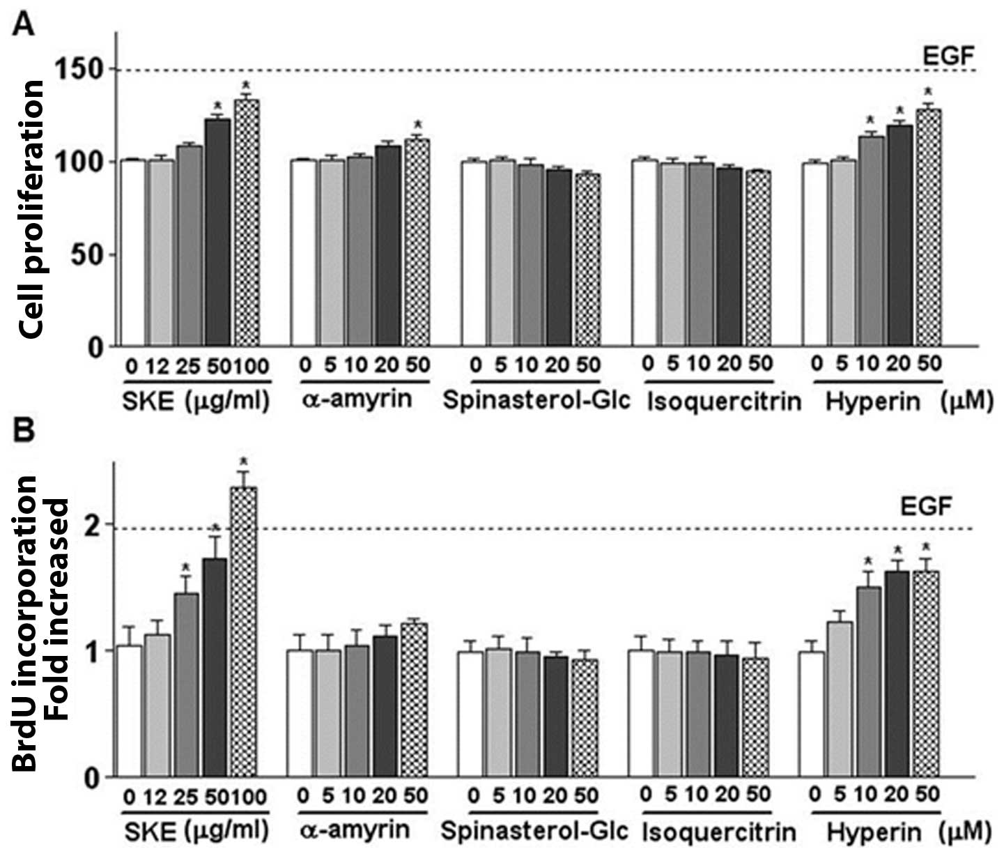
using colorimetric MTT assay. As shown in Fig. 1A, SKE significantly enhanced NHDF
growth by approximately 30% at a concentration of 100 μg/ml
(p<0.05) compared to that of the control untreated cells. In
addition, we examined the effects of SKE on DNA synthesis of NHDFs
by BrdU incorporation assay. As shown in Fig. 1B, DNA synthesis of NHDFs treated
with SKE increased by approximately 2.8-fold compared with the
untreated control cells.
Effects of phytochemicals from S. koreana
leaves on the proliferation of skin fibroblasts
Previous studies have reported on the isolation of
various phytochemicals, such as α-amyrin,
3-O-β-D-glucopyranosyl-spinasterol (spinasterol-Glc), and
isoquercitrin from methanol extracts by the successive partitioning
of the extracts with EtOAc, n-BuOH (13). Thus, in this study, we examined
the effects of isolated compounds, such as spinasterol-Glc,
α-amyrin and isoquercitrin on the proliferation of skin
fibroblasts. Our results revealed that spinasterol-Glc and
isoquercitrin did not affect the growth of NHDFs. α-amyrin appeared
to slightly stimulate the proliferation of NHDFs (Fig. 1A and B), which may not account for
the stimulatory effects of SKE. In an attempt to find a compound
that stimulates NHDF proliferation, we further fractionated the
subfraction originating from fractions 15 and 16 (13), which resulted in the separation of
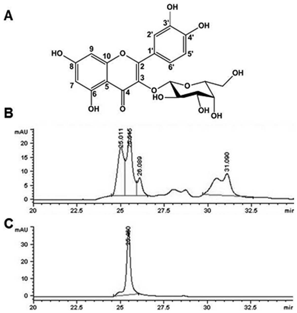
an additional compound. The structure of the compound, which is
shown in Fig. 2A, was determined
to be hyperin by nuclear magnetic resonance (NMR) spectroscopy
(data not shown). HPLC of the EtOAc subfraction revealed that
hyperin is a major compound in the fraction (Fig. 2B and C). We evaluated the effects
of hyperin on fibroblast proliferation and our results revealed
that hyperin strongly stimulated the proliferation of fibroblasts
as compared to the control cells (Fig. 1A). Hyperin increased the
proliferation of fibroblasts by at least 1.3-fold and stimulated
DNA synthesis by approximately 1.8-fold at a concentration of 50 μM
(Fig. 1A and B), which was almost
comparable to the effects of SKE.
Effects of SKE and phytochemicals from
SKE on the migration of skin fibroblasts
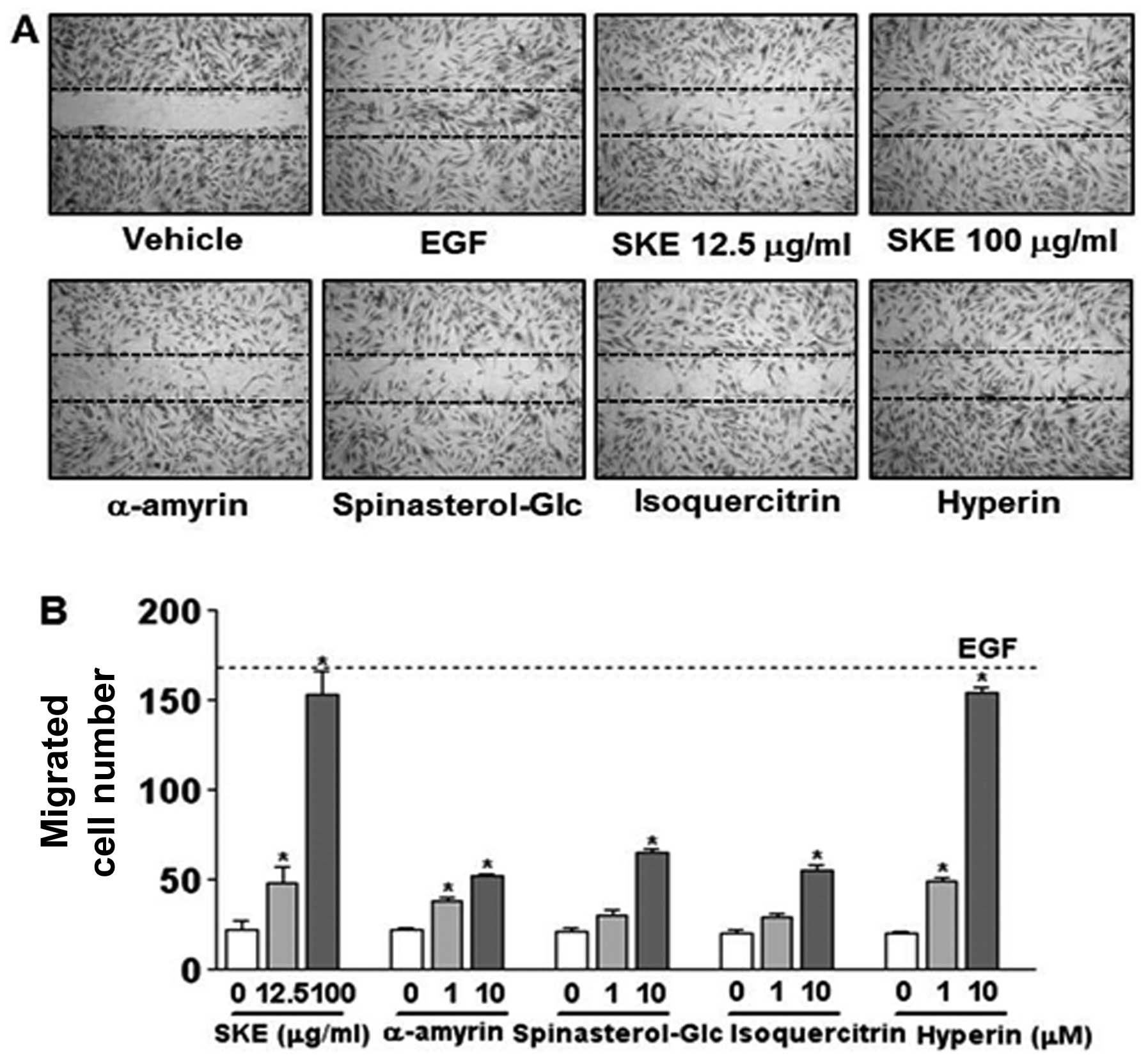
We then investigated the effects of SKE and its
phytochemicals on the migration of NHDFs and observed that SKE
enhanced the migration of human skin fibroblasts (Fig. 3A and B). When a confluent
monolayer of NHDFs was scratched, the migration of cells into the
wounded area was significantly increased in the presence of SKE
compared to the migration of cells in medium alone 6 h after
wounding. SKE at 100 μg/ml effectively increased the migration of
human fibroblasts, which was comparable to the migration induced by
EGF. It is therefore plausible that SKE may promote tissue repair
not only by increasing proliferation but also enhancing the
migration of skin fibroblasts.
We further examined the effects of its
phytochemicals on the migration of fibroblasts by a wound closure
assay. Among the compounds tested, hyperin induced the most
prominent stimulatory effect on the migration of NHDFs on the wound
areas. When a confluent monolayer was scratched, the migration of
the cells into the wounded area was markedly increased in the
presence of 10 μM of hyperin compared to the migration of cells in
medium alone 6 h after wounding. However, α-amyrin, spinasterol-Glc
and isoquercitrin showed only moderate or very weak activities in
promoting the migration of fibroblasts compared to hyperin
(Fig. 3A and B). Our results
demonstrated that hyperin markedly stimulated fibroblast migration.
After 6 h, cell migration was enhanced in response to hyperin (10
μM) by 3- to 5-fold over migration in the presence of medium alone
(Fig. 3B). Additionally, the
levels of NHDF migration induced by 10 μM of hyperin were similar
to the those observed for the positive control, EGF (10 ng/ml). Our
results revealed that hyperin stimulated the migration and DNA
synthesis in NHDFs, which promoted wound healing in the animal
wound model.
SKE induces the proliferation of
fibroblasts through the activation of the mitogen-activated protein
kinase (MAPK) and Akt signaling pathways
The activation of MAPK and Akt signaling pathways is
known to be involved in the migration and proliferation of skin
fibroblasts (15). Thus, we
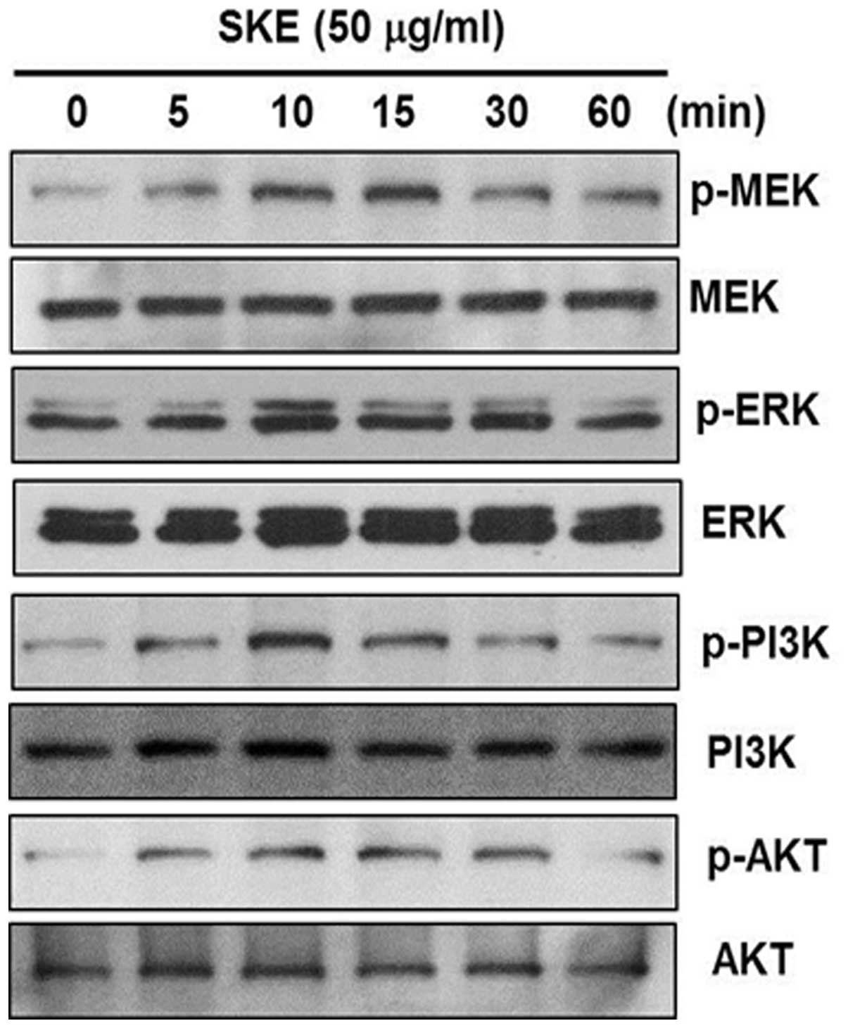
investigated the effects of SKE on the phosphoryration of PI3K/Akt
and MAPK/ERK1/2. The serum-deprived NHDFs displayed a barely
detectable phosphorylation in the absence of SKE (Fig. 4). However, when SKE was added,
ERK1/2, MEK, PI3K and Akt were phosphorylated as early as 5 min
after the addition of SKE. The phosphorylation reached a maximum
level at 10 to 15 min after exposure, and then decreased. These
results suggest that SKE enhances the proliferation and migration
of fibroblasts through the activation of the ERK1/2 and PI3K
signaling pathways.
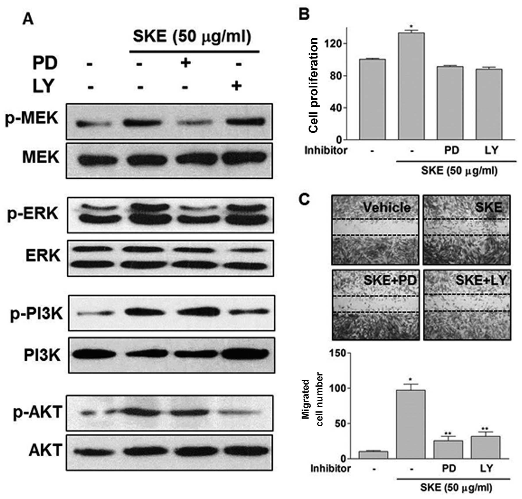
Inhibition of MEK/ERK1/2 and PI3K/Akt
decreases SKE- induced proliferation and migration of
fibroblasts
To determine the role of the MEK/ERK1/2 and PI3K/Akt
signaling cascades in SKE-induced NHDF proliferation and migration,
we analyzed the phosphorylation of signaling molecules in the
presence of specific signaling inhibitors, such as PI3K inhibitor
(LY294002) and MEK inhibitor (PD98059). The SKE-induced ERK
activation was inhibited by PD98059, but not by LY294002,
indicating that ERK phosphorylation is mediated by SKE-dependent
MEK activation. Akt activation was inhibited by LY294002, but not
PD98059, suggesting that SKE activates the PI3K/Akt pathway
(Fig. 5A). We then examined the
effects of these inhibitors on the SKE-induced fibroblast
proliferation and migration. The treatment of NHDFs with PD98059
and Ly294002 resulted in a significant decrease in the
proliferation and migration of fibroblasts (Fig. 5B and C).
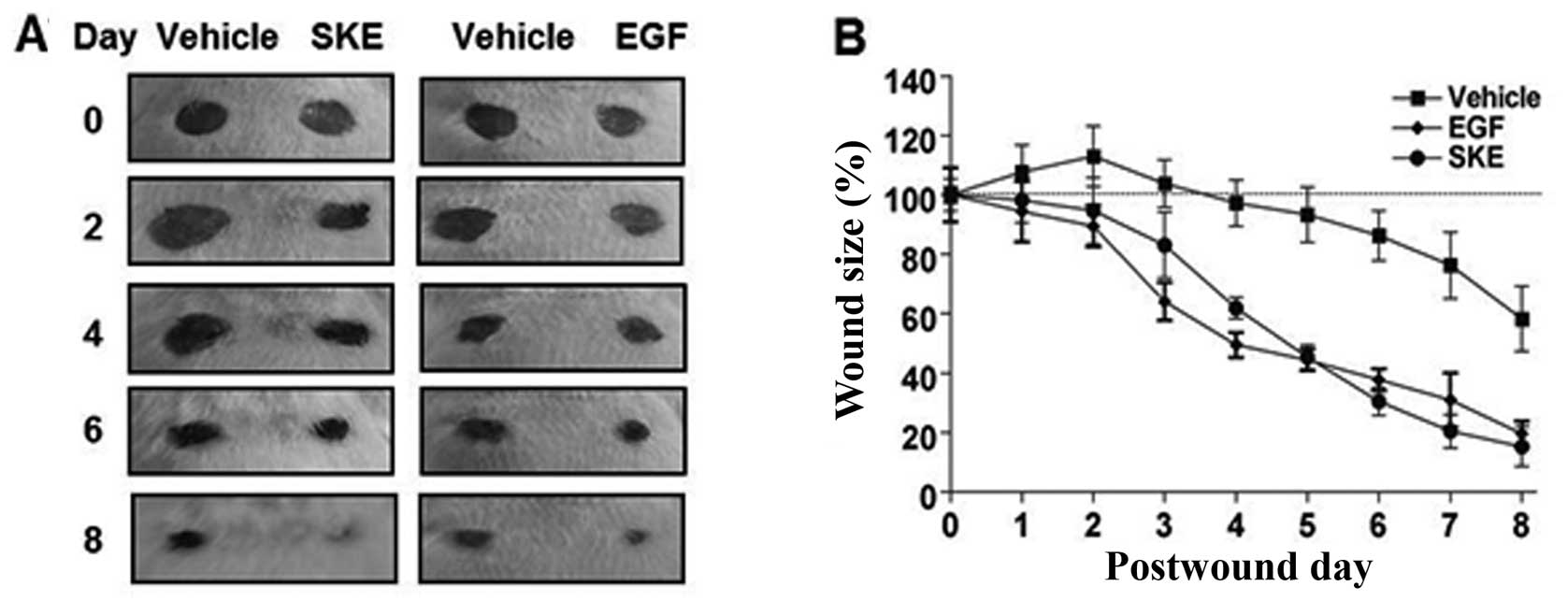
Effects of SKE on wound healing in
mice
Wounds on the skin of mice were treated with SKE,
EGF or PBS each day, and then wound repair was observed for 8 days.
The square measure of each wound was determined on the basis of the
pixel number and was then converted to the percentage of wound
size. In the SKE-treated mice, wounds healed from a mean area of
24.86±2.12 mm2 to a mean area of 4.34±0.39
mm2 (Fig. 6A and
Table I). The process of wound
healing as a function of time was then depicted as an X-Y plot
(Fig. 6B). Eight days after the
wound puncture, the wounds were reduced to approximately 83% of the
wound sizes on the first day (p<0.05) (Fig. 6B). The wound healing activity of
SKE was comparable to that of EGF at the concentrations indicated.
The wound sizes of SKE-treated skin markedly decreased compared to
the wound sizes of PBS-treated skin, indicating that SKE
accelerates wound healing in vivo.
 | Table IEffects of the topical application of
methanol extract of Stewartia koreana (SKE) on punch wounds
on the backs of mice. |
Table I
Effects of the topical application of
methanol extract of Stewartia koreana (SKE) on punch wounds
on the backs of mice.
| Mean lesion
diameter (mm2) |
|---|
|
|
|---|
| Treatment | Day 0 | Day 2 | Day 4 | Day 6 | Day 8 |
|---|
| Vehicle (PBS
alone) | 24.77±2.15 | 33.15±3.13 | 27.18±3.15 | 20.71±2.42 | 12.11±1.92 |
| SKE (200
μg/mice) | 24.86±2.12 | 22.53±3.17 | 16.25±2.19 | 10.02±1.16 | 4.34±0.39 |
| EGF (200
ng/mice) | 25.01±3.21 | 18.28±2.76 | 14.31±3.01 | 12.04±1.72 | 5.86±0.64 |
Our data demonstrated that the healing process of
the SKE-treated wounds was accelerated after 3 days, when
epithelization and provisional matrix formation begins (1,3).
The topical application of SKE enhanced cutaneous wound healing,
which appeared to be completed in 8 days. Our results showed that
the original tissue regeneration was much greater in skin wounds
treated with SKE than in the control wounds. Moreover, the healing
activity of SKE was almost comparable to that of EGF, which is
known to accelerate the wound healing process.
Discussion
A number of studies have reported that various plant
extracts contain wound healing activities (7–9,16,17). Skin cell behaviors, such as the
spreading and migration of fibroblasts, are major determinants of
the wound closure rate. In the present study, we demonstrate that
SKE stimulates the growth and migration of NHDFs. We also
demonstrate that, among the SKE isolated compounds from the
methanol extract of SKE, hyperin was the one that most strongly
stimulated the growth and migration of fibroblastss. Our results
demonstrated that the SKE-induced fibroblast proliferation and
migration were mediated by the ERK and PI3K signaling pathways.
Furthermore, we showed that the topical application of SKE enhanced
the cutaneous wound healing, which appeared to be completed in 8
days. The healing process of the SKE-treated wounds was accelerated
after 3 days, when epithelization, angiogenesis and provisional
matrix formation began (1). Our
results revealed that the original tissue regeneration was much
greater in the skin wounds treated with SKE than in the control
wounds, and that the healing activity of SKE was comparable to that
of EGF.
The stimulatory effects of SKE on fibroblast
proliferation and migration seem to be mediated by MEK/ERK, as well
as the PI3K/Akt signaling pathways. The phosphorylation of MEK/ERK
and PI3K/Akt were barely detectable under the serum-free conditions
and the phosphorylation of these molecules was increased after the
treatment of fibroblasts with SKE. The inhibition of the ERK
pathway by PD98059 and of the PI3K pathway by LY294002 markedly
reduced fibroblast proliferation and migration, indicating that the
ERK and PI3K pathways are involved in the SKE-induced cell growth
and migration. Our results are consistent with those of previous
studies, suggesting that the phosphorylation of Akt and ERK1/2
signaling pathways plays an important role in the promotion of
proliferation and migration of fibroblasts (15,18,19). The major function of the ERK and
PI3K signaling pathways is to stimulate gene transcription involved
in proliferation and migration of cells, including fibroblasts
(18,19).
We previously isolated a number of compounds from
SKE, including α-amyrin, isoquercitrin and spinasterol-Glc.
α-amyrin belongs to the triterpenoid family (13), which is known to promote ECM
accumulation and have an anti-inflammatory activity (20–22). α-amyrin is one of the most
abundant substances in SKE and is presumed to contribute to the
wound healing activity of SKE by suppressing inflammation in the
wounded areas and enhancing the production of ECM; however, in this
study, it did not promote the proliferation and migration of
fibroblasts. Spinasterol-Glc has been shown to strongly inhibit the
production of nitric oxide (NO) and proinflammatory cytokines in
lipopolysaccharide (LPS)-stimulated macrophages (23). Furthermore, spinasterol-Glc
promotes the production of procollagen in UVB-irradiated human
dermal fibroblast cells (13),
and suppresses the production of thymuns- and activation-regulated
chemokine (TARC/CCL17) in human keratinocytes (24). Previous studies have reported that
isoquercitrin exhibits anti-inflammatory and antioxidant activities
(25).
In the present study, we isolated hyperin (also
known as hyperoside or quercetin 3-O-β-D-galactoside) from
SKE and showed that it stimulated the proliferation and migration
of dermal fibroblasts in a dose-dependent manner. Hyperin belongs
to the flavonoid family, which is mainly found in Hypericum
perforatum L. (26). It has
been previously isolated from various plants and demonstrated to
exert multiple bioactivites, including protection from oxidative
damage (27), protective effects
against neurotoxicity induced by amyloid β-protein (28), and both anti-inflammatory and
anti-fungal activities (29–31). The wound healing activities of the
Hypericum perforatum L. extract have been demonstrated in
cultured NIH3T3 fibroblasts and in in vivo animal models
(32,33). However, to the best of our
knowledge, the biological activity of hyperin in stimulating dermal
fibroblasts has not been demonstrated to date. Our study
demonstrated that hyperin promoted the proliferation and migration
of fibroblast, indicating that it can contribute to the wound
healing acitivites of plant extracts, such as S. koreana.
Taken together, our data demonstrate that S. koreana leaves
contain various phytochemicals, including hyperin, spinasterol-Glc,
α-amyrin and isoquercitrin, which are beneficial for skin wound
healing.
In conclusion, this study demonstrates that SKE
promotes the proliferation and migration of human fibroblasts in
vitro, which may contribute to the enhancement of the wound
healing process. Consequently, we identified hyperin as an active
constituent and a major promoter of fibroblast proliferation and
migration. The methanol extracts of leaves from S. koreana
enhanced skin wound healing in an animal model. Thus, S.
koreana leaves and their isolated compound, hyperin, may be
potentially useful for the treatment of wounds and for the
development of agents contributing to the topical treatment of
wounds.
Acknowledgements
This study was supported by the Kyung Hee University
(S&P project: 20120726 and sabbatical year project: 20101559),
Republic of Korea.
References
|
1
|
Baum CL and Arpey CJ: Normal cutaneous
wound healing: clinical correlation with cellular and molecular
events. Dermatol Surg. 31:674–686. 2005. View Article : Google Scholar : PubMed/NCBI
|
|
2
|
Werner S, Krieg T and Smola H:
Keratinocyte-fibroblast interactions in wound healing. J Invest
Dermatol. 127:998–1008. 2007. View Article : Google Scholar : PubMed/NCBI
|
|
3
|
Brainman-Wiksman LB, Solomonik I, Spira R
and Tennenbaum T: Novel insights into wound healing sequence of
events. Toxicol Pathol. 35:767–779. 2007. View Article : Google Scholar : PubMed/NCBI
|
|
4
|
Grabley S and Thiericke R: Bioactive
agents from natural sources: trends in discovery and application.
Adv Biochem Eng Biotechnol. 64:101–154. 1999.PubMed/NCBI
|
|
5
|
Biswas TK and Mukherjee B: Plant medicines
of Indian origin for wound healing activity: a review. Int J Low
Extrem Wounds. 2:25–39. 2003. View Article : Google Scholar : PubMed/NCBI
|
|
6
|
Davis SC and Perez R: Cosmeceuticals and
natural products: wound healing. Clin Dermatol. 27:502–506. 2009.
View Article : Google Scholar : PubMed/NCBI
|
|
7
|
Adetutu A, Morgan WA and Corcoran O:
Ethnopharmacological survey and in vitro evaluation of
wound-healing plants used in South-western Nigeria. J
Ethnopharmacol. 137:50–56. 2011. View Article : Google Scholar : PubMed/NCBI
|
|
8
|
Agyare C, Lechtenberg M, Deters A,
Petereit F and Hensel A: Ellagitannins from Phyllanthus
muellerianus (Kuntze) Exell: Geraniin and furosin stimulate
cellular activity, differentiation and collagen synthesis of human
skin keratinocytes and dermal fibroblasts. Phytomedicine.
18:617–624. 2011.
|
|
9
|
Hayouni EA, Miled K, Boubaker S, et al:
Hydroalcholic extract based-ointment from Punica granatum L.
peels with enhanced in vivo healing potential on dermal wounds.
Phytomedicine. 18:976–984. 2011.PubMed/NCBI
|
|
10
|
Lee TH, Kwak HB, Kim HH, et al: Methanol
extracts of Stewartia koreana inhibit cyclooxygenase-2
(COX-2) and inducible nitric oxide synthase (iNOS) gene expression
by blocking NF-kappaB transactivation in LPS-activated RAW 264.7
cells. Mol Cells. 23:398–404. 2007.
|
|
11
|
Park CK, Kim HJ, Kwak HB, et al:
Inhibitory effects of Stewartia koreana on osteoclast
differentiation and bone resorption. Int Immunopharmacol.
7:1507–1516. 2007.
|
|
12
|
Lee TH, Lee GW, Kim CW, et al:
Stewartia koreana extract stimulates proliferation and
migration of human endothelial cells and induces neovasculization
in vivo. Phytother Res. 24:20–25. 2010. View Article : Google Scholar
|
|
13
|
Lee TH, Lee SM, Lee DY, et al: A
glycosidic spinasterol from Koreana Stewartia promotes
procollagen production and inhibits matrix metalloproteinase-1
expression in UVB-irradiated human dermal fibroblasts. Biol Pharm
Bull. 34:768–773. 2011.PubMed/NCBI
|
|
14
|
Safer JD, Crawford TM and Holick MF:
Topical thyroid hormone accelerates wound healing in mice.
Endocrinology. 146:4425–4430. 2005. View Article : Google Scholar : PubMed/NCBI
|
|
15
|
Etscheid M, Beer N and Dodt J: The
hyaluronan-binding protease upregulates ERK1/2 and PI3K/Akt
signalling pathways in fibroblasts and stimulates cell
proliferation and migration. Cell Signal. 17:1486–1494. 2005.
View Article : Google Scholar : PubMed/NCBI
|
|
16
|
Balekar N, Nakpheng T, Katkam NG and
Srichana T: Wound healing activity of
ent-kaura-9(11),16-dien-19-oic acid isolated from Wedelia
trilobata (L.) leaves. Phytomedicine. 19:1178–1184. 2012.
View Article : Google Scholar : PubMed/NCBI
|
|
17
|
Zhang Q, Fong CC, Yu WK, et al: Herbal
formula Astragali Radix and Rehmanniae Radix exerted
wound healing effect on human skin fibroblast cell line Hs27 via
the activation of transformation growth factor (TGF-β) pathway and
promoting extracellular matrix (ECM) deposition. Phytomedicine.
20:9–16. 2012.
|
|
18
|
Pericacho M, Velasco S, Prieto M, et al:
Endoglin haploinsufficiency promotes fibroblast accumulation during
wound healing through Akt activation. PLoS One. 8:e546872013.
View Article : Google Scholar : PubMed/NCBI
|
|
19
|
Sepe L, Ferrari MC, Cantarella C, Fioretti
F and Paolella G: Ras activated ERK and PI3K pathways
differentially affect directional movement of cultured fibroblasts.
Cell Physiol Biochem. 31:123–142. 2013. View Article : Google Scholar : PubMed/NCBI
|
|
20
|
Maquart FX, Chastang F, Simeon A, et al:
Triterpenes from Centella asiatica stimulate extracellular
matrix accumulation in rat experimental wounds. Eur J Dermatol.
9:289–296. 1999.
|
|
21
|
Biskup E, Gołębiowski M, Gniadecki R,
Stepnowski P and Łojkowska E: Triterpenoid α-amyrin stimulates
proliferation of human keratinocytes but does not protect them
against UVB damage. Acta Biochim Pol. 59:255–260. 2012.
|
|
22
|
Matos I, Bento AF, Marcon R, Claudino RF
and Calixto JB: Preventive and therapeutic oral administration of
the pentacyclic triterpene α,β-amyrin ameliorates dextran sulfate
sodium-induced colitis in mice: the relevance of cannabinoid
system. Mol Immunol. 54:482–492. 2013.PubMed/NCBI
|
|
23
|
Lee TH, Jung M, Bang MH, Chung DK and Kim
J: Inhibitory effects of a spinasterol glycoside on
lipopolysaccharide-induced production of nitric oxide and
proinflammatory cytokines via down-regulating MAP kinase pathways
and NF-κB activation in RAW264.7 macrophage cells. Int
Immunopharmacol. 13:264–270. 2012.PubMed/NCBI
|
|
24
|
Jung M, Lee TH, Bang MH, et al:
Suppression of thymus- and activation-regulated chemokine
(TARC/CCL17) production by 3-O-β-D-glucopyanosylspinasterol via
blocking NF-κB and STAT1 signaling pathways in TNF-α and
IFN-γ-induced HaCaT keratinocytes. Biochem Biophys Res Commun.
427:236–241. 2012.PubMed/NCBI
|
|
25
|
Rogerio AP, Dora CL, Andrade EL, et al:
Anti-inflammatory effect of quercetin-loaded microemulsion in the
airways allergic inflammatory model in mice. Pharmacol Res.
61:288–297. 2009. View Article : Google Scholar : PubMed/NCBI
|
|
26
|
Zou Y, Lu Y and Wei D: Antioxidant
activity of a flavonoid-rich extract of Hypericum perforatum
L. in vitro. J Agric Food Chem. 52:5032–5039. 2004. View Article : Google Scholar : PubMed/NCBI
|
|
27
|
Piao MJ, Kang KA, Zhang R, et al:
Hyperoside prevents oxidative damage induced by hydrogen peroxide
in lung fibroblast cells via an antioxidant effect. Biochim Biophys
Acta. 1780:1448–1457. 2008. View Article : Google Scholar : PubMed/NCBI
|
|
28
|
Zeng KW, Wang XM, Ko H, et al: Hyperoside
protects primary rat cortical neurons from neurotoxicity induced by
amyloid β-protein via the PI3K/Akt/Bad/Bcl(XL)-regulated
mitochondrial apoptotic pathway. Eur J Pharmacol. 67:45–55.
2011.PubMed/NCBI
|
|
29
|
Lee S, Jung SH, Lee YS, et al:
Antiinflammatory activity of hyperin from Acanthopanax
chiisanensis roots. Arch Pharm Res. 27:628–632. 2004.
View Article : Google Scholar : PubMed/NCBI
|
|
30
|
Li S, Zhang Z, Cain A, et al: Antifungal
activity of camptothecin, trifolin, and hyperoside isolated from
Camptotheca acuminata. J Agric Food Chem. 53:32–37. 2005.
View Article : Google Scholar
|
|
31
|
Lee S, Park HS, Notsu Y, et al: Effects of
hyperin, isoquercitrin and quercetin on lipopolysaccharide-induced
nitrite production in rat peritoneal macrophages. Phytother Res.
22:1552–1556. 2008. View
Article : Google Scholar : PubMed/NCBI
|
|
32
|
Süntar IP, Akkol EK, Yilmazer D, et al:
Investigations on the in vivo wound healing potential of
Hypericum perforatum L. J Ethnopharmacol. 127:468–477.
2010.
|
|
33
|
Dikmen M, Oztürk Y, Sagratini G, et al:
Evaluation of the wound healing potentials of two subspecies of
Hypericum perforatum on cultured NIH3T3 fibroblasts.
Phytother Res. 25:208–214. 2011.PubMed/NCBI
|