Introduction
Cancer, the leading cause of mortality worldwide,
can be defined by 6 hallmarks, including uncontrollable growth,
immortality and the ability to invade other tissues (1). The primary tumor eventually
transforms into a malignant phenotype through a series of
progressive physiological changes. The malignant phenotype leads to
the spread of the tumors through metastasis. The invasion and
development of a secondary tumor involves a series of sequential
steps, such as intravasation into the circulatory system and
extravasation to a secondary site (2). Not all tumor cells successfully
metastasize to distant sites, forming new tumors. Although
metastases cause 90% of human cancer-related deaths, they are not
responsible for the development of the primary tumor (10).
Tumor cell dissemination, epithelial-mesenchymal
transition (EMT), invasion and cell migration are the initial and
critical steps of the metastatic cascade (3,4).
EMT results in the secretion of extracellular matrix (ECM)
protein-degrading enzymes, which in turn facilitate the invasion
and migration of tumor cells by remodeling the ECM within the tumor
micro-environment. Extracellular proteinases, such as matrix
metalloproteinases (MMPs), initiate the changes required for tumor
metastasis, with the degradation of the basement membrane (type IV
collagen) playing a key role during invasion and metastasis
(5). MMPs and zinc-dependent
endopeptidases, members of a large family of proteases, are known
to carry out vital functions in normal physiology, including tissue
remodeling and organ development (6–8).
By contrast, tumor cells overexpress MMPs or dysregulate their
activities. Specifically, MMP-1, -2, -3, -7, -9, -13 and -14
actively contribute to tumor progression (9). MMPs not only modify the tumor
microenvironment, but also inhibit the apoptosis of cancer cells by
cleaving and clearing death receptors, and eventually misguiding
the tumor immune surveillance (10). Thus, MMPs have long been a target
of cancer chemotherapy. However, various types of MMP inhibitors
have failed to increase the survival rate. It is, therefore,
important to explore the advanced antitumor functions and
underlying mechanisms of action of MMP inhibitors (11).
Scutellarein (5,6,7,4′-tetrahydroxyflavone), a
flavone found in the perennial herb, Scutellaria
lateriflora, has a range of biological activities (12). The high bioavailability of
scutellarein has been attributed to its improved solubility
compared with scutellarin (40,5,6-trihydroxyflavone-7-glucuronide),
which is the most comprehensively studied active ingredient of the
herb. In addition, scutellarin is mainly absorbed in the form of
its hydrolyzed product, scutellarein, which is more active than
scutellarin (13). A previous
study demonstrated the significant anticancer activity of
scutellarein (the main constituent of Scutellaria barbata
extract) in human colon cancer cell lines (14). In the present study, we
investigated the potential of scutellarein to attenuate the
development of fibrosarcoma in vivo, as well as and its
inhibitory effects on the migration and invasion of HT1080
fibrosarcoma cells in vitro.
Materials and methods
Drugs
Scutellarein (purity ≥98%) and
3-(4,5-dimethylthiazol-2-yl)-2,5-diphenyltetrazolium-bromide (MTT)
were purchased from Sigma-Aldrich Inc. (St. Louis, MO, USA). HT1080
human fibrosarcoma cells were purchased form the American Type
Culture Collection (ATCC, Manassas, VA, USA). Dulbecco’s modified
Eagle’s medium (DMEM), fetal bovine serum (FBS), penicillin,
streptomycin, trypsin, phosphate-buffered saline (PBS) with calcium
chloride and magnesium chloride were obtained from Gibco-Life
Technologies (Carlsbad, CA, USA). Culture inserts used for wound
healing assay were obtained from ibidi GmbH (Munich, Germany). The
BD Cycletest Plus DNA reagent kit and Matrigel™ used for the
invasion assay were purchased from BD Biosciences (Franklin Lakes,
NJ, USA). Primary antibodies for western blot analysis were as
follows: rabbit anti-human MMP2 (#sc-10736; Santa Cruz
Biotechnology, Santa Cruz, CA, USA), goat anti-human MMP9
(#sc-6840; Santa Cruz Biotechnology), goat anti-human MMP14
(#sc-12367; Santa Cruz Biotechnology), rabbit anti-human pIκB
(#2859s; Cell Signaling Technology, Danvers, MA, USA), rabbit
anti-human IκB (#10268-1-AP; ProteinTech, Chicago, IL, USA), rabbit
anti-human NF-κB (#10745-1-AP; ProteinTech), rabbit anti-human
histone-H3 (#17168-1-AP; ProteinTech). Goat anti-rabbit IgG
peroxidase conjugate (#a9169; Sigma-Aldrich, and rabbit anti-goat
IgG peroxidase conjugate (#a5420; Sigma-Aldrich) were used as
secondary antibodies. All other chemicals and reagents used in the
study were of analytical grade and commercially available.
Cell culture
HT1080 human fibrosarcoma cells were maintained in
DMEM supplemented with 10% FBS, 100 U/ml streptomycin and 100 U/ml
penicillin at 37°C in a humidified incubator containing 5%
CO2. The cells were used after a minimum of 5 passages
and experiments were performed at confluence.
Cell viability (MTT) assay
The cytotoxic effects of scutellarein towards HT1080
cells were evaluated by MTT assay as previously described in the
study by Chen et al (15).
Briefly, the cells were plated in 48-well plates at a density of
1×10⁴ cells/well and incubated overnight. The cells were
then washed with fresh medium and incubated with 200 μl of fresh
medium containing 2 different concentrations (10 and 50 μm/l) of
scutellarein dissolved in 10% dimethyl sulfoxide (DMSO) for 24 h.
DMSO was used as the vehicle control in this experiment. The final
concentration of DMSO in the culture medium was <0.1% (v/v).
Following incubation, 20 μl of MTT solution (1 mg/ml) were added to
each well followed by incubation for a further 4 h at 37°C, after
which the MTT solution in each well was aspirated and 100 μl of
DMSO were added to dissolve the formazan crystals. The optical
density of each well was measured at 490 nm using a microplate
reader (Tecan Group Ltd., Grödig, Austria). The cytotoxicity of the
compound, scutellarein, was compared with that of the vehicle,
DMSO.
Cancer cell migration assay
The culture inserts, consisting of 2 reservoirs
separated by a 500-mm thick wall, were placed in a 24-well plate.
An equal amount (70 μl) of HT1080 cell suspension (5×10⁵
cells/ml) was added to each reservoir followed by incubation at
37°C. After the cells attached completely (10 h), the culture
inserts were gently removed and the wells were filled with
serum-free culture medium containing 0.2% bovine serum albumin
(BSA) in the presence or absence of scutellarein. The control of
the experiment was set by the addition of DMSO alone. The gap
between 2 cell layers was observed under a microscope (IX71;
Olympus, Tokyo, Japan) immediately and after 6 h of treatment.
Colony forming assay
The cells were maintained in DMEM supplemented with
10% FBS, 1% penicillin/streptomycin and 1×10³ cells were
plated in a 3.5-cm dish followed by incubation at 37°C overnight.
Various concentrations of the compound, scutellarein (10 and 50
μm/l), were added to each dish followed by incubation for 15 days.
The cells treated with the vehicle alone (DMSO) were maintained as
the negative control. After 15 days, each well was washed with 1 ml
PBS followed by the addition of 1 ml crystal violet solution (1%
crystal violet and 10% ethanol). After 10 min of incubation, the
excess crystal violet was washed out with PBS and the stained
colonies were counted.
In vitro invasion assay
The invasion of the HT1080 cells was monitored in
vitro using BD Matrigel™-coated 24-well Transwell units.
Briefly, the Matrigel-coated Transwells were washed thoroughly with
PBS and dried before use. The cells (200 μl; 2.5×10⁵
cells/ml of serum-free medium) were added to each filter chamber
followed by incubation for 4 h for complete attachment. The cells
were subsequently treated with various concentrations of
scutellarein. The Transwells were placed into the lower chamber
containing 750 μl of serum with DMEM and incubated for 36 h. The
medium in the upper chamber was then removed and washed twice with
PBS. The cells were fixed by the addition of formaldehyde and
permeabilized with 100% methanol. The methanol-permeablilized cells
were stained with crystal violet (Sigma-Aldrich). The excess stain
was removed by washing twice with PBS. Non-invasive cells on the
upper side of the Transwell were scraped off with a cotton swab and
cells on the bottom side were observed under a microscope (IX71;
Olympus). For quantification, the cells stained with crystal violet
were dissolved in acetic acid and absorbance was read at 600 nm
using a Bio-Rad 680 microplate reader (Bio-Rad Laboratories,
Hercules, CA, USA).
Cell cycle analysis
The analysis of the cell cycle of HT1080 cells was
carried out according to the manufacturer’s instructions (BD
Cycletest assay, BD Biosciences). Briefly, the HT1080 cells in the
exponential growth phase were seeded in a 6-well plate at a density
of 2×10⁵ cells/ml and incubated overnight at 37°C. The
culture medium was changed to 10% FBS supplemented with DMEM
containing 10 and 50 μm/l of scutellarein or the vehicle (DMSO)
followed by 24 h of incubation at 37°C. Following incubation, the
cells were harvested into 15-ml tubes and centrifuged (800 rpm, 5
min) at room temperature. The harvested cells were fixed by the
addition of 90% methanol, drop-wise to the cell pellet, and the
cell suspension was incubated for 30 min at 4°C. A cell pellet was
obtained by centrifugation at 800 rpm followed by washing twice
with PBS. The pellet was then resuspended in propidium iodide and
incubated at 37°C for 1 h. Data were analyzed using a
fluorescence-activated cell sorting machine (FACSCalibur flow
cytometer; BD Biosciences). The acquired FACS data were analyzed by
ModFit LT software (Verity Software House, Inc., Topsham, ME,
USA).
Measurement of MMP-2 and MMP-9 activity
by gelatin zymography
The HT1080 cells (2×10⁵ cells/ml) in DMEM
containing 10% FBS were seeded in 24-well plates and incubated
overnight at 37°C. The following day, the culture medium was
changed to serum-free, fresh medium containing 10 and 50 μm/l of
scutellarein or vehicle (DMSO), and incubated for 24 h at 37°C.
Following incubation, the conditioned medium was collected, and the
protein content was determined by Bradford assay. Equal amounts of
protein were electrophoresed on 10% SDS-PAGE containing 0.1%
gelatin. The electrophoresed gel was then washed with 50 mM
Tris-HCl (pH 7.5) containing 2.5% Triton X-100, followed by
incubation overnight at 37°C in a developing buffer containing 10
mM CaCl2, 50 mM Tris-HCl and 150 mM NaCl. Following
incubation, the gel was stained with 0.5% coomassie brilliant blue
dye in 30% methanol and 10% acetic acid to visualize the areas of
gelatin hydrolyzed by the MMPs. Finally, the bands were observed by
removing the stain using water.
Quantitative reverse
transcription-polymerase chain reaction (RT-qPCR)
RT-qPCR was performed to detect the mRNA expression
levels of MMP-2, MMP-9 and MMP-14 in the HT1080 cells treated with
the compound, scutellarein, and the vehicle according to the
manufacturer’s instructions (Invitrogen, Carlsbad, CA, USA).
Briefly, the cells were grown in a 60-mm culture dish, treated with
scutellarein (10 and 50 μm/l) or the vehicle (DMSO) and incubated
for 6 h at 37°C. Total cellular RNA was extracted from the treated
cells by lysing the cells with TRIzol reagent (Gibco, Carlsbad, CA,
USA) followed by centrifugation at 12,000 rpm for 15 min at 4°C
with chloroform (in 5:1 ratio). The supernatant was centrifuged
with isopropanol (in 1:1 ratio) at 10,000 rpm for 10 min. The RNA
pellet was washed with 75% ethanol and solubilized with diethyl
pyrocarbonate (DEPC)-treated water. RNA was quantified by measuring
the absorbance at 260 nm using a NanoDrop ND-1000 spectrophotometer
(Thermo Fisher Scientific, Waltham, MA, USA). Single-stranded cDNA
was prepared using a Reverse Transcription system (Promega,
Madison, WI, USA). The mRNA expression of the target gene was
determined by SYBR-Green assays. SYBR-Green qPCR SuperMix-UDG was
purchased from Invitrogen. Quantitative PCR was performed using an
Applied Biosystems 7300 Sequence Detection system. All experiments
were performed in triplicate. The relative gene expression levels
were calculated using the 2−ΔΔCT analysis tool. The
primers used for PCR were as follows: MMP-2 forward,
5′-CTGCGGTTTTCTCGAATCCATG-3′ and reverse,
5′-GTCCTTACCGTCAAAGGGGTATCC-3′; MMP-9 forward,
5′-GAGGCGCTCATGTACCCTATGTAC-3′ and reverse,
5′-GTTCAGGGCGAGGACCATAGAG-3′; MMP-14 forward,
5′-CTTCCGTGGAAACAAGTACTACCGT-3′ and reverse,
5″-ATCCCTTCCCAGACTTTGATGTTC-3″; and β-actin (ACTB; internal
control) forward, 5″-CCCTGGCACCCA GCAC-3″ and reverse,
5″-GCCGATCCACACGGAGTAC-3″.
Western blot analysis
The expression levels of the proteins of interest in
the treated HT1080 cells were analyzed using standard western blot
analysis. In this procedure, the treated cells were lysed with RIPA
buffer (Sigma-Aldrich) on ice for 5 min. The cytosolic and nuclear
fractions were separated using the Nuclear/Cytosol fractionation
kit (BioVision Technologies, Exton, PA, USA) following the
manufacturer’s instructions, where necessary. Cell lysates (20 μg
of total proteins) were separated by SDS-PAGE and electroblotted
onto nitrocellulose membranes. The membranes were blocked with 5%
BSA for 2 h at room temperature and incubated with different
antibodies overnight at 4°C. This was followed by incubation with
relevant secondary antibodies for 1 h at room temperature. Protein
bands were visualized using the Chemiluminescent ECL assay kit
(Amersham Pharmacia Biosciences) and an LAS-3000®
Luminescent image analyzer. Protein expression levels were
quantitatively determined using ImageJ software (National
Institutes of Health, Bethesda, MD, USA). β-actin and
glyceraldehyde-3-phosphate dehydrogenase (GAPDH) were used as
internal references for protein expression in the treated HT1080
cells.
Animal experiments
The effects of scutellarein on tumor progression
were monitored using a nude mouse model. Male Balb/c nude mice (5
weeks old) were purchased form SLAC Laboratory Animal Co.
(Shanghai, China), and all mice were maintained according to animal
welfare regulations and protocols approved by the Institutional
Animal Care and Use Committee of Tongji University (Shanghai,
China). The HT1080 cells (1×10⁷ cells/mouse) were
injected subcutaneously into the right forelimb axillary region of
Balb/c nude mice to generate tumors. After the tumors had grown,
the mice were randomly separated into 3 groups and treated with
scutellarein (0.05 and 0.5 μg/g) via intraperitoneal injection. The
control group in the experiment was treated with the same amount of
PBS. The mice were sacrificed by cervical dislocation after 20 days
and the tumor weight and volume of each mouse were evaluated.
Statistical analysis
All the results are representative of 3 or more
independent experiments, with the data expressed as the means ± SD.
Differences between the control and treatment groups were analyzed
using the Student’s t-test with SPSS 17.0 software. A P-value
<0.05 was considered to indicate a statistically significant
difference.
Results
Anti-proliferative effects of
scutellarein against HT1080 fibrosarcoma cells
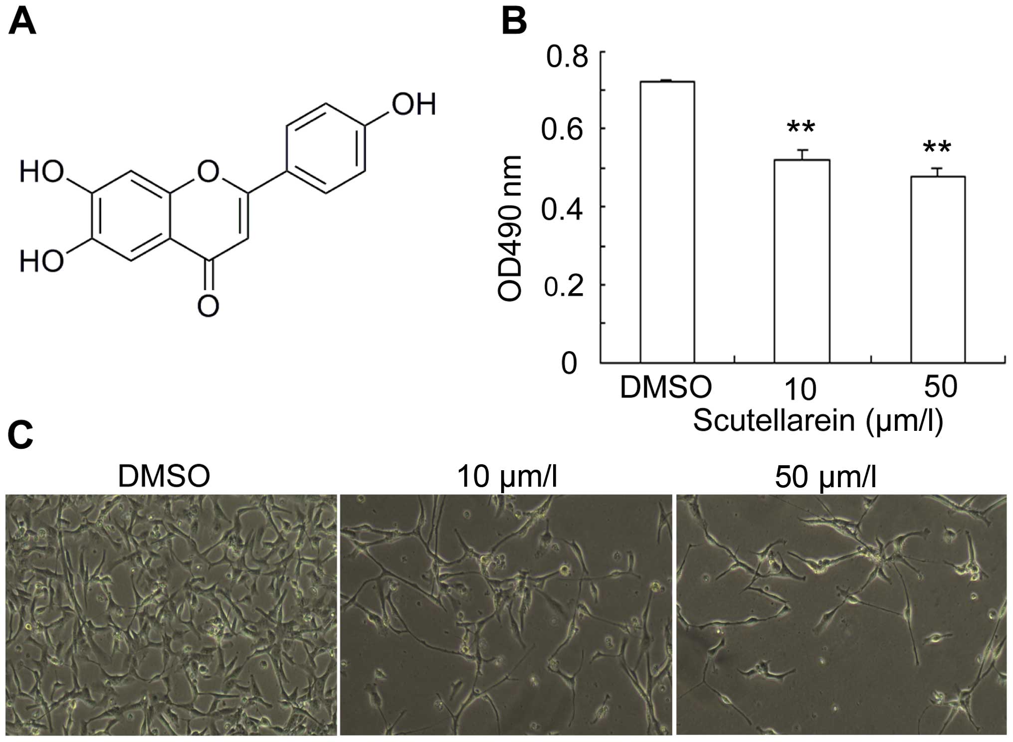
Scutellarein, found in Scutellaria
lateriflora and other members of the genus Scutellaria,
is a member of the flavone group of phenolic compounds (Fig. 1A). The viability of the HT1080
fibrosarcoma cells in the presence of scutellarein at various
concentrations and the vehicle (DMSO) was assessed with MTT assay.
In comparison to the DMSO-treated cells, the cells treated with 10
and 50 μm/l of scutellarein showed a 27.6 and 32.8% reduction in
cell viability, respectively (Fig.
1B). The suppressive effects of scutellarein on the viability
of the HT1080 fibrosarcoma cells clearly demonstrates its
anti-proliferative activity (Fig.
1C).
Scutellarein inhibits cell colony
formation and promotes apoptosis
To further investigate the anti-proliferative
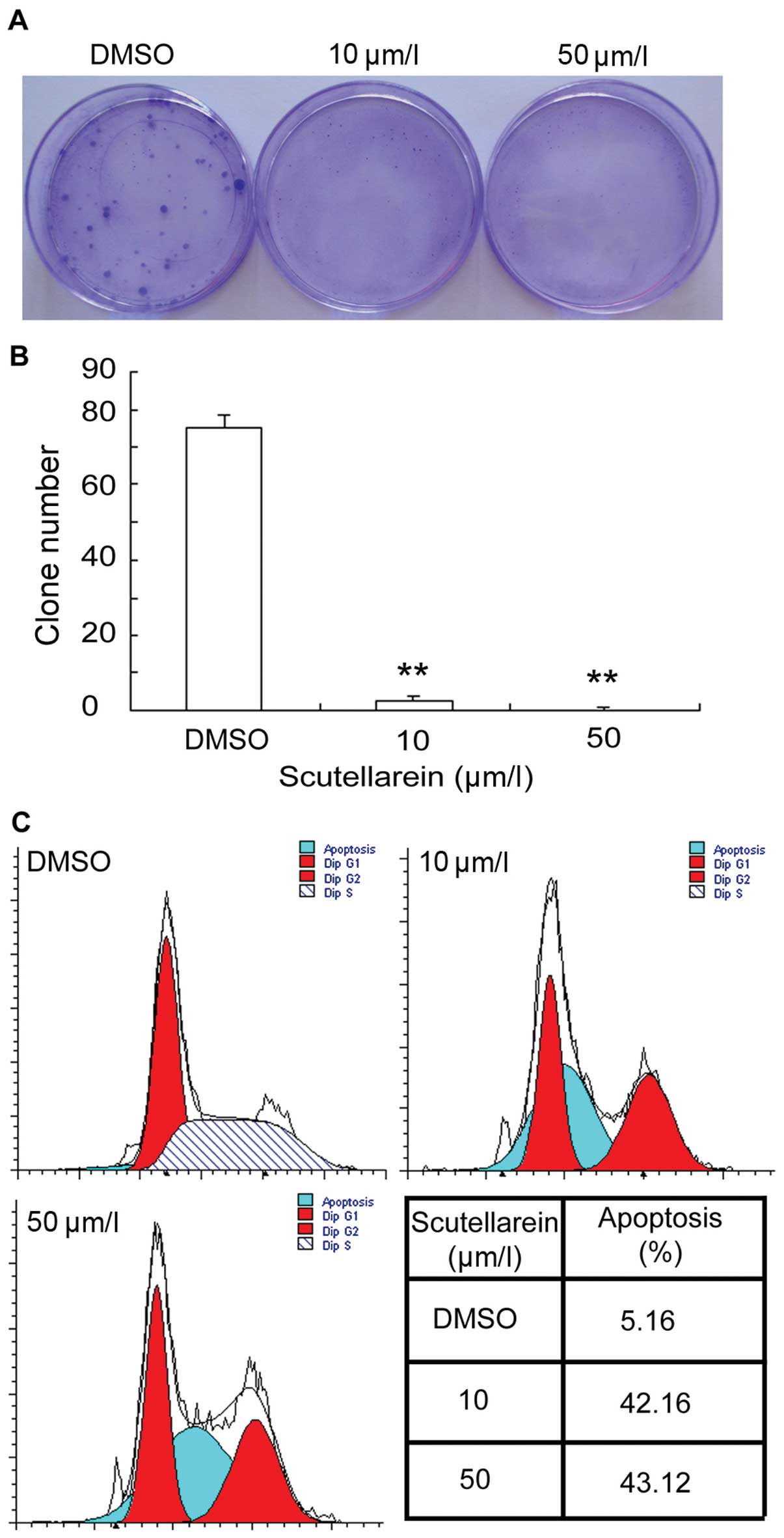
effects of scutellarein on human fibrosarcoma, we performed a cell
colony formation assay. The plates with scutellarein-treated cells
showed few cell clones, whereas the vehicle (DMSO)-treated cell
plates presented a clear colony formation of HT1080 cells (Fig. 2A). In addition, the quantification
analysis revealed that the average cell clone number of the cells
treated with scutellarein at concentrations of 10 and 50 μm/l was
2.6 and 0.2, respectively, while the DMSO treated group displayed a
cell clone number of 75 (Fig.
2B). Furthermore, cell apoptosis detection assay was performed
to determine whether scutellarein promotes the apoptosis of
fibrosarcoma cells. Flow cytometric analysis revealed that the rate
of apoptosis following treatment with 10 μm/l scutellarein was
42.16%; this increased to 43.12% following treatment with 50 μm/l
scutellarein, while for the DMSO-treated cells, the rate of
apoptosis was 5.16%. These results clearly indicate that
scutellarein significantly increases the number of apoptotic cells
(Fig. 2C).
Scutellarein attenuates fibrosarcoma in
vivo
Based on our results demonstrating that scutellarein
inhibited proliferation and promoted apoptosis in vitro, we
wished to determine whether this compound attenuates the
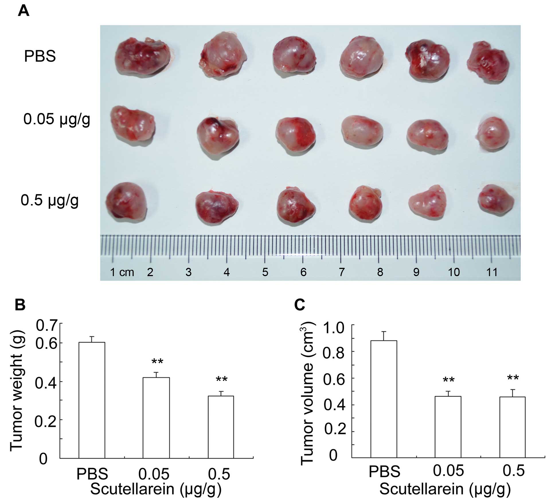
development of fibrosarcoma in vivo. Fibrosarcoma formation
was induced by injecting HT1080 cells into Balb/c nude mice. At 20
days following the injection of the cancer cells, the tumor-bearing
mice were sacrificed by cervical dislocation and the solid tumors
were removed and arranged with their weights measured and analyzed
(Fig. 3A). The injection of
scutellarein at doses of 0.05 and 0.5 μg/g, suppressed the tumor
weight by 30.25 and 46.17%, respectively, in comparison to the
control group treated with PBS (Fig.
3B). The tumors were also measured using a caliper, and the
tumor volume was calculated as the arithmetic mean of 2
perpendicular diameters. Statistical analysis revealed that
treatment with scutellarein at a dose of 0.5 μg/g reduced the tumor
volume by 47.4% when compared with the PBS-treated control group.
Similar results were observed following treatment with a lower
concentration (0.05 μg/g) of scutellarein (tumor volume was reduced
by 47%; Fig. 3C).
Scutellarein decreases metastasis in
vitro
To evaluate the antimetastatic potential of
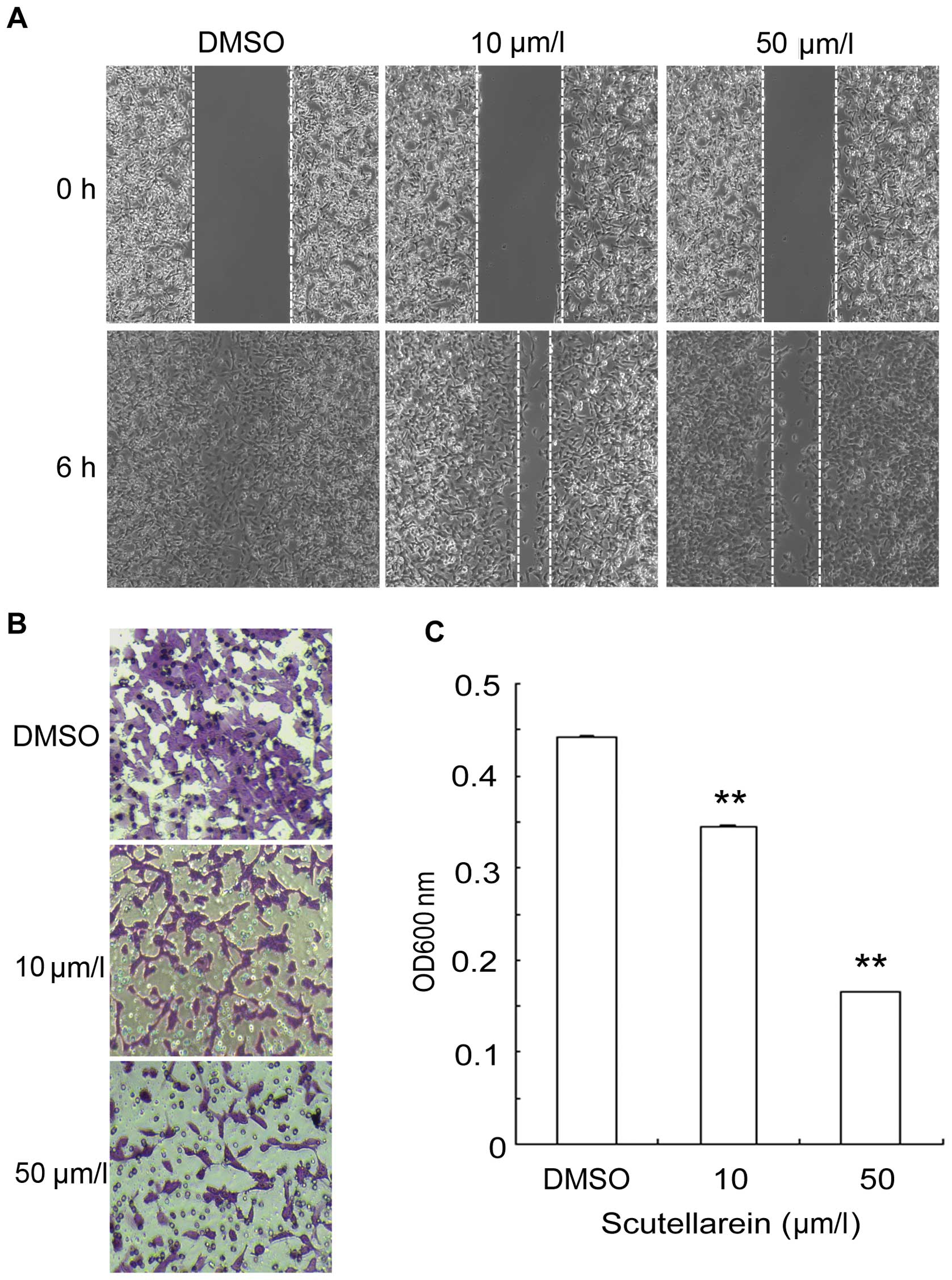
scutellarein, cell migration assay and invasion assay were
performed. The vehicle (DMSO)-treated HT1080 cells migrated towards
the empty area after 6 h of incubation. However, the
scutellarein-treated cells showed a significant dose-dependent
reduction in their migration ability, suggesting that scutellarein
reduced the HT1080 cell migration (Fig. 4A). Due to the ability of cells to
invade through Matrigel, the effects of scutellarein on the
invasion of HT1080 cells were determined by a standardized cell
Transwell invasion assay. A plethora of HT1080 cells treated with
the vehicle invaded the Matrigel, while the scutellarein-treated
cells showed a decrease in cell invasion in a dose-dependent manner
(Fig. 4B). Optical absorbance at
600 nm was measured based on crystal violet staining of the cancer
cells. Quantification analysis indicated that the highest
concentration of scutellarein used, induced a 3-fold decrease in
cell invasion (Fig. 4C). Taken
together, these observations indicate that scutellarein has the
potential to decrease cancer cell metastasis in vitro.
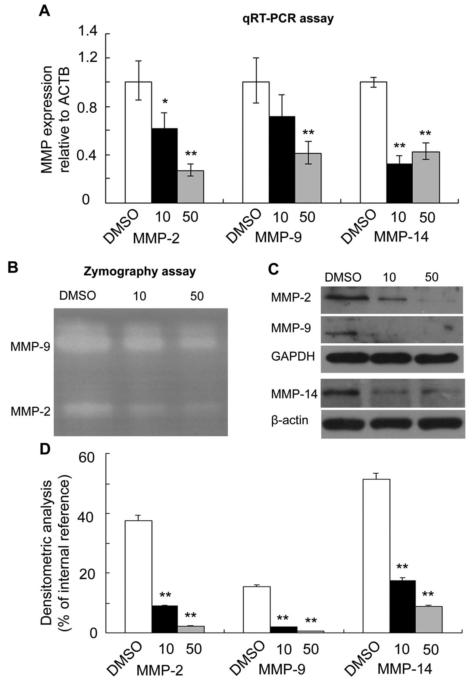
Effect of scutellarein on the expression
and activity of MMPs in HT1080 cells
To explore the mechanisms of the
scutellarein-mediated inhibition of cell migration, the gene
expression levels of cell migration-related proteins, MMP-2, -9 and
-14, were analyzed by qRT-PCR. The expression level of human
β-actin (ACTB) was used as an internal control. As compared to the
vehicle (DMSO)-treated cells, the expression levels of MMP-2, -9
and -14 were significantly reduced following treatment with
scutellarein. However, the inhibition efficiency varied among these
genes. The inhibition efficiency of scutellarein on the activity of
MMP-2 and -9 was dose-dependent. By contrast, the inhibition
efficiency of 10 μm/l scutellarein on MMP-14 activity was 67%,
while that of 50 μm/l scutellarein was 58%, suggesting a
non-dose-dependent pattern (Fig.
5A). The effects of scutellarein on the activity of gelatinases
(MMP-2 and -9) was confirmed by performing gelatin zymography,
which clearly indicated that scutellarein inhibited both MMP-2 and
-9 activity in a dose-dependent manner (Fig. 5B). Western blot analysis, carried
out to ascertain the levels of MMP-2, -9 and 14, revealed that
MMP-2 and -9 protein expression was suppressed by treatment with
scutellarein in a dose-dependent manner, as compared to the
vehicle-treated control. This result was in agreement with the
results of RT-qPCR. However, in contrast to the RT-qPCR data, the
protein level of MMP-14 was inhibited by treatment with
scutellarein in a dose-dependent manner (Fig. 5C).
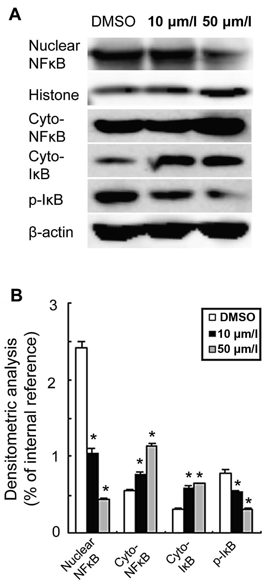
Scutellarein inhibits the activation of
the nuclear factor κ-light-chain-enhancer of activated B cells
(NF-κB) signaling pathway
Cell migration and proliferation are controlled by
complex cellular signaling pathways. The NF-κB signaling pathway is
crucial for migration and cell survival (16,17). The effects of scutellarein on the
regulation of the NF-κB signaling pathway were determined by
western blot analysis. The NF-κB transcription factor is
translocated from the cytosol to the nucleus upon activation. The
nuclear NF-κB levels were significantly suppressed following
treatment with scutellarein and the inhibition was dose-dependent
(Fig. 6A). Quantification
analysis revealed that, at the highest concentration of
scutellarein, the nuclear NF-κB levels were reduced by up to 5-fold
compared to the vehicle (DMSO)-treated cells (Fig. 6B). The cytosolic NF-κB levels also
increased in a dose-dependent manner. Furthermore, the cytosolic
levels of IκB and p-IκB were analyzed. The results indicated that
treatment with scutellarein increased the cytosolic IκB levels in a
dose-dependent manner, suppressing IκB phosphorylation. In
conclusion, these data suggest that scutellarein regulates the
proliferation and migration of HT1080 cells by inhibiting the
activation of the NF-κB signaling pathway.
Discussion
Natural products have been the single most
productive source for the development of drugs, particularly as
anticancer agents (18,19). Scutellarin, clinically used in the
treatment of acute cerebral infarction and paralysis in China, is a
natural product extracted from the Chinese herb, Erigeron
breviscapus (Vant.) Hand-Mazz (20). Previous studies have demonstrated
that scutellarin has anticancer properties (21,22). However, the water-solubility and
bioavailability of scutellarin is low (23). It has been reported that
scutellarein has relatively better solubility, bioavailability and
bio-activity compared to scutellarin (13). Based on these observations, the
present study was designed to investigate the ability of
scutellarein as a lead compound to attenuate the development of
human fibrosarcoma by using in vitro and in vivo
models. Our results revealed that scutellarein has the potential to
inhibit tumor formation and the metastasis of fibrosarcoma
cells.
Metastasis, accounting for over 90% of
cancer-related deaths worldwide, is an extremely complex phenomenon
mediated by a large number of signaling cascades (24,25). The progression of metastasis
involves cell migration from the primary tumor site, invasion of
adjacent tissues, entry into and transport through the blood
vascular system, reaching secondary sites, tumor growth and
proliferation (26). Metastasis
has been one of the most popular targets of anticancer studies,
particularly for exploring therapeutic targets (27,28). Through the analysis of
scutellarein-mediated suppression of cancer cell invasion and
migration at the molecular level, the present study proved that
scutellarein is a candidate compound for use in the prevention of
cancer metastasis.
MMPs, including MMP-2, -9 and -14, which are
overexpressed in tumor cells, aid migration and invasion by
modifying the cellular microenvironment. Hence, small molecules or
compounds inhibiting the expression of MMPs may prove to be
beneficial to cancer therapy. However, several of the previously
identified MMP inhibitors have failed in clinical trials, perhaps
due to the large number of proteinases mediating enhanced tumor
growth and progression (29).
Therefore, the suppression of tumor growth is one of the critical
factors in the treatment of cancer. Cancer cell growth and
progression may occur due to inadequate apoptosis, which is
initiated by extracellular receptor signaling and the proteolytic
cascade (30). Accordingly, the
induction of apoptosis in conjunction with the suppression of MMPs
may be a potent mechanism for cancer treatment. In the present
study, our results revealed that scutellarein not only decreased
the expression of MMP-2, -9 and -14, but also induced apoptosis by
suppressing the proliferation of HT1080 cells, thereby attenuating
tumor development and metastasis.
The activation of NF-κB transcription factors is
associated with several aspects of tumorigenesis, including cancer
cell survival and proliferation, the prevention of apoptosis and an
increase in the metastatic potential of tumor cells (31). Moreover, NF-κB expression in the
nucleus contributes to the activation of MMP-2 and MMP-9, which
play critical roles in cancer metastasis (32). Our results clearly demonstrated
that the compound, scutellarein, potently inhibited the activation
and nuclear translocation of NF-κB. Therefore, scutellarein may
further decrease the expression of MMP-2, -9 and -14 through the
NF-κB signaling pathway, thus inhibiting the migration and invasion
of cancer cells.
In conclusion, scutellarein effectively suppressed
the invasion and migration of HT1080 cells by inhibiting the
expression and activity of MMP-2, -9 and -14, which are crucial
enzymes in cancer metastasis. Scutellarein reduced cell
proliferation by inducing the apoptosis of HT1080 cells.
Furthermore, scutellarein suppressed tumor development in a nude
mice injected with HT1080 cells, which demonstrated the stability
and activity of scutellarein in vivo.
Acknowledgements
The present study was supported by grants from the
Ministry of Science and Technology of China (2010CB945600 and
2011CB965100), the National Natural Science Foundation of China
(81271498, 81202550 and 81100673), the Shanghai Science Foundation
(no. 11PJ1407800) and the Ministry of Education of China
(IRT1168).
Abbreviations:
|
MMP
|
matrix metalloproteinase
|
|
NF-κB
|
nuclear factor κ-light-chain-enhancer
of activated B cells
|
|
EMT
|
epithelial-mesenchymal transition
|
|
ECM
|
extracellular matrix
|
|
MTT
|
3-(4,5-
dimethylthiazol-2-yl)-2,5-diphenyltetrazolium bromide
|
|
DMEM
|
Dulbecco’s modified Eagle’s medium
|
|
DMSO
|
dimethyl sulfoxide
|
|
BSA
|
bovine serum albumin
|
|
FBS
|
fetal bovine serum
|
|
PBS
|
phosphate-buffered saline
|
|
RT-qPCR
|
quantitative reverse transcription-
polymerase chain reaction
|
|
DEPC
|
diethyl pyrocarbonate
|
References
|
1
|
Hanahan D and Weinberg RA: Hallmarks of
cancer: the next generation. Cell. 144:646–674. 2011. View Article : Google Scholar : PubMed/NCBI
|
|
2
|
Joyce JA and Pollard JW:
Microenvironmental regulation of metastasis. Nat Rev Cancer.
9:239–252. 2009. View
Article : Google Scholar : PubMed/NCBI
|
|
3
|
Psaila B and Lyden D: The metastatic
niche: adapting the foreign soil. Nat Rev Cancer. 9:285–293. 2009.
View Article : Google Scholar : PubMed/NCBI
|
|
4
|
Weigelt B, Peterse JL and van’t Veer LJ:
Breast cancer metastasis: markers and models. Nat Rev Cancer.
5:591–602. 2005. View
Article : Google Scholar : PubMed/NCBI
|
|
5
|
Liotta LA: Tumor invasion and metastases:
role of the basement membrane. Warner-Lambert Parke-Davis Award
lecture. Am J Pathol. 117:339–348. 1984.PubMed/NCBI
|
|
6
|
Egeblad M and Werb Z: New functions for
the matrix metalloproteinases in cancer progression. Nat Rev
Cancer. 2:161–174. 2002. View
Article : Google Scholar : PubMed/NCBI
|
|
7
|
Nagase H and Woessner JF Jr: Matrix
metalloproteinases. J Biol Chem. 274:21491–21494. 1999. View Article : Google Scholar : PubMed/NCBI
|
|
8
|
Visse R and Nagase H: Matrix
metalloproteinases and tissue inhibitors of metalloproteinases:
structure, function, and biochemistry. Circ Res. 92:827–839. 2003.
View Article : Google Scholar : PubMed/NCBI
|
|
9
|
Deryugina EI and Quigley JP: Matrix
metalloproteinases and tumor metastasis. Cancer Metastasis Rev.
25:9–34. 2006. View Article : Google Scholar : PubMed/NCBI
|
|
10
|
Mehlen P and Puisieux A: Metastasis: a
question of life or death. Nat Rev Cancer. 6:449–458. 2006.
View Article : Google Scholar : PubMed/NCBI
|
|
11
|
Nuti E, Tuccinardi T and Rossello A:
Matrix metalloproteinase inhibitors: new challenges in the era of
post broad-spectrum inhibitors. Curr Pharm Des. 13:2087–2100. 2007.
View Article : Google Scholar : PubMed/NCBI
|
|
12
|
Gao R, Zhu BH, Tang SB, Wang JF and Ren J:
Scutellarein inhibits hypoxia- and moderately-high glucose-induced
proliferation and VEGF expression in human retinal endothelial
cells. Acta Pharmacol Sin. 29:707–712. 2008. View Article : Google Scholar : PubMed/NCBI
|
|
13
|
Li NG, Song SL, Shen MZ, Tang YP, Shi ZH,
Tang H, Shi QP, Fu YF and Duan JA: Mannich bases of scutellarein as
thrombin-inhibitors: design, synthesis, biological activity and
solubility. Bioorgan Med Chem. 20:6919–6923. 2012. View Article : Google Scholar
|
|
14
|
Goh D, Lee YH and Ong ES: Inhibitory
effects of a chemically standardized extract from Scutellaria
barbata in human colon cancer cell lines, LoVo. J Agr Food Chem.
53:8197–8204. 2005. View Article : Google Scholar
|
|
15
|
Chen G, Shi X, Sun C, Li M, Zhou Q, Zhang
C, Huang J, Qiu Y, Wen X, Zhang Y, et al: VEGF-mediated
proliferation of human adipose tissue-derived stem cells. PloS One.
8:e736732013. View Article : Google Scholar : PubMed/NCBI
|
|
16
|
Helbig G, Christopherson KW 2nd,
Bhat-Nakshatri P, Kumar S, Kishimoto H, Miller KD, Broxmeyer HE and
Nakshatri H: NF-kappaB promotes breast cancer cell migration and
metastasis by inducing the expression of the chemokine receptor
CXCR4. J Biol Chem. 278:21631–21638. 2003. View Article : Google Scholar : PubMed/NCBI
|
|
17
|
Luo JL, Kamata H and Karin M:
IKK/NF-kappaB signaling: balancing life and death - a new approach
to cancer therapy. J Clin Invest. 115:2625–2632. 2005. View Article : Google Scholar : PubMed/NCBI
|
|
18
|
Harvey AL: Natural products in drug
discovery. Drug Discov Today. 13:894–901. 2008. View Article : Google Scholar : PubMed/NCBI
|
|
19
|
Koehn FE and Carter GT: The evolving role
of natural products in drug discovery. Nat Rev Drug Discov.
4:206–220. 2005. View
Article : Google Scholar : PubMed/NCBI
|
|
20
|
Pan ZW, Feng TM, Shan LC, Cai BZ, Chu WF,
Niu HL, Lu YJ and Yang BF: Scutellarin-induced
endothelium-independent relaxation in rat aorta. Phytother Res.
22:1428–1433. 2008. View
Article : Google Scholar : PubMed/NCBI
|
|
21
|
Chan JY, Tan BK and Lee SC: Scutellarin
sensitizes drug-evoked colon cancer cell apoptosis through enhanced
caspase-6 activation. Anticancer Res. 29:3043–3047. 2009.PubMed/NCBI
|
|
22
|
Li H, Huang D, Gao Z, Lv Y, Zhang L, Cui H
and Zheng J: Scutellarin inhibits cell migration by regulating
production of αvβ6 integrin and E-cadherin in human tongue cancer
cells. Oncol Rep. 24:1153–1160. 2010.PubMed/NCBI
|
|
23
|
Qian LH, Li NG, Tang YP, Zhang L, Tang H,
Wang ZJ, Liu L, Song SL, Guo JM and Ding AW: Synthesis and
bio-activity evaluation of scutellarein as a potent agent for the
therapy of ischemic cerebrovascular disease. Int J Mol Sci.
12:8208–8216. 2011. View Article : Google Scholar : PubMed/NCBI
|
|
24
|
Chaffer CL and Weinberg RA: A perspective
on cancer cell metastasis. Science. 331:1559–1564. 2011. View Article : Google Scholar : PubMed/NCBI
|
|
25
|
Muller A, Homey B, Soto H, Ge NF, Catron
D, Buchanan ME, McClanahan T, Murphy E, Yuan W, Wagner SN, et al:
Involvement of chemokine receptors in breast cancer metastasis.
Nature. 410:50–56. 2001. View
Article : Google Scholar : PubMed/NCBI
|
|
26
|
Fidler IJ: Timeline - the pathogenesis of
cancer metastasis: the ‘seed and soil’ hypothesis revisited. Nat
Rev Cancer. 3:453–458. 2003. View
Article : Google Scholar : PubMed/NCBI
|
|
27
|
Eccles SA and Welch DR: Metastasis: recent
discoveries and novel treatment strategies. Lancet. 369:1742–1757.
2007. View Article : Google Scholar : PubMed/NCBI
|
|
28
|
Geiger TR and Peeper DS: Metastasis
mechanisms. Biochim Biophys Acta. 1796:293–308. 2009.PubMed/NCBI
|
|
29
|
Coussens LM, Fingleton B and Matrisian LM:
Matrix metalloproteinase inhibitors and cancer: trials and
tribulations. Science. 295:2387–2392. 2002. View Article : Google Scholar : PubMed/NCBI
|
|
30
|
Kessenbrock K, Plaks V and Werb Z: Matrix
metalloproteinases: regulators of the tumor microenvironment. Cell.
141:52–67. 2010. View Article : Google Scholar : PubMed/NCBI
|
|
31
|
Perkins ND: The diverse and complex roles
of NF-κB subunits in cancer. Nat Rev Cancer. 12:121–132.
2012.PubMed/NCBI
|
|
32
|
Orlowski RZ and Baldwin AS: NF-κB as a
therapeutic target in cancer. Trends Mol Med. 8:385–389. 2002.
View Article : Google Scholar : PubMed/NCBI
|