Introduction
Age-related hearing loss (AHL), also known as
presbycusis, is the most common form of hearing loss and the
predominant age-related neurodegenerative disease affecting
approximately 40% of individuals by the age of 65 years (1). Thus far, there is no effective
treatment available for AHL. The irreversible loss of cochlear hair
cells in the inner ear is one of the main causes of AHL in both
aging humans and in animal models (2–4),
and decreasing the loss of cochlear hair cells may present an
attractive therapeutic strategy for the treatment of AHL.
MicroRNAs (microRNAs or miRs) are non-coding RNAs,
18–25 nucleotides in length, which regulate the expression of
target mRNAs, as well as influence cellular senescence and aging
(5–7). Recently, alterations in the
expression of the miR-29 family and the miR-34 family have been
documented during aging in the mammalian inner ear (8,9).
Of interest is that miR-29b has been shown to be involved in
cellular senescence and the apoptosis of nerve cell lines, the
brain and the liver during aging (10,11). However, the functions of miR-29b
regarding apoptosis are not yet fully understood. Sirtuin 1 (SIRT1)
is a nicotinamide adenine dinucleotide (NAD)-dependent deacetylase
that acts as a sensor to regulate the intracellular oxidative
stress status by the deacetylation of its substrates, including
proliferator-activated receptor-gamma coactivator 1α (PGC-1α), a
transcriptional coregulator that binds to numerous transcription
factors to promote mitochondrial biogenesis and oxidative
metabolism (12–14). Oxidative stress, which is mainly
caused by mitochondrial dysfunction, is known to play a causal role
in AHL through the induction of apoptosis (15,16). Of note, SIRT1 has been confirmed
to be a direct target of miR-29b (17).
In this study, we hypothesized that
miR-29b/SIRT1/PGC-1α signaling may play a role in hair cell death
and AHL pathogenesis, and that strategies aimed at inhibiting
miR-29b activity or restoring SIRT1 function, may prove to be be
beneficial in the treatment of AHL. To test our hypothesis,
cochlear miR-29b/SIRT1/PGC-1α expression was examined in C57BL/6
mice, a mouse model of AHL. Additionally, the potential effects of
miR-29b on the expression of SIRT1 and PGC-1α and the underlying
mechanisms were assessed using HEI-OC1 inner ear cells.
Materials and methods
Animals and groups
Sixty C57BL/6 mice were procured from the Laboratory
Animal Center of the Fourth Military Medical University (Xian,
China) and divided into 2 groups as follows: a 'young' group (1–2
months of age, 30 mice) and an 'old' group (12–16 months of age, 30
mice), and were fed standard chow. Hearing tests were conducted on
all mice, and cochlear tissues were collected for hair cell
counting. All procedures involving animals were conducted in
accordance with the Guidelines for Animal Experiments approved by
the Ethics Committee for Animal Studies of the Fourth Military
Medical University.
Auditory brainstem response (ABR)
All mice were anesthetized with an intraperitoneal
injection mixture that contained 100 mg/kg ketamine and 10 mg/kg
xylazine. ABR measurements were performed by inserting subdermal
needle electrodes at the vertex (active), under the left ear
(reference), and under the right ear (ground). Tucker-Davis
Technologies (TDT System III, Alachua, FL, USA) hardware and
software were used to generate acoustic signals and to process the
responses. Ten-millisecond (msec) tone bursts with a 1 msec rise or
fall time were presented at 4, 8, 16 and 32 kHz at a rate of
21.1/sec. The average response to 1,000 stimuli was obtained by
reducing the sound intensity at 5 dB intervals near the threshold,
which was defined as the lowest stimulation decibel level at which
a positive wave in the evoked response trace was evident.
Tissue preparation and isolation of
cochlear tissue mitochondria
After the ABR recordings, the mice were decapitated,
and the cochleae were removed and fixed by immersion in 4%
paraformaldehyde in 0.1 mM phosphate-buffered saline (PBS, pH 7.4)
overnight at 4°C, followed by being decalcified in 4% sodium
ethylenediaminetetraacetic acid for 4 days. Following incubation in
30% sucrose overnight, the cochleae were cut into 10-mm thick
slices and stored at −20°C. For RNA and protein preparations, the
cochlear tissues were dissected with small forceps, snap-frozen in
liquid nitrogen and stored at −80°C. The mitochondria were isolated
from the cochleae using differential centrifugation and TOM22
immunomagnetic affinity isolation. Briefly, the cochleae were
dounce homogenized in isolation medium (IM) containing 225 mM
sucrose, 75 mM mannitol, 1 mM EGTA, 5 mM HEPES, and complete Mini,
EDTA-free protease inhibitor cocktail (Roche Diagnostics,
Indianapolis, IN, USA) adjusted to pH 7.4. The homogenate was
centrifuged at 1,000 × g for 10 min, the supernatant collected and
the pellet was resuspended in IM. Following a second centrifugation
at 1,000 × g for 10 min, the pooled supernatants were centrifuged
at 8,000 × g for 10 min. The pellet containing the mitochondria was
further purified using anti-TOM22 immunomagnetic affinity isolation
(Miltenyi Biotech, Bergisch Gladbach, Germany). Purified
mitochondria were lysed in 100 mM Tris-HCl with 4% (w/v) SDS and
0.1 M DTT adjusted to pH 7.6 using brief sonication and incubation
at 95°C for 5 min.
Nuclear staining and terminal
deoxynucleotidyl transferase dUTP nick-end labeling (TUNEL)
assay
Following fixation with 4% paraformaldehyde, the
organ of Corti, which was isloated from the cochleae under a
dissecting microscope, was rinsed with PBS, immersed in 0.5% Triton
X-100 for 15 min at room temperature, incubated with
4′,6-diamidino-2-phenylin-dole (DAPI, 10 mg/ml; Sigma, St. Louis,
MO, USA) for 10 min, and mounted on glass slides in 50% glycerol.
Some surface preparations of the organ of Corti were incubated with
100 ml of TUNEL reaction mixture (Roche Diagnostics) containing
dUTP-FITC and terminal deoxynucleotidyl transferase at 37°C in the
dark for 60 min, followed by incubation with propidium iodide (PI)
staining solution (10 mg/ml PI in PBS) for 30 min at room
temperature. The samples were observed and imaged using an Olympus
BX63 microscope (Olympus, Ishikawa, Japan). Hair cell counts were
identified by the presence of a nucleus and performed in two
locations: at 10–20% (outer hair cells) and 65–70% (inner hair
cells) of the total cochlear duct distance from the apex, which
approximately corresponds to the frequency range of 7–8 or 32–36
kHz (18). Hair cells were
counted in 6 mice from each group and were considered to be
degenerated if the cell nuclei were absent.
HEI-OC1 cell culture and cell groups
The HEI-OC1 cells were a gift from Dr. Federico
Kalinec (House Ear Institute, Los Angeles, CA, USA) and were
cultured in Dulbecco's modified Eagle's medium supplemented with
10% fetal bovine serum (both from Gibco, Gaithersburg, MD, USA) at
33°C and 7% CO2. The cells were divided into 7 different
groups as follows: the control group, the
H2O2 group and 5 transfection groups. The
cells in the transfection groups were transfected with a miR-29b
mimic, a negative control (pre-miRNA), or a miR-29b inhibitor
(GenePharma, Shanghai, China). Full-length SIRT1 was amplified by
PCR using cDNA, and was confirmed by sequencing and subcloning into
a plk0.1 expression vector (Invitrogen, Carlsbad, CA, USA). The
HEI-OC1 cells were placed in a 6-well plate and transfected with
plk0.1-scramble or plk0.1-SIRT1 using Lipofectamine 2000
(Invitrogen). Following transfection, the HEI-OC1 cells were
exposed to 50 µM H2O2 for 1 h.
Measurement of mitochondrial membrane
potential
The mitochondrial dye,
5,5′,6,6′-tetrachloro-1,1′,3,3′-tetramethyl-benzimidazolylcarbocyanine
iodide (JC-1), was used to determine the mitochondrial membrane
potential. JC-1 exhibits potential-dependent accumulation in the
mitochondria, which is indicated by a fluorescence emission shift
from green (530 nm) to red (590 nm). Accordingly, mitochondrial
depolarization is indicated by a decrease in the red/green
fluorescence intensity ratio. In brief, HEI-OC1 cells were cultured
in a 24-well plate and subjected to the different treatments as
described above, followed by incubation with 10 mg/ml JC-1 for 30
min at 37°C. The cells were then scanned using a FlexStation 3
multi-mode microplate reader (Molecular Devices, Silicon Valley,
USA). The fluorescence ratio (590 to 530 nm) was used for
quantitative analysis.
Cell proliferation assay
After the concentration was adjusted to
2×10⁶ cells/well, the cells were seeded in 96-well
plates and then stained at the indicated time points with 100
µl sterile MTT (Molecular Probes, Eugene, OR, USA) for 4 h
at 37°C. The supernatant was removed and 200 µl dimethyl
sulfoxide (DMSO) (Invitrogen) was added for 30 min to dissolve the
formazan crystals. The spectrometric absorbance at a wavelength of
490 nm was measured on a SpectraMax M5 microplate reader (Molecular
Devices). Each sample was tested in triplicate and all experiments
were performed 3 times.
Apoptosis assay
Flow cytometric analysis with Annexin V-FITC/PI
double staining was performed using an Annexin V-FITC apoptosis
detection kit (Bio-Rad, Hercules, CA, USA) to measure cell
apoptosis. All groups of HEI-OC1 cells were incubated for 72 h, and
the cells were then harvested and centrifuged at 1,200 × g for 5
min, followed by washing twice with ice-cold PBS. Following the
addition of 5 µl of Annexin V, 5 µl of PI and 500
µl of PBS to the cells, the cells were incubated at room
temperature for 15 min in the dark. Cell apoptosis was analyzed on
a FACS can flow cytometer (CyAn ADP; Beckman Coulter, Brea, CA,
USA). Annexin V-FITC positive and PI-negative cells were scored as
apoptotic. Double-stained cells were considered as either necrotic
or late apoptotic cells.
Reverse transcription-quantitative
(real-time) polymerase chain reaction (RT-qPCR)
Total RNA was isolated from the cells using TRIzol
Reagent (Invitrogen) according to the manufacturer's instructions,
and 1 mg total RNA was reverse transcribed using the ReverTra-Plus™
kit (Toyobo, Osaka, Japan). The primer sequences used for
amplification were as follows: SIRT1 forward,
5′-AAAGGAATTGGTTCATTTATCAGAG-3′ and reverse,
5′-TTGTGGTTTTTCTTCCACACA-3′; PGC-1α forward,
5′-AAACTTGCTAGCGGTCCTCA-3′ and reverse, 5′-TGGCTGGTGCCAGTAAGAG-3′;
and β-actin forward, 5′-TGAACGGGAAGCTCACTGG-3′ and reverse,
5′-GCTTCACCACCTTCTTGATGTC-3′. Comple mentary DNA samples were
amplified using SYBR Premix Ex Taq (Tli RNaseH Plus; Takara, Otsu,
Japan) and detected with the Roche LightCycler 480 real-time PCR
system. β-actin was used as an internal control for SIRT1 and
PGC-1α normalization.
For the analysis of miR-29b expression, enriched
small RNAs were isolated from the cochlear tissues using TRIzol
reagent, with 500 ng of RNA being reverse transcribed using
specific miRNA stem-loop primers and a PrimeScript RT reagent kit
(Takara). Mature miRNA expression was measured with Takara Taq
version 2.0 plus dye (Takara) according to the manufacturer's
instructions, and the miRNA levels were normalized to U6 small
nuclear RNA expression.
Western blot analysis
The cochlear tissues and cultured HEI-OC1 cells were
homogenized in ice-cold radioimmuno-precipitation assay lysis
buffer (Santa Cruz Biotechnology, Inc., Santa Cruz, CA, USA) for 30
min and centrifuged at 12,000 × g at 4°C for 10 min, and the
supernatants were collected. Protein concentrations were determined
using a protein assay dye reagent (Bio-Rad, Hercules, CA, USA).
Protein samples (50 mg) were resolved by sodium dodecyl sulfate
polyacrylamide gel-electrophoresis (SDS-PAGE), and proteins were
transferred onto polyvinylidene fluoride membranes (Millipore,
Billerica, MA, USA) and blocked with 5% non-fat dry milk in PBS
with 0.1% Tween-20 (PBS-T). The membranes were incubated with
anti-SIRT1 (1:1,000; sc-74504) or anti-PGC-1α (1:1,000; sc-13067)
(both from Santa Cruz Biotechnology, Inc.) antibodies overnight,
washed 3 times (10 min each) with PBS-T, and incubated with goat
anti-mouse IgG (1:5,000; F020220, Sigma) for 1 h. Following
extensive washing of the membranes with 1X PBS-T, the
immunoreactive bands were visualized by enhanced chemiluminescence
(Millipore). Band intensities were quantified by densitometric
analysis using NIH ImageJ software, and β-actin (1:2,000; sc-47778,
Santa Cruz Biotechnology, Inc.) was used as a loading and an
internal control to enable sample normalization.
Statistical analysis
All experiments were independently repeated at least
3 times. One-way analysis of variance (ANOVA) with Fisher's post
hoc test and Student's t-test was used for statistical analysis.
Values of p<0.05 were considered to indicate statistically
significant differences.
Results
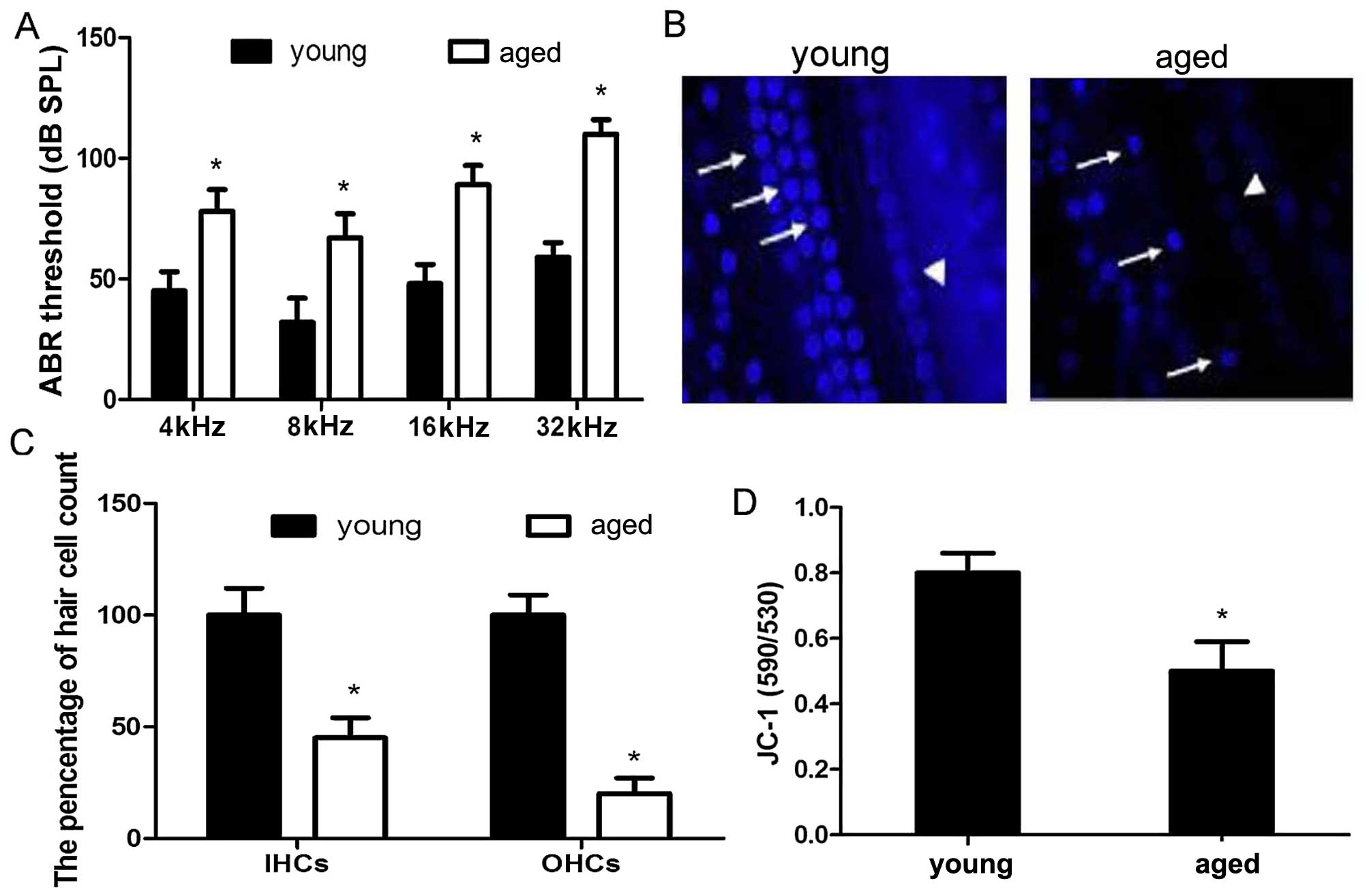
Progressive hearing threshold shifts and
hair cell loss
In an attempt to elucidate the mechanisms
responsible for the progression of AHL, hearing function in young
and aged C57BL/6 mice was evaluated by ABR, which is an objective
electro-physiological test to determine hearing function. The
average thresholds from the aged mice (78±9 dB at 4 kHz, 67±10 dB
at 8 kHz, 89±8 dB at 16 kHz and 110+6 dB at 32 kHz) were
significantly elevated compared with those from the young mice
(45±8 dB at 4 kHz, 32±10 dB at 8 kHz, 48±8 dB at 16 kHz and 59±6 dB
at 32 kHz) at all tested frequencies, indicating that the aged
C57BL/6 mice developed significant hearing loss (Fig. 1A). After the ABR measurements, the
cochleae were processed for surface preparations and quantitative
hair cell counts. The percentage inner hair cells and outer hair
cells in the aged mice was significantly decreased compared with
the young mice in the apical cochlear turn (Fig. 1B and C).
Mitochondrial dysfunction induced by
aging in the C57BL/6 mice
To confirm whether mitochondrial dysfunction is
involved during aging, the mitochondrial dye, JC-1, was used to
determine the mitochondrial membrane potential in the cochleae. A
significant decrease in mitochondrial membrane potential was
observed in thecochleae of the aged C57BL/6 mice compared with the
young ones, indicating the appearance of mitochondrial dysfunction
in the aged C57BL/6 mice (Fig.
1D).
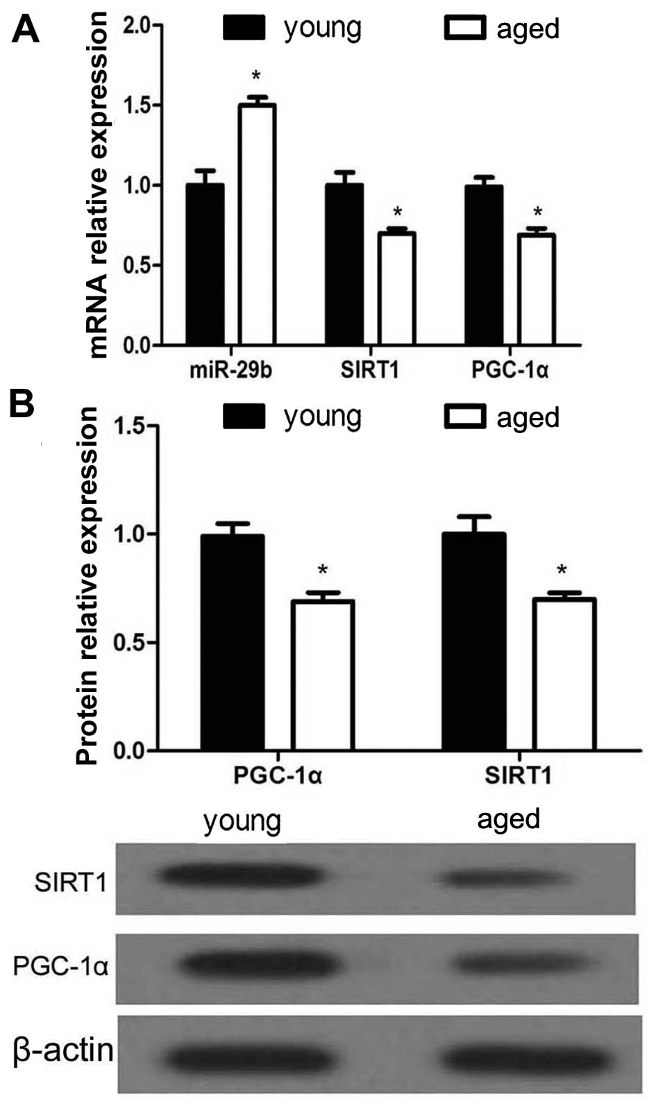
Expression of miR-29b, SIRT1 and PGC-1α
in the cochleae during aging
To further elucidate the mechanisms responsible for
hearing loss, RT-qPCR was used to examine the miR-29b, SIRT1 and
PGC-1α mRNA expression levels in the cochleae of young and aged
C57BL/6 mice, while the protein expression levels of SIRT1 and
PGC-1α were examined by western blot analysis. The results revealed
that miR-29b expression was significantly upregulated in the aged
C57BL/6 mice compared with the young mice (Fig. 2). Conversely, the mRNA and protein
expression levels of SIRT1 and PGC-1α were markedly decreased in
the cochleae of the aged mice.
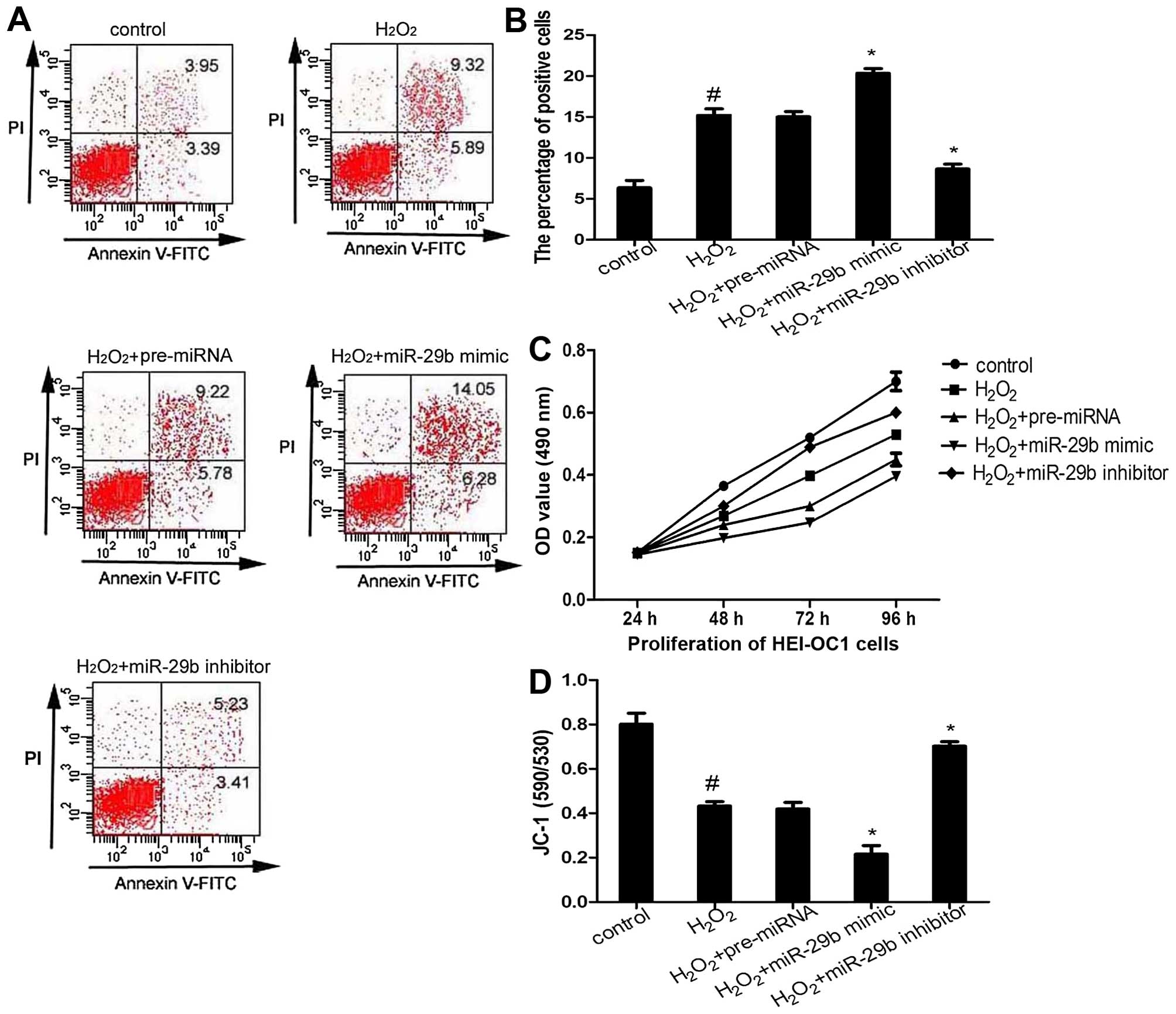
Effect of miR-29b on the apoptosis and
proliferation of HEI-OC1 cells
It is already known that
H2O2-induced oxidative stress promotes cell
apoptosis (19). In this study,
to examine the effects of miR-29b on cell survival, HEI-OC1 cells
transfected with the miR-29b mimic or miR-29b inhibitor were
exposed to 50 µM H2O2 for 1 h, and
then apoptosis was measured by flow cytometric analysis. The
results revealed that the overexpression of miR-29b promoted cell
apoptosis, whereas transfection with the miR-29b inhibitor
decreased apoptosis compared with the H2O2
group (Fig. 3A and B). The
results of MTT assay demonstrated that transfection with the
miR-29b mimic significantly suppressed HEI-OC1 cell proliferation
compared with the H2O2 group, whereas
transfection with the miR-29b inhibitor promoted cell proliferation
(Fig. 3C).
miR-29b overexpression induces
mitochondrial dysfunction
To determine whether miR-29b is involved in
mitochondrial dysfunction, the mitochondrial membrane potential was
determined using the mitochondrial dye, JC-1, in all groups of
HEI-OC1 cells. Transfection of the cells with the miR-29b mimic
exacerbated the decrease in H2O2-induced
mitochondrial membrane potential observed in the
H2O2 group (Fig. 3D). By contrast, transfection with
the miR-29b inhibitor attenuated the
H2O2-induced mitochondrial dysfunction. These
results indicate that the overexpression of miR-29b induces
mitochondrial dysfunction.
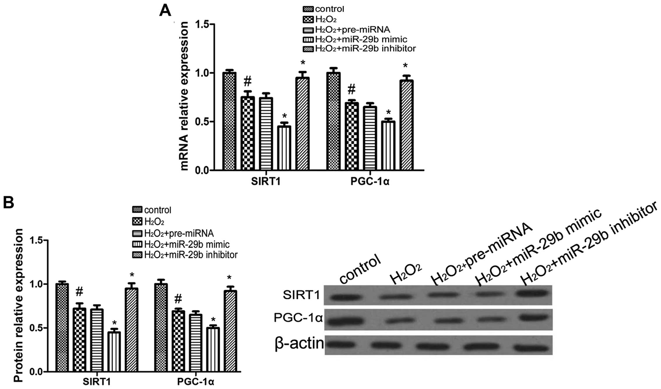
miR-29b modulates the expression of SIRT1
and PGC-1α in HEI-OC1 cells
To test the hypothesis that miR-29b modulates SIRT1
and PGC-1α expression, the mRNA and protein expression levels of
SIRT1 and PGC-1α were measured by RT-qPCR and western blot
analysis, respectively in the HEI-OC1 cells following transfection
with the miR-29b mimic or the miR-29b inhibitor. The overexpression
of miR-29b suppressed the mRNA and protein expression of SIRT1 and
PGC-1α compared with the H2O2 group, whereas
transfection with the miR-29b inhibitor promoted the expression of
SIRT1 and PGC-1α (Fig. 4). These
findings suggest that miR-29b modulates the expression of SIRT1 and
PGC-1α in HEI-OC1 cells.
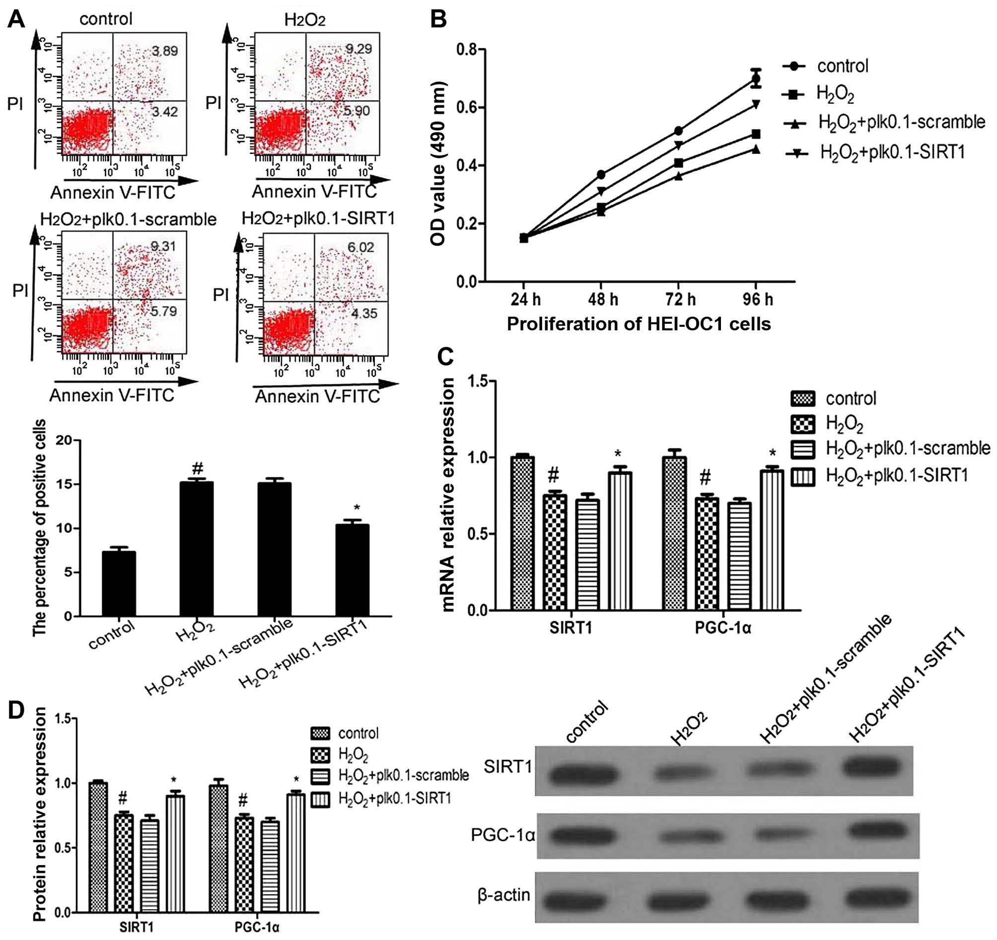
Effect of SIRT1 overexpression on PGC-1α
expression, as well as on HEI-OC1 cell apoptosis and
proliferation
To examine the effects of SIRT1 on PGC-1α
expression, as well as on HEI-OC1 cell apoptosis and proliferation,
the HEI-OC1 cells were transfected with plk0.1-scramble or
plk-0.1-SIRT1 using Lipofectamine 2000 in DMEM.
The overexpression of SIRT1 significantly suppressed
cell apoptosis and promoted cell proliferation, and also promoted
the mRNA level and protein expression of PGC-1α compared with the
H2O2 group (Fig. 5). These results suggest that SIRT1
modulates the expression of PGC-1α, as well as the apoptosis and
proliferation of HEI-OC1 cells.
Discussion
Aging is one of the most prominent factors which
influence the onset and progression of multiple diseases, along
with mitochondrial dysfunction, which is induced by oxidative
stress. AHL, a common age-related disease, often occurs in those
aged 65 years and above. A previous study demonstrated that the
expression of miR-29b was upregulated in AHL (5). In the present study, our results
also confirmed that miR-29b was overexpressed in the cochleae of
aging C57BL/6 mice, which is the most widely used mouse model for
the study of aging and age-associated diseases (20). Of note, in our study, there was a
significant decrease in the counts of hair cells and increased
mitochondrial dysfunction in the cochleae of the aged C57BL/6 mice,
which is in agreement with the findings of previous studies
indicating that hair cell apoptosis is a key contributor to the
development of AHL (4,21). SIRT1-mediated PCG-1α deacetylation
is necessary for the activation of mitochondrial fatty acid
oxidation genes (22). In this
study, an age-related decrease in SIRT1 and PGC-1α expression along
with mitochondrial dysfunction was noted in the cochleae of aged
mice. Of note, SIRT1 has been confirmed to be a direct target of
miR-29b (23). Thus, we
hypothesized that miR-29b/SIRT1/PGC-1α signaling may play a crucial
role during hair cell death and in the pathogenesis of AHL.
HEI-OC1, a conditionally immortalized mouse cell
line derived from the postnatal organ of Corti, has been
extensively used to elucidate pathways of hair cell pathology
(24,25). In this study, HEI-OC1 cells were
divided into different groups (control, H2O2,
H2O2 + miR-29b mimic,
H2O2 + pre-miRNA, H2O2
+ miR-29b inhibitor). It is well known that oxidative stress plays
a causal role in AHL through the induction of apoptosis (4,26).
In this study, MTT and apoptosis assays indicated that
H2O2 significantly suppressed cell
proliferation and promoted apoptosis compared to the control group.
However, the overexpression of miR-29b enhanced the
H2O2-mediated suppression of cell
proliferation and the promoting effect on cell apoptosis, while the
knockdown of miR-29b attenuated these effects. Analysis of
mitochondrial membrane potential using the mitochondrial dye, JC-1,
indicated that the overexpression of miR-29b significantly
decreased mitochondrial membrane potential, which exacerbated
H2O2-induced mitochondrial dysfunction. These
results indicated that the overexpression of miR-29b promoted the
loss of hair cells and mitochondrial dysfunction.
SIRT1 is recognized as a lifespan modulator and is
implicated in preventing a number of age-related diseases (27,28), such as cancer, Alzheimer's disease
and type 2 diabetes (29,30). As a NAD-dependent deacetylase,
SIRT1 also interacts with PGC-1α, which plays a critical role in
apoptosis, energy homeostasis, longevity and mitochondrial function
(31,32). In this study, in the cochleae of
aged C57BL/6 mice, the expressions levels of SIRT1 and PGC-1α were
significantly decreased. A previous study demonstrated that SIRT1
was the direct target gene of miR-29 (18). Moreover, in this study,
transfection of HEI-OC1 cells with miR-29b mimic suppressed the
expression of SIRT1 and PGC-1α, while the knockdown of miR-29b
significantly upregulated the mRNA level and protein expression of
SIRT1 and PGC-1α. Furthermore, the overexpression of SIRT1
significantly increased the expression of PGC-1α in HEI-OC1 cells
and suppressed cell apoptosis, and promoted cell proliferation.
Thus, it can be concluded that the overexpression of miR-29b
induces mitochondrial dysfunction and hair cell apoptosis through
the downregulation of the expression of SIRT1 and PGC-1α during
aging.
In conclusion, the findings of this study support
the hypothesis that miR-29b/SIRT1/PGC-1α signaling is involved in
the development and progression of AHL. miR-29b appears to modulate
mitochondrial dysfunction and apoptosis through SIRT1/PGC-1α
signaling in HEI-OC1 cells. Overall, miR-29b/SIRT1/PGC-1α signaling
may present an attractive pharmacological target for the
development of novel drugs for the treatment of AHL reatment.
Acknowledgments
The present study was supported by grants from the
National Natural Science Foundation of China (no. 81271070, no.
81271077 and no. 81470695) and the Science and Technology Research
and Development Program of Shaanxi Province (no. 2014SF2-08).
References
|
1
|
Xiong H, Pang J, Yang H, Dai M, Liu Y, Ou
Y, Huang Q, Chen S, Zhang Z, Xu Y, et al: Activation of
miR-34a/SIRT1/p53 signaling contributes to cochlear hair cell
apoptosis: Implications for age-related hearing loss. Neurobiol
Aging. 36:1692–1701. 2015. View Article : Google Scholar : PubMed/NCBI
|
|
2
|
Yamasoba T, Lin FR, Someya S, Kashio A,
Sakamoto T and Kondo K: Current concepts in age-related hearing
loss: Epidemiology and mechanistic pathways. Hear Res. 303:30–38.
2013. View Article : Google Scholar : PubMed/NCBI
|
|
3
|
Kidd Iii AR and Bao J: Recent advances in
the study of age-related hearing loss: A mini-review. Gerontology.
58:490–496. 2012. View Article : Google Scholar : PubMed/NCBI
|
|
4
|
Someya S, Xu J, Kondo K, Ding D, Salvi RJ,
Yamasoba T, Rabinovitch PS, Weindruch R, Leeuwenburgh C, Tanokura M
and Prolla TA: Age-related hearing loss in C57BL/6J mice is
mediated by Bak-dependent mitochondrial apoptosis. Proc Natl Acad
Sci USA. 106:19432–19437. 2009. View Article : Google Scholar : PubMed/NCBI
|
|
5
|
Zhang Q, Liu H, McGee J, Walsh EJ, Soukup
GA and He DZ: Identifying microRNAs involved in degeneration of the
organ of corti during age-related hearing loss. PLoS One.
8:e627862013. View Article : Google Scholar : PubMed/NCBI
|
|
6
|
Lee S, Choi E, Cha MJ, Park AJ, Yoon C and
Hwang KC: Impact of miRNAs on cardiovascular aging. J Geriatr
Cardiol. 12:569–574. 2015.PubMed/NCBI
|
|
7
|
Olivieri F, Albertini MC, Orciani M, Ceka
A, Cricca M, Procopio AD and Bonafè M: DNA damage response (DDR)
and senescence: Shuttled inflamma-miRNAs on the stage of
inflammaging. Oncotarget. 6:35509–35521. 2015.PubMed/NCBI
|
|
8
|
Andl T and Botchkareva NV: MicroRNAs
(miRNAs) in the control of HF development and cycling: The next
frontiers in hair research. Exp Dermatol. 24:821–826. 2015.
View Article : Google Scholar : PubMed/NCBI
|
|
9
|
Ushakov K, Rudnicki A and Avraham KB:
MicroRNAs in sensorineural diseases of the ear. Front Mol Neurosci.
6:522013. View Article : Google Scholar
|
|
10
|
Fenn AM, Smith KM, Lovett-Rache AE,
Guerau-de-Arellano M, Whitacre CC and Godbout TP: Increased
micro-RNA 29b in the aged brain correlates with the reduction of
insulin-like growth factor-1 and fractalkine ligand. Neurobiol
Aging. 34:2748–2758. 2013. View Article : Google Scholar : PubMed/NCBI
|
|
11
|
Kamran F, Andrade AC, Nella AA, Clokie SJ,
Rezvani G, Nilsson O, Baron J and Lui JC: Evidence That
up-regulation of microRNA-29 contributes to postnatal body growth
deceleration. Mol Endocrinol. 29:921–932. 2015. View Article : Google Scholar : PubMed/NCBI
|
|
12
|
Lee YH, Chen HY, Su LJ and Chueh PJ:
Sirtuin 1 (SIRT1) deacetylase activity and NAD+/NADH
ratio are imperative for capsaicin-mediated programmed cell death.
J Agric Food Chem 6. 3:7361–7370. 2015. View Article : Google Scholar
|
|
13
|
Liu H, Sheng M, Liu Y, Wang P, Chen Y,
Chen L, Wang W and Li B: Expression of SIRT1 and oxidative stress
in diabetic dry eye. Int J Clin Exp Pathol. 8:7644–7653.
2015.PubMed/NCBI
|
|
14
|
Tan M, Tang C, Zhang Y, Cheng Y, Cai L,
Chen X, Gao Y, Deng Y and Pan M: SIRT1/PGC-1α signaling protects
hepatocytes against mitochondrial oxidative stress induced by bile
acids. Free Radic Res. 49:935–945. 2015. View Article : Google Scholar
|
|
15
|
Kwon DN, Park WJ, Choi YJ, Gurunathan S
and Kim JH: Oxidative stress and ROS metabolism via down-regulation
of sirtuin 3 expression in Cmah-null mice affect hearing loss.
Aging (Albany NY). 7:579–594. 2015. View Article : Google Scholar
|
|
16
|
Someya S, Yu W, Hallows WC, Xu J, Vann JM,
Leeuwenburgh C, Tanokura M, Denu JM and Prolla TA: Sirt3 mediates
reduction of oxidative damage and prevention of age-related hearing
loss under caloric restriction. Cell. 143:802–812. 2010. View Article : Google Scholar : PubMed/NCBI
|
|
17
|
Zhou L, Xu DY, Sha WG, Shen L, Lu GY, Yin
X and Wang MJ: High glucose induces renal tubular epithelial injury
via Sirt1/NF-kappaB/microR-29/Keap1 signal pathway. J Transl Med.
13:3522015. View Article : Google Scholar : PubMed/NCBI
|
|
18
|
Viberg A and Canlon B: The guide to
plotting a cochleogram. Hear Res. 197:1–10. 2004. View Article : Google Scholar : PubMed/NCBI
|
|
19
|
Wang G, Tang C, Yan G and Feng B: Gene
expression profiling of H9c2 cells subjected to HO-induced
apoptosis with/without AF-HF001. Biol Pharm Bull. 39:207–214. 2015.
View Article : Google Scholar
|
|
20
|
Han X, Ge R, Xie G, Li P, Zhao X, Gao L,
Zhang H, Wang O, Huang F and Han F: Caspase-mediated apoptosis in
the cochleae contributes to the early onset of hearing loss in A/J
mice. ASN Neuro. 7:72015. View Article : Google Scholar
|
|
21
|
Op de Beeck K, Schacht J and Van Camp G:
Apoptosis in acquired and genetic hearing impairment: The
programmed death of the hair cell. Hear Res. 281:18–27. 2011.
View Article : Google Scholar : PubMed/NCBI
|
|
22
|
McCarty MF, DiNicolantonio JJ and O'Keefe
JH: Ketosis may promote brain macroautophagy by activating Sirt1
and hypoxia-inducible factor-1. Med Hypotheses. 85:631–639. 2015.
View Article : Google Scholar : PubMed/NCBI
|
|
23
|
Xu Z, Zhang L, Fei X, Yi X, Li W and Wang
Q: The miR-29b-Sirt1 axis regulates self-renewal of mouse embryonic
stem cells in response to reactive oxygen species. Cell Signal.
26:1500–1505. 2014. View Article : Google Scholar : PubMed/NCBI
|
|
24
|
Chen FQ, Zheng HW, Hill K and Sha SH:
Traumatic noise activates Rho-family GTPases through transient
cellular energy depletion. J Neurosci. 32:12421–12430. 2012.
View Article : Google Scholar : PubMed/NCBI
|
|
25
|
Kim HJ, Lee JH, Kim SJ, Oh GS, Moon HD,
Kwon KB, Park C, Park BH, Lee HK, Chung SY, et al: Roles of NADPH
oxidases in cisplatin-induced reactive oxygen species generation
and ototoxicity. J Neurosci. 30:3933–3946. 2010. View Article : Google Scholar : PubMed/NCBI
|
|
26
|
Jiang H, Talaska AE, Schacht J and Sha SH:
Oxidative imbalance in the aging inner ear. Neurobiol Aging.
28:1605–1612. 2007. View Article : Google Scholar
|
|
27
|
Zeng Y and Yang K: Sirtuin 1 participates
in the process of age-related retinal degeneration. Biochem Biophys
Res Commun. 468:167–172. 2015. View Article : Google Scholar : PubMed/NCBI
|
|
28
|
Maiese K: SIRT1 and stem cells: In the
forefront with cardiovascular disease, neurodegeneration and
cancer. World J Stem Cells. 7:235–242. 2015. View Article : Google Scholar : PubMed/NCBI
|
|
29
|
Herskovits AZ and Guarente L: SIRT1 in
neurodevelopment and brain senescence. Neuron. 81:471–483. 2014.
View Article : Google Scholar : PubMed/NCBI
|
|
30
|
Sebastián C, Satterstrom FK, Haigis MC and
Mostoslavsky R: From sirtuin biology to human diseases: An update.
J Biol Chem. 287:42444–42452. 2012. View Article : Google Scholar : PubMed/NCBI
|
|
31
|
Seo S, Lee MS, Chang E, Shin Y, Oh S, Kim
IH and Kim Y: Rutin increases muscle mitochondrial biogenesis with
AMPK activation in high-fat diet-induced obese rats. Nutrients.
7:8152–8169. 2015. View Article : Google Scholar : PubMed/NCBI
|
|
32
|
Gerhart-Hines Z, Rodgers JT, Bare O, Lerin
C, Kim SH, Mostoslavsky R, Alt FW, Wu Z and Puigserver P: Metabolic
control of muscle mitochondrial function and fatty acid oxidation
through SIRT1/PGC-1alpha. EMBO J. 26:1913–1923. 2007. View Article : Google Scholar : PubMed/NCBI
|