Introduction
Atopic dermatitis is a skin disease with
inflammatory, pruritic, chronic and relapsing symptoms and up to
20% of children are affected worldwide (1). Atopic dermatitis is caused by
unbalanced Th cells as Th2 cykokine increase, and is characterized
by increased serum levels of IgE and peripheral eosinophilia
(2). Atopic dermatitis is well
known as a chronic inflammatory disease, as it is caused by
defective skin barrier function (3) and by the overexpression of
inflammatory factors, such as cyclooxygenase-2 (COX-2) and
inducible nitric oxide (NO) synthase (iNOS) (4). Inflammatory processes are mediated
through COX-2 and iNOS which generate NO and prostaglandin
E2 (5). Macrophages,
generally stimulated by lipopolysaccharide (LPS), also play major
role in inflammation as a self-defense against innate immunity
(6).
Artemisia capillaris (A. capillaris;
Ac) belongs to the family of Asteraceae and the genus
Artemisia, which has been traditionally used as a medicinal
herb and as a hepatoprotective, analgesic and antipyretic agent in
Asia (7). It also has been
reported to have various functions against inflammation (6), cancer (8), Helicobacter pylori infection
(9) and hepatotoxicity (10). Recently, it was reported that 70%
ethanol extract of Ac exerted inhibitory effects on atopic
dermatitis-like skin lesions via the downregulation of serum
histamine content and IgE expression (11). In our laboratory, we have been
eager to screen out useful plant resources for industrial purposes.
In order to improve the bioavailability of natural ingredients, the
fermentation technique had been used to enhance their activity, as
well as to reduce their toxicity. Even though submerged
fermentation is a more common process in microbial production,
solid fermentation is more effective as regards productivity and
bioconversion (12). Moreover, it
has been well documented that solid fermentation is a simple, easy
and economical process which requires limited facilities; however,
it is a time-consuming process compared to liquid fermentation. It
has been previously demonstrated that solid state fermented black
bean by Aspergillus species (13), and solid fermented wheat grain by
Grifola species enhanced the antioxidant activity of black
bean (13,14). Chickpea, which is a type of bean,
has also been shown to exert anti-hyperglycemic effects following
solid-state bioconversion by Rhizopus oligosporus (15). These are good examples that
non-degradable biomass can be converted into valuable biomaterials
by solid fermentation using fungi, yeast and bacteria, which can
enhance the bioavailability of natural resources.
The present study demonstrated that Ac,
following solid state fermentation with Ganoderma lucidum (G.
lucidum; Gl), had the potent potential to inhibit atopic
dermatitis-related symptoms in an animal model of
2,4-dinitrofluorobenzene (DNFB)-induced atopic dermatitis. To the
best of our knowledge, there are no available studies to date
examining the effects of Ac following solid fermentation on
atopic dermatitis. Solid fermentation is considered an innovative
process with good pharmaceutical potential as the antioxidant
capacity of Ac increases following solid fermentation with
various mycelia. In this study, we focused on Gl, which has
great potential to improve the antioxidant capacity, in order to
investigate whether Ac has the potential to alleviate
inflammation-related symptoms before and after solid
fermentation.
We hypothesized that following the solid
fermentation Ac leaves with Gl will result in the
production of active compounds which may have the potential to
enhance the anti-inflammatory effects of Ac on atopic
dermatitis.
Materials and methods
Materials and reagents
DNFB (Sigma, St. Louis, MO, USA; dissolved in
polyethylene glycol) was sterile filtered using a syringe filter
0.45 µm (Pall Life Sciences, Port Washington, NY, USA) and
used for the induction of atopic dermatitis in mice. Polyethylene
glycol (P3265; Sigma) was used as a vehicle for application on the
mouse ear surface.
Animal care
C57BL/6 mice (6 weeks of age, male, weighing between
20 and 23 g, 5 mice per group) were purchased from Samtaco Korea
(Osan, Korea). The mice were allowed to acclimatize for 7 days in
an air-ventilated animal room at a temperature of 22+1°C and a
humidity of 65+5% under a 12-h light/dark cycle. All animal
experiments were carried out according to the guidelines of the
Committee of the International Association for the Study of pain
Research and Ethical Issues (16)
and following the approval (permission no. KNU-2014-0145) of the
KNU Animal Ethics Committee (Chair, Dr Hee-Kyung Jin).
Solid fermentation of Gl on Ac
leaves
The plant was obtained from a local supplier at
Andong, Korea and Gl was donated from Farmbios Co., Ltd.
(Daegu, Korea). Fermentation was carried out as previously
described by Shin et al (17), with a slight modification. In
brief, the mycelia of Gl was cultured on potato dextrose
broth (Difco Co., Detroit, MI, USA) at 25°C in a shaking incubator
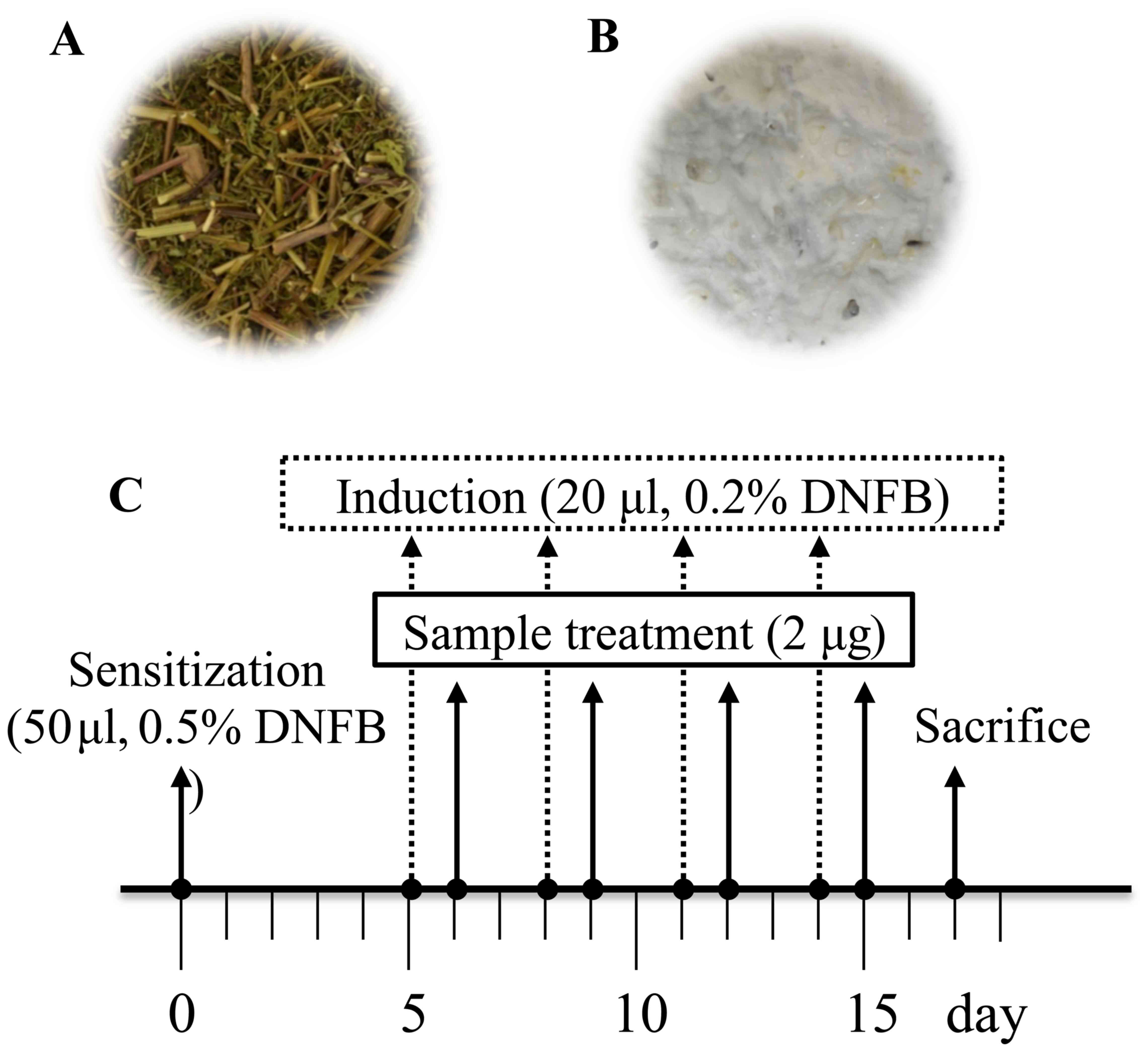
for 20 days. A. capillaris was dried and autoclaved at 121°C
for 20 min (Fig. 1A), and 10 g of
the grown mycelia with potato dextrose agar medium was then adapted
into 200 g of A. capil-laris. Even though the mycelia were
in liquid potato dextrose agar medium, they were rapidly dried
(<5% moisture) and the mixed materials were subjected to solid
fermentation. This fermentation was performed at 25°C for 2 weeks,
and we finally observed the white fruit body of G. lucidum
(Fig. 1B). The fermented product
was extracted with distilled water ina shaking incubator in at 25°C
for 24 h, and it was then lyophilized (MCTD85; Il-Shin,
Gyeonggi-do, Korea) following filtration. The voucher specimens of
the plant and fermented samples have been deposited in the
Laboratory of Enzyme Biotechnology, Kyungpook National University,
Daegu, Korea.
Animal model of DNFB-induced atopic
dermatitis
We created an animal model of DNFB-induced atopic
dermatitis as previously described, with some modifications
(18,19). In brief, 50 µl of 0.5%
(w/v) DNFB solution (Sigma; dissolved in polyethylene glycol) was
applied on the abdominal skin of mice for primary sensitization. At
5 days following sensitization, 20 µl 0.2% (w/v) DNFB was
applied to the ear skin at 3-day intervals. Treatment with 20
µl of the samples (final concentration, 100 µg/ml)
was carried out 1 day following exposure to DNFB (Fig. 1C). In total, DNFB and the
treatment samples were applied 4 times and we observed
inflammation-related symptoms, such as swelling or scabs on the
mouse ear surface. At 2 days following the final sample
application, the mice were sacrificed and the ear organs were
isolated. The experimental groups were divided into the NT (vehicle
alone) group, DNFB (DNFB treatment alone) group, Ac (DNFB
treatment and thereafter application of aqueous fraction of
Ac) group, Gl (DNFB treatment and thereafter
application of aqueous fraction of Gl) group and
afAC/Gl (DNFB treatment and thereafter application of
aqueous fraction produced by solid fermentation of Gl on
Ac) group, as described in Fig. 1.
Hematoxyline and eosin (H&E) staining
and immunohis-tochemistry
H&E staining was carried out as previously
described (20). In brief, the
ear tissues were entirely cut and fixed with 10% formalin solution
in phosphate-buffered saline (PBS) for at least 12 h. In order to
maintain the linear shape of the ear tissue, it was placed between
2 coverslips following formalin fixation. The organ sections were
fixed in paraffin lengthwise and transferred onto slides.
Subsequently, 3% hydrogen peroxide in methanol solution was applied
to prevent endogenous peroxidase activity and the slides were
stained with H&E (HHS32 and HT110332΄ Sigma). To perform
immunohistochemical analysis, each slide was treated with a 10%
normal goat serum for 1 h, and the slides were then incubated
overnight at 4°C with rabbit anti-mouse antibodies against iNOS
(ab15326), endothelial NOS (eNOS; ab5589) and neuronal NOS (nNOS;
ab72428) (all from Abcam, Cambridge, MA, USA), as previously
described (21). To observe the
immunohistochemical sections, a Nikon microscope and camera
(Eclipse, TE-2000U; Nikon, Tokyo, Japan) were used.
Evaluation of atopic dermatitis-related
symptoms
Various factors of atopic responses (ear swelling,
ear epidermal swelling, total cell count and eosinophil count) were
measured on the H&E-stained sections. First, ear thickness or
ear epidermal thickness was calculated using ImageJ software [1.48
version, National Institutes of Health (NIH)]. Next, total cell and
eosinophil counts were determined. Due to the of unique morphology
of eosinophils, total cells were counted first, and then eosinophil
cells were observantly counted under a microscope (Eclipse,
TE-2000U; Nikon) at ×400 magnification.
Measurement of NO levels and cytotoxicity
assay
NO production and cell viability were assayed using
the RAW264.7 cell line, which was obtained from American Type
Culture Collection (Cat. no. TIB-71; Manassas, VA, USA). The cells
were seeded as 5×10⁴ cells/well with 200 µl of
medium [10% fetal bovine serum (FBS; Hyclone, Lagan, UT, USA) in
Dulbecco's modified Eagle's medium (DMEM; Hyclone)] in a 96-well
plate. The cells were pre-incubated in a 96-well plate for 4 h, and
this was followed by the addition of lipopolysaccharide (LPS, 1
mg/ml, L2880; Sigma) and the treatment samples and incubation at 5%
CO2, 37°C for 24 h. To detect NO production, 100
µl of supernatants from the cell culture plate were moved to
a new 96-well plate, and 100 µl of Griess reagent (G4410;
Sigma) were then added. According to the reaction between NO
product and Griess reagent, the color changed yellow to pink and
the absorbance was then measured at 520 nm on a spectrophotometer
(VICTOR3; Perkin Elmer, Wellesley, MA, USA). To measure the
cytotoxicity of the treatment samples, cell culture medium was
removed from a 96-well cell culture plate which had remained
following NO assay. Subsequently, 100 µl of DMEM with 0.5
mg/ml methyl-thiazolyldiphenyltetrazolium bromide (MTT, M2128)
solution were added followed by incubation for 30 min. The
supernatants were removed and the plates were washed twice with
PBS. Thereafter, the produced formazan crystals from the MTT
solution were dissolved into DMSO and cell viability was assessed
by measuring the absorbance at 595 nm on a spectrophotometer.
Reverse transcription-polymerase chain
reaction (PCR) for mRNA expression
To determine the effects of A. capillaris on
mRNA expression in RAW264.7 cells, the cells were seeded at
5×10⁶ cells/well in a 6-well plate (BD Falcon, NJ, USA).
The cells were treated with LPS (1 µg/ml) for 4 h and with
the sample extracts at 30 µg/ml for 5 h. Following the
removal of the cell supernatant, RNA was isolated using
TRIzol® reagent (Invitrogen, New York, NY, USA). Total
RNA was reverse transcribed into cDNA using RT-PCR master mix
(Qiagen, Valencia, CA, USA). cDNA was amplified with manufactured
primers as follows: matrix metalloproteinase (MMP)-2 forward,
ACCAGAACACCATCGAGACC and reverse, AAAGCATCATCCACGGTTTC; MMP-7
forward, GAGTGCCAGATGTTGCAGAA and reverse, CCATCAAAGGGGAAGCTGT;
MMP-9 forward, GGGTTTCTGTCCAGACCAAG and reverse,
GGATGCCGTCTATGTCGTCT; MMP-12 forward, TTGATGGCAAAGGTGGTACA and
reverse, CGAAATGTGCTGGGGTTAAG; MMP-14 forward, CCGCCATGCAAAAGTTCTAT
and reverse, GCCTTGATCTCAGTCCCAAA, MMP-19 forward,
GACATTCGCCTCTCTTTCCA and reverse, AGGTCCCCTCAGTCCAGAGT; and
glyceraldehyde 3-phosphate dehydrogenase (GAPDH) forward,
ATGTTCCAGTATGACTCCAC and reverse, GCCAAAGTTGTCATGGATGA (Bioneer,
Daejeon, Korea) and detected using 1% agarose gel by ChemiDOC™
XRS+ Molecular Imager (Bio-Rad, Hercules, CA, USA). The
band density from the esults of the PCR product was calculated
using Image Lab software (5.0 version; Bio-Rad).
High-performance liquid chromatography
photodiode array (HPLC-PDA) analysis
Cholorogenic acid and caffeic acid were analyzed as
described in a previous study (11). HPLC analysis was performed using a
Waters 2695 HPLC system (Waters Co., Milford, MA, USA) fitted with
a binary pump, an autosampler, a column oven and a PDA detector.
The HPLC system was monitored by a computer equipped with Empower
software (Waters Co.). Chromatographic separation was performed on
a HYPERSIL ODS C18 column (250×4.6 mm, 5 µm; Thermo
Scientific, Waltham, MA, USA). The chromatographic binary mobile
phase consisted of 1% (v/v) aqueous acetic acid (A) and 1% (v/v)
acetic acid in acetonitrile (B) for chlorogenic acid analysis. The
gradient flow rates were as follows: 0–5 min, 0–10% B; 5–30 min,
10–50% B; 30–35 min, 50–50% B; 35–40 min, 50-10% B. The flow rate
and injection volume were 1.0 ml/min and 10 µl,
respectively. The detection wavelength was set at 320 nm. Caffeic
acid separated at a flow rate of 0.5 ml/min; solvent A was
acetonitrile and solvent B was acetic acid solution (dilute 20 ml
of glacial acetic acid to 1,000 ml with DI water, pH 2.6). Elution
was performed according to the following conditions: 0–5 min, 5% A;
25–30 min, 35% A; 35–40 min 90% A; 40.1–41 min, 5% A and monitored
at 280 nm.
Statistical analysis
Data are expressed as the means ± standard
deviation. Statistical significance was determined by one-way ANOVA
with Tukey's post-hoc test using the SPSS 21.0 program (SPSS, Inc.,
Chicago, IL, USA). The critical level for significance was set at
P<0.05.
Results and Discussion
Effects of aqueous fraction produced by
the solid fermentation of Gl on Ac (afAc/Gl) on a mouse model of
DNFB-induced atopic dermatitis
To determine the inhibitory effects of
afAc/Gl against atopic dermatitis-related symptoms, we
established a mouse model of DNFB-induced atopic dermatitis
(Fig. 1C) and analyzed the data.
First, we conducted an observation of the ear surface, as atopic
dermatitis is characterized by the appearance of inflammation. In
the DNFB-treated mice, due to scratching of the ears, we found that
the skin had swelled and scabs had formed on the DNFB-applied
tissue sites. However, we found that the sample-treated mice,
particularly those treated with afAc/Gl, exhibited less
scratching behavior, ear swelling and scabs. At the end of the
experimental period, the ears of the mice in the DNFB-treated group
had become twisted in appearance due to the inflammatory response
in the body, whereas in the sample-treated groups, the ears of the
mice were only moderately twisted in appearance; in particular, the
ears of the mice in the afAc/Gl-treated group were almost
similar in appearance to those of the control group (Fig. 2A). To obtain more reliable
positive data, we assessed ear thickness on H&E-stained slides
using ImageJ software. The paraffin-embedded ear tissues exhibited
incremental thickness form the edge of the outer site of the ear
towards the inside; therefore, we selected the same section and
averaged the length for further investigation. The whole appearance
of the paraffin-embedded ear tissue was observed under a microscope
at a magnification of ×40 (Fig.
2B), and half the ear tissue on cartilage was observed at ×400
magnification (Fig. 2C). The
results revealed that ear thickness in the afAc/Gl-treated
mice decreased by 32%, while that of the Gl-treated mice
decreased by 13%; ear thickness in the Ac-treated mice did
not differ compared to the NT group (Fig. 2D). In addition, ear epidermal
thickness in the afAc/Gl-treated mice decreased by 37%,
which was the most signicant among the sample treatment groups.
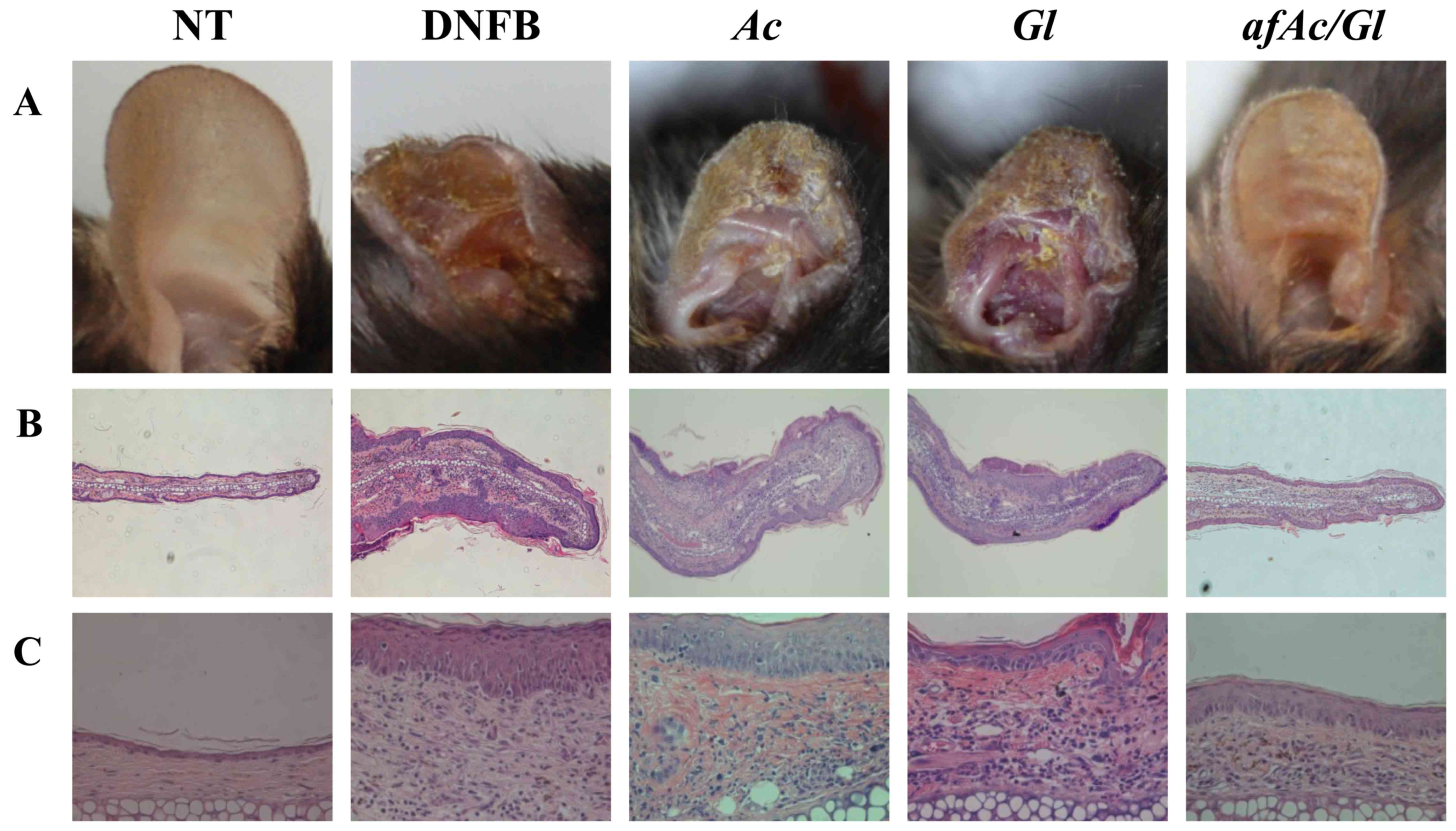 | Figure 2Inhibitory effect of an aqueous
extract of Artemisia capillaris following fermentation with
Ganoderma lucidum against 2,4-dinitrofluorobenzene
(DNFB)-induced atopic dermatitis-related symptoms in C57BL/6 mice.
Apart from the NT group, all groups were treated 0.2% (w/v) DNFB on
the ear surface (concentration of 30 µg/ml). NT, no
treatment group; Ac, Artemisia capillaris; Gl,
Ganoderma lucidum; afAc/Gl, aqueous fraction produced
by solid fermentation of Ganoderma lucidum on Artemisia
capillaris. (A) Images of ears of mice. (B and C) Observation
of atopic dermatitis-related symptoms by H&E staining, (B) ×40
magnification and (C) ×400 magnification. (D) Assessment of atopic
dermatitis-related symptoms reduction by afAc/Gl. Ear
thickness (first row), ear epidermal thickness (second row), and
total cell (third row), eosinophil (forth row) counts were assessed
as described in the Materials and methods. Data were randomly
extracted 5 times from each paraffin-embedded section, and
calculated using ImageJ software. The values of quantitative
measurement were adjusted to those of the DNFB-treated group. Data
are expressed as the means ± standard deviations (n=5);
*P<0.05. |
In atopic dermatitis, eosinophil numbers depend on
various factors, such as platelet-activating factor (PAF),
cytokines, leucotrienes and prostanoids (22). Therefore, eosinophil numbers can
be predicted by increased itching, which may not be observed in
normal skin. As shown by our results, eosinophils were observed at
limited numbers in the control group; however, the eosinophil
numbers were markedly inreased in the DNFB-treated group. Treatment
with afAc/Gl decreased the number of eosinophils by
approximately 74% compared to those of the DNFB-treated group
(Fig. 2D). These results suggest
that the solid fermentation of afAc by Gl may be used to
enhance the anti-inflammatory potential in atopic dermatitis.
afAc/Gl extract exhibits enhanced
anti-inflammatory potential in atopic dermatitis by reducing eNOS
expression in vivo
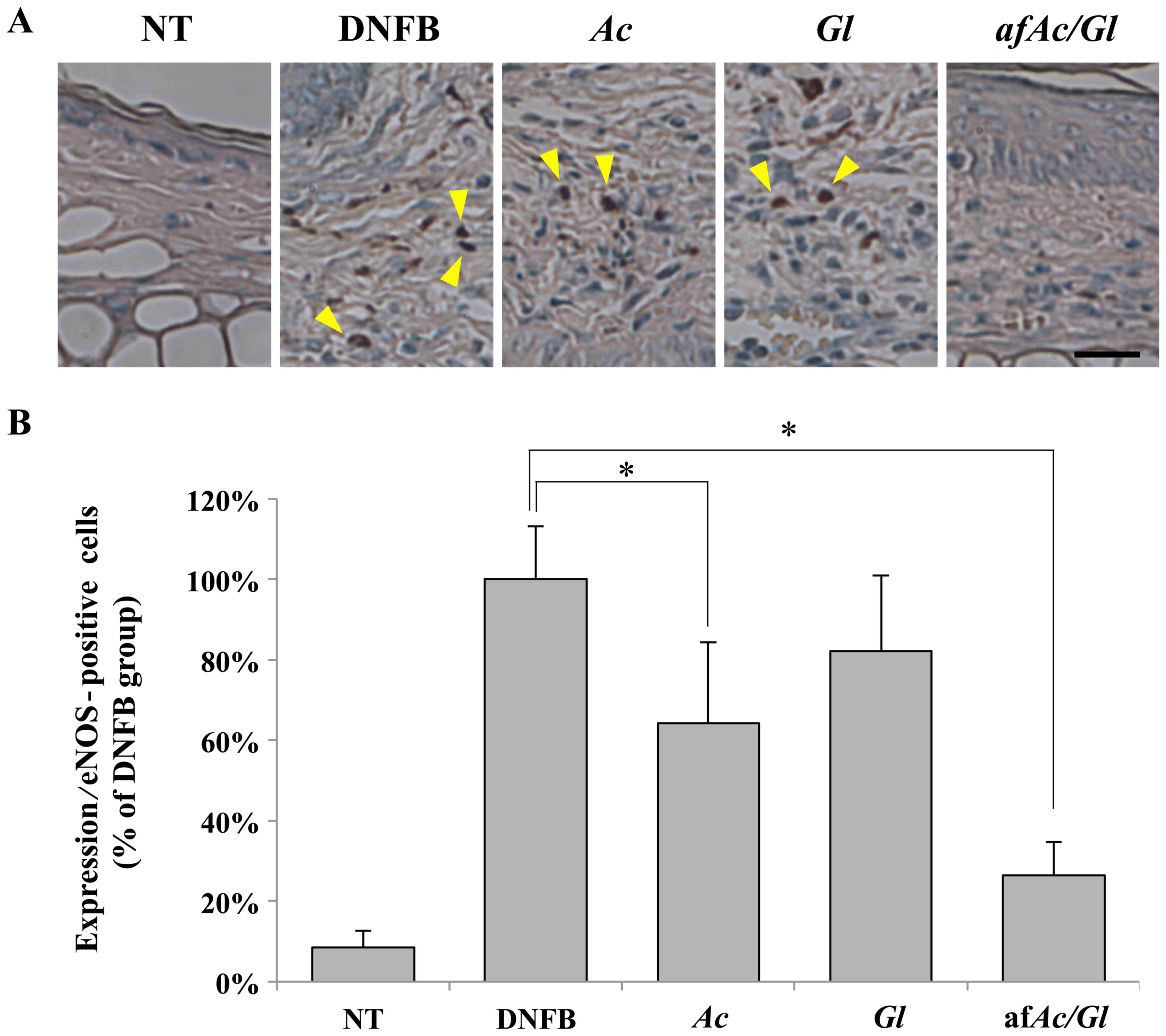
We then investigated whether eNOS plays a pivotal
role in downregulating the protein expression in skin tissues
exposed to DNFB. Protein expression was determined by
immunohistochemical microscopic analysis at a magnification of ×400
(Fig. 3A). We first counted the
number of eNOS-positive cells on overall paraffin-embedded ear
tissues. The results revealed that the tissue of the
afAc/Gl-treated mice did not exhibit any eNOS-positive
cells, as opposed to the tissue of the DNFB-, Ac- or
Gl-treated mice (Fig. 3A,
compare the number of arrowheads). Thus, eNOS expression was
increased by DNFB and afAc/Gl treatment attenuated this
effect. As shown in Fig. 3B, the
number of eNOS-positive cells in the afAc/Gl-treated group
was signifi-cantly decreased compared to the DNFB group. Treatment
with Ac also decreased the numer of eNOS-positive cells, but
not as significantly as afAc/Gl. Similarly, iNOS was also
excessively expressed in the DNFB-treated group (Fig. 3C and D). However, only treatment
with afAc/Gl significantly decreased the number of
iNOS-positive cells. The expression of iNOS did not exhibit any
significant changes between the groups (data not shown).
NO is an important molecule in body homeostasis as a
secondary signal mediator, and it is synthesized from L-arginine by
NOS [nNOS (NOS1), iNOS (NOS2) and eNOS (NOS3)]. iNOS is mainly
well-known as an inflammatory mediator with a function similar to
COX-2, which is generally upregulated in inflammation (23–25). It has been well-documented that
iNOS is upregulated during the inflammatory response in various
cancer cells exposed to LPS, while nNOS (NOS1) and eNOS (NOS3) are
expressed constitutively (23).
iNOS is a key enzyme among the NOSs, although the three NOSs have
been shown to be involved in various inflammation-related diseases
(26). Recently, it was
demonstrated that, in dogs with D. farinae-induced atopic
dermatitis, eNOS was strongly expressed along with iNOS, NADPH
oxidase 4 (NOX4), transforming growth factor-β1 (TGF-β1), COX-2 and
myeloperoxidase (MPO), suggesting that these molecules are crossly
associated with the cascade for the metabolic pathway (27). It has been reported that eNOS and
iNOS are expressed in skin biopsies of patients with atopic
dermatitis (28). On the other
hand, in another study on colitis, eNOS-transgenic mice exhibit
lower weight loss, less mortality, less inflammation-induced
damage, les colonic myeloperoxidase activity and a lower expression
of tumor necrosis factor-α compared to WT mice (29). Therefore, the study of eNOS
expression has yielded conflicting results; however, there seems to
be a lower expression of eNOS with a less active inflammatory
response, which are taken together with our data on Fig. 3B.
Asthma is very similar to atopic dermatitis by
comparison of the molecular mechanisms and symptoms. In a previous
study, NOS-deficient mice were treated with ovalbumin to induce
airway inflammation, and thereafter airway hyperresponsiveness was
evaluated using methacholine response. In that study, the
iNOS-deficient mice exhibited similar results to the WT mice, but
the nNOS-deficient mice exhibited significantly less airway
responsiveness than the WT mice (30). Overall, these data suggest that
NOSs are major factors inatopic dermatitis; we confirmed that eNOS,
as opposed to iNOS, was a more critical controlling factor in
atopic dermatitis-related symptoms.
afAc/Gl downregulates MMP expression in
vitro
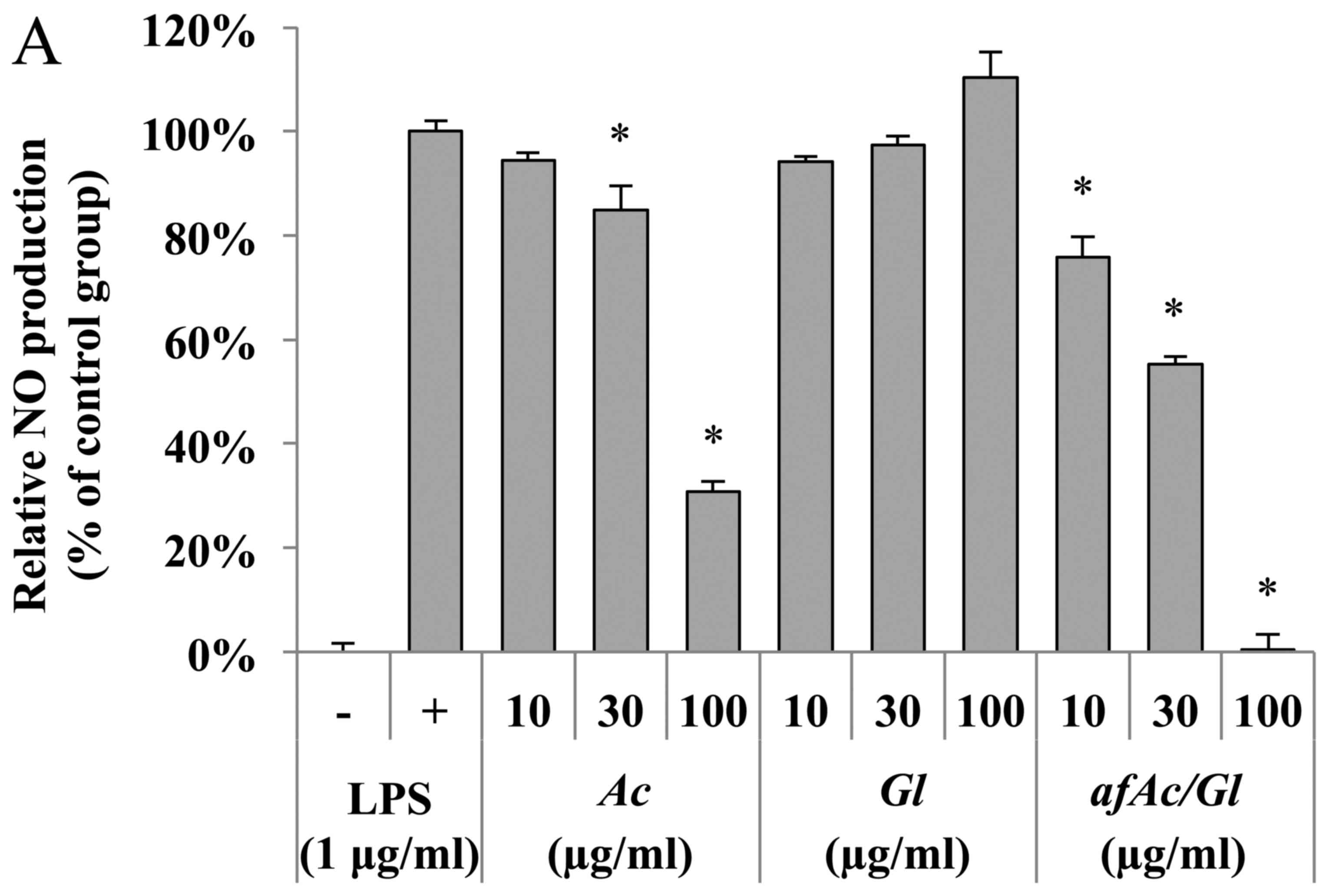
We then also used RAW264.7 cells to prove which
molecular target(s) is regulated by the fraction. Anti-inflammatory
effects can be examined by NO production, which was induced by LPS
(500 ng/ml). As a result, no toxicity was observed from all samples
by the MTT assay. The afAc/Gl (most potent inhibitory
activity at 100 µg/ml) inhibited NO activity more
significantly compared with Ac (69% inhibitory activity),
although Gl had no significant effect (Fig. 4A, compare bars 6–9).
MMPs are also a family of zinc-dependent enzymes
that regulate the extracellular matrix as key factors in
inflammation and disease (31).
We found that afAc/Gl extract regulated the expression of
MMP-2, -7, -9, -12, -14 and -19 in RAW264.7 cells; however,
Ac had a lesser effect on MMP expression compared
toafAc/Gl, even though Ac significantly decreased
MMP-9 expression (Fig. 4B).
MMP-1a-MMP-29 have been shown to be expressed in various cell
lines, indicating that these proteases are associated with
inflammatory events in various cell populations. Among these, MMP-2
and -9 have been shown to be associated with allergic inflammation
(32). A previous study
demonstrated that MMP-2 was expressed in the epidermis and dermis
from patients with atopic dermatitis, while MMP-9 was not
upregulated (33). However,
another study demonstrated that MMP-9 was upregulated in a mouse
model of DNFB-induced atopic dermatitis, and the IL-31 and T-bet
gene were also expressed in the ear epidermis (34). MMP-2 and -12 have been shown to be
upregulated in the lung by inspiratory resistive breathing
(35). Another study demonstrated
that the mRNA levels of MMP-1, MMP-7, MMP-10, MMP-14 and MMP-19
were increased independently of COX-2 in monocytes stimulated with
LPS (36). Therefore, our results
on the MMP regulation pattern imply that afAc/Gl exerts
potent effects against atopic dermatitis via the inhibition MMP-2,
-7, -9, -12, -14 and -19. Moreover, in a previous study,
MMP-9-knockout mice exhibited increased levels of IL-4, and IL-13
in lung tissue, as shown by immunohistochemical analysis (37), resulting in an increase in
eosinophil and neutrophils counts in bronchioalveolar lavage fluid
from MMP-9 knockout mice. MMP-2 knockout mice have been shown to
exhibit increased asthmatic symptoms and asphyxiation induced by
allergens (38). Moreover,
MMP-19-deficient mice have been shown to exhibit exacerbated
eosinophilic inflammation in bronchoalveolar lavage fluid and
bronchial tissue following allergen challenge (39). Thus, these data indicate that some
MMPs are critical to the maintenance of lung tissue, and may
protect against asthmatic symptoms. Therefore, MMPs are not only
upregulated by allergic inflammation, but also maintaine
tissue-specific metabolism in the lungs. Further investigations on
the role of MMPs in atopic dermatitis or asthma are warranted in
order to develop appropriate curative therapy and/or of preventive
food ingredients.
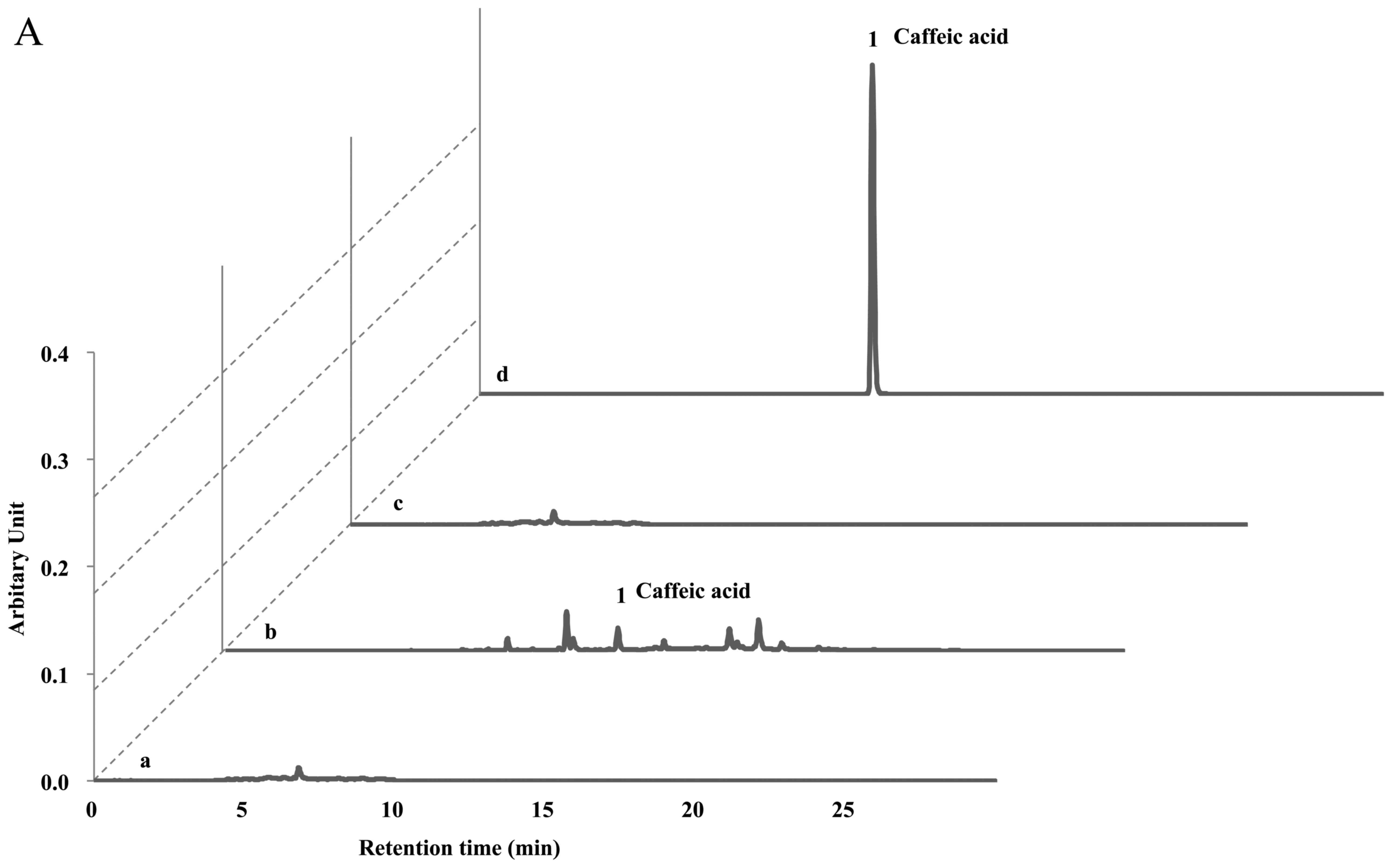
HPLC analyses of afAc/Gl
The protective effects of the Ac extract (70%
ethanol) against atopic dermatitis have previously been reported
(11). Ac contains potent
compounds as anti-atopic or inflammatory constituents, such as
hyperoside, isoquercetrin, chlorogenic acid, isochlorogenic acid,
caffeic acid and scoparone. Therefore, we investigated whether the
solid fermentation process can enhance the anti-atopic activity by
converting these compounds into more potent ingredients.
Surprisingly, we found that the afAc/Gl extract did not show
any peaks on the HPLC analysis sheet, although the Ac
extract contained these compounds (Fig. 5). Since the Gl mycelium
weight was <5% in the afAc/Gl extract and the Gl
extract showed no activity against atopic dermatitis, we predicted
that the active compounds had increased following solid
fermentation by converting the compounds, such as chlorogenic acid
and caffeic acid. Taken together, these results strongly suggest
that isolation and purification of converted compounds is
effective, in that converted compound(s) can be used as anti-atopic
agent(s), as well as cosmetic ingredient(s) without any
toxicity.
In conclusion, the solid fermentation of the
Ac extract with Gl enhanced the anti-inflammatory
effects on atopic dermatitis. In mice with DNFB-induced atopic
dermatitis, treatment with afAc/Gl led to a 32% decrease in
ear thickness, and a decrease in eosinophil number of 74%, which
was approximately 2-7-fold more effective than the Ac
extract without fermentation. Additionally, the numbers of iNOS-
and eNOS-positive cells were decreased by treatment with
afAc/Gl, as shown by immunohistochemical analysis. The
afAc/Gl extract was more effective than Ac on NO
inhibition, as well as on the inhibition of MMP-2, -7, -9, -12, -14
and -19 mRNA expression in RAW264.7 cells. As shown by HPLC
analysis, we found that the Ac extract contained caffeic
acid, catechin and chlorogenic acid, while the afAc/Gl
extract did not. Taken together, we hypothesized that these and
other polyphenolic compounds had been changed into novel
biomaterial(s) with anti-inflammatory potential by solid
fermentation, although we aim to perform further investigations to
identify the specific anti-inflammatory ingredient(s) in the
afAc/Gl extract. Importantly however, we demonstrate that
solid fermentation is an innovative technology for the enhancement
of bioconversion during the processing of natural products.
Acknowledgments
This study was supported by the Basic Science
Research Program through the National Research Foundation of Korea
(NRF) funded by the Ministry of Science, ICT and Future Planning
(NRF-2014R1A2A2A01006882).
References
|
1
|
Ring J, Alomar A, Bieber T, Deleuran M,
Fink-Wagner A, Gelmetti C, Gieler U, Lipozencic J, Luger T, Oranje
AP, et al European Dermatology Forum (EDF); European Academy of
Dermatology and Venereology (EADV); European Federation of Allergy
(EFA); European Task Force on Atopic Dermatitis (ETFAD); European
Society of Pediatric Dermatology (ESPD); Global Allergy and Asthma
European Network (GA2LEN): Guidelines for treatment of atopic
eczema (atopic dermatitis) part I. J Eur Acad Dermatol Venereol.
26:1045–1060. 2012. View Article : Google Scholar : PubMed/NCBI
|
|
2
|
Leung DY, Boguniewicz M, Howell MD, Nomura
I and Hamid QA: New insights into atopic dermatitis. J Clin Invest.
113:651–657. 2004. View
Article : Google Scholar : PubMed/NCBI
|
|
3
|
Harper JI, Godwin H, Green A, Wilkes LE,
Holden NJ, Moffatt M, Cookson WO, Layton G and Chandler S: A study
of matrix metalloproteinase expression and activity in atopic
dermatitis using a novel skin wash sampling assay for functional
biomarker analysis. Br J Dermatol. 162:397–403. 2010. View Article : Google Scholar
|
|
4
|
Jeong MS, Choi SE, Kim JY, Kim JS, Kim EJ,
Park KH, Lee do I, Joo SS, Lee CS, Bang H, et al: Atopic
dermatitis-like skin lesions reduced by topical application and
intraperitoneal injection of hirsutenone in NC/Nga mice. Clin Dev
Immunol. 2010:6185172010. View Article : Google Scholar :
|
|
5
|
Kim JB, Han AR, Park EY, Kim JY, Cho W,
Lee J, Seo EK and Lee KT: Inhibition of LPS-induced iNOS, COX-2 and
cytokines expression by poncirin through the NF-kappaB inactivation
in RAW 264.7 macrophage cells. Biol Pharm Bull. 30:2345–2351. 2007.
View Article : Google Scholar : PubMed/NCBI
|
|
6
|
Hong SH, Seo SH, Lee JH and Choi BT: The
aqueous extract from Artemisia capillaris Thunb. inhibits
lipopolysaccharide-induced inflammatory response through preventing
NF-kappaB activation in human hepatoma cell line and rat liver. Int
J Mol Med. 13:717–720. 2004.PubMed/NCBI
|
|
7
|
Jang E, Shin MH, Kim KS, Kim Y, Na YC, Woo
HJ, Kim Y, Lee JH and Jang HJ: Anti-lipoapoptotic effect of
Artemisia capillaris extract on free fatty acids-induced HepG2
cells. BMC Complement Altern Med. 14:2532014. View Article : Google Scholar : PubMed/NCBI
|
|
8
|
Feng G, Wang X, You C, Cheng X, Han Z,
Zong L, Zhou C and Zhang M: Antiproliferative potential of
Artemisia capillaris polysaccharide against human nasopharyngeal
carcinoma cells. Carbohydr Polym. 92:1040–1045. 2013. View Article : Google Scholar : PubMed/NCBI
|
|
9
|
Lee JH, Park EK, Uhm CS, Chung MS and Kim
KH: Inhibition of Helicobacter pylori adhesion to human gastric
adenocarcinoma epithelial cells by acidic polysaccharides from
Artemisia capillaris and Panax ginseng. Planta Med. 70:615–619.
2004. View Article : Google Scholar : PubMed/NCBI
|
|
10
|
Lee HI, Seo KO, Yun KW, Kim MJ and Lee MK:
Comparative study of the and Artemisia hepatoprotective efficacy of
Artemisia iwayomogi capillaris on ethanol-administered mice. J Food
Sci. 76:T207–T211. 2011. View Article : Google Scholar
|
|
11
|
Ha H, Lee H, Seo CS, Lim HS, Lee JK, Lee
MY and Shin H: Artemisia capillaris inhibits atopic dermatitis-like
skin lesions in Dermatophagoides farinae-sensitized Nc/Nga mice.
BMC Complement Altern Med. 14:1002014. View Article : Google Scholar : PubMed/NCBI
|
|
12
|
Martins S, Mussatto SI, Martínez-Avila G,
Montañez-Saenz J, Aguilar CN and Teixeira JA: Bioactive phenolic
compounds: Production and extraction by solid-state fermentation. A
review Biotechnol Adv. 29:365–373. 2011. View Article : Google Scholar
|
|
13
|
Lee IH, Hung YH and Chou CC: Solid-state
fermentation with fungi to enhance the antioxidative activity,
total phenolic and anthocyanin contents of black bean. Int J Food
Microbiol. 121:150–156. 2008. View Article : Google Scholar
|
|
14
|
Postemsky P and Curvetto N: Enhancement of
wheat grain antioxidant activity by solid state fermentation with
Grifola spp. J Med Food. 17:543–549. 2014. View Article : Google Scholar : PubMed/NCBI
|
|
15
|
Sánchez-Magaña LM, Cuevas-Rodríguez EO,
Gutiérrez-Dorado R, Ayala-Rodríguez AE, Valdez-Ortiz A,
Milán-Carrillo J and Reyes-Moreno C: Solid-state bioconversion of
chickpea (Cicer arietinum L.) by Rhizopus oligosporus to improve
total phenolic content, antioxidant activity and hypoglycemic
functionality. Int J Food Sci Nutr. 65:558–564. 2014. View Article : Google Scholar : PubMed/NCBI
|
|
16
|
Charlton E: Ethical guidelines for pain
research in humans. Committee on Ethical Issues of the
International Association for the Study of Pain. Pain. 63:277–278.
1995. View Article : Google Scholar : PubMed/NCBI
|
|
17
|
Shin YK, Son HU, Kim JM, Heo JC, Lee SH
and Kim JG: Cinnamomum cassia bark produced by solid-state
fermentation with Phellinus baumii has the potential to alleviate
atopic dermatitis-related symptoms. Int J Mol Med. 35:187–194.
2015.
|
|
18
|
Bhol KC and Schechter PJ: Topical
nanocrystalline silver cream suppresses inflammatory cytokines and
induces apoptosis of inflammatory cells in a murine model of
allergic contact dermatitis. Br J Dermatol. 152:1235–1242. 2005.
View Article : Google Scholar : PubMed/NCBI
|
|
19
|
Choi EM and Hwang JK: Antiinflammatory,
analgesic and antioxidant activities of the fruit of Foeniculum
vulgare. Fitoterapia. 75:557–565. 2004. View Article : Google Scholar : PubMed/NCBI
|
|
20
|
Sato Y, Mukai K, Watanabe S, Goto M and
Shimosato Y: The AMeX method. A simplified technique of tissue
processing and paraffin embedding with improved preservation of
antigens for immunostaining. Am J Pathol. 125:431–435.
1986.PubMed/NCBI
|
|
21
|
Haines DM and Chelack BJ: Technical
considerations for developing enzyme immunohistochemical staining
procedures on formalin-fixed paraffin-embedded tissues for
diagnostic pathology. J Vet Diagn Invest. 3:101–112. 1991.
View Article : Google Scholar : PubMed/NCBI
|
|
22
|
Ständer S and Steinhoff M: Pathophysiology
of pruritus in atopic dermatitis: An overview. Exp Dermatol.
11:12–24. 2002. View Article : Google Scholar : PubMed/NCBI
|
|
23
|
Patruno A, Franceschelli S, Pesce M,
Maccallini C, Fantacuzzi M, Speranza L, Ferrone A, De Lutiis MA,
Ricciotti E, Amoroso R, et al: Novel aminobenzyl-acetamidine
derivative modulate the differential regulation of NOSs in LPS
induced inflammatory response: Role of I3K/Akt pathway. Biochim
Biophys Acta. 1820:2095–2104. 2012. View Article : Google Scholar : PubMed/NCBI
|
|
24
|
Hollá LI, Bucková D, Kuhrová V,
Stejskalová A, Francová H, Znojil V and Vácha J: Prevalence of
endothelial nitric oxide synthase gene polymorphisms in patients
with atopic asthma. Clin Exp Allergy. 32:1193–1198. 2002.
View Article : Google Scholar : PubMed/NCBI
|
|
25
|
Yao S, Pandey P, Ljunggren-Rose A and
Sriram S: LPS mediated injury to oligodendrocytes is mediated by
the activation of nNOS: Relevance to human demyelinating disease.
Nitric Oxide. 22:197–204. 2010. View Article : Google Scholar
|
|
26
|
Moncada S and Higgs EA: Endogenous nitric
oxide: Physiology, pathology and clinical relevance. Eur J Clin
Invest. 21:361–374. 1991. View Article : Google Scholar : PubMed/NCBI
|
|
27
|
Jee MK, Im YB, Choi JI and Kang SK:
Compensation of cATSCs-derived TGFβ1 and IL10 expressions was
effectively modulated atopic dermatitis. Cell Death Dis.
4:e4972013. View Article : Google Scholar
|
|
28
|
Rowe A, Farrell AM and Bunker CB:
Constitutive endothelial and inducible nitric oxide synthase in
inflammatory dermatoses. Br J Dermatol. 136:18–23. 1997. View Article : Google Scholar : PubMed/NCBI
|
|
29
|
Okaniwa N, Sasaki M, Mizushima T,
Ogasawara N, Funaki Y, Joh T and Kasugai K: eNOS plays an important
role in the regulation of colonic inflammation: A novel therapeutic
target and a predictive marker for the prognosis of ulcerative
colitis. Free Radic Res. 49:35–44. 2015. View Article : Google Scholar
|
|
30
|
De Sanctis GT, MacLean JA, Hamada K, Mehta
S, Scott JA, Jiao A, Yandava CN, Kobzik L, Wolyniec WW, Fabian AJ,
et al: Contribution of nitric oxide synthases 1, 2, and 3 to airway
hyper-responsiveness and inflammation in a murine model of asthma.
J Exp Med. 189:1621–1630. 1999. View Article : Google Scholar : PubMed/NCBI
|
|
31
|
Vandenbroucke RE and Libert C: Is there
new hope for therapeutic matrix metalloproteinase inhibition? Nat
Rev Drug Discov. 13:904–927. 2014. View Article : Google Scholar : PubMed/NCBI
|
|
32
|
Kim DY, Park BS, Hong GU, Lee BJ, Park JW,
Kim SY and Ro JY: Anti-inflammatory effects of the R2 peptide, an
inhibitor of transglutaminase 2, in a mouse model of allergic
asthma, induced by ovalbumin. Br J Pharmacol. 162:210–225. 2011.
View Article : Google Scholar :
|
|
33
|
Jin H, Oyoshi MK, Le Y, Bianchi T, Koduru
S, Mathias CB, Kumar L, Le Bras S, Young D, Collins M, et al:
IL-21R is essential for epicutaneous sensitization and allergic
skin inflammation in humans and mice. J Clin Invest. 119:47–60.
2009.
|
|
34
|
Heo JC, Nam DY, Seo MS and Lee SH:
Alleviation of atopic dermatitis-related symptoms by Perilla
frutescens Britton. Int J Mol Med. 28:733–737. 2011.PubMed/NCBI
|
|
35
|
Toumpanakis D, Noussia O, Sigala I,
Litsiou E, Loverdos K, Zacharatos P, Karavana V, Michailidou T,
Magkou C, Zhou Z, et al: Inspiratory resistive breathing induces
MMP-9 and MMP-12 expression in the lung. Am J Physiol Lung Cell Mol
Physiol. 308:L683–L692. 2015. View Article : Google Scholar : PubMed/NCBI
|
|
36
|
Reel B, Sala-Newby GB, Huang WC and Newby
AC: Diverse patterns of cyclooxygenase-independent
metalloproteinase gene regulation in human monocytes. Br J
Pharmacol. 163:1679–1690. 2011. View Article : Google Scholar : PubMed/NCBI
|
|
37
|
Corry DB, Kiss A, Song LZ, Song L, Xu J,
Lee SH, Werb Z and Kheradmand F: Overlapping and independent
contributions of MMP2 and MMP9 to lung allergic inflammatory cell
egression through decreased CC chemokines. FASEB J. 18:995–997.
2004.PubMed/NCBI
|
|
38
|
Corry DB, Rishi K, Kanellis J, Kiss A,
Song Lz LZ, Xu J, Feng L, Werb Z and Kheradmand F: Decreased
allergic lung inflammatory cell egression and increased
susceptibility to asphyxiation in MMP2-deficiency. Nat Immunol.
3:347–353. 2002. View
Article : Google Scholar : PubMed/NCBI
|
|
39
|
Gueders MM, Hirst SJ, Quesada-Calvo F,
Paulissen G, Hacha J, Gilles C, Gosset P, Louis R, Foidart JM,
Lopez-Otin C, et al: Matrix metalloproteinase-19 deficiency
promotes tenascin-C accumulation and allergen-induced airway
inflammation. Am J Respir Cell Mol Biol. 43:286–295. 2010.
View Article : Google Scholar
|