Introduction
Camellia japonica L. (C. japonica) is
a tree belonging to the Theaceae family and is cultivated and has
been used in traditional medicine in Japan, China, and Korea for
the treatment of stomachic illness, bleeding and inflammation
(1,2). The usage of the seed and flower from
C. japonica was first reported in ʻDongui Bogamʼ which was
thought to have been written by the doctor of oriental medicine,
Heo Jun in 1613 (3).
C. japonica is a perennial herb that grows in
forests in the southern region of Korea. Recently, the northern
limits of C. japonica have widely increased due to the
greenhouse effect on Earth's atmosphere in South Korea (4). Additionally, the provincial
governments of South Korea have made many efforts to industrialize
C. japonica in the field of food and medicinal sources. From
2004, green tea from C. japonica has been developed as a
food source in Korea so called ʻDong-Baek Chaʼ which was
commercialized in 2007 (5).
However, the specific and beneficial usage of the leaf as a
functional food source and/or plant medicine has not yet been
determined.
Chemically, this species has shown to be constituted
of triterpenes, saponins, glycosylated flavonoids and tannins
(1,6–8).
Several of these derivatives have been shown to exhibit biological
activity, such as antioxidant (7), antifungal (9), anti-viral (10) and cytotoxic potential (8).
In our preliminary experiments, we screened
approximately 500 plant extracts and selected the extract of C.
japonica leaf as a xanthine oxidase (XO) inhibitor to find an
alternative medicine for hyperuricemia. We investigated the active
constituents and the biological activities of extracts from C.
japonica leaf for the development of possible alternatives to
allopurinol.
Hyperuricemia is a condition in which the levels of
uric acid in the blood exceed the normal range, which is due to the
regular intake of food with a high purine content and is invariably
accompanied by gout, chronic kidney disease and other metabolic
syndromes (11). Uric acid is the
final metabolite of the purine compound, which is formed by the
oxidation of hypoxanthine to xanthine and xanthine to uric acid by
XO (12). The overproduction of
uric acid by XO leads to hyperuricemia, which is the main cause of
gout. Gout is a metabolic disorder that is closely associated with
high levels of uric acid in the body, which can cause inflammation,
gouty arthritis and uric acid nephrolithiasis (13). Gout has been reported to afflict
>2 million men and women in the US alone (14). The prevalence of gout is rapidly
increasing in other regions, including Northeast Asia (15), likely due to changes in dietary
habits.
The aim of the present study was to investigate the
potential of extract of C. japonica leaf (ECJL) to inhibit
XO and to act as an antioxidant in vitro and in vivo.
Moreover, the extract was investigated for its effects on the
elimination of uric acid in serum and the inhibition of XO in mice
with potassium oxonate-induced hyperuricemia. Screening of the
extract for its XO inhibitory activity followed by their potential
to reduce serum uric acid levels and XO activity may play an
important role in identifying a potent chemical entity for the
treatment of gout and related inflammatory disorders. To clarify
the biological marker in ECJL, we describe the identification of
bioactive constituents that include rutin, vitamin E,
all-trans-squalene, neophytadiene, linolenic acid, n-hexadecanoic
acid, n-octacosane, n-eicosane, and 6,9-pentadecadien-1-ol using
gas chromatography-mass spectrometry (GC-MS) and high-performance
liquid chromatography (HPLC) analysis.
Materials and methods
Plant materials
The leaves of C. japonica were provided from
Jeollanamdo Wando Arboretum, in Jeonnam, Korea. C. japonica
leaves were collected on Joyag island, Korea (126°56′50.07″E
longitude and 34°22′31.27″N latitude). A voucher specimen
(MNUCSS-CJ-01) was deposited at Mokpo National University (Muan,
Korea). The leaves were separated for use in the present study. The
air-dried and powdered C. japonica leaf (10 g) was extracted
twice with ethanol (100 ml) at room temperature for 3 days. The
resultant ethanol solution was evaporated, dried and stored at
−50°C. The sample was used for in vitro and in vivo
experiments, and for the analysis of the active constituents.
Animals
Male ICR mice (4 weeks old) were purchased from
Orient Bio Co. (Sungnam, Korea). The mice were retained in a clean
room at a temperature of 20–23°C with 12-h light (07:00–19:00) and
dark (19:00–07:00) cycles, and a relative humidity of 50±5%. The
mice were housed in ventilated mice cages (Tecniplast, West
Chester, PA, USA) under filtered and pathogen-free air, with food
(Agribrands Purina Korea, Inc., Seoul, Korea) and water available
ad libitum. All animal experiments were carried out
according to the guidelines of the Animal Investigation Committee
of Jeonnam Bioindustry Foundation (Naju, Korea) (approval no.
JINR1517).
Profiling of constituents by GC-MS
analysis
The analytical methods for the analysis based on
GC-MS have been previously reported (16). The analysis of scanned organic
compounds was performed using an Agilent 7890 Gas Chromatograph
System, coupled to a quadrupole Agilent 5975C electron ionization
(EI) (70 eV) mass spectrometric detector (Agilent Technologies,
Inc., Palo Alto, CA, USA). An Agilent HP-5MS fused silica capillary
column (30 m × 0.25 mm i.d., 0.25 µm film thickness) was
utilized for the identification of organic compounds. Briefly GC-MS
was tuned using perfluorotribuyl-amine (PFTBA) by mass fragments of
69.0, 219.0, 502.0 m/z under EI conditions. The GC oven was heated
using the following program: isothermal at 65°C for 10 min and 10
min−1 to 300°C with He as the carrier gas. The transfer
line was heated at 300°C, and the mass spectrometer was then
operated in scan mode (50–550 amu). All mass spectra were compared
with the data system library (NIST 2008). The summarized operation
parameters for the GC are shown in Table I.
 | Table IAnalysis condition of GC-MS. |
Table I
Analysis condition of GC-MS.
| Parameter | Condition |
|---|
| Column | Agilent HP-5MS
fused silica capillary column (30 m × 0.25 mm i.d., 0.25 µm
film thickness) |
| Carrier | Helium |
| Split | 1:5 |
| Injection
volume | 1 µl |
| MS source | 230°C |
| MS quad | 150°C |
| Thermal aux | 300°C |
| Electron
ionization | 70 eV |
| Mass range | 50–550 amu |
| Scan method | Full scan |
| Rate
(°C/min) | Value
(°C) | Hold time
(min) |
|---|
| Analytical
temperature |
| Initial | | 65 | 10 |
| Ramp | 10 | 300 | 22 |
| Total | 55.5 | | |
Profiling of constituents by HPLC
analysis
Constituent profiling of ECJL was performed with
HPLC. All HPLC analyses were performed using the Alliance 2695 HPLC
System (Waters Corp., Millford, MA, USA) equipped with a photodiode
array detector. The Agilent ZORBAX Extend-C18 (5 µm, 150 mm
l. × 5 mm i.d.) analytical column was used with a mobile phase
consisting of solvent A (acetonitrile) and solvent B (water
containing 0.2% phosphoric acid). Gradient elution (from 10/90 to
100/0, v/v) was performed at a flow rate of 1.0 ml/min (Table II). Column temperature was
maintained at 25°C. The detection wavelength was set at 270 nm for
rutin. The solvent was filtered through a 0.22-µm filter and
degassed. The sample injection volume was 10 µl.
| Table IIAnalytical conditions of HPLC for
analysis of ECJL. |
Table II
Analytical conditions of HPLC for
analysis of ECJL.
| Parameters | Conditions |
|---|
| Column | Zorbax extended-C18
(C18, 4.6 mm × 150 mm, 5 µm) |
| Flow rate | 1 ml/min |
| Injection
volumn | 10 µl |
| UV detection | 270 nm |
| Run time | 30 min |
| Time (min) | % Aa | % Bb |
|---|
| Gradient | 0 | 90 | 10 |
| 7 | 90 | 10 |
| 15 | 70 | 30 |
| 22 | 70 | 30 |
| 25 | 0 | 100 |
| 30 | 90 | 10 |
| 35 | 90 | 10 |
2,2-Diphenyl-1-picrylhydrazyl (DPPH) free
radical assay
The determination of the antioxidant activity of
ECJL was performed by the DPPH radical scavenging method. DPPH
radicals have an absorption maximum of 517 nm, which disappears
with reduction by an antioxidant compound. ECJL solution (1 ml)
containing 1–20 mg of ECJL was added to a 0.4 mM DPPH ECJL solution
(1 ml). The solution was mixed and allowed to react at room
temperature in the dark for 10 min. The absorbance at 517 nm was
measured using a microplate reader (PerkinElmer, Inc., Waltham, MA,
USA). The radical scavenging activity was calculated as a
percentage using the following equation: DPPH radical scavenging
activity (%) = [1 − (Asample/Ablank)] ×100.
The DPPH free radical scavenging activities of the samples were
compared in terms of their IC50 (µg/ml) values,
as previously described (17).
Determination of reducing power
The reducing power of ECJL was determined according
to a modified reducing power assay method. The sample (0.1 ml) was
added to 0.2 M sodium phosphate buffer (0.5 ml) and 1% potassium
ferricyanide (0.5 ml), and this mixture was incubated at 50°C for
20 min. Following incubation, 10% trichloroacetic acid solution
(0.5 ml) was added to the reaction mixture, and it was centrifuged
at 12,000 rpm for 10 min. The supernatant was mixed with distilled
water (0.5 ml) and a 0.1% iron (III) chloride solution (0.1 ml),
and the absorbance at 700 nm of the resulting solution was
measured. The reducing powers of the samples were expressed as
vitamin C equivalents, as previously described (17).
Determination of total phenolic
content
The total phenolic content was determined by
Folin-Ciocalteu assay, as previously described (18). Water solution (1 ml) containing 5
mg of ECJL or standard was mixed with 1 ml of 2% sodium carbonate
solution and 1 ml of 10% Folin-Ciocalteu's phenol reagent. After 10
min, the absorbance was measured at 750 nm using a microplate
reader (PerkinElmer, Inc.). The measurement was compared to
calibration curve of gallic acid. The results were expressed as
milligrams of gallic acid equivalents per gram of sample, as
previously described (17).
Determination of XO inhibitory activity
in vitro
The XO inhibitory activity was measured by
monitoring uric acid formation in a XO system, as previously
described (19). The assay system
consisted of 0.6 ml phosphate buffer (100 mM; pH 7.4), 0.1 ml
sample, 0.1 ml XO (0.2 U/ml) and 0.2 ml xanthine (1 mM; dissolved
in 0.1 N NaOH). The reaction was initiated by adding the enzyme
with or without inhibitors, and the change in absorbance of the
mixture at 290 nm for 15 min was measured against a reagent blank.
A 0.2 ml aliquot of 1 N HCl was used to terminate the enzymatic
reaction. Allopurinol was used as a positive control.
Establishment of the mouse model of
hyperuricemia and drug administration
Six groups of mice (n=5 for each group) were treated
once daily for 7 days as follows: the mice in the 2 negative
control groups (NOR and HU groups) received 0.3% CMC-Na aqueous
solution; the mice in the positive control group (ALLO group)
received allopurinol solution at a dose of 10 mg/kg; the mice in
the ECJL 30, ECJL 100, and ECJL 300 groups received the ECJL
solution at doses of 30, 100 and 300 mg/kg, respectively.
Hyperuricemia was induced in the mice by potassium oxonate, a
uricase inhibitor (20). Briefly,
ECJL (30, 100 and 300 mg/kg) or allopurinol (10 mg/kg) were
dissolved in 0.3% CMC-Na aqueous solution. The resultant drug
solutions were orally administered once per day for 7 days. Food,
except water, was withdrawn from the mice at 1.5 h prior to drug
administration, and the mice were intraperitoneally injected with
potassium oxonate (300 mg/kg) 1 h before the final drug
administration on the 7th day in order to induce hyperuricemia.
Blood samples were collected via the tail vein of the mice at 1 h
after the final drug administration on the 7th day. The blood
samples were allowed to clot for approximately 1 h at room
temperature and then centrifuged at 10,000 × g for 15 min to obtain
serum. The serum samples were stored at −80°C until use. The serum
uric acid concentration was measured using standard diagnostic kits
(Abcam, Cambridge, UK). Each assay was performed in triplicate.
Determination of XO inhibitory activity
in vivo
The residual activity of XO in the mouse liver and
plasma were spectrophotometrically determined by monitoring uric
acid formation from xanthine (12). The mouse livers (0.5 g) were
homogenized in an 1 ml aliquot of 50 mM sodium phosphate buffer (pH
7.4). The homogenates were centrifuged at 3,000 × g for 10 min at
4°C. After removing the lipid layer, the supernatant was
centrifuged at 10,000 × g for 60 min at 4°C. The resultant
supernatant was then used for determining XO residual activity and
the total protein concentration. A 0.12 ml aliquot of xanthine
solution (250 mM) was added to a test tube containing 10 µl
liver homogenate and 0.54 ml potassium oxonate solution (1 mM) in
50-mM sodium phosphate buffer (pH 7.4) that were previously
incubated at 35°C for 15 min. The reaction was termined after 0 and
30 min by the addition of a 0.1 ml aliquot of 0.6 M HCl.
Thereafter, the test tube was centrifuged at 3,000 × g for 5 min,
and the absorbance of the supernatant was measured at 295 nm using
a UV/VIS spectrophotometer (Biochrom Libra S12; serial no. 116997;
Biochrom Ltd., Cambridge, UK). The total protein concentration was
determined spectrophotometrically by the Bradford method (21). XO activity was expressed as
micromoles of uric acid formed per minute (U) per milligram
protein.
Statistical analysis
A value of P<0.05 was considered to indicate a
statistically significant difference and was determined using a
t-test between the two means for the unpaired data or an ANOVA
(post hoc test: Tukey's multiple range test) among the 3 or more
means for the unpaired data. All data were expressed as the means ±
standard deviation and rounded to 2 decimal places.
Results
Antioxidant activity and total phenolic
contents of ECJL
The antioxidant potential of ECJL was determined by
DPPH and reducing power assays. Plants have been reported to be
useful sources of phytochemicals, such as phenolics, and various
health benefits, such as antioxidant activity have been suggested
(22). In addition, the various
antioxidant activities of natural resources significantly correlate
with their major compound contents, such as polyphenols. Therefore,
in this study, we determined the total phenolic contents of the
extract obtained from ECJL.
The DPPH scavenging effect is a widely used method
to evaluate the free radical scavenging ability of plant extracts.
Table III shows the measured
DPPH radical scavenging activity. A low IC50 value
indicates a strong antioxidant activity in ECJL. The
IC50 value of the extract was 38.53±0.72
µg/ml.
| Table IIIAntioxidant activities of ECJL. |
Table III
Antioxidant activities of ECJL.
| DPPH
IC50
(µg/ml) | Total
phenol
(mg/g eq. gallic acid) | Reducing
power
(ascorbic acid eq. µg/50 µg ECJL) |
|---|
| ECJL | 38.53±0.72 | 46.6±1.6 | 13.34±1.7 |
| Ascorbic acid | 6.95±0.3 | | |
Fe3+ was transformed into Fe2+
in the presence of extracts to measure the reductive capability. At
50 µg/ml of the extract, the value, which is expressed in
vitamin C equivalents, of the reducing power of vitamin C was
13.34±1.7 µg/50 µg ECJL.
Total phenol compounds, as determined by the
Folin-Ciocalteu method, as previously described (17), are reported as gallic acid
equivalents by reference to a standard curve (r2=0.999).
Table III shows the total
phenolic content of the extract. The amount of the phenolic
compounds in the extract was 46.6±1.6 mg/g eq. gallic acid.
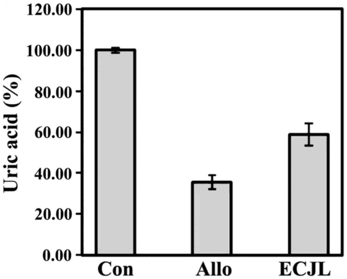
XO inhibitory activity of ECJL in
vitro
Fig. 1 shows the
XO inhibitory activity of ECJL. XO inhibitory activity was
expressed as the suppression rate of uric acid production.
Allopurinol (as a positive control) at a concentration of 30
µg/ml significantly inhibited XO activity by 64.6±3.4%.
Notably, the dose-dependent XO inhibitory activity of ECJL
significantly increased. ECJL at a concentration of 2 mg/ml
inhibited XO activity by 41.3±5.5%.
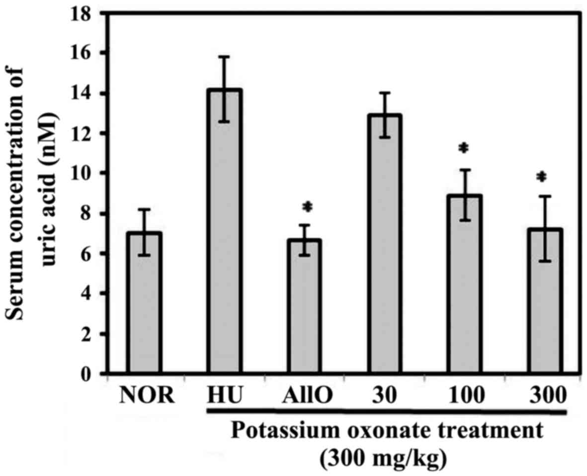
Anti-hyperuricemic effects of the extract
in vivo: serum uric acid levels
Fig. 2 shows the
effects of ECJL on the serum uric acid levels in mice with
potassium oxonate-induced hyperuricemia. The concentrations were
significantly higher in the mice with hyperuricemia compared to the
normal mice, indicating that the mouse model of hyperuricemia was
successfully established at 1 h after the intraperitoneal injection
of potassium oxonate, a urate oxidase inhibitor, which is
consistent with the findings of a previous study (20). The serum uric acid concentrations
in the normal mice were comparable with those in the hyperuricemic
mice that were administered allopurinol or the 3 different doses of
ECJL (30, 100 and 300 mg/kg over a period of 7 days.
In our in vivo model, the level of serum uric
acid was effectively increased by potassium oxonate at a dose of
300 mg/kg via intraperitoneal injection (14.19±1.62 µM). The
serum levels of uric acid in the allopurinol-treated group and ECJL
treatment groups (30, 100, 300 mg/kg) were 6.65±0.74, 12.89±1.13,
8.89±1.17 and 7.23±1.63 µM, respectively. Allopurinol at a
dose of 10 mg/kg suppressed the serum uric acid levels by 53% and
ECJL at a dose of 300 mg significantly suppressed the uric acid
levels by 49.1%, similar to allopurinol.
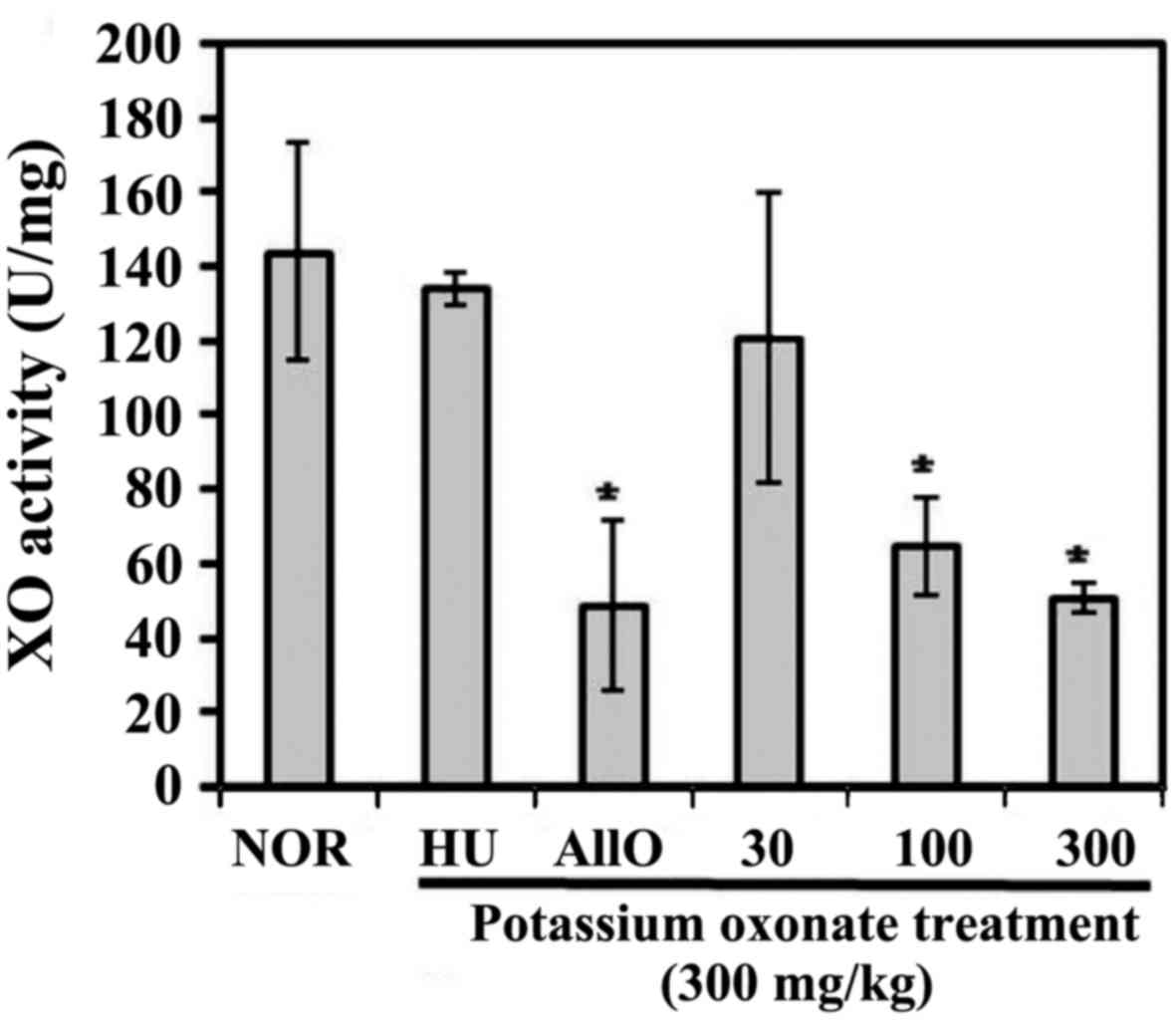
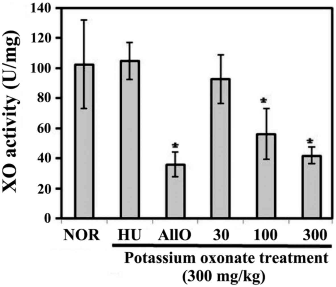
Anti-hyperuricemic effects in vivo:
hepatic and serum XO activity
Figs. 3 and
4 show the effects of ECJL on the
hepatic and serum XO activity in the mice with potassium
oxonate-induced hyperuricemia. Pre-treatment with allopurinol for 1
week (oral administration) and ECJL at the doses of 100 and 300
mg/kg significantly reduced hepatic XO activity by 66.1, 55.2 and
64.7%, respectively (Fig. 3).
Similarly, the serum XO activity in the mice pre-treated orally
with allopurinol and ECJL at the doses of 100 and 300 mg/kg was
reduced by 65, 45.2 and 59.3%, respectively.
However, there were no significant differences in
hepatic and plasma XO activity between the normal and hyperuricemic
control groups. Taken together, our findings demonstrated that ECJL
at the dose of 300 mg/kg exerted XO inhibitory activity similar to
that of allopurinol at a dose of 10 mg/kg.
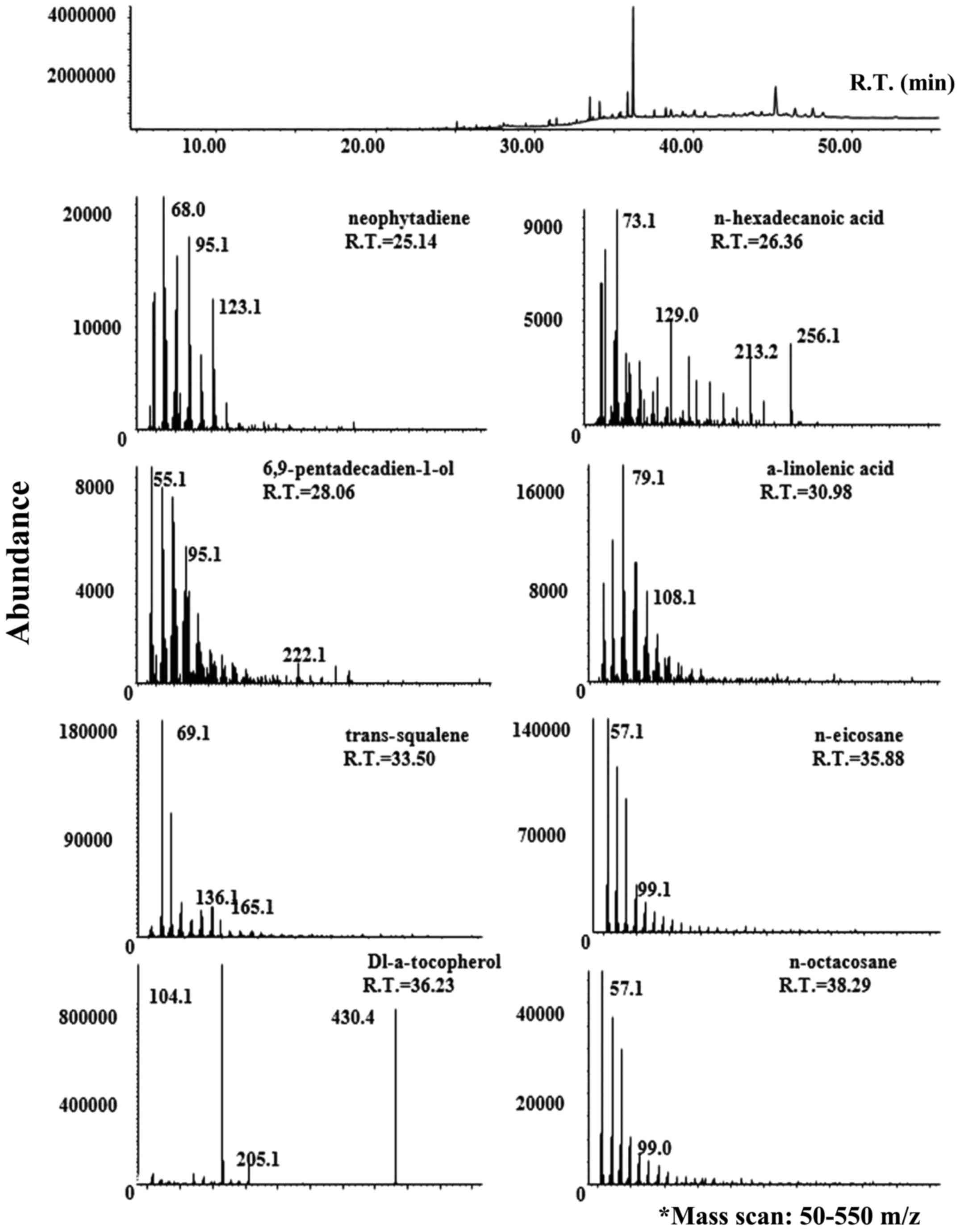
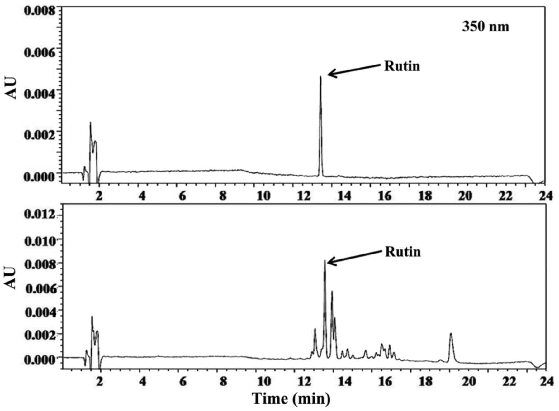
XO inhibitory activity of the main
components identified from ECJL
In the present study, we identified active compounds
related to XO inhibitory activity from C. japonica leaf
using GC-MS and liquid chromatography analysis. We identified one
XO inhibitor, namely rutin (5.87%) using liquid chromatography. We
identified 8 active compounds, which were vitamin E (25.35%)
n-eicosane (10.2%), neophytadiene (0.91%), all trans-squalene
(3.32%), n-octacosane (2.65%), 6,9-pentadecadien-1-ol (1.51%),
α-linolenic acid (1.41%), and n-hexadecanoic acid (0.61%) using
GC-MS.
Figs. 5 and
6 show typical GC-MS and HPLC
chromatograms, respectively, which show the phytochemical
constituents. After clarifying the active substances, we expected
that the potent XO inhibitory activity of ECJL was due to the
synergism of antioxidant and XO inhibitory substances.
Discussion
In our preliminary experiments, we screened 500
plant extracts, and selected ECJL as a candidate for an XO
inhibitor. We fractionated the extract using solvent extraction and
found that the ethanol fraction (ECJL) exhibited the most prominent
XO inhibitory activity in water/ethanol extraction. The ethanol
fraction exhibited strong DPPH radical scavenging activity and
reducing power (unpublished data). Based on these results, the
ethanol fraction was selected for further in vitro and in
vivo experiments.
Our results revealed that total phenolics were
enriched in ECJL. Phenolics have been shown to possess antioxidant
and anti-inflammatory activities (23).
A number of phenolics have been shown to possess
antioxidant and XO inhibitory activity, and to have the ability to
decrease uric acid levels in serum. Thus, the solvent extraction
condition was optimized with respect to the antioxidant and XO
inhibitory activity (unpublished data). Table III shows that ECJL contained
phenolic compounds (46.6±1.6 mg/g eq. gallic acid).
Hyperuricemia is an abnormal condition which
involves high levels of uric acid, and the major factor is known to
be the overproduction of uric acid by XO (24). In general, the extract that has
antioxidant activity is closely related to XO inhibitory activity
and the recovery of oxidative damage. The breakdown of purine
metabolism is known to be responsible for hyperuricemia.
Hyperuricemia is mainly caused by decreased renal uric acid
excretion or excessive hepatic uric acid generation (25). XO can catalyze the oxidation of
hypoxanthine to uric acid, primarily in the liver (26). Gout is a representative disease
that is closely related to hyperuricemia. Excessive levels of serum
uric acid are the main risk factors for uric acid crystal
deposition in joints and kidneys, resulting in uric acid
nephrolithiasis and gouty arthritis (27). Thus, the serum uric acid level and
hepatic XO activity were evaluated to examiney the
anti-hyperuricemic effects of ECJL that was prepared in this
study.
Figs. 2Figure 3–4 show that ECJL at doses of 100 and 300
mg/kg significantly reduced the serum uric acid levels and
inhibited the hepatic and serum XO activity. These results clearly
indicate that the oral administration of ECJL markedly attenuates
the hyperuricemic state in mice. In particular, there were no
significant differences in hepatic and serum XO activity between
the normal and hyperuricemic control groups. This result suggests
that intraperitoneal pre-treatment with potassium oxonate, a known
uricase inhibitor, may not affect XO activity in mice. Thus, ECJL
controls hyperuricemia via the inhibition of XO in the liver and
serum in potassium oxonatetreated mice.
The mechanisms of action of ECJL could be explained
through analytical study of the active constituents in ECJL.
Fig. 5 shows that we identified
the important bioactive markers related to antioxidant activity and
hyperuricemi, such as vitamin E (25.35%), neophytadiene (0.91%),
all trans-squalene (3.32%), α-linolenic acid (1.41%), n-eicosane
(10.2%), n-octacosane (2.65%), 6,9-pentadecadien-1-ol (1.51%), and
n-hexadecanoic acid (0.61%) using GC-MS.
Vitamin E was the major compound in ECJL. Catignani
et al found that the increase in XO activity in the liver
was due to the level of vitamin E and involved the accumulation of
the enzyme protein in vitamin E-deficient rabbits (28). Mohd Fahami et al reported
that palm vitamin E also reduced XO acitivity in gastric regions
(29). Thus, vitamin E may be the
first key compound as a strong antioxidant agent and XO regulator
in ECJL.
Squalene has been reported to possess antioxidant
properties. Huang et al reported that squalene is a highly
effective oxygen-scavenging agent (30).
Among the identified fatty acids, linolenic acid was
analyzed. Park et al reported that linolenic acid
downregulated inducible nitric oxide synthase (iNOS) and
cyclooxygenase-2 (COX-2) expression and thereby reduced nitric
oxide (NO) and prostaglandin E2 production in lipopolysaccharide
(LPS)-activated RAW264.7 cells. These findings indicate the
potential therapeutic use of linolenic acid as an anti-inflammatory
agent (31). Linolenic acid is
not closely related to hyperuricemia. Besides, linolenic acid
affects secondary inflammatory damage, such as uric acid
nephrolithiasis and gouty arthritis.
Fig. 6 shows that
we identified rutin as an XO inhibitor using bioassay-guided liquid
chromatography. The content of rutin in ECJL was determined to be
5.87% (w/w). A previous study reported that rutin at the dose of
50–100 mg/kg significantly decreased the biomarkers of serum urate,
creatinine and blood urea nitrogen and serum, and kidney uromodulin
levels in hyperuricemic mice (32). Azuma et al reported that 5
flavonoids were purified from the buthanol extract of C.
japonica leaf and identified them as quercetin, kaempferol,
apigenin, rutin and quercetrin (33). Previous studies have reported that
quercetin inhibits XO activity in a competitive manner, while
apigenin inhibits it in a mixed manner (20,34). As shown in a previous sudy, rutin,
when administered orally to mice with oxonate-induced
hyperuricemia, was able to draw out dose-dependent hypouricemic
effects by exerting significant inhibitory effects on xanthine
dehydrogenase/xanthine oxidase (XDH/XO) activities (35). In the present study, we identified
rutin by liquid chromatography. Considering previous reports of
active markers and purification techniques, further studies are
warranted for the isolation and identification of active compounds
from ECJL.
Allopurinol is commonly used to treat and control
hyperuricemia. However, its use can cause side-effects to patients,
and these are reported as the main cause for the termination of
anti-hyperuricemic therapy. This necessitates an alternative herbal
medicine that is therapeutically effective for hyperuricemia and
gout (12). In the present study,
we aimed to evaluate the anti-hyperuricemic activities of ECJL, and
to determine the optimal dosage in experimental models in
vivo to predict oral administration in humans. ECJL was
effective in reducing uric acid levels and inhibiting liver/serum
XO activity, which indicates that ECJL may exert its
anti-hyperuricemic effects mainly through this mechanism. ECJL
reduced uric acid levels at two evaluated doses, at 100–300 mg/kg,
significantly inhibiting liver and serum XO activity.
In general, we considered the oral intake of dried
Camellia japonica leaf of 10 g daily for the treatment of
gout. Due to the yield of extract being 14.17%, the oral dose for a
human weighing 60 kg is 1,417 mg/day (23.61 mg/kg/day). The
conversion factor between humans and mice is 12.33 (36). Therefore, if the effective dose
for mice is 300 mg/kg/day, the human equivalent dose is 1,459.85
mg/60 kg/day as ECJL, or 10.3 g/60 kg/day as dried Camellia
japonica leaf. In the present study, mice with hyporuricemia
were treated with 30, 100 and 300 mg/kg ECJL and the effective oral
dose was drawn in the range of 100–300 mg/kg. We concluded that the
oral intake of 3–10 g of C. japonica leaf is beneficial to
prevent and/or decrease the possibility of the occurrence of
hyperuricemia related disease.
Taken together, we found beneficial effects of ECJL
from the results of biological evaluation through antioxidant assay
and XO assay in vitro and in vivo. We also identified
the active compounds that contribute to the antioxidant,
anti-inflammatory, and XO inhibitory properties of ECJL from the
results of GC-MS and HPLC analysis. In the future, we aim to
determine whether ECJL has beneficial effect on gouty
arthritis.
In conclusion, in the present study, C.
japonica leaf from Korea was selected based on the possibility
of its development for plant medicine and/or functional food
materials as an anti-hyperuricemic agent. Our study revealed that
the ethanol extract of C. japonica leaf was a positive
sample which exerts a potent XO inhibitory effect similar to that
of allopurinol. This study is a significant contribution to the
knowledge of bioactive markers from the C. japonica leaf as
potential sources for the medical industry, and presents data to
explain the effects of ECJL in the treatment of hyperuricemia.
References
|
1
|
Yoshikawa M, Murakami T, Yoshizumi S,
Murakami N, Yamahara J and Matsuda H: Bioactive saponins and
glycosides. V. Acylated polyhydroxyolean-12-ene triterpene
oligoglycosides, camelliasaponins A1, A2, B1, B2, C1, and C2, from
the seeds of Camellia japonica L: Structures and inhibitory
activity on alcohol absorption. Chem Pharm Bull (Tokyo).
44:1899–1907. 1996. View Article : Google Scholar
|
|
2
|
Yoshikawa M, Morikawa T, Asao Y, Fujiwara
E, Nakamura S and Matsuda H: Medicinal flowers. XV. The structures
of noroleanane- and oleanane-type triterpene oligoglycosides with
gastroprotective and platelet aggregation activities from flower
buds of Camellia japonica. Chem Pharm Bull (Tokyo). 55:606–612.
2007. View Article : Google Scholar
|
|
3
|
Heo J: Dongui Bogam. Naeuiwon edition.
Bubinmunhwasa publisher; Seoul, Korea: 1613
|
|
4
|
Cha SS, Lee KE, Lee SH, Choi MJ and Shim
JK: Decomposition and nutrient dynamics of leaf litter of Camellia
japonica L. in Korea. Korean J Environ Ecol. 30:110–117. 2016.In
Korean. View Article : Google Scholar
|
|
5
|
Hwang EJ, Cha YJ, Park MH, Lee JW and Lee
SY: Cytotoxicity and chemosensitizing effect of Camellia (Camellia
japonica) tea extracts. J Korean Soc Food Sci Nutr. 33:487–493.
2004.In Korean. View Article : Google Scholar
|
|
6
|
Yang JL, Ha TK, Dhodary B, Pyo E, Nguyen
NH, Cho H, Kim E and Oh WK: Oleanane triterpenes from the flowers
of Camellia japonica inhibit porcine epidemic diarrhea virus (PEDV)
replication. J Med Chem. 58:1268–1280. 2015. View Article : Google Scholar : PubMed/NCBI
|
|
7
|
Onodera K, Hanashiro K and Yasumoto T:
Camellianoside, a novel antioxidant glycoside from the leaves of
Camellia japonica. Biosci Biotechnol Biochem. 70:1995–1998. 2006.
View Article : Google Scholar : PubMed/NCBI
|
|
8
|
Thao NT, Hung TM, Lee MK, Kim JC, Min BS
and Bae K: Triterpenoids from Camellia japonica and their cytotoxic
activity. Chem Pharm Bull (Tokyo). 58:121–124. 2010. View Article : Google Scholar
|
|
9
|
Nagata T, Tsushida T, Hamaya E, Enoki N,
Manabe S and Nishino C: Camellidins, antifungal saponins isolated
from Camellia japonica. Agric Biol Chem. 49:1181–1186. 1985.
|
|
10
|
Park JC, Hur JM, Park JG, Hatano T,
Yoshida T, Miyashiro H, Min BS and Hattori M: Inhibitory effects of
Korean medicinal plants and camelliatannin H from Camellia japonica
on human immunodeficiency virus type 1 protease. Phytother Res.
16:422–426. 2002. View
Article : Google Scholar : PubMed/NCBI
|
|
11
|
Zhao M, Zhu D, Sun-Waterhouse D, Su G, Lin
L, Wang X and Dong Y: In vitro and in vivo studies on adlay-derived
seed extracts: Phenolic profiles, antioxidant activities, serum
uric acid suppression, and xanthine oxidase inhibitory effects. J
Agric Food Chem. 62:7771–7778. 2014. View Article : Google Scholar : PubMed/NCBI
|
|
12
|
Lemos Lima Rde C, Ferrari FC, de Souza MR,
de Sá Pereira BM, de Paula CA and Saúde-Guimarães DA: Effects of
extracts of leaves from Sparattosperma leucanthum on hyperuricemia
and gouty arthritis. J Ethnopharmacol. 161:194–199. 2015.
View Article : Google Scholar
|
|
13
|
Chu YH, Chen CJ, Wu SH and Hsieh JF:
Inhibition of xanthine oxidase by Rhodiola crenulata extracts and
their phytochemicals. J Agric Food Chem. 62:3742–3749. 2014.
View Article : Google Scholar : PubMed/NCBI
|
|
14
|
Kramer HM and Curhan G: The association
between gout and nephrolithiasis: The National Health and Nutrition
Examination Survey III, 1988–1994. Am J Kidney Dis. 40:37–42. 2002.
View Article : Google Scholar : PubMed/NCBI
|
|
15
|
Choi HJ, Lee CH, Lee JH, Yoon BY, Kim HA,
Suh CH, Choi ST, Song JS, Joo HY, Choi SJ, et al: Current gout
treatment and flare in South Korea: Prophylactic duration
associated with fewer gout flares. Int J Rheum Dis. Aug
27–2014.Epub ahead of print. View Article : Google Scholar
|
|
16
|
Bae MS, Shin JS, Lee KY, Lee KH and Kim
YJ: Long-range transport of biomass burning emissions based on
organic molecular markers and carbonaceous thermal distribution.
Sci Total Environ. 466–467:56–66. 2014. View Article : Google Scholar
|
|
17
|
Kim YH, Cho ML, Kim DB, Shin GH, Lee JH,
Lee JS, Park SO, Lee SJ, Shin HM and Lee OH: The antioxidant
activity and their major antioxidant compounds from Acanthopanax
senticosus and A. koreanum. Molecules. 20:13281–13295. 2015.
View Article : Google Scholar : PubMed/NCBI
|
|
18
|
Seo JH, Kim JE, Shim JH, Yoon G, Bang MA,
Bae CS, Lee KJ, Park DH and Cho SS: HPLC analysis, optimization of
extraction conditions and biological evaluation of Corylopsis
coreana Uyeki flos. Molecules. 21:942016. View Article : Google Scholar : PubMed/NCBI
|
|
19
|
Arimboor R, Rangan M, Aravind SG and
Arumughan C: Tetrahydroamentoflavone (THA) from Semecarpus
anacardium as a potent inhibitor of xanthine oxidase. J
Ethnopharmacol. 133:1117–1120. 2011. View Article : Google Scholar
|
|
20
|
Huo LN, Wang W, Zhang CY, Shi HB, Liu Y,
Liu XH, Guo BH, Zhao DM and Gao H: Bioassay-guided isolation and
identification of xanthine oxidase inhibitory constituents from the
leaves of Perilla frutescens. Molecules. 20:17848–17859. 2015.
View Article : Google Scholar : PubMed/NCBI
|
|
21
|
Bradford MM: A rapid and sensitive method
for the quantitation of microgram quantities of protein utilizing
the principle of protein-dye binding. Anal Biochem. 72:248–254.
1976. View Article : Google Scholar : PubMed/NCBI
|
|
22
|
Perron NR and Brumaghim JL: A review of
the antioxidant mechanisms of polyphenol compounds related to iron
binding. Cell Biochem Biophys. 53:75–100. 2009. View Article : Google Scholar : PubMed/NCBI
|
|
23
|
Liu LM, Cheng SF, Shieh PC, Lee JC, Chen
JJ, Ho CT, Kuo SC, Kuo DH, Huang LJ and Way TD: The methanol
extract of Euonymus laxiflorus, Rubia lanceolata and Gardenia
jasminoides inhibits xanthine oxidase and reduce serum uric acid
level in rats. Food Chem Toxicol. 70:179–184. 2014. View Article : Google Scholar : PubMed/NCBI
|
|
24
|
Wu XH, Yu CH, Zhang CF, Anderson S and
Zhang YW: Smilax riparia reduces hyperuricemia in mice as a
potential treatment of gout. Am J Chin Med. 42:257–259. 2014.
View Article : Google Scholar : PubMed/NCBI
|
|
25
|
Gurwitz JH, Kalish SC, Bohn RL, Glynn RJ,
Monane M, Mogun H and Avorn J: Thiazide diuretics and the
initiation of anti-gout therapy. J Clin Epidemiol. 50:953–959.
1997. View Article : Google Scholar : PubMed/NCBI
|
|
26
|
Ardan T, Kovaceva J and Cejková J:
Comparative histochemical and immunohistochemical study on xanthine
oxidoreductase/xanthine oxidase in mammalian corneal epithelium.
Acta Histochem. 106:69–75. 2004. View Article : Google Scholar : PubMed/NCBI
|
|
27
|
Grassi D, Ferri L, Desideri G, Di Giosia
P, Cheli P, Del Pinto R, Properzi G and Ferri C: Chronic
hyperuricemia, uric acid deposit and cardiovascular risk. Curr
Pharm Des. 19:2432–2438. 2013. View Article : Google Scholar :
|
|
28
|
Catignani GL, Chytil F and Darby WJ:
Vitamin E deficiency: Immunochemical evidence for increased
accumulation of liver xanthine oxidase. Proc Natl Acad Sci USA.
71:1966–1968. 1974. View Article : Google Scholar : PubMed/NCBI
|
|
29
|
Mohd Fahami NA, Ibrahim IA, Kamisah Y and
Mohd Ismail N: Palm vitamin E reduces catecholamines, xanthine
oxidase activity and gastric lesions in rats exposed to
water-immersion restraint stress. BMC Gastroenterol. 12:542012.
View Article : Google Scholar : PubMed/NCBI
|
|
30
|
Huang ZR, Lin YK and Fang JY: Biological
and pharmacological activities of squalene and related compounds:
Potential uses in cosmetic dermatology. Molecules. 14:540–554.
2009. View Article : Google Scholar : PubMed/NCBI
|
|
31
|
Park SY, Seetharaman R, Ko MJ, Kim DY, Kim
TH, Yoon MK, Kwak JH, Lee SJ, Bae YS and Choi YW: Ethyl linoleate
from garlic attenuates lipopolysaccharide-induced pro-inflammatory
cytokine production by inducing heme oxygenase-1 in RAW264.7 cells.
Int Immunopharmacol. 19:253–261. 2014. View Article : Google Scholar : PubMed/NCBI
|
|
32
|
Chen YS, Hu QH, Zhang X, Zhu Q and Kong
LD: Beneficial effect of rutin on oxonate-induced hyperuricemia and
renal dysfunction in mice. Pharmacology. 92:75–83. 2013. View Article : Google Scholar : PubMed/NCBI
|
|
33
|
Azuma CM, dos Santos FCS and Lago JHG:
Flavonoids and fatty acids of Camellia japonica leaves extract. Rev
Bras Farmacogn. 21:1159–1162. 2011. View Article : Google Scholar
|
|
34
|
Pauff JM and Hille R: Inhibition studies
of bovine xanthine oxidase by luteolin, silibinin, quercetin, and
curcumin. J Nat Prod. 72:725–731. 2009. View Article : Google Scholar : PubMed/NCBI
|
|
35
|
Zhu JX, Wang Y, Kong LD, Yang C and Zhang
X: Effects of Biota orientalis extract and its flavonoid
constituents, quercetin and rutin on serum uric acid levels in
oxonate-induced mice and xanthine dehydrogenase and xanthine
oxidase activities in mouse liver. J Ethnopharmacol. 93:133–140.
2004. View Article : Google Scholar : PubMed/NCBI
|
|
36
|
Shin JW, Seol IC and Son CG:
Interpretation of animal dose and human equivalent dose for drug
development. J Korean Orient Med. 31:1–7. 2010.
|