Introduction
Ultrasound contrast agents (UCAs) are widely used in
clinical practice in order to enhance the diagnostic capability and
utility of traditional ultrasound imaging modes. The UCAs currently
used commercially are usually designed to serve as blood pool
agents with diameters of 1–8 μm and cannot pass through the
blood vessel wall into the tumor tissue to achieve the molecular
imaging of tumor tissue (1).
Additionally the vascular endothelial gap in tumors is
approximately 380–780 nm, which is much wider than that of normal
tissue (2,3). In order to ensure that UCAs pass
through the tumor tissue, researchers have paid more attention to
the preparation of nanosized UCAs, due to their potential for the
extravascular molecular imaging of tumors (4).
Although nanosized UCAs achieve passive imaging for
tumors via enhanced permeability and retention (EPR), the specific
capability of nanosized UCA to bind with tumor tissue is still
limited (5). The technique which
involves labeling nanoparticles with specific ligands can generate
targeted nanoparticles capable of binding to specific tissues or
tumors (6–8). Following intravenous injection, the
nanoparticles aggregate specifically in the target tissues via the
blood circulation, thus allowing the ultrasonography-based specific
imaging of pathogenic changes at a molecular or cellular level
(9).
Folate receptor (FR) binds folic acid (FA) with high
affinity and mediates its intracellular transport via
receptor-mediated endocytosis (10). There are three isoforms of FR
(FRα, FRβ and FRγ) and type α is the major isoform mediating folate
transport, and is overexpressed on the surface of various types of
tumor, including pancreatic, prostate, lung, head and neck, breast
and ovarian cancer, and mesothelioma (11–19). Based on its limited expression and
restricted distribution pattern in normal tissue, FRα is the most
widely studied member of the FR family, and various strategies for
targeting FR, which is overexpressed in cancer, have been developed
(20,21).
However, very few techniques involving ultrasound
nanoparticles coupled with FR targeting and layer-by-layer (LbL)
self-assembly have been described, to the best of our knowledge
(22,23). In the present study, we developed
nanosized and highly efficient targeted nanoparticles modified with
FA and polyethylene glycol (PEG) to verify the hypothesis that
targeted nanoparticles can enhance the diagnostic value of
ultrasound in FR-overexpressing tumors.
Materials and methods
Materials
N-Hydroxysuccinimide (NHS) were supplied by
Sinopharm Chemical Reagent (Shanghai, China). Perfluorooctyl
bromide (PFOB), FA and
N-(3-Dimethylaminopropyl)-N′-ethylcarbodiimide hydrochloride (EDC)
were obtained from Sigma-Aldrich (St. Louis, MO, USA). PEG
(NH2-PEG3400) was obtained from Xibao
Biochemicals (Shanghai, China). Chitosan (CS; degree of
deacetylation 95%) was obtained from Haidebei Marine Bioengineering
(Shandong, China). Alginate (ALG) was obtained from Yuanye Biotech
(Shanghai, China). Anti-folate binding protein antibody (EPR4708)
was obtained from Abcam (Cambridge, UK). Fluorescein isothiocyanate
(FITC), DiI and immobilon-P transfer membrane were purchased from
Solarbio (Beijing, China). Cy7 NHS ester (MW 720.94) was obtained
from FANBO Biochemicals (Beijing, China). The PrimeScript™ RT
reagent kit with gDNA eraser and SYBR Premix Ex Taq were obtained
from Takara (Shiga, Japan).
Cell culture and animal preparation
Bel7402 (human liver cancer cells), SW620 (human
colorectal cancer cells) and A549 (human lung cancer cells) were
purchased from the Cell Bank of Chinese Academy of Sciences
(Shanghai, China). BALB/c 3T3 fibroblasts were purchased from the
Cell Bank of Zhongshan University (Guangzhou, China). The cells
were cultured in RPMI-1640 medium supplemented with 10%
heat-inactivated fetal bovine serum at 37°C, 5% CO2 cell
incubator.
BALB/c nude male mice (n=54; 5–6 weeks old; body
weight, 17–21 g; SLAC Laboratory Animals, Shanghai, China) kept in
a specific pathogen-free environment were used as described below.
All animal experiments were approved by the Animal Ethics Committee
of the Medical School of Ningbo University (Ningbo, China) in
accordance with the National Institutes of Health Guide for the
Care and Use of Laboratory Animals and the ARRIVE guidelines. Under
sterile conditions, logarithmic-phase Bel7402, SW620 (FR
overexpressing cancer cells) and A549 (FR underexpressing cancer
cells) were used to prepare cell suspensions at a density of
1×10⁷ cells/ml; BALB/c 3T3 fibroblasts in the
logarithmic phase growth were used prepare cell suspensions of
5×10⁶ cells/ml. The mixture containing each type of
cancer cell suspension (9:1 volume radio, 100 μl each mouse)
was subcutaneously inoculated into the 5–6-week-old male BALB/c
nude mice. Twenty animals were used for each xenograft tumor type.
The xenografts were monitored daily with a venire caliper until the
subcutaneous tumors reached 0.5–1.0 cm in diameter for 16–31 days
after implantation.
Detection of FR expression in 3 types of
cancer cells and xenografts
FR mRNA expression in cancer cell
lines detected by reverse transcription-quantitative PCR
(RT-qPCR)
RT-qPCR was used to measure the mRNa expression of
FRα in the 3 cancer cell lines. Total RNA was extracted from the 3
types of cancer cells using TRIzol reagent protocol (Invitrogen,
Carlsbad, CA, USA). Reverse transcription was performed using 500
ng of total RNA and the PrimeScript™ RT reagent kit according to
the manufacturer's instructions, and PCR was performed using the
following primers: human FRα (sense strand,
5′-AGGTGCCATCTCTCCACAGT-3′ and antisense,
5′-GAGGACAAGTTGCATGAGCA-3′; cDNA amplicon size: 135 bp; Tm, 60°C);
human GAPDH (sense, 5′-TTAAAAGCAGCCCTGGTGAC-3′ and antisense,
5′-CTCTGCTCCTCCTGTTCGAC-3′; cDNA amplicon size: 138 bp; Tm, 60°C).
cDNA was measured by using SYBR-Green RT-PCR in a StepOnePlus
Real-Time PCR system (Thermo Fisher Scientific, Inc., Waltham, MA,
USA) at 95°C for 30 sec, 40 cycles of 95°C for 5 sec and 60°C for
34 sec. All PCR reactions were performed at least in triplicate and
the mRNA levels were represented as relative quantification, which
was calculated using relative expression of FR (NFRα) =
2−ΔΔCq, where Cq = ΔCq sample − ΔCq calibrator (ΔCq = Cq
of target gene − Cq of GAPDH).
Western blot analysis and
immunohistochemical staining of FR in tumor tissues
Each tumor xenograt tissue (300 mg) was lysed on ice
for 30 min in 1 ml radioimmunoprecipitation assay protein lysis
buffer (RIPA) followed by the addition of 10 μl PMSF. All
tumor tissue lysates were then transferred into a 1.5 ml tube and
centrifuged for 15 min at 15,000 rpm at 4°C. The supernatant was
transferred to a new 1.5 ml centrifuge tube. A bicinchoninicacid
(BCA) kit (ComWin Biotech, Beijing, China) was then used to
determine the protein concentration. Additionally, the samples were
supplemented with 5X sodium dodecyl-polyacrylamide gel
electrophoresis (SDS-PAGE) loading buffer, mixed and boiled for 5
min. Twenty micrograms of total protein were separated by SDS-PAGE
and transferred onto a polyvinylidene fluoride (PVDF) membrane. The
membrane was blocked with 5% albumin bovine in TBST at room
temperature for 1 h. Subsequently the blots were incubated with
1:5,000 dilution (0.145 μg/ml) anti-folate binding protein
monoclonal antibody (EPR4708; ab125030; Abcam) at 4°C overnight,
followed by the addition of 1:500 dilution of goat anti-rabbit IgG
HRP-conjugated secondary antibody (CW0103S; ComWin Biotech) at room
temperature for 2 h; the membrane was washed 3 times with TBST
after each antibody incubation.
To detect FR expression by immunohistochemical
staining, each tumor-bearing nude mouse was anesthetized
intraperitoneally with 1% sodium pentobarbital and sacrificed when
the subcutaneous tumors reached the required volume. After that,
the tumors were completely removed from the xenografts and frozen
by liquid nitrogen immediately. The fresh frozen blocks of the
removed tumors were dissected into serial sections of approximately
4-μm-thick using a freezing microtome (CM1860; Leica
Microsystems, Wetzlar, Germany). Each section was covered on a
clean glass slide and fixed by 4% polyformaldehyde for 10 min. The
slides were then washed in phosphate-buffered saline (PBS), and
soaked in 3% hydrogen peroxide methanol solution for 15 min. Each
slide was incubated with 5% goat serum albumin for 20 min to block
non-specific antigen sites, and rabbit anti-folate binding protein
monoclonal antibody (EPR4708; ab125030; Abcam) was added. These
slides were placed inside a humidified chamber for 30 min at room
temperature, then washed with PBS buffer for 5 min and dried.
Subsequently, the streptavidin-biotin complex immunocytochemistry
protocol was used for staining and mild re-dyeing with hematoxylin.
Subsequently, the slides were dehydrated, covered slipped with
mounting, and observed under a microscope (NIB-100; Olympus, Tokyo,
Japan). The results were considered as positive when claybank
particles were observed in the cytoplasm, cell membrane or
nucleus.
Preparation of FR-targeted
nanoparticles (FRNPs) and blank nanoparticles (NPs)
The FR-targeted PFOB (FRNPs) were prepared to
combine FA- and PEG-modified targeted shells with a PFOB nanocore
template using the LbL assembly technology. PFOB nanocore templates
were prepared by thin-film hydration and the ultrasonic
emulsification method, as previously described (24,25). Firstly, egg lecithin and
cholesterol (2:1 M radio) were dissolved into an organic solvent
mixture of chloroform. The organic solvent was removed from the
organic mixture in arotary evaporator (Yarong, Shanghai, China) at
50°C under a vacuum. The resulting thin film (100 mg) was hydrated
by using purified water (5 ml) and then alginate (100 mg) was added
to to the solution. Subsequently, the mixture was supplemented with
PFOB (220 μl) in a dropwise manner and sonicated at a
certain ultrasound intensity to form the PFOB nanocore template.
The resulting PFOB nanocore templates also served as blank NPs for
use in subsequent experiments.
FA conjugation to CS and ALG modified with
NH2-PEG were carried out to form crosslinking between
amino and carboxyl groups in the presence of EDC and NHS, which
served as targeted shells, as previously described (23). Briefly, the synthesis of FA-CS was
as follows: FA (220.7 mg) dissolved in 10 ml of DMSO was activated
with EDC (287.6 mg) and NHS (172.6 mg) at room temperature for 4 h,
and the activated solution was then added in a dropwise manner to
20 ml of 2% acetic acid aqueous solution containing CS (161 mg) and
stirred for 20 h at room temperature in the dark. Also, the
synthesis of PEG-ALG was as follows: ALG (176 mg) dissolved in 10
ml of DMSO was activated with EDC (46 mg) and NHS (27.6 mg)at room
temperature for 4 h, then the activated solution was then added in
a dropwise manner to 20 ml of 2% acetic acid aqueous solution
containing PEG (273.2 mg) and stirred for 20 h at room temperature
in the dark. The reaction products was dialyzed using dialysis
membrane (molecular weight cut-off of 8–10 kDa) to remove unreacted
substance and lyophilized.
Polymer (FA-CS and PEG-ALG) and PFOB liposome
solutions were prepared at a concentration of 10 mg/ml and 30
mg/ml. Subsequently, 1 ml polymer (FA-CS) solution was added to the
liposome dispersions and vortexed for 10 min. The mixture was then
sonicated for 5 min and kept aside for PEG-ALG polymer solution
addition (26). The above steps
were repeated 3 times and the resulting products were incubated
with 2 ml polymer (FA-CS) solution for 30 min. The FRNPs were then
obtained by centrifugation (12,000 × g, 10 min, 4°C) and washed 3
times using purified water. The FRNPs was dispersed in 5% w/v
mannitol aqueous solution by low-frequency ultrasound and filtrated
by 0.45 μm membrane filtration to remove large
impurities.
Characterization of nanoparticles
Particle size and zeta potential
Firstly the appearance of FRNP emulsions was
observed using a light microscope (NIB-100; Olympus). Particle
size, polydispersity index (PDI) and zeta potential were obtained
using a Malvern Zetasizer Nano ZS (Malvern Instrument, Malvern, UK)
and size measurements were performed at 20°C. The results were
expressed as the mean diameter and size distribution of FRNPs,
blank NPs obtained from 3 measurements. The standard deviation of
the size and the PDI were also given. Before each measurement, 200
μl of nanoparticles suspension were diluted into 1 ml of
PBS.
Transmission electron microscopy
(TEM)
The morphological characterization and internal
structure of FRNPs and blank NPs were observed using a transmission
electron microscopy JEOL 2100 HR (Jeol, Tokyo, Japan). Suspensions
of FRNPs and blank NPs were deposited on filter papers to blot off
the excess solution and air-dried by using an incandescent lamp
before observation. Images were acquired using a high-resolution
camera, Advantage Orius 831 (Gatan, Pleasanton, CA, USA).
Targeted binding assay of
nanoparticles in vitro
Logarithmic-phase Bel7402, SW620 and A549 cells were
stained with DiI before use. The stained cells were then seeded on
coverslips placed in 12-well cell plates at a density of
2×10⁴ cells/well divided into 2 groups and cultured for
24 h. FITC-labeled FRNPs (1×10¹³ particles/ml, 20
μl) were added to the first group, which served as the
targeted group. FITC-labeled blank NPs (1×10¹³
particles/ml, 10 μl) were added to the second group, which
served as the control group. After the cells were incubated with
the nanoparticles for 6 h, the coverslips were washed 3 times with
PBS, fixed with 4% paraformaldehyde and finally washed with PBS.
Subsequently, the coverslips were mounted onto glass slides and
observed under a fluorescence microscope (DMIRB; Leica
Microsystems).
Ultrasound targeted imaging of FRNPs
in vivo
Nine animals were used for each xenograft tumor type
in ultrasound imaging. The tumor-bearing nude mice were
anesthetized intraperitoneally with 1% sodium pentobarbital for
imaging, and the surfaces of both the tumor and probe were covered
with a 6-mm-thick coupling agent. A LA332 broadband linear
ultrasound probe connected to an Mylab90 ultrasound system (Esaote,
Genova, Italy) was used to perform B mode ultrasound imaging of the
xenografts. Once the cross-section of a xenograft was fully
observed, the probe was immobilized and the parameters of
ultrasonography mode were set up (mechanical index 0.7; gain 80%).
Each type of test tumor-bearing nude mice were divided into 3
groups: group A received 200 μl FRNPs, group B received 200
μl blank NPs and group C received 200 μl Sonovue. The
contrast agents were injected into the test animals via the tail
vein. The ultrasound images of the xeongrafts were collected prior
to the injection and at 1, 5, 10, 20, 40, 80, 120 and 160 min
post-injection; the imaging date were collected to calculate the
increased imaging intensity at different time-points.
Fluorescence imaging of FRNPs in
vivo
To validate the targeting capability of FRNPs in the
other routine and investigate the biodistribution of FRNPs, we
labeled the shell of FRNPs with Cy7 fluorescent dye (27) and each xenograft was intravenously
injected with 200 μl Cy7-labeled FRNPs. Six animals were
used for each type of xenograft. Following administration by
intravenous injection, the time-dependent biodistribution of the
FRNPs in the xenografts was imaged using the Kodak In-vivo Imaging
System FX Pro (excitation, 610 nm; emission, 790 nm; exposure time,
10 sec; Carestream Health, Rochester, NY, USA). The fluorescence
images were then analyzed based on their spectral patterns using
Carestream Molecular Imaging software. Scans were performed at 5,
30 min, 1, 1.5, 3 and 12 h post-injection, after which the
tumor-bearing mices were sacrificed. The tumors, heart, liver,
spleen, lungs, kidneys and intestines were harvested and imaged to
estimate the tissue distribution of FRNPs. To compare the targeting
ability of FRNPs in different tumors, the target-to-background
ratio (TBR) at different time points was obtained using the
Carestream MI software to calculate regions of interest (ROI)
functions. The ROIs were selected manually by drawing regions on
the in vivo fluorescent images. The TBR was calculated using
the following formula: TBR = SIT/SIM, where
SIT is the average signal intensity of the tumor and
SIM is the average signal intensity of the contra
lateral thigh muscle. This procedure was carried out as previously
described by Alencar et al (28).
Histological analysis
Twelve animals (each type of xenografts taken from 4
mice) were divided into 2 groups, including the targeted group and
the control group. The targeted group was intravenously injected
with 200 μl Cy7-labeled FRNPs, whereas the control group
received 200 μl physiological saline. At 2 h post-injection,
the mice were sacrificed. Tumor tissues were carefully removed and
fixed with 4% paraformaldehyde at pH 7.4 for 4 days. The tumors
were then washed with PBS, then embeded with optimal cutting
temperature (OCT) and plalced into liquid nitrogen for storage.
Frozen tissue blocks were dissected into 4-μm-thick sections
and placed on clean glass slides. The slides were dehydrated,
coverslipped and mounted, and examined under a fluorescence
microscope (DMIRB; Leica Microsystems). Red fluorescence spots were
observed with a long-pass 515–560 nm emission filter.
Statistical analysis
The experimental data were analyzed using
Statistical Package for Social Science (SPSS) 19.0 software (SPSS
Inc., Chicago, IL, USA). All the quantitative data were expressed
as the means ± standard deviation. The FRNPs and blank NB
ultrasound indicator data from in vivo imaging were obtained
and analyzed using Image-Pro Plus 6.0 software (Media Cybernetics
Inc., Rockville, MD, USA). The ultrasound indicators of the
nanoparticles in the 3 types of xenografts were analyzed by
analysis of variance (ANOVA). A P-value <0.05 was considered to
indicate a statistically significant difference. Histograms and the
curve with non-linear regression were plotted using GraphPad Prism
5.0 software (GraphPad Software, La Jolla, CA, USA).
Results
Synthesis of the nanoparticles
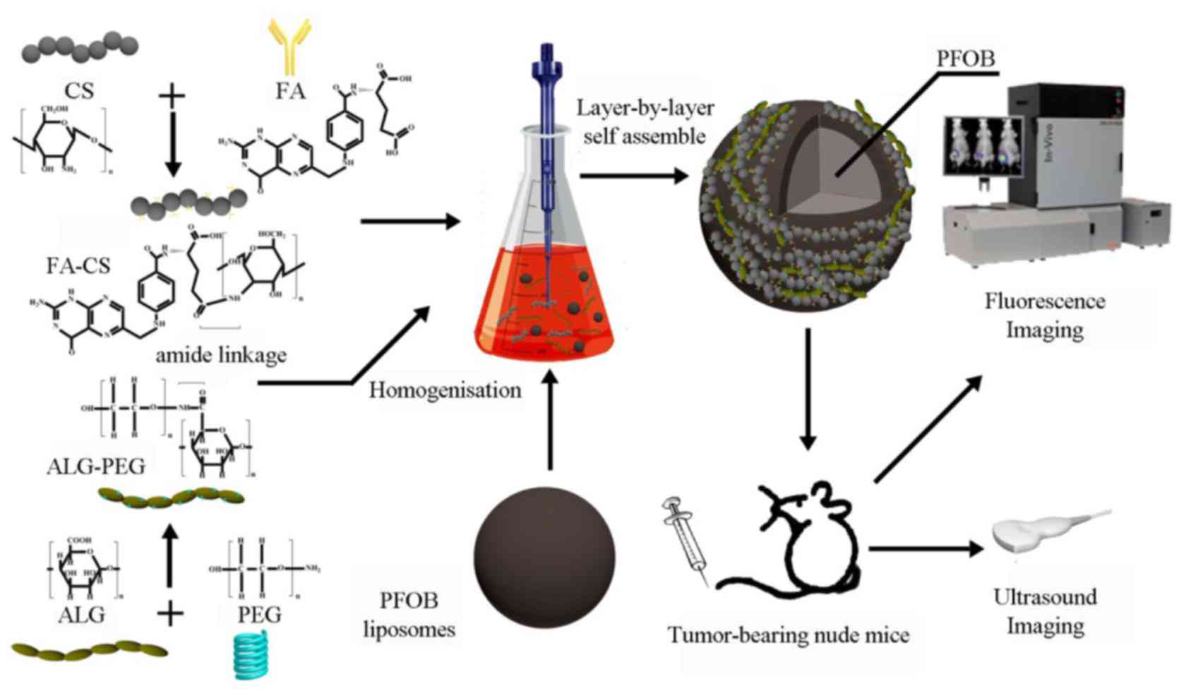
In this study, we used PFOB as the core of the
contrast agents, which is a classical, highly biocompatible
alternative for nanosized ultrasound contrast agents. The synthesis
of PEG-ALG and FA-CS was conducted using the carbodiimide method.
Furthermore, we attempted to develop FR-targeted PFOB nanoparticles
enveloped in a targeted shell consisting of PEG-ALG and FA-CS and
explored the related characteristics, targeting ability,
biodistribution in vivo and ultrasound contrast-enhancing
potential and behavior. The process of LbL self-assembly is shown
in Fig. 1.
Particle size and zeta potential
characterization
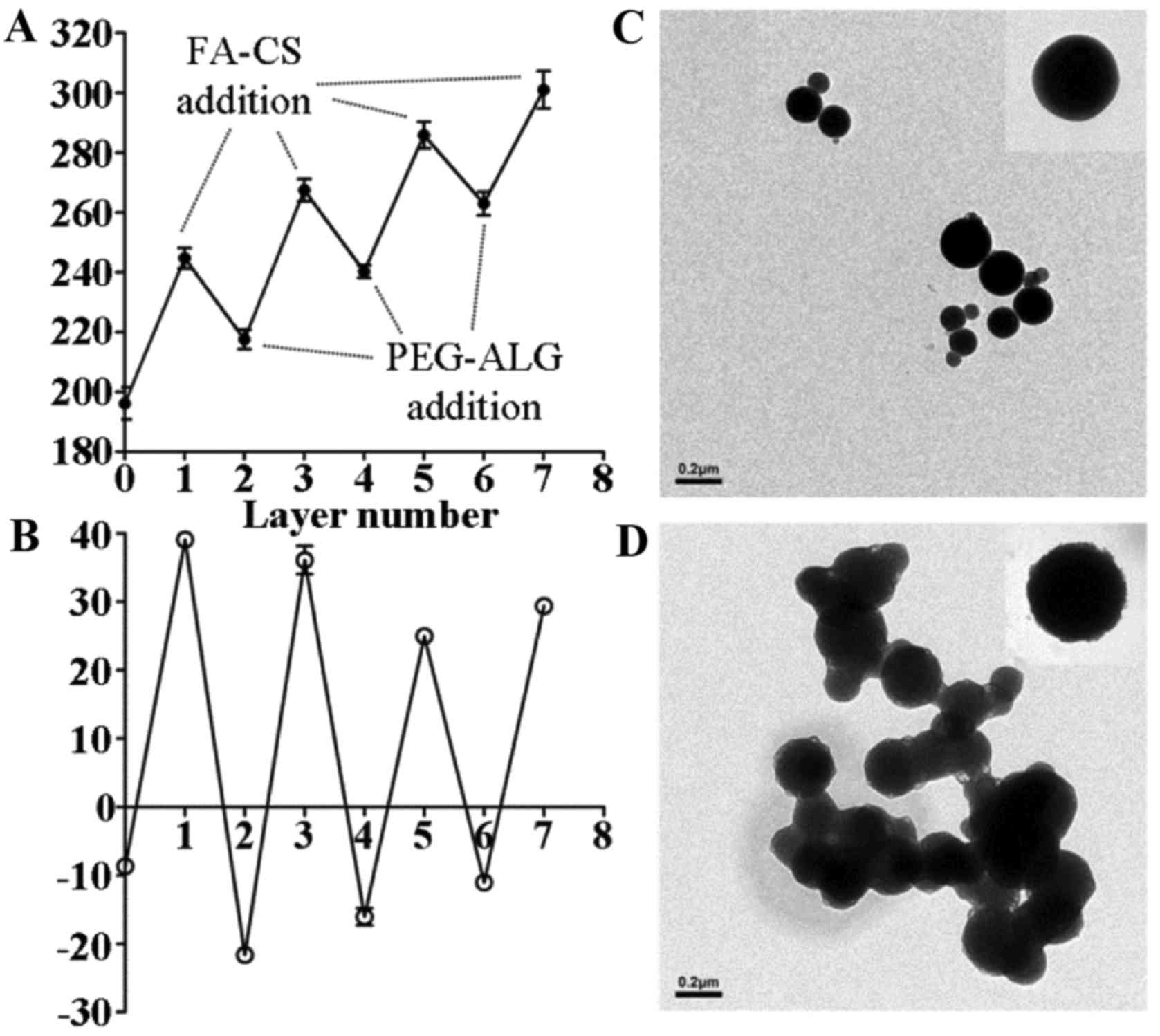
The data of dynamic light scattering (DLS) analysis
indicated that the alternate polymer assembly was accompanied by a
gradual increase in particle size (Fig. 2A). Prior to LbL fabrication, blank
NPs (PFOB liposomes) exhibited an average particle size of
196.2±9.3 nm. In the LbL assembly process, the overall size of the
nanoparticles decreased after each addition of PEG-ALG. This
indicated that the strong ionic electrostatic interaction between
PEG-ALG and FA-CS contributed to the formation of a mesh shell on
the surface of PFOB liposome. After the process was completed, the
final size of the FRNPs reached an average particle size of
301±10.8 nm. Deposition of alternate polymers was accompanied by a
reversal of surface potential (Fig.
2B), which indicated that a solid monolayer had been deposited.
An abrupt rise in the liposome surface potential (from –8.66±0.68
to 39.1±0.43 mV) was observed after addition of the first (FA-CS)
layer. However, the similar surface potential was restored upon
addition of the second (PEG-ALG) layer.
Morphological analysis
TEM was used to observe the morphology and structure
of the FRNPs directly. The blank NPs exhibited well-defined
spherical shape with a smooth surface and small size (Fig. 2C). The FRNPs exhibited a rough
surface and increased average diameter after the addition of
polymers by LbL assembly (Fig.
2D): a darker phosphotungstic acid-stained core and a
lesser-stained shell were visible. This indicated that the addition
of polymer layers resulted in the formation of compact core-shell
NPs.
Estabslishment of tumor xenograft animal
model
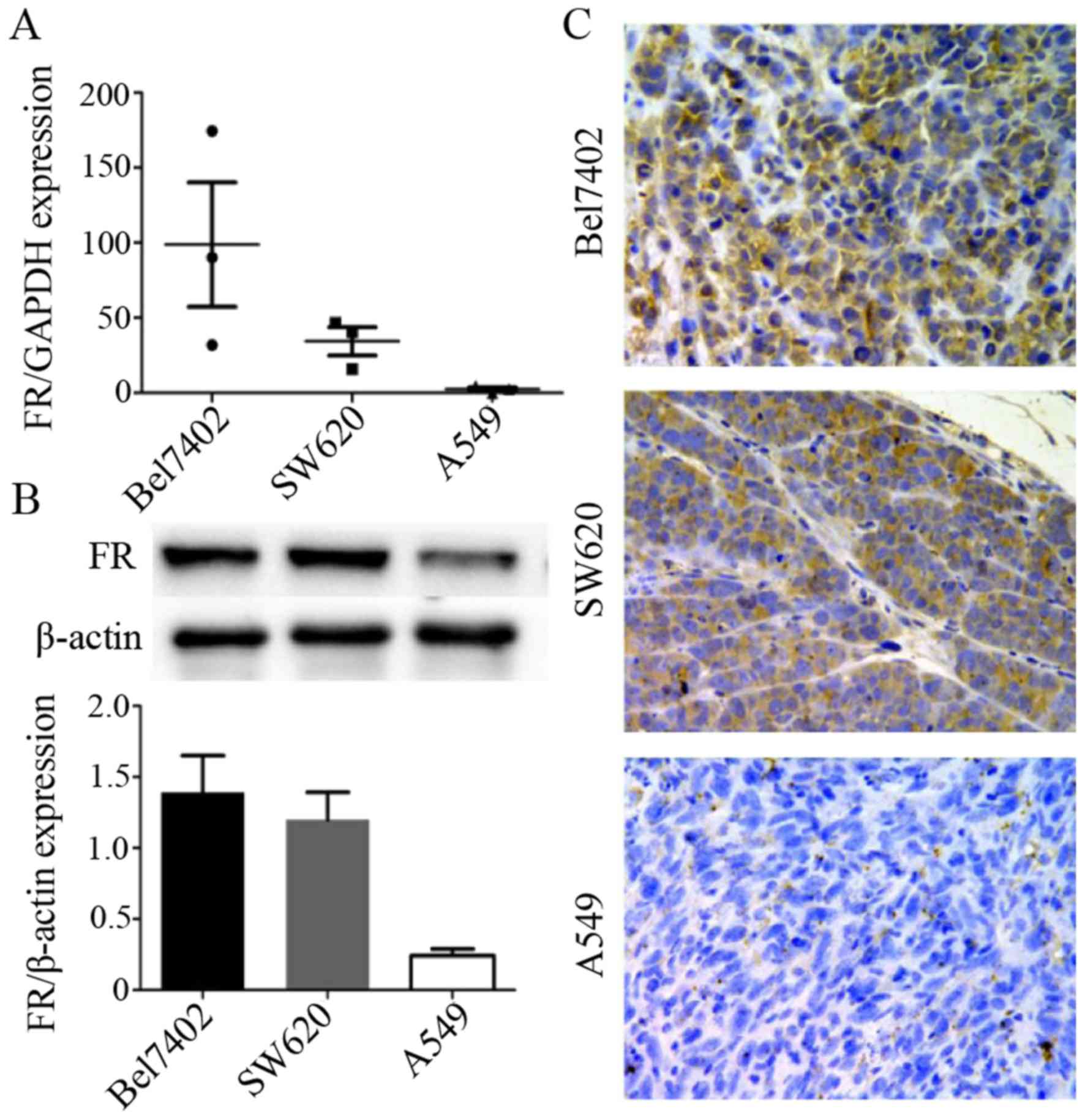
Firstly, RT-qPCR was used to examine FR expression
in the Bel7402, SW620 and A549 cancer cells. The results presented
in Fig. 3A showed that among
these 3 types of cancer cells,the Bel7402 cells expressed the
highest level of FR, followed by the SW620 cells. The A549 cells
exhibited the lowest expression level of FR. Subsequently, we
developed certain types of tumor xenografts with these cancer cell
lines by injecting the cell suspesions into mice and determined FR
expression in the tumor tissue of these 3 types of xenografts (for
each type of xenograft, we randomly selected 3 animals).
The results of western blot analysis (Fig. 3B) and immunohistochemical staining
(Fig. 3C) of the tumor tissues
were consistent with the results of RT-qPCR for the FR mRNA level
in the 3 cancer cell lines. FR expression in the Bel7402 and SW620
tumor xenografts was much higher than that in the A549 tumor
xenografts. This indicated that the animal models were successfully
created and that the xenografts conformed to the quirement for the
experiments of ultrasound targeted imaging and biodistribution
in vivo.
Uptake of nanoparticles by the cancer
cells
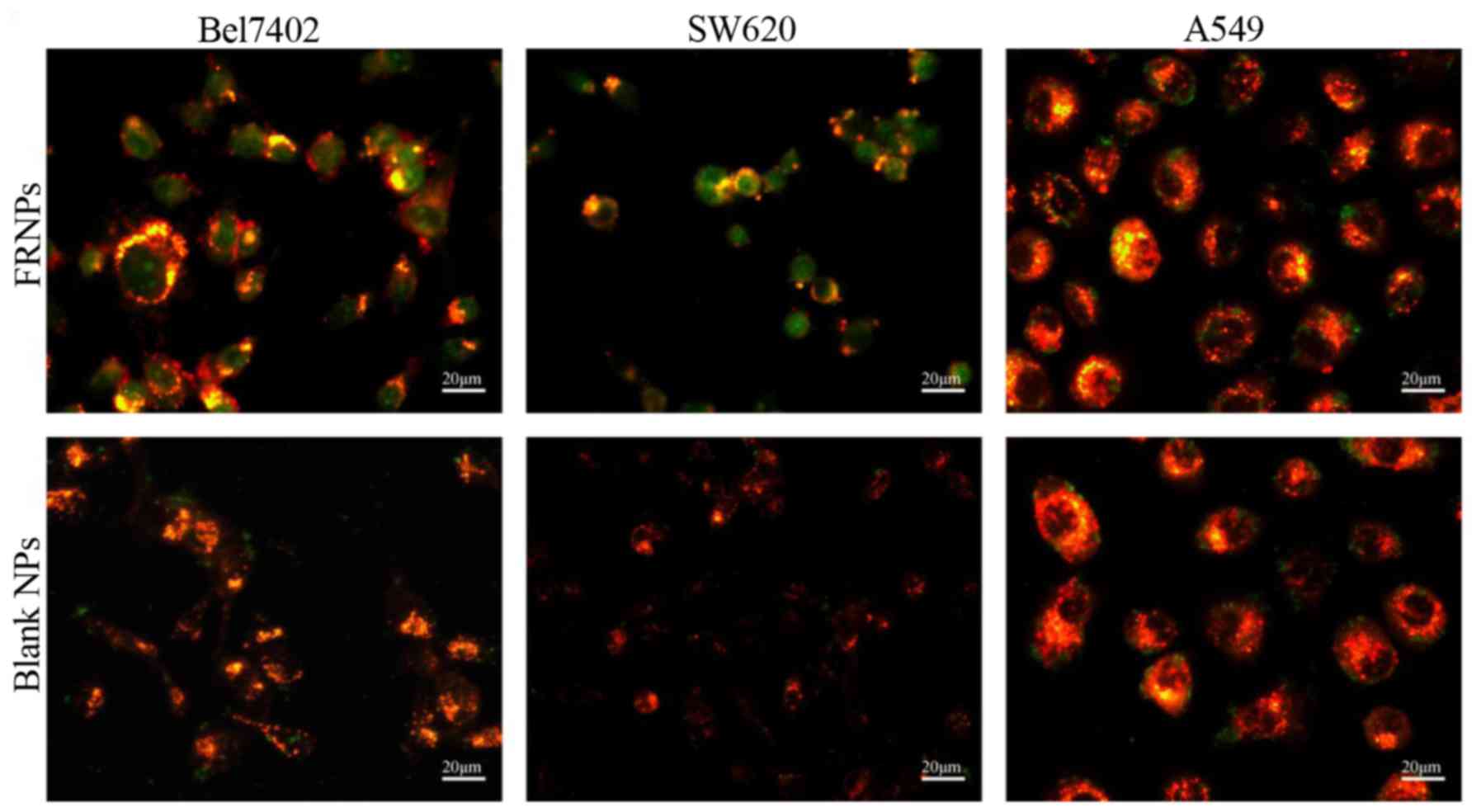
Since the FITC-labeled FRNPs and blank NPs were
developed to bind to FR and trigger receptor-mediated
internalization in tumor cells, we compared the uptake of the
targeted group and the control group in the Bel7402 cells, SW620
cells and A549 cells lines (Fig.
4). In the targeted group, a large number of green fluorescent
spots representing the FRNPs were absorbed into the cytoplasm of
Bel7402 and SW620 cells, whereas few green spots remained on the
cell membrane of the A549 cells. Furthermore, in the control group,
few green fluorescent spots representing the blank NPs were also
observed on the 3 types of cell membrane. These results
demonstrated that the FRNPs had a greater affinity to the Bel7402
cells and SW620 cells, in which FR was overexpressed.
Ultrasound targeted imaging in vivo
The peak enhancement of tumor
ultrasound imaging results
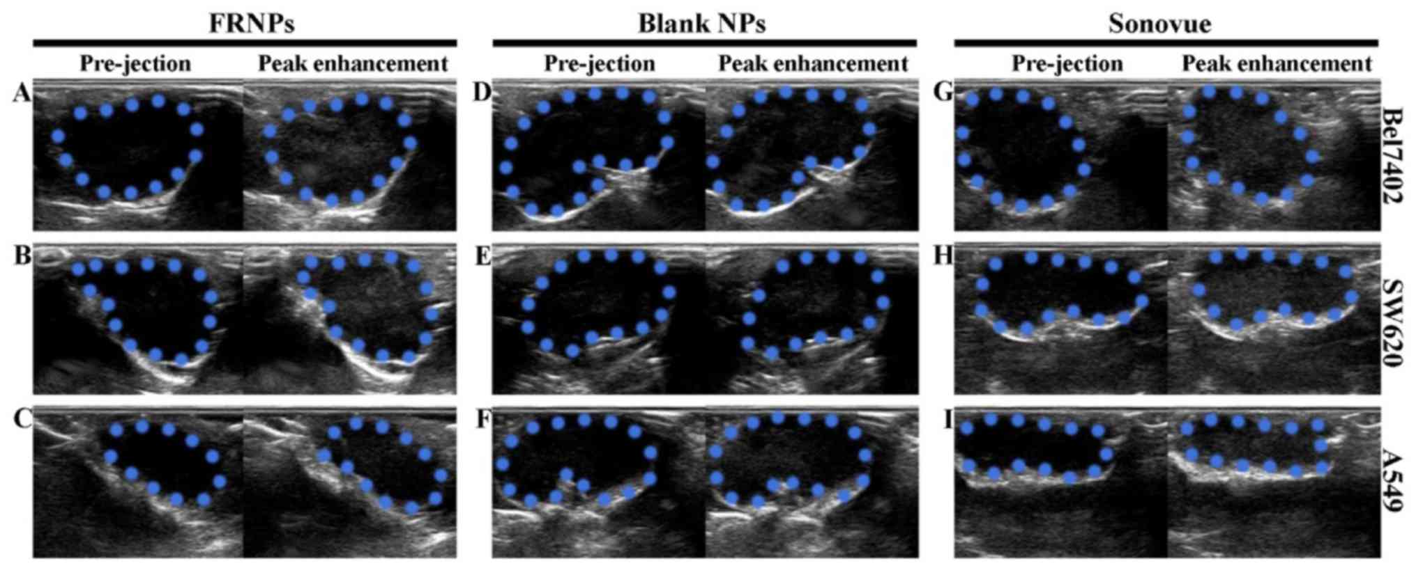
The peak enhancement of tumor imaging is shown in
Fig. 5. The ultrasonographic
images among the 3 types of xenografts revealed that the
enhancement of Sonovue was higher than the enhancement achieved
with the FRNPs and blank NPs. However, the enhancement of the FRNPs
in both the Bel7402 and SW620 tumor xenografts was apparently
higher than the enhancement achieved with the blank NPs at the peak
level. In the A549 tumor xenografts, the imaging results of the
FRNPs and blank NPs were comparable at the peak level of
enhancement. Thus, the blank NPs achieved the worst performance on
tumor-enhanced imaging, and both the FRNPs and Sonovue obtained
excellent tumor-enhanced imaging for FR overexpressing tumors.
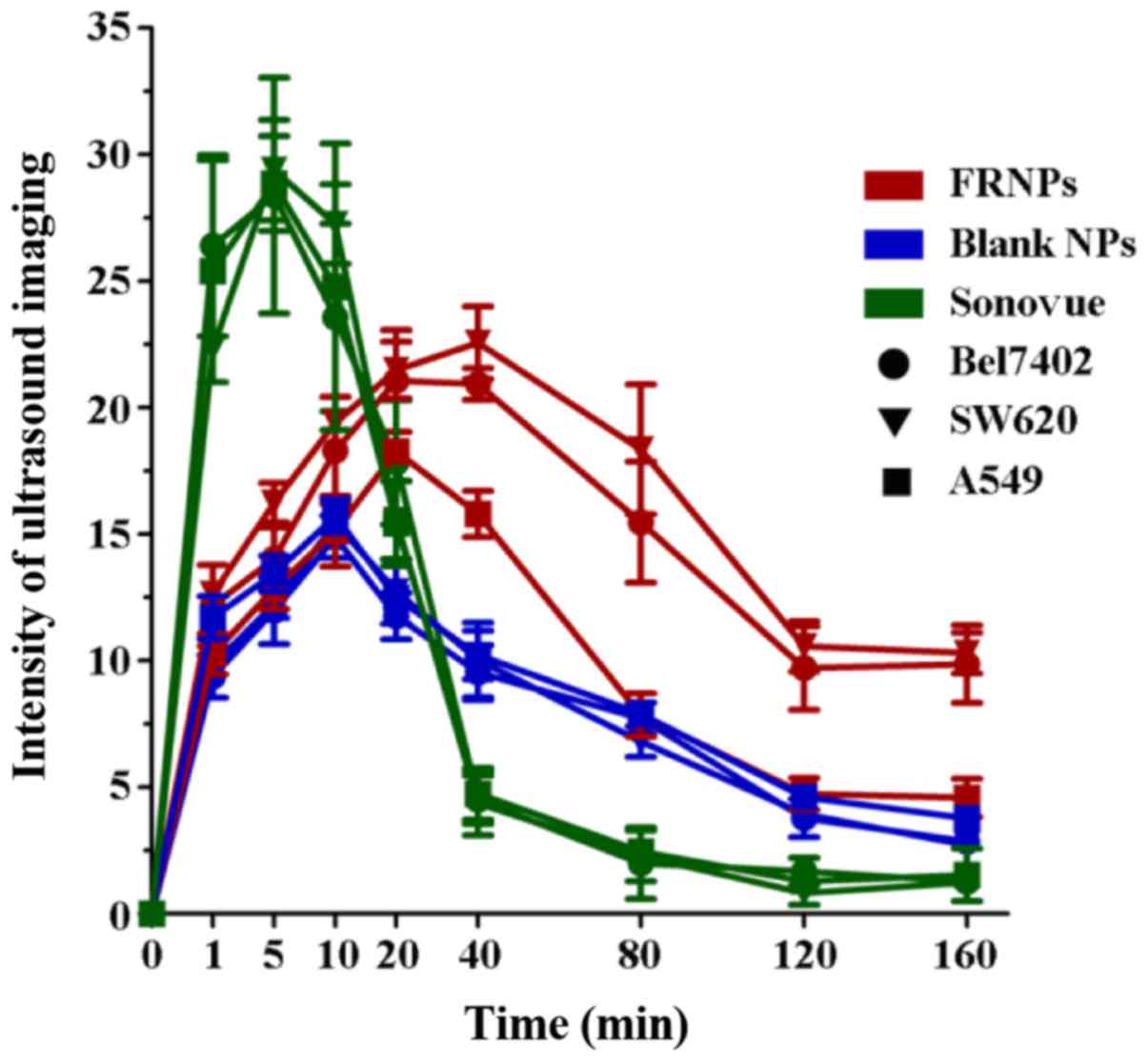
Variation trend in the intensity value
of tumor-enhanced imaging
Imaging Pro Plus 6.0 software was used to analyze
the grey value of the ultrasound images. The difference in the grey
valve of images collected at pre-injection and post-injection
served as the intensity value of tumor imaging by 3 types of
contrast agents in the xenografts. The time-intensity curve is
shown in Fig. 6, illustrating the
change in ultrasonic enhancement in areas of interest (AOI) of the
tumors collected from the xenografts. From 1 to 10 min
post-injection, the imaging of the tumors was significantly
enhanced in the Sonovue group, and the intensity value of tumor
imaging in this group was considerably higher than that of the
other groups (P<0.001), with no clear differences among the 3
types of xenografts (P=0.996). At 20 min post-injection, the
intensity value in the Sonovue group decreased rapidly, and almost
disappeared within 40 min post-injection. However, the intensity
value in the FRNPs and blank NP groups gradually increased after
the injection and reached the peak levels at 10 and 20 min
post-injection. In the FRNP group, the intensity value of the
Bel7402 and SW620 tumor xenografts was significantly higher than
that of the A549 tumor xenografts from 20 to 160 min post-injection
(P=0.012). Until the time point of 160 min post-injection, the
enhancement of the FRNPs in the Bel7402 tumor xenografts and SW620
tumor xenografts remained at a high level of intensity, and this
suggested that the FRNPs exhibited a longer duration of effective
enhanced ultrasound imaging. In the blank NP groups, the mean grey
value exhibited no significant difference among the 3 types of
xenografts at all time points (P=0.694). According to the results
of ultrasound imaging in vivo, the images from the FR
overexpressing tumors (Bel7402 tumor xenografts and SW620 tumor
xenografts) displayed a higher increased intensity value than the
images from the low expressing tumors (A549 xenografts). However,
we did not find similar situations such as these after the
injection of blank NPs and Sonovue. These results revealed that the
FRNPs achieved specific enhanced imaging for FR overexpressing
tumors and the effects of tumor imaging were significantly superior
to those of Sonovue at 40 min post-injection.
Targeting ability and biodistribution of
FRNPs in vivo
The fluorescence images of Cy7-labeled FRNPs were
obtained using the Kodak In-vivo Imaging System FX PRO at different
time points following injection with Cy7-labeled FRNPs. The Cy7
fluorescence dye, which has absorption and emission maxima around
747 and 774 nm, belongs to the class of NIR fluorescence dyes. NIR
fluorescence imaging has many advantages, including relatively
minimal autofluorescence, low tissue absorption and scatter, which
results in greater tissue penetration than visible optical
fluorescent dyes for in vivo imaging applications within the
650–900 nm NIR window (29,30). Thus, the fluorescent images of
Cy7-labeled nanoparticles can determine the in vivo
biodistribution and tumor selective effect of nanoparticles
(31–34).
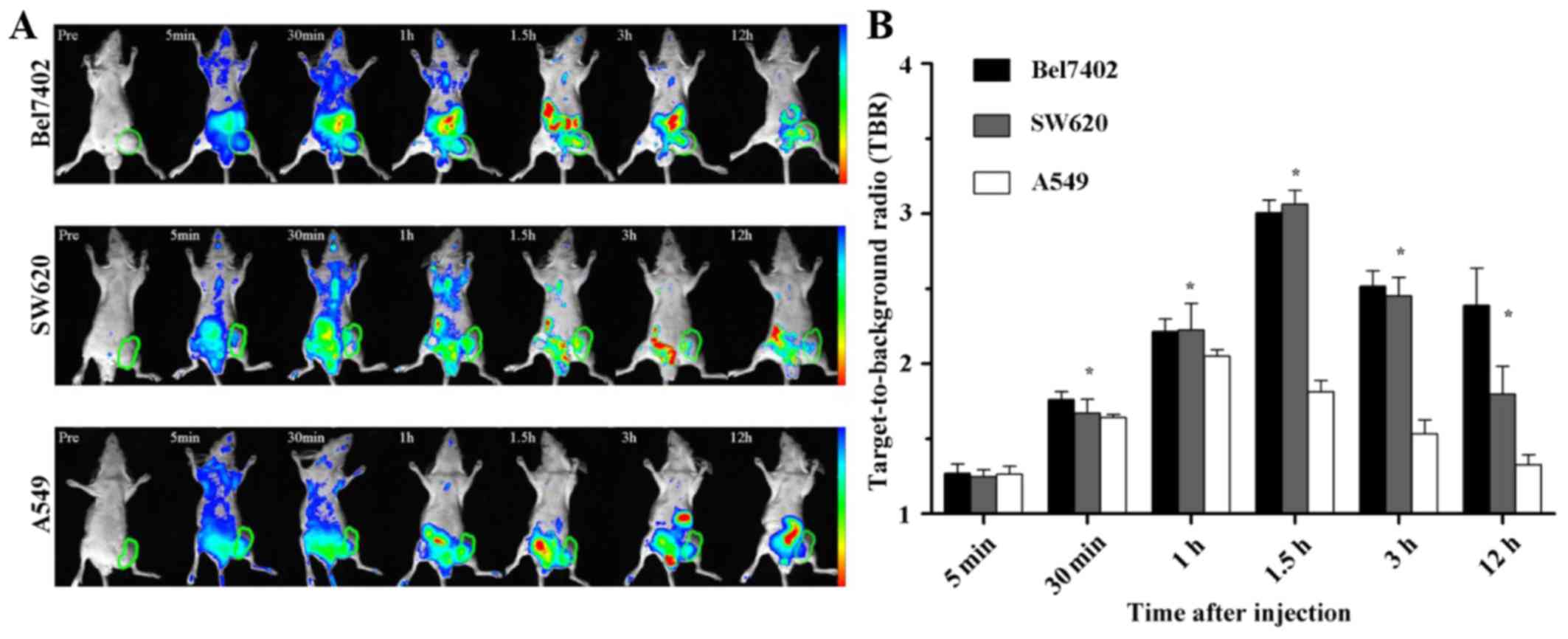
The fluorescence imaging of xenografts were as shown
in Fig. 7A, indicating the
distribution of FRNPs and its aggregation in tumor region. At 1 and
5 min after the intravenous injection of the fluorescent particles,
the images of xenografts were covered with different fluorescent
signal strength and there were no differences in fluorescence
intensity within tumor regions among the 3 types of xenografts. The
fluorescence intensity of the tumor regions in the Bel7402 and
SW620 tumor xenografts increased significantly from 30 min to 2 h
post-injection, and high levels of fluorescence intensity remained
until 12 h. By contrast, less tumor fluorescence accumulation was
observed in the A549 xenografts at the same time points. The
fluorescence intensity of the tumors and muscles was detected at
the different time points in order to semi-quantitatively analyze
the targeting ability of the FRNPs to the FR overexpressing tumor
tissues. The average of TBR between the tumor and adjacent normal
thigh muscle is shown in Fig.
7B.
To further determine the fluorescent signals in
different tissues, the mice were sacrificed at 12 h post-injection
and the heart, liver, spleen, lung, kidneys, intestines and tumors
were excised, washed with PBS and exposed to fluorescence imaging.
As shown in Fig. 8 for the ex
vivo fluorescence imaging of different tissues, the fluorescent
signal in the tumors of the Bel7402 and SW620 tumor xenografts was
very strong, whereas little signal intensity was visible in the
tumors of the A549 tumor xenografts. To semi-quantitatively analyze
the targeting ability of the FRNPs, the fluorescence intensity in
the tumors was measured. The average fluorescent signal intensity
of the tumors excised from the Bel7402, SW620 and A549 tumor
xenografts was (2.4±0.1)×1010,
(2.3±0.02)×1010, (1.24±0.03)×1010
photons/sec/mm2 (the fluorescence images of tumors can
be seen on the right side in the bottom row of Fig. 8A, B and C). The differences of
fluorescent signal intensity among the 3 types of xenografts were
statistically significant (P=0.027). These results were consistent
with the outcomes from the in vivo ultrasound imaging. The
ex vivo tumor imaging results also demonstrated that the
FRNPs exhibited relatively high tumor-targeted distribution in the
FR overexpressing tumors.
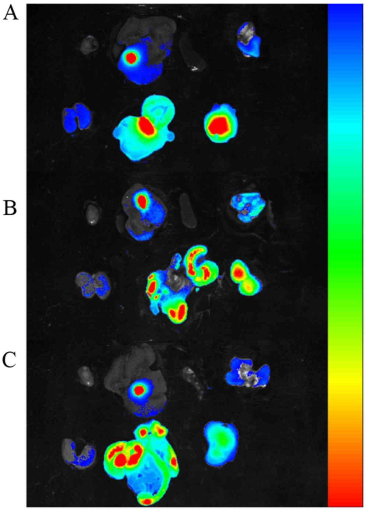 | Figure 8Ex vivo NIR imaging of other
organs and tumor tissues at 12 h post-injection, collected from 3
types of xenografts, (A) Bel7402, (B) SW620, (C) A549 tumor
xenografts. (A, B and C) Organs and tumor tissue are included in
proper sequence in the top row and bottom row. Top row, heart,
liver, spleen, lung were arranged from left to right; bottom row,
kidneys, intestines, tumor tissue were arranged from left to right.
The range from blue to red represents the changes in fluorescence
intensity from weak to strong. |
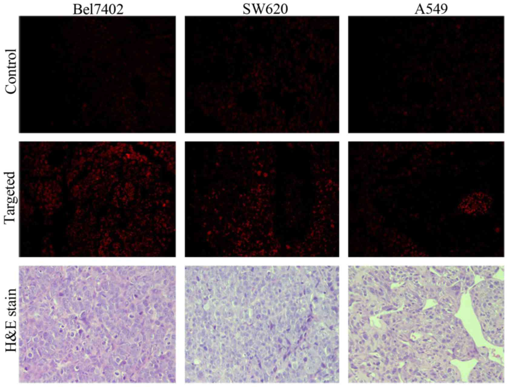
Histological analysis
To further verify the fluorescence imaging results
and confirm the presence of FRNPs in tumor tissues, the frozen
tissue slices of the tumor tissues were collected and fixed at 90
min post-injection of Cy7-labeled FRNPs (Fig. 9). Only a weak fluorescent signal,
corresponding to autofluorescence was observed in the tumor slices
of the control group. In the targeted group, we observed that the
fluorescent signals of high intensity (red spots) were converged on
some areas that seemed like the blood vessels of tumor tissues,
whereas fluorescent signals of extravascular areas remained weak in
the slices of A549 xenografts. By contrast, more and brighter
fluorescent signals of extravascular areas in the slices of the
Bel7402 xenografts and SW620 xenografts were observed. These
results revealed that the FRNPs passed through the endothelial gap
of the tumor blood vessel and combined with the tumor tissues. The
targeting ability to FR overexpressing tumors and PEGylated surface
against the 'cleaning up' by the reticuloendothelial system (RES)
led to the accumulation of FRNPs in FR overexpressing tumors. The
H&E-stained slices of tumor tissue were observed and typical
pathological mitotic figures were evident and did not exhibit any
significant differences between the 2 groups.
Discussion
With the development of contrast agents,
contrast-enhanced ultrasonography has played on important role in
the detection and differentiation of tumors. Although many types of
ultrasound contrast agents have been developed for clinical
application, some clinical studies have indicated that the contrast
agents currently used are limited by insufficient sensitivity and
specificity (1,4,35).
Nowadays, the emerging of molecular imaging
introduces a new avenue for tumor detection at the molecular level
with ultrasonography. However the application of molecular
ultrasound imaging is required to develop nanosized contrast
agents, of which the particle size is smaller than the contrast
agents recently used (7,36).
Currently, PFOB nanoparticles have been reported to
be applied to molecular ultrasound imaging in vivo,
including several molecular targeted medicine applications
(37–40). The application of PFOB
nanoparticles into the field of ultrasonography has been limited to
low-intensity of ultrasound reflection due to nanosized particle
size and the nanoparticles may also be less liable to non-specific
signal enhancement events since larger concentrations (more binding
events) are required to produce conspicuity (41,42).
Furthermore, FR is highly expressed in various human
malignancies and it has been served as a potential targeted ligand
for tumor imaging and antitumor therapy due to the high affinity to
bind with FA. Thus, in this study, we conducted a constructive
attempt to apply the polymers of FA-CS and PEG-ALG to the
preparation of the FR overexpressed tumor targeted nanoparticles
(FRNPs) by using the LbL assembly technique, which maintained the
advantages of FR-targeting affinity and resistance against the
macrophage of RES due to PEG modification.
In this study, the targeted shell consisted of FA-CS
and PEG-ALG, which were easily obtained by the carbodiimide method
and did not need complex modifications. We introduced the LbL
assembly technique to the process of core-shell enveloping, which
improved the quality and effectiveness of targeted shell. As shown
in Fig. 2A, the particle size
decreased from 244.7±6.0 to 217.6±5.6 nm and the surface potential
was reversed (from 39.1±0.4 to −21.6±1.3) after the first addition
of PEG-ALG (layer 2). This indicated that a mesh shell on the
surface of PFOB liposome was forming by the strong ionic
electrostatic interaction between PEG-ALG and FA-CS. In the TEM
images (Fig. 2D), we could see
that FRNPs exhibited a rough surface and retained larger particle
size, whereas the blank NPs (PFOB liposomes) maintained a smooth
surface and a small size. Although the different layers could not
distinguished clearly, a darker phosphotungstically acid-stained
core and a lesser-stained shell were visible. After the LbL
assembly was completed, the FRNPs maintained FR-targeting function
(FA modified) and solvent stability (the electrostatic repulsive
force and the PEGyalted polymers that further extend the repulsive
force and prevent aggregation). In the assembly process, we made an
attempt to keep the particle size in an optimal range by careful
adjustment of the polymer radios.
In the targeted binding assay in vitro, the
FRNPs were specifically taken up by the Bel7402 and SW620 cancer
cells (FR overexpressing cancer cell lines). Subsequently, we
investigated the ultrasongraphy for xenograft models and found that
the FRNPs obtained specific ultrasound enhanced imaging for FR
overexpressing tumors. Although the enhancement of FRNPs was
slightly weaker than that of Sonovue, it maintained a longer
enhanced duration in FR overexpressing tumors. Furthermore, in the
fluorescence imaging of the xenograft models, the ultrasound
imaging results of the FRNPs were confirmed. As shown in Fig. 7, that fluorescence intensity in
the FR overexpressing tumor region was significantly stronger at 30
min post-injection, and the ex vivo fluorescence imaging of
tumors and other organs (Fig. 8)
revealed that the intestines and tumors possessed a higher
intensity of fluorescent signal. These results indicated that FRNPs
could accumulate in FR overexpressed tumor and passed out through
the endothelial gap of tumor blood vessel for targeting to FR
overexpressed tumors.
In conclusion, in this study, we developed a novel
strategy of ultrasonic nanoparticles modified with FA and PEG for
FR overexpressingtumor imaging. FRNPs were successfully synthesized
with the LbL assembly technique and maintained suitable
physicochemical properties. Our data illustrated that FRNPs
exhibited a respectable ability to target the certain tumors in
vitro and in vivo. At present, there are certain
deficiencies and limits in tumor-targeted imaging. FRNPs may thus
have potential for early diagnosis and targeted therapy of FR
overexpressing tumors. The nanoparticles and these methods of
preparation may prove to be useful for further medical and
biological applications.
Acknowledgments
The present study was supported by grants from the
Social Development Major Project in Ningbo, China (no.
2012C5013).
References
|
1
|
Perera RH, Hernandez C, Zhou H, Kota P,
Burke A and Exner AA: Ultrasound imaging beyond the vasculature
with new generation contrast agents. Wiley Interdiscip Rev Nanomed
Nanobiotechnol. 7:593–608. 2015. View Article : Google Scholar : PubMed/NCBI
|
|
2
|
Casciaro S, Soloperto G, Greco A, Casciaro
E, Franchini R and Conversano F: Effectiveness of functionalized
nanosystems formultimodal molecular sensing and imaging in
medicine. IEEE Sens J. 6:2305–2312. 2013. View Article : Google Scholar
|
|
3
|
Maeda H, Wu J, Sawa T, Matsumura Y and
Hori K: Tumor vascular permeability and the EPR effect in
macromolecular therapeutics: A review. J Control Release.
65:271–284. 2000. View Article : Google Scholar : PubMed/NCBI
|
|
4
|
Hahn MA, Singh AK, Sharma P, Brown SC and
Moudgil BM: Nanoparticles as contrast agents for in-vivo
bioimaging: Current status and future perspectives. Anal Bioanal
Chem. 399:3–27. 2011. View Article : Google Scholar
|
|
5
|
Kobayashi H, Turkbey B, Watanabe R and
Choyke PL: Cancer drug delivery: Considerations in the rational
design of nanosized bioconjugates. Bioconjug Chem. 25:2093–2100.
2014. View Article : Google Scholar : PubMed/NCBI
|
|
6
|
Orocio-Rodríguez E, Ferro-Flores G,
Santos-Cuevas CL, Ramírez FM, Ocampo-García BE, Azorín-Vega E and
Sánchez-García FM: Two novel nanosized radiolabeled analogues of
somatostatin for neuroendocrine tumor imaging. J Nanosci
Nanotechnol. 15:4159–4169. 2015. View Article : Google Scholar : PubMed/NCBI
|
|
7
|
Toy R, Bauer L, Hoimes C, Ghaghada KB and
Karathanasis E: Targeted nanotechnology for cancer imaging. Adv
Drug Deliv Rev. 76:79–97. 2014. View Article : Google Scholar : PubMed/NCBI
|
|
8
|
Wilson KE, Wang TY and Willmann JK:
Acoustic and photo-acoustic molecular imaging of cancer. J Nucl
Med. 54:1851–1854. 2013. View Article : Google Scholar : PubMed/NCBI
|
|
9
|
Gessner R and Dayton PA: Advances in
molecular imaging with ultrasound. Mol Imaging. 9:117–127.
2010.PubMed/NCBI
|
|
10
|
Zhao R, Matherly LH and Goldman ID:
Membrane transporters and folate homeostasis: Intestinal absorption
and transport into systemic compartments and tissues. Expert Rev
Mol Med. 11:e42009. View Article : Google Scholar : PubMed/NCBI
|
|
11
|
Shen F, Wu M, Ross JF, Miller D and Ratnam
M: Folate receptor type gamma is primarily a secretory protein due
to lack of an efficient signal for glycosylphosphatidylinositol
modification: Protein characterization and cell type specificity.
Biochemistry. 34:5660–5665. 1995. View Article : Google Scholar : PubMed/NCBI
|
|
12
|
Kane MA: The role of folates in squamous
cell carcinoma of the head and neck. Cancer Detect Prev. 29:46–53.
2005. View Article : Google Scholar : PubMed/NCBI
|
|
13
|
Hartmann LC, Keeney GL, Lingle WL,
Christianson TJ, Varghese B, Hillman D, Oberg AL and Low PS: Folate
receptor overexpression is associated with poor outcome in breast
cancer. Int J Cancer. 121:938–942. 2007. View Article : Google Scholar : PubMed/NCBI
|
|
14
|
O'Shannessy DJ, Yu G, Smale R, Fu YS,
Singhal S, Thiel RP, Somers EB and Vachani A: Folate receptor alpha
expression in lung cancer: Diagnostic and prognostic significance.
Oncotarget. 3:414–425. 2012. View Article : Google Scholar : PubMed/NCBI
|
|
15
|
Parker N, Turk MJ, Westrick E, Lewis JD,
Low PS and Leamon CP: Folate receptor expression in carcinomas and
normal tissues determined by a quantitative radioligand binding
assay. Anal Biochem. 338:284–293. 2005. View Article : Google Scholar : PubMed/NCBI
|
|
16
|
Assaraf YG, Leamon CP and Reddy JA: The
folate receptor as a rational therapeutic target for personalized
cancer treatment. Drug Resist Updat. 17:89–95. 2014. View Article : Google Scholar : PubMed/NCBI
|
|
17
|
Wu M, Gunning W and Ratnam M: Expression
of folate receptor type α in relation to cell type, malignancy, and
differentiation in ovary, uterus, and cervix. Cancer Epidemiol
Biomarkers Prev. 8:775–782. 1999.PubMed/NCBI
|
|
18
|
Kalli KR, Oberg AL, Keeney GL,
Christianson TJ, Low PS, Knutson KL and Hartmann LC: Folate
receptor alpha as a tumor target in epithelial ovarian cancer.
Gynecol Oncol. 108:619–626. 2008. View Article : Google Scholar : PubMed/NCBI
|
|
19
|
Bueno R, Appasani K, Mercer H, Lester S
and Sugarbaker D: The α folate receptor is highly activated in
malignant pleural mesothelioma. J Thorac Cardiovasc Surg.
121:225–233. 2001. View Article : Google Scholar : PubMed/NCBI
|
|
20
|
Lu Y, Sega E and Low PS: Folate
receptor-targeted immunotherapy: Induction of humoral and cellular
immunity against hapten-decorated cancer cells. Int J Cancer.
116:710–719. 2005. View Article : Google Scholar : PubMed/NCBI
|
|
21
|
Siafaka P, Betsiou M, Tsolou A, Angelou E,
Agianian B, Koffa M, Chaitidou S, Karavas E, Avgoustakis K and
Bikiaris D: Synthesis of folate-pegylated polyester nanoparticles
encapsulating ixabepilone for targeting folate receptor
overexpressing breast cancer cells. J Mater Sci Mater Med.
26:2752015. View Article : Google Scholar
|
|
22
|
Liu X, Zhao J, Guo D, Wang Z, Song W, Chen
W and Zhou J: Synthesis and evaluation of perfluorooctylbromide
nanoparticles modified with a folate receptor for targeting ovarian
cancer: in vitro and in vivo experiments. Int J Clin Exp Med.
8:10122–10131. 2015.PubMed/NCBI
|
|
23
|
Zhou J, Romero G, Rojas E, Ma L, Moya S
and Gao C: Layer by layer chitosan/alginate coatings on
poly(lactide-co-glycolide) nanoparticles for antifouling protection
and Folic acid binding to achieve selective cell targeting. J
Colloid Interface Sci. 345:241–247. 2010. View Article : Google Scholar : PubMed/NCBI
|
|
24
|
Elhissi AM, O'Neill MA, Roberts SA and
Taylor KM: A calori-metric study of dimyristoylphosphatidylcholine
phase transitions and steroid-liposome interactions for liposomes
prepared by thin film and proliposome methods. Int J Pharm.
320:124–130. 2006. View Article : Google Scholar : PubMed/NCBI
|
|
25
|
Salem HF, Ahmed SM, Hassaballah AE and
Omar MM: Targeting brain cells with glutathione-modulated
nanoliposomes: In vitro and in vivo study. Drug Des Devel Ther.
9:3705–3727. 2015. View Article : Google Scholar : PubMed/NCBI
|
|
26
|
Barnett BP, Ruiz-Cabello J, Hota P,
Ouwerkerk R, Shamblott MJ, Lauzon C, Walczak P, Gilson WD, Chacko
VP, Kraitchman DL, et al: Use of perfluorocarbon nanoparticles for
non-invasive multimodal cell tracking of human pancreatic islets.
Biomaterials. 35:9984–9994. 2014.
|
|
27
|
Kim J, Lee CM, Jeong HJ and Lee KY: In
vivo tumor accumulation of nanoparticles formed by ionic
interaction of glycol chitosan and fatty acid ethyl ester. J
Nanosci Nanotechnol. 11:1160–1166. 2011. View Article : Google Scholar : PubMed/NCBI
|
|
28
|
Alencar H, Funovics MA, Figueiredo J,
Sawaya H, Weissleder R and Mahmood U: Colonic adenocarcinomas:
Near-infrared microcatheter imaging of smart probes for early
detection - study in mice. Radiology. 244:232–238. 2007. View Article : Google Scholar : PubMed/NCBI
|
|
29
|
Hawrysz DJ and Sevick-Muraca EM:
Developments toward diagnostic breast cancer imaging using
near-infrared optical measurements and fluorescent contrast agents.
Neoplasia. 2:388–417. 2000. View Article : Google Scholar
|
|
30
|
Zheng C, Zheng M, Gong P, Jia D, Zhang P,
Shi B, Sheng Z, Ma Y and Cai L: Indocyanine green-loaded
biodegradable tumor targeting nanoprobes for in vitro and in vivo
imaging. Biomaterials. 33:5603–5609. 2012. View Article : Google Scholar : PubMed/NCBI
|
|
31
|
Hama Y, Koyama Y, Choyke PL and Kobayashi
H: Two-color in vivo dynamic contrast-enhanced pharmacokinetic
imaging. J Biomed Opt. 12:0340162007. View Article : Google Scholar : PubMed/NCBI
|
|
32
|
Nakamura T, Kawano K, Shiraishi K,
Yokoyama M and Maitani Y: Folate-targeted gadolinium-lipid-based
nanoparticles as a bimodal contrast agent for tumor fluorescent and
magnetic resonance imaging. Biol Pharm Bull. 37:521–527. 2014.
View Article : Google Scholar : PubMed/NCBI
|
|
33
|
Chung EJ, Mlinar LB, Sugimoto MJ, Nord K,
Roman BB and Tirrell M: In vivo biodistribution and clearance of
peptide amphiphile micelles. Nanomedicine. 11:479–487. 2015.
View Article : Google Scholar
|
|
34
|
Chung EJ, Cheng Y, Morshed R, Nord K, Han
Y, Wegscheid ML, Auffinger B, Wainwright DA, Lesniak MS and Tirrell
MV: Fibrin-binding, peptide amphiphile micelles for targeting
glioblastoma. Biomaterials. 35:1249–1256. 2014. View Article : Google Scholar
|
|
35
|
Blomqvist L, Carlsson S, Gjertsson P,
Heintz E, Hultcrantz M, Mejare I and Andrén O: Limited evidence for
the use of imaging to detect prostate cancer: A systematic review.
Eur J Radiol. 83:1601–1606. 2014. View Article : Google Scholar : PubMed/NCBI
|
|
36
|
Sivasubramanian M, Hsia Y and Lo LW:
Nanoparticle-facilitated functional and molecular imaging for the
early detection of cancer. Front Mol Biosci. 1:152014. View Article : Google Scholar : PubMed/NCBI
|
|
37
|
Giraudeau C, Geffroy F, Mériaux S,
Boumezbeur F, Robert P, Port M, Robic C, Le Bihan D, Lethimonnier F
and Valette J: 19F molecular MR imaging for detection of brain
tumor angiogenesis: In vivo validation using targeted PFOB
nanoparticles. Angiogenesis. 16:171–179. 2013. View Article : Google Scholar
|
|
38
|
Vu-Quang H, Vinding MS, Xia D, Nielsen T,
Ullisch MG, Dong M, Nielsen NC and Kjems J: Chitosan-coated
poly(lactic-co-glycolic acid) perfluorooctyl bromide nanoparticles
for cell labeling in (19)F magnetic resonance imaging. Carbohydr
Polym. 136:936–944. 2016. View Article : Google Scholar
|
|
39
|
Watanabe T, Kimura Y and Ono T:
Microfluidic fabrication of monodisperse polylactide microcapsules
with tunable structures through rapid precipitation. Langmuir.
29:14082–14088. 2013. View Article : Google Scholar : PubMed/NCBI
|
|
40
|
Decato S, Bemis T, Madsen E and Mecozzi S:
Synthesis and characterization of perfluoro-tert-butyl
semifluorinated amphiphilic polymers and their potential
application in hydrophobic drug delivery. Polym Chem. 5:6461–6471.
2014. View Article : Google Scholar : PubMed/NCBI
|
|
41
|
Marsh JN, Hall CS, Scott MJ, Fuhrhop RW,
Gaffney PJ, Wickline SA and Lanza GM: Improvements in the
ultrasonic contrast of targeted perfluorocarbon nanoparticles using
an acoustic transmission line model. IEEE Trans Ultrason
Ferroelectr Freq Control. 49:29–38. 2002. View Article : Google Scholar : PubMed/NCBI
|
|
42
|
Marsh JN, Partlow KC, Abendschein DR,
Scott MJ, Lanza GM and Wickline SA: Molecular imaging with targeted
perfluorocarbon nanoparticles: Quantification of the concentration
dependence of contrast enhancement for binding to sparse cellular
epitopes. Ultrasound Med Biol. 33:950–958. 2007. View Article : Google Scholar : PubMed/NCBI
|