Introduction
Articular cartilage has limited healing potential
once injured. Autologous chondrocyte implantation (ACI) has emerged
as a novel approach to cartilage repair through the use of
harvested chondrocytes (1,2).
During the process of ACI, expansion of the chondrocytes from the
donor tissue in vitro is indispensable (3). However, this approach is restricted
by the limited cell numbers and the dedifferentiation of the
chondrocytes when they are cultured in vitro.
Dedifferentiated chondrocytes are characterized by a marked
increase in collagen type I (Col I)and a decrease in
cartilage-specific markers, including collagen type II (Col II) and
aggrecan (AGC) (4).
Numerous pieces of evidence indicate that cell-cell
and cell-extracellular matrix (ECM) directly influence cell
signaling via cell adhesion molecules, including integrins and
cadherins, which are crucial for cell functions (5–7).
Scaffolds not only serve as templates for cells to facilitate cell
motility, ECM synthesis and physiological storage of bioactive
molecules (8), but also provide
the advantage of guidance for cell differentiation by affecting
cell-cell and cell-ECM interactions (9). Similar to the structure of hyaline
cartilage, three-dimensional (3D) scaffolds, particularly the
hydrogels, are highly recommended (10). It is generally accepted that the
3D environment favors the maintenance of the chondrocyte phenotype
and supports redifferentiation of dedifferentiated articular
chondrocytes (11–13). However, cells are encapsulated in
matrix and cannot be harvested for further implantation. The
difficulty of taking the cells out of the 3D matrices with their
viability and functions intact therefore remains.
As an alternative, monolayer culture using an
elaborate cellular microenvironment that is created mimicking the
in vivo situation may assist in maintaining the cell
phenotype and prevent dedifferentiation (14). Collagen is widely used for
cartilage regeneration (15,16), not only as it resembles the ECM of
natural cartilage, but also as it has weak antigenicity,
biodegradability and superior biocompatibility (17,18). The collagen membranes have
provided a monolayer culture system, which facilitated the
expanding of chondrocyte cells (19), and have been used for autologous
chondrocyte implantation (20,21). Without the addition of growth
factors, mesenchymal stem cells cultured in collagen beads can
differentiate into chondrocytes and form hyaline cartilage similar
to native cartilage (22,23). However, pure collagen has poor
mechanical strength and less favorable bioactivity (24), which does not meet the criteria
for cartilage tissue engineering. As cartilage is a weight-bearing
tissue, the scaffold for cartilage engineering should be of an
appropriate mechanical strength. Therefore, to improve the
mechanical properties of collagen, the introduction of inorganic
material is an optimal choice. Hydroxyapatite (HA) has been
reported to reinforce the mechanical and biological properties of
collagen (25,26), and also showed potential for
cartilage repair (27). In
particular, nano-sized HA particles are superior to micrometric
particles due to the higher surface/volume ratio, better dispersion
and the possibility of mixing smaller amounts of HAs in the
collagen matrix (28). Compared
with mechanical or ultrasonic stirring, which always results in
micro-HA, the in situ hybridization method enables the
creation of nano-HA, with the advantage of obtaining a homogeneous
biocomposite scaffold with well-distributed nano-HA particles
(29). A previous study showed
that collagen-HA by way of in situ hybridization can enhance
the mechanical strength and facilitate chondrocyte growth (25). Thus, we hypothesize that
nano-HA/collagen (nHC) film may be favorable cell substrate for
in vitro culture of chondrocytes.
In the present study, nano-HA particles were
synthesized innovatively in situ in collagen solution,
forming nHC film. To verify its capacity in preventing chondrocytes
from dedifferentiation in vitro, nHC film was used as a
substrate for chondrocytes. Furthermore, chondrocyte proliferation
on the novel gel films was compared with that of pure collagen and
an HC film on which HA particles were prepared by physical mixing.
The analysis of cell viability, morphology, phenotype, protein
level and biological function were implemented. With the intention
to preserve cell phenotype and promote cell growth to ensure
sufficient and functional cells, this study may provide novel
insights for cell-based therapy.
Materials and methods
Materials and chemicals
COLIA1 of calf origin and nano-HA powder were
purchased from the Engineering Research Center in Biomaterials,
Sichuan University (Chengdu, Sichuan, China). Chemicals of
analytical grade, including calcium nitrate
[Ca(NO3)2], ammonium hydroxide
(NH3•H2O), diammonium phosphate
[(NH4)2HPO4·12H2O],
glacial acetic acid, calcium chloride and sodium hydroxide were all
purchased from Tianjin Kemiou Chemical Reagent Co., Ltd. (Tianjin,
China). Type II collagenase, proteinase K solution, Hoechst 33258
and 1,9-dimethylmethylene blue (DMMB) were purchased from
Sigma-Aldrich; Merck KGaA (Darmstadt, Germany). Dulbecco’s modified
Eagle’s medium (DMEM) was obtained from GE Healthcare Life Sciences
(Logan, UT, USA). Fetal bovine serum (FBS) was procured from
Hangzhou Sijiqing Biological Engineering Materials Co., Ltd.
(Hangzhou, China). Anti-COLIA1 and -COL1A2 antibodies and the
immunohistochemistry (IHC) kit were purchased from Wuhan Boster
Biological Technology, Ltd. (Wuhan, China). Phosphate-buffered
saline (PBS) and trypsin was obtained from Beijing Solarbio Science
and Technology Co., Ltd. (Beijing, China). A total of 20 healthy
newborn Sprague-Dawley (SD) rats (age, 3–5 days; weight, 10–12 g;
random gender) were obtained from the Experimental Animal Center of
Guangxi Medical University (Nanning, China). The rats were housed
in an SPF level lab under controlled a temperature of 20–24°C and a
relative humidity of 50–60% with a 12/12 h light-dark cycle. All
the rats had free access to formula food and water.
Preparation of nHC hydrogel in situ
All the chemicals were stored at 4°C in sterile
conditions (25). To prepare the
nHC hydrogel, COLIA1 solution was neutralized by 1 mol glacial
acetic acid and 1 mol sodium hydroxide, with a final concentration
of 10 mg/ml. Next, 1 mol/l
(NH4)2HPO4·12H2O was
added into the collagen solution. Ca(NO3)2 (1
mol/l) was used to adjust the ratio of Ca/P to a ratio of 1.67 and
then NH3·H2O was titrated slowly to regulate
the pH value up to 9, with maintenance for 2 h. The solution would
be neutralized by 1 mol glacial acetic acid and 1 mol sodium
hydroxide. Once the pH value was 7, the solution would be
gelatinized at 25°C. The HC gel was fabricated by the physical
mixing of nano-HA powders into collagen solution in a ratio of 10
mg:1 ml.
Isolation and culture of
chondrocytes
This study was performed strictly according to the
recommendations in the Guide for the Care and Use of Laboratory
Animals of the National Institutes of Health. The protocol was
approved by the Ethics Committee of Animal Experiments of Guangxi
Medical University. All surgery was performed under sodium
pentobarbital anesthesia, and all efforts were made to minimize
suffering. Cartilage was isolated from newborn SD rats (3–5 days
old) in sterile conditions. The new born SD rats were sacrificed by
an intravenous injection of a euthanasia solution. Articular
cartilage was isolated from the joints of the limbs in sterile
conditions. Soft tissue attached to the surface of the cartilage
was removed using 0.25 mg/ml trypsin at 37°C for 30 min. The
cartilage was then shred into 1 mm3 pieces with
ophthalmic scissors. The tissue pieces were digested in 1 mg/ml
type II collagenase for 3 h. Finally, chondrocytes were collected
and cultures on dishes in DMEM with 10% FBS. Subsequent to
passaging 2 times, the cells were seeded at a density of
2.4×10⁴/cm2 on dishes paved with nHC, HC or
COLIA1 gel. In the control group, chondrocytes were cultured in 10%
FBS containing 10 ng/ml transforming growth factor-β (TGF-β)
(Peprotech, Inc., Rocky Hill, NJ, USA), 50 mg/ml
insulin-transferring-selenium, 100 nmol/l dexamethasone and 50
µg/ml vitamin C (all Sigma-Aldrich; Merck KGaA). Experiments
were performed at 2, 4 and 6 days of culture.
Proliferation of chondrocytes
The proliferation of the cells was observed by
methyl thiazolyl tetra zolium (MTT) assay (Sigma-Aldrich; Merck
KGaA). Briefly, chondrocytes (5,000 cells/well) were cultured in
96-well microplates coated with collagen, HC or nHC film for 1, 2,
3, 4, 5 and 6 days. MTT (20 µl; 5 mg/ml) was added into the
culture medium (200 µl) in each well. Following incubation
for 4 h at 37°C, the medium supernatant was removed carefully and
dimethyl sulfoxide was added (150 µl/well). The optical
density (OD) was detected by a microplate reader at 490 nm.
Cells adhesion
A scanning electron microscope (SEM) was used to
analyze the adhesion of the cells on the film surface. Slides
coated with gel film seeded with chondrocytes were fixed in
glutaraldehyde for 24 h at 4°C. Following dehydration, drying and
gold spraying in order, the film surfaces and cell-matrix
interaction were studied with an SEM (Hitachi, Ltd., Tokyo, Japan)
operating at 3 kV, and images were captured.
Cell viability examination
Chondrocyte viability on the gel film was measured
utilizing a live-dead viability assay kit (Invitrogen; Thermo
Fisher Scientific, Inc., Waltham, MA, USA). Subsequent to being
washed in PBS, the chondrocytes were incubated in 1 ml PBS
containing 0.5 µl calcein-acetoxymethyl (calcein-AM) and 2
µl propidium iodide (PI) in the dark. After 5 min, the
viability of the cells was visualized with a laser scanning
confocal microscope (NIS Elements, version 2.1, Nikon A1; Nikon,
Tokyo, Japan) and images were captured.
Morphological analysis
Subsequent to being cultured for 2, 4 or 6 days, the
chondrocytes were fixed in 4% (w/v) para-formaldehyde (Beijing
Solarbio Science and Technology Co., Ltd.) for 30 min. Hematoxylin
and eosin (H&E; Beyotime, Shanghai, China) was used to dye the
nuclei and cytoplasm, respectively, at room temperature for 5 min.
Finally, samples were gradually dehydrated in 75% alcohol (10 sec),
95% alcohol (10 sec), anhydrousalcohol (10 sec) (Yong Da, Tianjing,
China) successively at room temperature. They were them dried in a
drying oven (DHG-9140; Jing Hong, Shanghai, China) at 60°C for 30
sec, and sealed with neutral gum (Beijing Solarbio Science and
Technology Co., Ltd.) and observed using a microscope (BX46;
Olympus, Tokyo, Japan).
Aggrecan secretion
Safranin O staining solution was used to detect the
AGC secretion of the chondrocytes seeded on gel film. Subsequent to
fixing and washing, the cells were incubated with Safranin O
(Beijing Solarbio Science and Technology Co., Ltd.) at room
temperature for 20 min. Next, the samples were gradually
dehydrated, sealed with neutral gum and observed. To further detect
the AGC secretion ability of the chondrocytes, the cells were
digested with proteinase K solution for 16 h at 60°C. The DNA
production was measured by spectrofluorometer using Hoechst 33258
dye at room temperature for 5 min in the dark. The
glycosaminoglycan (GAG) content was quantified by DMMB assay. GAG
production in each cell was normalized to the total DNA content of
the cells.
Immunohistochemical staining
To analyze the cartilage-like matrix production of
chondrocytes, COLIA1 and COL1A2 were detected by IHC using
monoclonal antibodies to the proteins of interest. The cells were
fixed in 4% (w/v) paraformaldehyde (Beijing Solarbio Science and
Technology Co., Ltd.) at room temperature for 30 min and then
treated with 0.01% Triton X-100 (Beyotime) for 10 min, and then
H2O2 (3%; Beyotime) was added to eliminate
endogenous peroxidase activity for 10 min. Goat serum (5%; Wuhan
Boster Biological Technology, Ltd.) was used to block the
non-specific binding sites at room temperature for 15 min. After 10
min, the samples were incubated with primary antibodies against
COLIA1 (1:100; BA0325; Wuhan Boster Biological Technology, Ltd.)
and COL1A2 (1:100; BA0533; Wuhan Boster Biological Technology,
Ltd.) at 4°C overnight. Subsequent to rewarming at room temperature
for 30 min, the cells were incubated with the secondary antibody
(1:100; BA1005; Wuhan Boster Biological Technology, Ltd.) for 10
min. A 3,3′-diaminobenzidine tetrahydrochloride kit (Wuhan Boster
Biological Technology, Ltd.) was used to visualize the antibody
binding at room temperature for 2 min. Hematoxylin was used to dye
the nuclei at room temperature for 15 sec. Finally, samples were
gradually dehydrated in 75% alcohol (10 sec), 95% alcohol (10 sec),
anhydrousalcohol (10 sec) (Yong Da) successively at room
temperature. After being dried in drying oven (DHG-9140; Jing Hong)
at 60°C for 30 sec, the samples were sealed with neutral gum
(Beijing Solarbio Science and Technology Co., Ltd.) and observed
using a microscope (BX46; Olympus).
Reverse transcription-quantitative
polymerase chain reaction (RT-qPCR) analysis
Total RNA was isolated from the cell/hydrogel
constructs using an extraction kit (Tiangen Biotech Co., Ltd.,
Beijing, China) at 6 days of culture. RNA was reverse transcribed
by First Strand cDNA Synthesis kit (Roche Diagnostics, Basel,
Switzerland) according to the manufacturer’s protocols. The cDNAs
were then subjected to RT-PCR to measure the expression of COLIA1
and COL1A2, and AGC. Briefly, according to the RT-PCR kit
(containing SYBR-Green I) protocol (Roche Diagnostics GmbH,
Mannheim, Germany), 1 µl RNA, 30 pmol forward and reverse
primers (Liuhe Genomics Technology Co., Peiking, China) and other
reagents in the kit were added into the reaction tube. The initial
denaturation stage was at 95°C for 5 min, followed by 35 cycles of
PCR. Each cycle occurred at 94°C for 40 sec, 56°C (AGC) or 57°C
(β-actin, COLIA1 and COL1A2) for 40 sec, and 72°C for 90 sec.
Finally, extension occurred at 72°C for 5 min. Relative
quantification was calculated using the 2−ΔΔCq method
(30) and all the results were
normalized to a house-keeping gene, β-actin. The sequence of
primers for β-actin, AGC, COLIA1 and COL1A2 were as follows:
β-actin forward, 5′-ATGATATCGCCGCGCTCGTCGT-3′ and reverse,
5′-CCTCGTCGCCCACATAGGAATC-3′; AGC forward,
5′-GAGTGGGCGGTGAGGAGGACAT-3′ and reverse,
5′-CTGCGGCGCCGTGGGGGAGA-3′; COLIA1 forward,
5′-CGGCGGTGGTTACGACTTTGGTT-3′ and reverse,
5′-GGGTTCTTCCGGGAGCCTTCAG-3′; and COL1A2 forward,
5′-GCCACCGTGCCCAAGAAGAACT-3′ and reverse,
5′-ACAGCAGGCGCAGGAAGCTCAT-3′.
Statistical analysis
Every experiment was conducted at least three times
for the different days, and samples were collected in triplicate
for each group. The statistical differences were measured by a
one-way analysis of variance. Firstly, homogeneity of variances was
calculated by Levene’s test (α=0.05). Secondly, there was
significant difference between each group if F>Fα (α=0.05),
P<0.05. Finally, multiple comparisons were conducted using the
Student-Newman-Keuls post hoc test.
Results
Chondrocyte proliferation
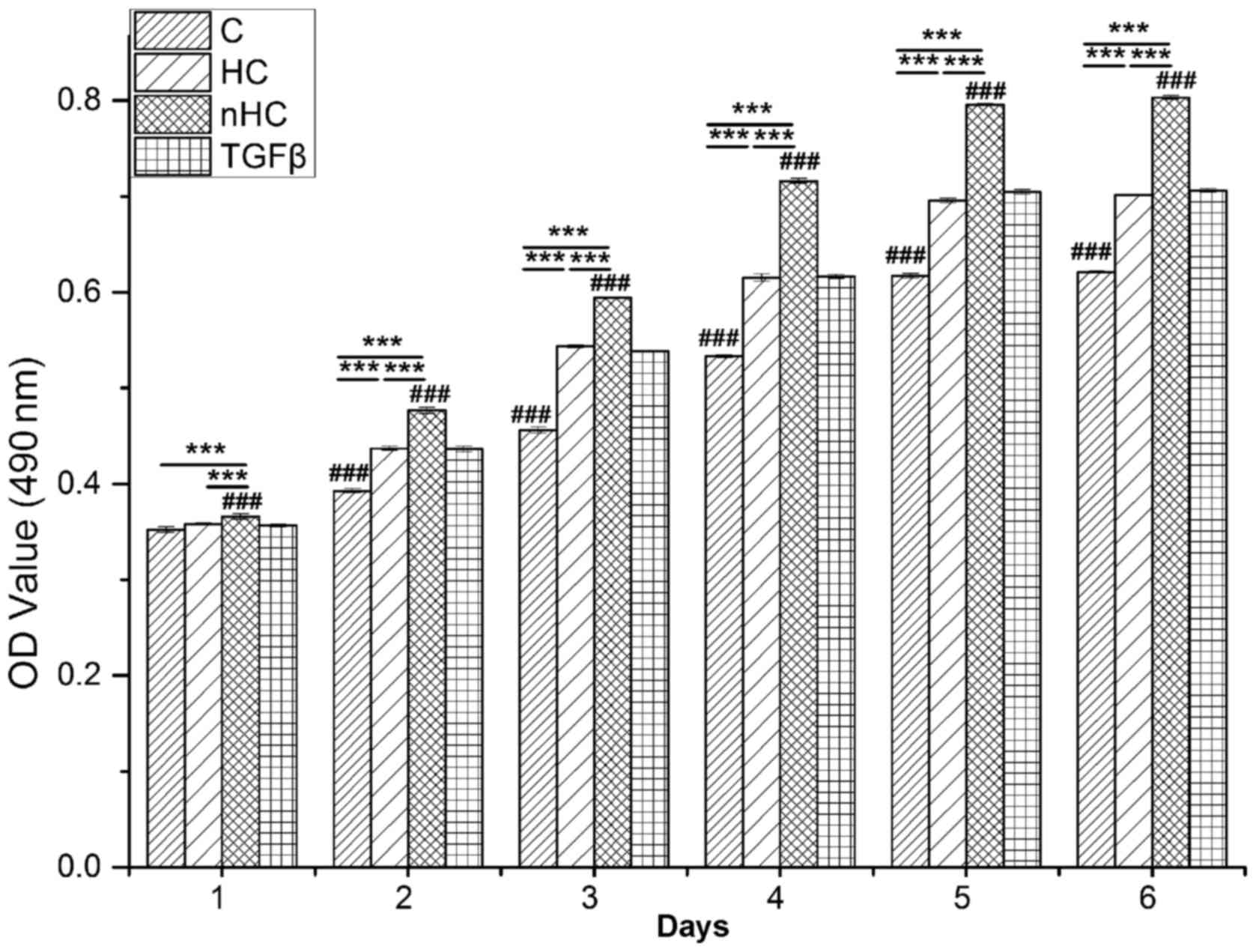
Proliferation of chondrocytes cultured on the gel
film was detected by MTT. The results showed that cells
proliferated in a time-dependent manner (Fig. 1). In the early culture period,
cells proliferated rapidly and then reached a plateau at 4 days.
Comparatively, the OD value of the nHC group was the highest
followed by that of the TGF-β group, the HC group and the collagen
group (P<0.05), which indicates that nHC gel supports cell
growth the most markedly. There was no significant difference in OD
value between the TGF-β group and the HC group.
Chondrocyte distribution
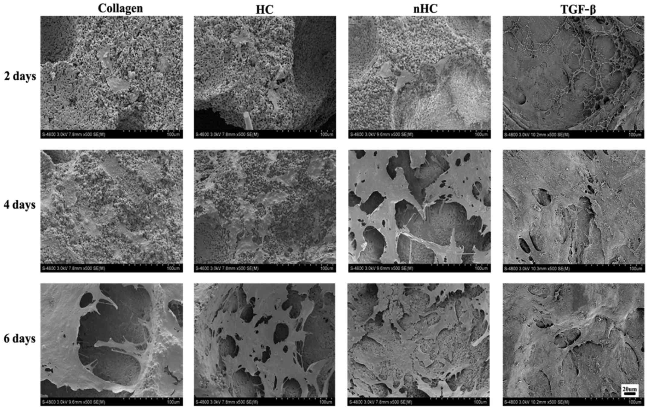
The chondrocyte distribution on the gel films was
observed under a SEM (Fig. 2).
The number of cells adhered to the nHC film was much higher than
that adhered to the others, suggesting that the nHC film could
better support cell growth.
Cells viability
The chondrocyte viability was examined by
calcein-AM/PI staining (Fig. 3)
with alive cells depicted in green and dead cells in red (Fig. 3A). The number of the cells in
green or red was calculated by Image-Pro Plus 6.0 (National
Institutes of Health, Bethesda, USA) respectively after importing
the pictures. The results showed that cells survival (Fig. 3B) on the nHC film was much higher
than that in the TGF-β group and on HC and collagen film, which was
consistent with the results of the MTT assay. Furthermore, the
viable to dead ratio (Fig. 3B) of
the cells cultured on nHC film was higher than that of the control
groups, indicating that the nHC film may serve as a substrate of
benefit for chondrocyte survival and proliferation.
General observation
Chondrocytes seeded on the gel film were measured by
H&E staining (Fig. 4). A
greater number of cell clusters formed by aggregation of
chondrocytes were observed in the nHC group.
Aggrecan secretion
To determinate the AGC secretion of chondrocytes
seeded on the film, samples were stained with Safranin O. The AGC
appeared orange following reaction with Safranin O. The results of
the Safranin O staining (Fig. 5A)
showed that the orange color was much more evident in the nHC group
than in the HC and collagen groups. The histograms in Fig. 5B show that the GAG and DNA
contents were highest in the nHC group compared with that in the
other collagen-based groups and even the positive group, which was
TGF-β added to the culture medium. However, the ratio of GAG to DNA
in the nHC group was lower than that of the TGF-β group, which
indicated that nHC film could serve as an optimal substrate for
chondrocyte growth, but that it could not maintain the phenotype
equal to using TGF-β.
 | Figure 5Aggrecan secretion. (A) AGC appeared
orange following reaction with safranin O. The results of the
safranin O staining show that the orange color is more evident in
the nHC group than in the HC and collagen groups. (B) The
histograms show that the GAGs and DNA contents were highest in the
nHC group than in the other collagen-based groups and even the
positive group, which was TGF-β added to culture medium. However,
the ratio of GAG to DNA in the nHC group was lower than in the
TGF-β group. The data are reported as the mean ± standard
deviation. *P<0.05, **P<0.01 and
***P<0.001 vs. the collagen-based groups;
#P<0.05, ##P<0.01 and
###P<0.001 for comparisons between the collagen-based
groups and the TGF-β group. TGF-β, transforming growth factor-β; C,
collagen; HC, hydroxyapatite/collagen; nHC, nano-HC; calcein-AM/PI,
acetoxymethyl/propidium iodide; GAGs, glycosaminoglycans. |
Immunohistochemical staining
Immunohistochemical staining was used to detect the
indicators of cartilage ECM, COLIA1 or COL1A2 (Fig. 6). The expression of COL1A2 was
stronger and that of COLIA1 was weaker in the nHC group compared
with that in the HC and pure collagen groups, indicating that the
chondrocyte phenotype and biological functions could be maintained
effectively when cells were cultured on nHC film for a long period
of time.
RT-qPCR analysis
RT-qPCR analysis of tissues cultured for 6 days
in vitro showed that there was a significant difference in
the gene expression of AGC, COLIA1 or COL1A2 among the
collagen-based hydrogels (P<0.05) (Fig. 7). COL1A2 exhibited higher mRNA
expression and COL-I exhibited expression in the nHC group compared
with the control groups, indicating that the ratio of COL1A2 to
COLIA1 was enhanced. Therefore, as a culture matrix, nHC gel may
increase the synthesis of cartilage marker and it is reasonable to
speculate that the chondrocyte phenotype with no dedifferentiation
was maintained in nHC film.
 | Figure 7RT-qPCR analysis. Cartilage matrix
relative gene expression was analyzed by RT-qPCR. The chondrocytes
were seeded on the nHC, HC and pure collagen films and cultured for
6 days. Gene expression in cells relative to the control group was
calculated by the 2−ΔΔCq method using the β-actin gene
as the internal control. The data are reported as the mean ±
standard deviation. *P<0.05, **P<0.01
and ***P<0.001 vs. the collagen-based groups;
#P<0.05, ##P<0.01 and
###P<0.001 for comparisons between the collagen-based
groups and the TGF-β group. HC, hydroxyapatite/collagen; nHC,
nano-HC; calcein-AM/PI, acetoxymethyl/propidium iodide; TGF-β,
transforming growth factor-β; COL-I, collagen type I; COL-II,
collagen type II; RT-qPCR, reverse transcription-quantitative
polymerase chain reaction. |
Discussion
The present study focused on the biomimetic
monolayer as a substrate for the in vitro culture of
chondrocytes to preserve phenotype and promote cell growth, in
order to provide a promising cell reservoir in cell-based therapy.
Based on our previous findings, we chose nHC film by way of in
situ synthesis and compared with collagen and mixed HC
films.
In the present study, the results showed that
collagen, HC and nHC films could support the growth of
chondrocytes, as demonstrated by cell proliferation assays,
morphological examinations and cell viability analyses. Compared
with the other collagen-based groups, nHC film showed superiority
with regard to growth promotion and phenotype maintenance for
chondrocytes. It is well known that chondrocytes are
anchorage-dependent and require attachment to the ECM in order to
survive and proliferate (31).
Collagen provides a more native surface for chondrocytes, as it is
the major component of cartilage ECM. Most importantly, collagen
possesses ligands that favor cellular attachment. It was previously
reported that collagen membranes seeded with chondrocytes appeared
to improve cartilage healing by improving composite histological
scores, cartilage GAG and DNA contents, and mechanical properties
in vivo (32). In
vitro, chondrocytes treated with collagen in 2D monolayer
culture maintained high expression of characteristic chondrocyte
markers, whereas the expression of the fibrocartilage marker was
minimal (33). Furthermore, in
the present study, expression of cartilage-specific genes and
proteins, including COL1A2 and AGC, was enhanced with the increase
in cell proliferation and growth, as demonstrated by MTT, RT-qPCR
and immunohistology. This indicates that collagen-based substrates
facilitate the maintenance of a differentiated phenotype for
chondrocytes. Thus, collagen-based films are preferable as cell
substrates for chondrocyte culture in vitro.
Further, the addition of HA particles, whether
micro- or nano-scale, were found to enhance cell attachment more
markedly than collagen alone. These results corroborate previous
studies showing that HC composites are preferable over collagen in
that HA strengthens the bioactivity of collagen (25). When combining collagen and HA, the
wettability and permeability of the composite could be improved,
resulting into culture medium penetration that is favorable for
cell growth (34). The HA
particles are spread on the film very evenly and this is expected
to increase the chances for cells to make contact with HA
particles, which may enhance cell-ECM interactions (35). Comparatively, nHC by way of in
situ synthesis is superior to the conventional blend-mixing HC
composites. Without the aggregation of nano-HA, nano-sized HA
synthesized in situ can provide a higher surface area for
cell growth, which may increase cellular proliferation (36,37). Unlike ultrasonic or mechanical
mixing, the in situ hybridization technique (29,38) ensures the crystallization of
nano-HA (39), with the advantage
of obtaining a homogeneous hybrid scaffold with uniformly
distributed nano-HA particles (25,40). Our previous study showed that
nano-HA particles synthesized in situ were bestowed with
enhanced mechanical properties and improved the biological activity
of the 3D scaffold (25). In the
present study, the nHC film encouraged cell proliferation and
prevented chondrocyte dedifferentiation, suggesting its role as a
favorable substrate for chondrocytes.
In conclusion, the present study investigated the
effect of collagen-based substrate on the growth and phenotype
maintenance of chondrocytes when expanded in vitro. As
evidenced by MTT assay, use of an SEM, calcein-AM/PI staining,
H&E staining, Safranin O staining, IHC and RT-qPCR, cell growth
and cartilage-specific ECM components, including AGC and COL1A2,
were promoted in the nHC film compared with that in the control.
Expression of COLIA1, an indicator of dedifferentiation, was
downregulated in the collagen-based film groups. nHC showed the
best performance among all the collagen-based groups. The nHC film
may serve as a cartilage-like ECM, and favors cell growth and
prevention of dedifferentiation. Taken together, these results show
that nHC composite film is a promising substrate for the culture of
chondrocytes for cell-based therapy.
Abbreviations:
|
ACI
|
autologous chondrocyte
implantation
|
|
AGC
|
aggrecan
|
|
calcein-AM
|
calcein-acetoxymethyl
|
|
Ca(NO3)2
|
calcium nitrate
|
|
Col I
|
type I collagen
|
|
Col II
|
type II collagen
|
|
DMEM
|
Dulbecco’s modified Eagle’s medium
|
|
DMMB
|
1,9-dimethylmethylene blue
|
|
ECM
|
extracellular matrix
|
|
FBS
|
fetal bovine serum
|
|
GAGs
|
glyco saminoglycans
|
|
HA
|
hydroxyapatite
|
|
HC
|
hydroxyapatite/collagen
|
|
H&E
|
hematoxylin and eosin
|
|
IHC
|
immunohistochemistry
|
|
MTT
|
methyl thiazolyl tetrazolium
|
|
nHC
|
nano-hydroxyapatite/collagen
|
|
NH3·H2O
|
ammonium hydroxide
|
|
(NH4)2HPO4·12H2O
|
diammonium phosphate
|
|
OD
|
optical density
|
|
PBS
|
phosphate-buffered solution
|
|
PI
|
propidium iodide
|
|
RT-qPCR
|
reverse transcription-quantitative
polymerase chain reaction
|
|
SD
|
Sprague-Dawley
|
|
SEM
|
scanning electron microscope
|
Acknowledgments
The authors thank Zhenhui Lu and Qin Liu for kindly
offering technical guidance for IHC staining.
Notes
[1]
Funding
This study was financially supported by the National
Science and Technology Pillar Program of China (grant no.
2012BAI42G00), the National Key Research and Development Program of
China (2016YFB0700804), the National Natural Science Fund of China
(grant no. 81472054), the Scientific and Technical Research Project
in Guangxi Universities (KY2015LX059), High Level Innovation Teams
and Outstanding Scholars in Guangxi Universities (the third batch)
and the Key Scientific Research Collaboration Program of Guangxi
Biomedical Collaborative Innovation Center (GCICB-SR-2017002).
[2] Availability
of data and materials
All data generated or analyzed during this study are
included in this published article.
[3] Authors’
contributions
Xianfang Jiang and Yanping Zhong contributed equally
to this study. Li Zheng designed the study. Xianfang Jiang
conducted the study. Yanping Zhong conducted the data analysis.
Jinmin Zhao revised the manuscript. All authors contributed to
discussion of the results and approved the final version.
[4] Ethics
approval and consent to participate
This study was carried out strictly according to the
recommendations in the Guide for the Care and Use of Laboratory
Animals of the National Institutes of Health. The protocol was
approved by the Ethics Committee of Animal Experiments of Guangxi
Medical University.
[5] Consent for
publication
Not applicable
[6] Competing
interests
The authors declare that they have no competing
interests.
References
|
1
|
Brittberg M, Lindahl A, Nilsson A, Ohlsson
C, Isaksson O and Peterson L: Treatment of deep cartilage defects
in the knee with autologous chondrocyte transplantation. N Engl J
Med. 331:889–895. 1994. View Article : Google Scholar : PubMed/NCBI
|
|
2
|
Chung C and Burdick JA: Engineering
cartilage tissue. Adv Drug Deliv Rev. 60:243–262. 2008. View Article : Google Scholar
|
|
3
|
Lee JI, Sato M, Kim HW and Mochida J:
Transplantatation of scaffold-free spheroids composed of
synovium-derived cells and chondrocytes for the treatment of
cartilage defects of the knee. Eur Cell Mater. 22:275–290. 2011.
View Article : Google Scholar : PubMed/NCBI
|
|
4
|
Vinatier C, Gauthier O, Fatimi A, Merceron
C, Masson M, Moreau A, Moreau F, Fellah B, Weiss P and Guicheux J:
An injectable cellulose-based hydrogel for the transfer of
autologous nasal chondrocytes in articular cartilage defects.
Biotechnol Bioeng. 102:1259–1267. 2009. View Article : Google Scholar
|
|
5
|
Knudson W and Loeser RF: CD44 and integrin
matrix receptors participate in cartilage homeostasis. Cell Mol
Life Sci. 59:36–44. 2002. View Article : Google Scholar : PubMed/NCBI
|
|
6
|
Weber GF, Bjerke MA and DeSimone DW:
Integrins and cadherins join forces to form adhesive networks. J
Cell Sci. 124:1183–1193. 2011. View Article : Google Scholar : PubMed/NCBI
|
|
7
|
Chen X and Gumbiner BM: Crosstalk between
different adhesion molecules. Curr Opin Cell Biol. 18:572–578.
2006. View Article : Google Scholar : PubMed/NCBI
|
|
8
|
Risbud MV and Sittinger M: Tissue
engineering: advances in in vitro cartilage generation. Trends
Biotechnol. 20:351–356. 2002. View Article : Google Scholar : PubMed/NCBI
|
|
9
|
Levorson EJ, Hu O, Mountziaris PM, Kasper
FK and Mikos AG: Cell-derived polymer/extracellular matrix
composite scaffolds for cartilage regeneration, part 2: construct
devitalization and determination of chondroinductive capacity.
Tissue Eng Part C Methods. 20:358–372. 2014. View Article : Google Scholar :
|
|
10
|
Callahan LA, Ganios AM, McBurney DL,
Dilisio MF, Weiner SD, Horton WE Jr and Becker ML: ECM production
of primary human and bovine chondrocytes in hybrid PEG hydrogels
containing type I collagen and hyaluronic acid. Biomacromolecules.
13:1625–1631. 2012. View Article : Google Scholar : PubMed/NCBI
|
|
11
|
Lin Z, Willers C, Xu J and Zheng MH: The
chondrocyte: biology and clinical application. Tissue Eng.
12:1971–1984. 2006. View Article : Google Scholar : PubMed/NCBI
|
|
12
|
Caron MM, Emans PJ, Coolsen MM, Voss L,
Surtel DA, Cremers A, van Rhijn LW and Welting TJ:
Redifferentiation of dedifferentiated human articular chondrocytes:
comparison of 2D and 3D cultures. Osteoarthritis Cartilage.
20:1170–1178. 2012. View Article : Google Scholar : PubMed/NCBI
|
|
13
|
Tekari A, Luginbuehl R, Hofstetter W and
Egli RJ: Chondrocytes expressing intracellular collagen type II
enter the cell cycle and co-express collagen type I in monolayer
culture. J Orthop Res. 32:1503–1511. 2014. View Article : Google Scholar : PubMed/NCBI
|
|
14
|
Mhanna R, Öztürk E, Schlink P and
Zenobi-Wong M: Probing the microenvironmental conditions for
induction of superficial zone protein expression. Osteoarthritis
Cartilage. 21:1924–1932. 2013. View Article : Google Scholar : PubMed/NCBI
|
|
15
|
Luo T and Kiick KL: Collagen-like peptides
and peptide-polymer conjugates in the design of assembled
materials. Eur Polym J. 49:2998–3009. 2013. View Article : Google Scholar : PubMed/NCBI
|
|
16
|
Guo Y, Yuan T, Xiao Z, Tang P, Xiao Y, Fan
Y and Zhang X: Hydrogels of collagen/chondroitin sulfate/hyaluronan
interpenetrating polymer network for cartilage tissue engineering.
J Mater Sci Mater Med. 23:2267–2279. 2012. View Article : Google Scholar : PubMed/NCBI
|
|
17
|
Lee CH, Singla A and Lee Y: Biomedical
applications of collagen. Int J Pharm. 221:1–22. 2001. View Article : Google Scholar : PubMed/NCBI
|
|
18
|
Goo HC, Hwang YS, Choi YR, Cho HN and Suh
H: Development of collagenase-resistant collagen and its
interaction with adult human dermal fibroblasts. Biomaterials.
24:5099–5113. 2003. View Article : Google Scholar : PubMed/NCBI
|
|
19
|
Kino-Oka M, Yashiki S, Ota Y, Mushiaki Y,
Sugawara K, Yamamoto T, Takezawa T and Taya M: Subculture of
chondrocytes on a collagen type I-coated substrate with suppressed
cellular dedifferentiation. Tissue Eng. 11:597–608. 2005.
View Article : Google Scholar : PubMed/NCBI
|
|
20
|
Gillogly SD and Wheeler KS: Autologous
chondrocyte implantation with collagen membrane. Sports Med
Arthrosc Rev. 23:118–124. 2015. View Article : Google Scholar : PubMed/NCBI
|
|
21
|
Hindle P, Hall AC and Biant LC: Viability
of chondrocytes seeded onto a collagen I/III membrane for
matrix-induced autologous chondrocyte implantation. J Orthop Res.
32:1495–1502. 2014. View Article : Google Scholar : PubMed/NCBI
|
|
22
|
Zheng L, Fan HS, Sun J, Chen XN, Wang G,
Zhang L, Fan YJ and Zhang XD: Chondrogenic differentiation of
mesenchymal stem cells induced by collagen-based hydrogel: an in
vivo study. J Biomed Mater Res A. 93:783–792. 2010.
|
|
23
|
Li YY, Cheng HW, Cheung KM, Chan D and
Chan BP: Mesenchymal stem cell-collagen microspheres for articular
cartilage repair: cell density and differentiation status. Acta
Biomater. 10:1919–1929. 2014. View Article : Google Scholar : PubMed/NCBI
|
|
24
|
Li W, Guo R, Lan Y and Zhang Y, Xue W and
Zhang Y: Preparation and properties of cellulose nanocrystals
reinforced collagen composite films. J Biomed Mater Res A.
102:1131–1139. 2014. View Article : Google Scholar
|
|
25
|
Zheng L, Jiang X, Chen X, Fan H and Zhang
X: Evaluation of novel in situ synthesized
nano-hydroxyapatite/collagen/alginate hydrogels for osteochondral
tissue engineering. Biomed Mater. 9:0650042014. View Article : Google Scholar : PubMed/NCBI
|
|
26
|
Laydi F, Rahouadj R, Cauchois G, Stoltz JF
and de Isla N: Hydroxyapatite incorporated into collagen gels for
mesenchymal stem cell culture. Biomed Mater Eng. 23:311–315.
2013.PubMed/NCBI
|
|
27
|
Wang X, Grogan SP, Rieser F, Winkelmann V,
Maquet V, Berge ML and Mainil-Varlet P: Tissue engineering of
biphasic cartilage constructs using various biodegradable
scaffolds: an in vitro study. Biomaterials. 25:3681–3688. 2004.
View Article : Google Scholar : PubMed/NCBI
|
|
28
|
Liang JZ: Reinforcement and quantitative
description of inorganic particulate-filled polymer composites.
Composites Part B Engineering. 51:224–232. 2013. View Article : Google Scholar
|
|
29
|
Fabbri P, Bondioli F, Messori M, Bartoli
C, Dinucci D and Chiellini F: Porous scaffolds of polycaprolactone
reinforced with in situ generated hydroxyapatite for bone tissue
engineering. J Mater Sci Mater Med. 21:343–351. 2010. View Article : Google Scholar
|
|
30
|
Livak and Schmittgen: Analysis of relative
gene expression data using real-time quantitative PCR and the
2-ΔΔCt method. Methods. 25:402–408. 2001. View Article : Google Scholar
|
|
31
|
Yashiki S, Umegaki R, Kino-Oka M and Taya
M: Evaluation of attachment and growth of anchorage-dependent cells
on culture surfaces with type I collagen coating. J Biosci Bioeng.
92:385–388. 2001. View Article : Google Scholar
|
|
32
|
Nixon AJ, Rickey E, Butler TJ, Scimeca MS,
Moran N and Matthews GL: A chondrocyte infiltrated collagen type
I/III membrane (MACI® implant) improves cartilage
healing in the equine patellofemoral joint model. Osteoarthritis
Cartilage. 23:648–660. 2015. View Article : Google Scholar : PubMed/NCBI
|
|
33
|
Smeriglio P, Dhulipala L, Lai JH, Goodman
SB, Dragoo JL, Smith RL, Maloney WJ, Yang F and Bhutani N: Collagen
VI enhances cartilage tissue generation by stimulating chondrocyte
proliferation. Tissue Eng Part A. 21:840–849. 2015. View Article : Google Scholar
|
|
34
|
Jia L, Duan Z, Fan D, Mi Y, Hui J and
Chang L: Human-like collagen/nano-hydroxyapatite scaffolds for the
culture of chondrocytes. Mater Sci Eng C. 33:727–734. 2013.
View Article : Google Scholar
|
|
35
|
Hunter KT and Ma T: In vitro evaluation of
hydroxyapatite-chitosan-gelatin composite membrane in guided tissue
regeneration. J Biomed Mater Res A. 101:1016–1025. 2013. View Article : Google Scholar
|
|
36
|
Chen J, Yu Q, Zhang G, Yang S, Wu J and
Zhang Q: Preparation and biocompatibility of nanohybrid scaffolds
by in situ homogeneous formation of nano hydroxyapatite from
biopolymer polyelectrolyte complex for bone repair applications.
Colloids Surf B Biointerfaces. 93:100–107. 2012. View Article : Google Scholar : PubMed/NCBI
|
|
37
|
Chen J, Zhang G, Yang S, Li J, Jia H, Fang
Z and Zhang Q: Effects of in situ and physical mixing on mechanical
and bioactive behaviors of nano hydroxyapatite-chitosan scaffolds.
J Biomater Sci Polym Ed. 22:2097–2106. 2011. View Article : Google Scholar
|
|
38
|
Zhang CY, Lu H, Zhuang Z, Wang XP and Fang
QF: Nano-hydroxyapatite/poly(L-lactic acid) composite synthesized
by a modified in situ precipitation: preparation and properties. J
Mater Sci Mater Med. 21:3077–3083. 2010. View Article : Google Scholar : PubMed/NCBI
|
|
39
|
Chen J, Nan K, Yin S, Wang Y, Wu T and
Zhang Q: Characterization and biocompatibility of nanohybrid
scaffold prepared via in situ crystallization of hydroxyapatite in
chitosan matrix. Colloids Surf B Biointerfaces. 81:640–647. 2010.
View Article : Google Scholar : PubMed/NCBI
|
|
40
|
Zhu W, Chen K, Lu W, Sun Q, Peng L, Fen W,
Li H, Ou Y, Liu H, Wang D, et al: In vitro study of nano-HA/PLLA
composite scaffold for rabbit BMSC differentiation under TGF-β1
induction. In Vitro Cell Dev Biol Anim. 50:214–220. 2014.
View Article : Google Scholar
|


