Introduction
Gastric cancer is a common and frequently occurring
malignancy worldwide. At present, systemic chemotherapy is the most
common clinical treatment for gastric cancer. Cisplatin (DDP) is a
well-known chemotherapeutic drug, that is widely used in
chemotherapy of multiple types of cancer, including gastric cancer
(1). However, development of
chemotherapy resistance significantly reduces its therapeutic
effect in gastric cancer. The regulatory molecules involved in
resistance in gastric cancer remain not fully elucidated and
require further study.
The heat shock proteins (HSPs) are a highly
conserved protein family consisting of several members. As
molecular chaperones, HSPs have been reported to be involved in the
folding, modification and degradation of intracellular proteins
(2,3). The stress-inducible HSP70 (also
known as HSPA1 or HSP72) is an important member of the HSP family.
Recent studies have demonstrated that HSP70 is expressed at
extremely low levels in unstressed normal cells, but is highly
expressed in a variety of cancers and correlated with tumor grade,
metastasis, chemotherapy resistance and poor prognosis (4–6).
Because of its tumor-related expression and important role in
cancer progression, HSP70 has become an attractive target for
cancer therapy (7,8).
The primary aim of the present study was to
investigate the effect of HSP70 in the cisplatin-induced apoptosis
of gastric cancer cells and to explore its molecular mechanism. The
results demonstrated that downregulating HSP70 expression enhanced
cisplatin-induced gastric cancer apoptosis via regulation of the
mitogen-activated protein kinase (MAPK) signaling pathway. The
present study provided an experimental basis and novel insights for
HSP70 as a target for cancer therapy.
Materials and methods
Reagents and antibodies
Cisplatin (purity, ≥99.9) was purchased from
Sigma-Aldrich (Merck KGaA, Darmstadt, Germany), and
pifithrin-µ (PES), SB203580, SP600125 and U0126 were
purchased from Selleck Chemicals (Houston, TX, USA). All of the
above reagents were dissolved in DMSO and then diluted with
sterilized PBS. The final concentration of DMSO in cultures was
<0.1%. Rabbit monoclonal antibodies against p38 (cat. no. 9219s;
1:1,000), phosphorylated (p)-p38 (Thr180/Tyr182; cat. no. 9215s;
1:1,000), extracellular signal-regulated kinase (ERK, cat. no.
9102s; 1:1,000), p-ERK (Thr202/Tyr204; cat. no. 4376s; 1:1,000),
c-Jun N-terminal kinase (JNK; cat. no. 9252s; 1:1,000), p-JNK
(Thr183/Tyr185; cat. no. 4671s; 1:1,000), p-SRC proto-oncogene
non-receptor tyrosine kinase (Src, Tyr416; cat. no. 6943s; 1:500),
p-AKT serine/threonine kinase 1 (Akt, Ser473; cat. no. 4060s;
1:500), p-inhibitor of κB (IκB, Ser32; cat. no. 2859s; 1:500),
poly-ADP-ribose-polymerase (PARP, cat. no. 9532s; 1:1,000),
pro-caspase-3 (cat. no. 9662s; 1:500), cleaved caspase-3 (cat. no.
9661s; 1:500), β-actin (cat. no. 4970s; 1:1,000), GAPDH (cat. no.
5174s; 1:1,000), and rat monoclonal antibody against HSP70 (cat.
no. 4573s; 1:1,000) were all from Cell Signaling Technology, Inc.
(Beverly, MA, USA). All secondary antibodies (cat. no. 926-32210 or
926-32211; 1:5,000) were purchased from LI-COR Biosciences
(Lincoln, NE, USA).
Cell culture
The human gastric cancer HGC-27 cell line was
obtained from GuangZhou Cellcook Biotech Co., Ltd. (Guangzhou,
China). Cells were cultured in RPMI-1640 medium (Gibco; Thermo
Fisher Scientific, Inc., Waltham, MA, USA) supplemented with 10%
fetal bovine serum (FBS; Gibco; Thermo Fisher Scientific, Inc.),
10% non-essential amino acids, 100 µg/ml streptomycin and
100 U/ml penicillin and incubated at 37°C in an atmosphere of 5%
CO2.
Plasmids and transfection
Green fluorescent protein (GFP)-labeled HSP70
overexpression plasmids and negative plasmids (empty vector control
plasmid; CMV-MCS-EGFP-SV40-Neomycin), and HSP70 short hairpin (sh)
RNA and negative plasmids (non-targeting shRNA plasmid;
hU6-MCS-CMV-GFP-SV40-Neomycin), were purchased from GeneChem Co.,
Ltd. (Shanghai, China). HGC-27 cells were seeded into 12-well cell
culture plates and cultured to 60–70% confluence, prior to
transfection with plasmids for the indicated times using
Lipofectamine® 3000 Reagent (Thermo Fisher Scientific,
Inc.). Operations were performed according to the manufacturers
instructions.
Annexin V/propidium iodide (PI) double
staining assay
Apoptosis rate was detected by Annexin V/PI double
staining (Jiancheng Bioengineering Institute, Nanjing, China).
Briefly, following treatment, HGC-27 cells were collected and
washed two times with PBS. Cells were resuspended in 500 µl
of binding buffer, and 5 µl PI and 5 µl Annexin
V-fluorescein isothiocyanate were added to the cells. Apoptosis
rate was detected by flow cytometry (BD Biosciences, Franklin
Lakes, NJ, USA).
DAPI staining
Following treatment, HGC-27 cells were washed three
times with PBS and fixed with 4% paraformaldehyde for 20 min at
room temperature. After rising with PBS, cells were stained with
DAPI for 3 min in the dark. The morphology of apoptotic cells was
examined using an inverted fluorescence microscope (Olympus
Corporation, Tokyo, Japan).
Western blotting
HGC-27 cells were washed three times with cold PBS
and then lysed in RIPA buffer containing protease inhibitors
(Beyotime Institute of Biotechnology, Haimen, China) for 30 min on
ice. Lysates were centrifuged (14,300 × g) at 4°C for 15 min, and
the supernatant was collected. The bicinchoninic acid assay was
used to measure protein concentration in the supernatant, and equal
amounts of total protein (50 µg) were loaded onto 12% gels
for SDS-PAGE. Separated proteins were transferred onto
nitrocellulose membranes (Pall Corporation, Port Washington, NY,
USA), which then were blocked with 5% skim milk for 1 h at room
temperature and then incubated with the indicated primary
antibodies overnight at 4°C. After washing three times with TBST,
membranes were incubated with IRDye800 fluorophore-conjugated
secondary antibody for 1 h at room temperature in the dark.
Antigen-antibody complexes were detected using a LI-COR Odyssey
Infrared Imaging System (LI-COR Biosciences) and LI-COR Odyssey
analysis software was used to quantify the proteins.
Cell Counting Kit-8 (CCK-8) assay
HGC-27 cells were seeded into 96-well cell culture
plates, and the following day they were treated with different
doses of PES for 24 h. CCK-8 was then added at 10 µl/well.
Following incubation for 2 h at 37°C and 5% CO2,
absorbance at 450 nm was measured using a Multiskan GO plate reader
(Thermo Fisher Scientific, Inc.). Each experiment was repeated
three times.
Statistical analysis
Data were expressed as mean ± standard deviation.
Statistical analysis was performed using SPSS17.0 software (SPSS,
Inc., Chicago, IL, USA). The results were compared using one-way
analysis of variance followed by a post hoc Tukey test for multiple
comparisons. P<0.05 was considered to indicate a statistically
significant difference.
Results
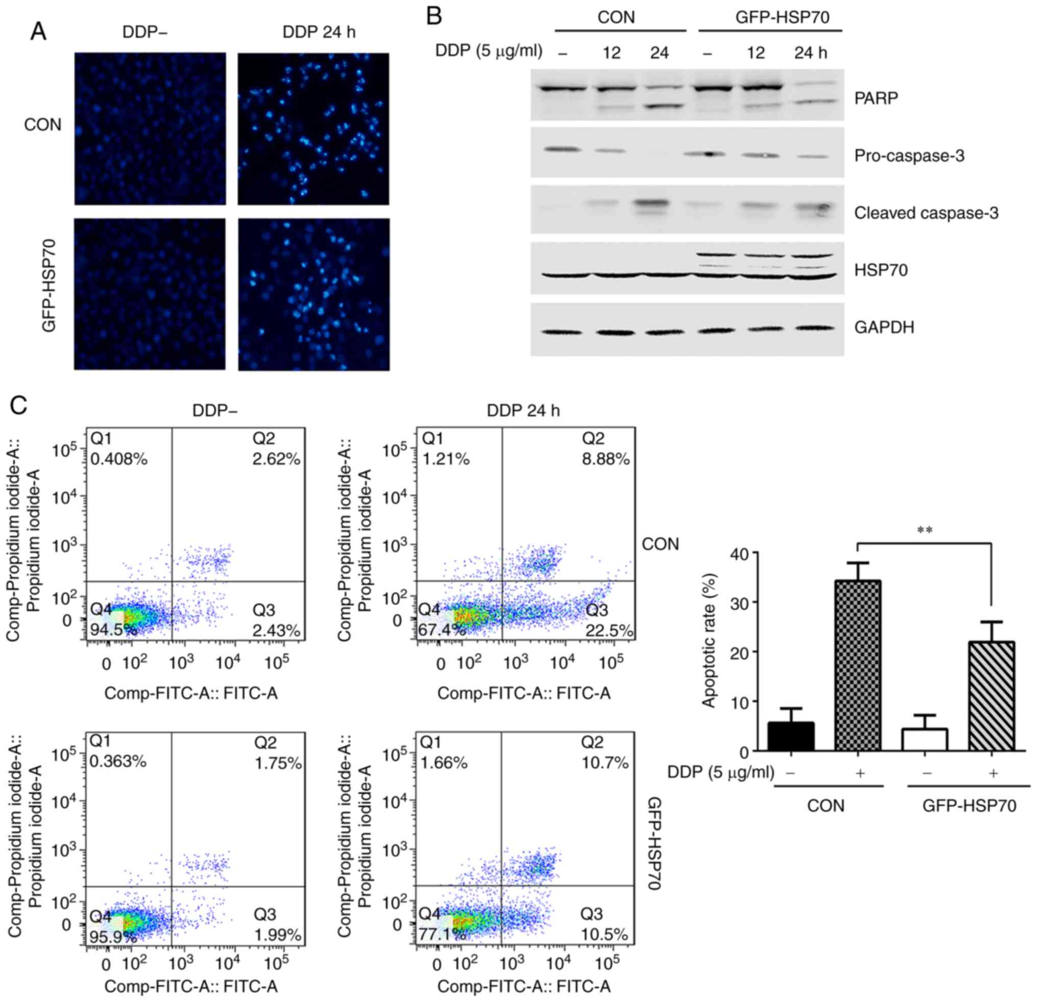
HSP70 overexpression antagonizes
cisplatin-induced HGC-27 cell apoptosis
HGC-27 cells were transfected with GFP-HSP70 or
control plasmid, and 24 h post-transfection they were stimulated
with cisplatin (5 µg/ml) for 24 h. The morphology of
apoptotic cells was observed with DAPI staining and the apoptosis
rate was determined by flow cytometry. The expression of
apoptosis-related proteins, including PARP, pro-caspase-3 and
cleaved caspase-3, were detected by western blotting. HGC-27 cells
transfected with control plasmid and not treated with cisplatin had
light blue, round nuclear morphology as evident by DAPI staining
(Fig. 1A). However, following
cisplatin treatment, the nuclei exhibited morphological changes
typical of apoptosis, including nuclear condensation and nuclear
fragmentation (Fig. 1A). These
typical cisplatin-induced morphological changes were clearly
reversed in HSP70-overexpressing HGC-27 cells (Fig. 1A). In addition, cisplatin-induced
expression of cleaved PARP and cleaved caspase-3 was decreased,
while pro-caspase-3 levels were increased, in
GFP-HSP70-overexpressing cells compared with cells transfected with
control plasmid (Fig. 1B). Flow
cytometry was also used to determine the apoptotic rate of HGC-27
cells induced by cisplatin treatment. The apoptotic rate was 31.38%
in control plasmid-transfected cells, while it was reduced to 21.2%
in HSP70-overexpressing cells (Fig.
1C).
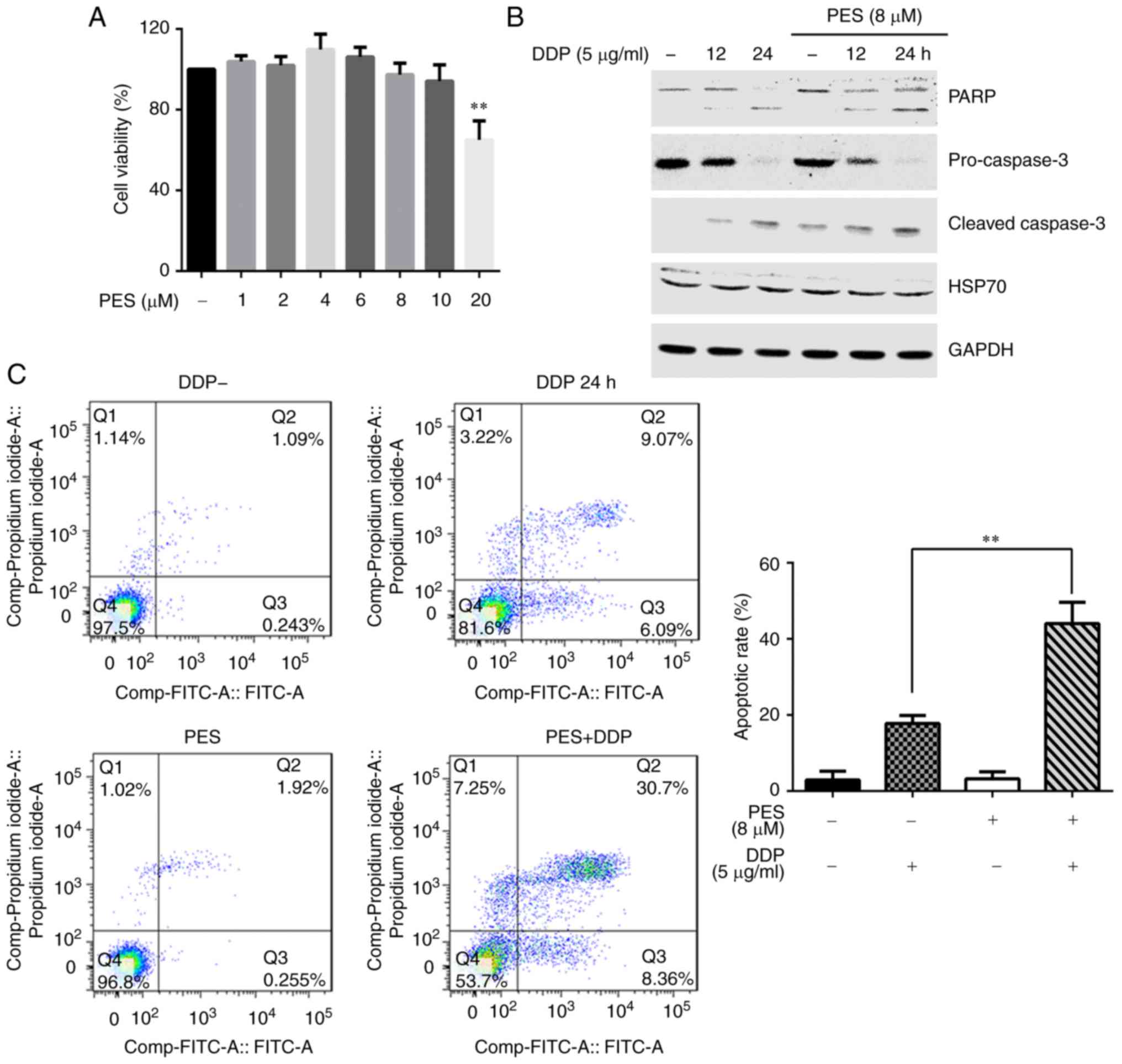
Downregulation of HSP70 or suppression of
its function enhances cisplatin-induced HGC-27 cell apoptosis
To further explore the role of HSP70 in
cisplatin-induced apoptosis, a HSP70 inhibitor, PES, was used to
treat the cells. PES interacts selectively with the
stress-inducible HSP70 protein and inhibits its function (2,9,10).
Firstly, the cytotoxicity of PES was determined using the CCK-8
assay. HGC-27 cells were treated with different concentrations of
PES (1, 2, 4, 6, 8, 10 or 20 µM) for 24 h, and cell
viability was analyzed by CCK-8 assay. PES did not affect cell
viability, even at a dose of 10 µM (Fig. 2A); the dose of 8 µM was
therefore selected for subsequent experiments. Secondly, the
effects of PES on cisplatin-induced cell apoptosis were examined.
Western blot analysis indicated that PES pretreatment increased the
expression of cleaved PARP and cleaved-caspase-3, and decreased the
levels of pro-caspase-3, compared with cells treated with cisplatin
alone (Fig. 2B). An Annexin V/PI
double staining assay indicated that the apoptotic rate in
cisplatin-stimulated cells was 18.2%, while inhibition of HSP70 by
PES increased this to 41.2% (Fig.
2C).
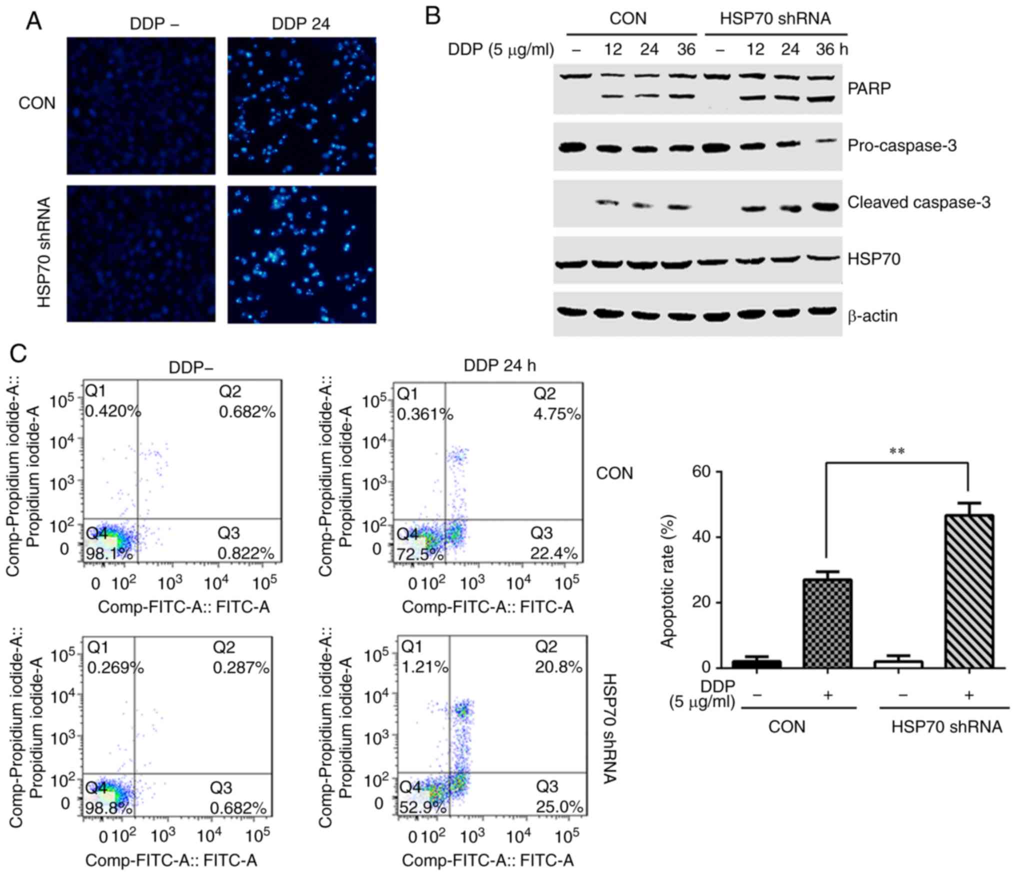
As a second method to suppress HSP70 function and
explore its effect on cisplatin-induced apoptosis, HSP70-specific
shRNA was transfected in the cells to down-regulate its expression.
The nuclear condensation and nuclear fragmentation induced by
cisplatin in HSP70 shRNA-transfected cells were obviously enhanced
compared with control plasmid-transfected HGC-27 cells (Fig. 3A). The levels of cleaved PARP and
cleaved caspase-3 following cisplatin stimulation were increased,
while pro-caspase-3 levels were decreased, in the HSP70
shRNA-transfected cells compared with control (Fig. 3B). Concurrently, the apoptotic
ratio was 45.8% in HSP70 shRNA-transfected cells and 27.15% in the
control (Fig. 3C). Taken
together, these results suggest that HSP70 serves a protective role
in cisplatin-induced HGC-27 cell apoptosis.
HSP70 affects cisplatin-induced MAPK
signaling pathway activation
MAPK and survival-related signaling pathways, such
as Akt and nuclear factor (NF)-κB, have an important role in the
proliferation, differentiation and apoptosis of tumor cells
(11–13). To investigate whether the
molecular mechanism of HSP70 regulation of cisplatin-induced
apoptosis is modulated by the MAPK, Akt or NF-κB signaling
pathways, HGC-27 cells were treated with cisplatin for different
amounts of time, and the phosphorylation of p38, ERK, JNK, Src, Akt
and IκB were monitored by western blotting. Cisplatin stimulation
resulted in a time-dependent increase in the phosphorylation of
p38, ERK and JNK, but had no effect on the total levels of p38, ERK
and JNK proteins (Fig. 4A).
Phosphorylation of Src, Akt and IκB was also detected, but this was
unaffected by cisplatin treatment (Fig. 4B). These results suggested that
the MAPK signaling pathway may be involved in cisplatin-induced
apoptosis.
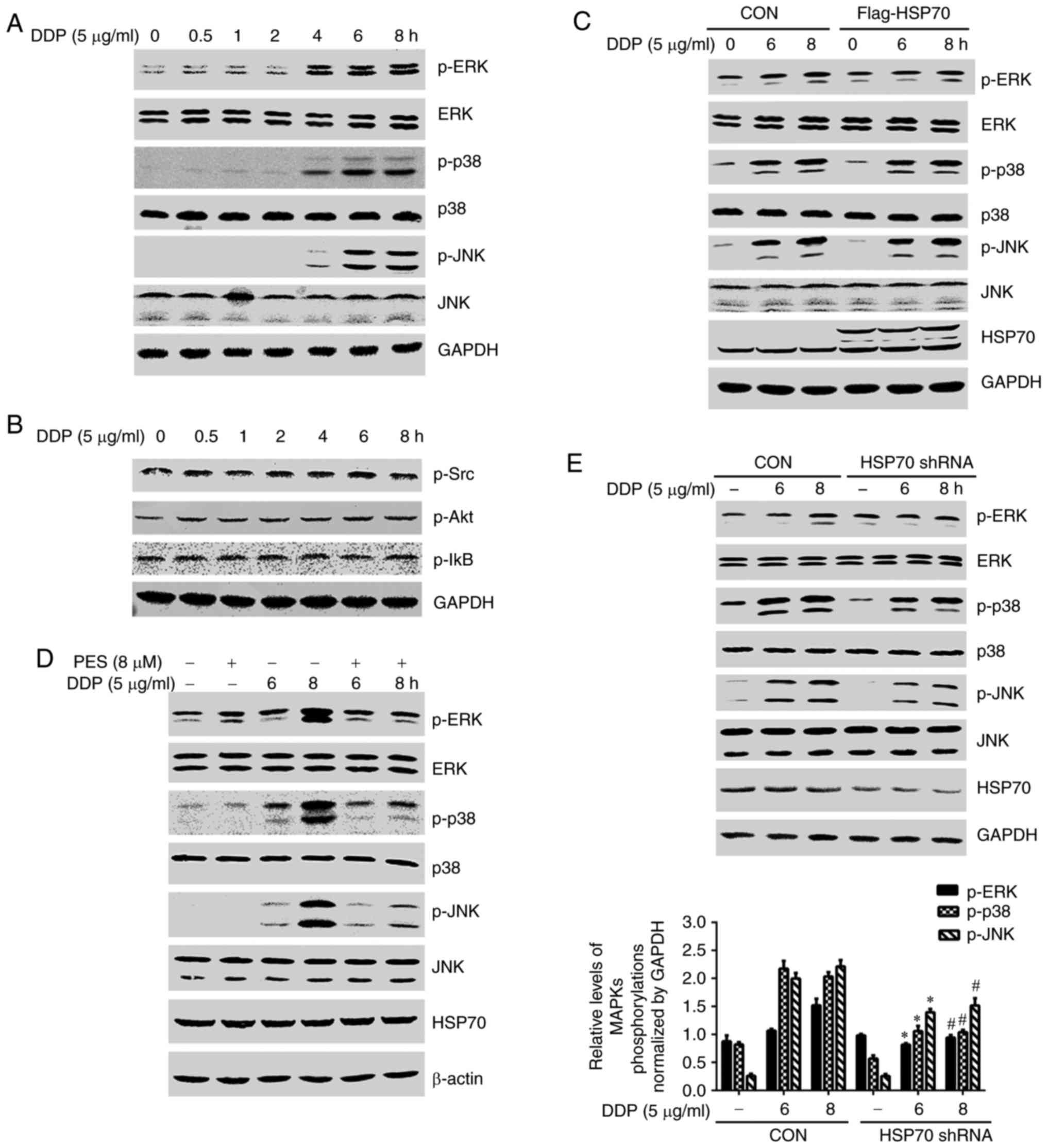 | Figure 4HSP70 affects cisplatin-induced MAPK
signaling pathway activation. (A) HGC-27 cells were treated with
cisplatin (5 µg/ml) for different amounts of time and the
phosphorylation and total protein levels of (A) p38, ERK and JNK,
and (B) Src, Akt and IκB, were monitored by western blotting. (C)
HSP70-overexpressing and control plasmids were transfected into
HGC-27 cells. (D) HGC-27 cells were pretreated with PES for 2 h and
then stimulated with cisplatin for 6 or 8 h. (E) HSP70-shRNA and
control plasmids were transfected into HGC-27 cells. After
transfection, the cells were treated with cisplatin for 6 or 8 h
and the phosphorylation and total protein levels of p38, ERK and
JNK were monitored by western blotting. *P<0.05
compared with control cells at 6 h; #P<0.05 compared
with control cells at 8 h. HSP70, heat shock protein 70; ERK,
extracellular signal-regulated kinase; JNK, c-Jun N-terminal
kinase; Src, SRC proto-oncogene non-receptor tyrosine kinase; Akt,
AKT serine/threonine kinase 1; IκB, inhibitor of κB; sh, short
hairpin; DDP, cisplatin; CON, control; p-, phosphorylated; PES,
pifithrin-µ. |
To further investigate the effects of HSP70 on
cisplatin-induced phosphorylation in the MAPK signaling pathway,
HSP70-overexpressing plasmids were transfected into HGC-27 cells.
HSP70 overexpression did not exert any effects on p38, ERK or JNK
phosphorylation compared with empty vector controls (Fig. 4C). Conversely, PES was used to
inhibit HSP70 function, and HSP70 inhibition resulted in a striking
reduction of cisplatin-induced phosphorylation of p38, ERK and JNK
(Fig. 4D). HSP70 shRNA
transfection also suppressed cisplatin-induced phosphorylation of
p38, ERK and JNK (Fig. 4E).
Collectively, these results suggest that HSP70 facilitates the
activation of p38, ERK and JNK in HGC-27 cells.
MAPK pathway inhibition enhances
cisplatin-induced HGC-27 cell apoptosis
To investigate the role of MAPK signaling in the
process of cisplatin-induced HGC-27 cell apoptosis, cells were
stimulated with cisplatin or cisplatin and a p38-specific inhibitor
(SB203580), a JNK specific inhibitor (SP600125) or an ERK1/2
inhibitor (U0126) for 24 h. All inhibitor pretreatments enhanced
the expression of cleaved PARP and cleaved caspase-3 and decreased
the levels of pro-caspase-3 induced by cisplatin (Fig. 5A). Additionally, compared with
cisplatin-only treated cells, apoptosis levels were significantly
elevated in cells stimulated with cisplatin and the inhibitors
(Fig. 5B). Finally, in order to
confirm the inhibitory effects of each inhibitor, the
phosphorylation levels of p38, ERK or JNK were respectively
detected. Fig 5C illustrates that
each inhibitor treatment suppressed its corresponding molecule,
while having little effect on HSP70 protein expression levels.
Collectively, these findings suggest that the MAPK signaling
pathway antagonized cisplatin-induced apoptosis in HGC-27
cells.
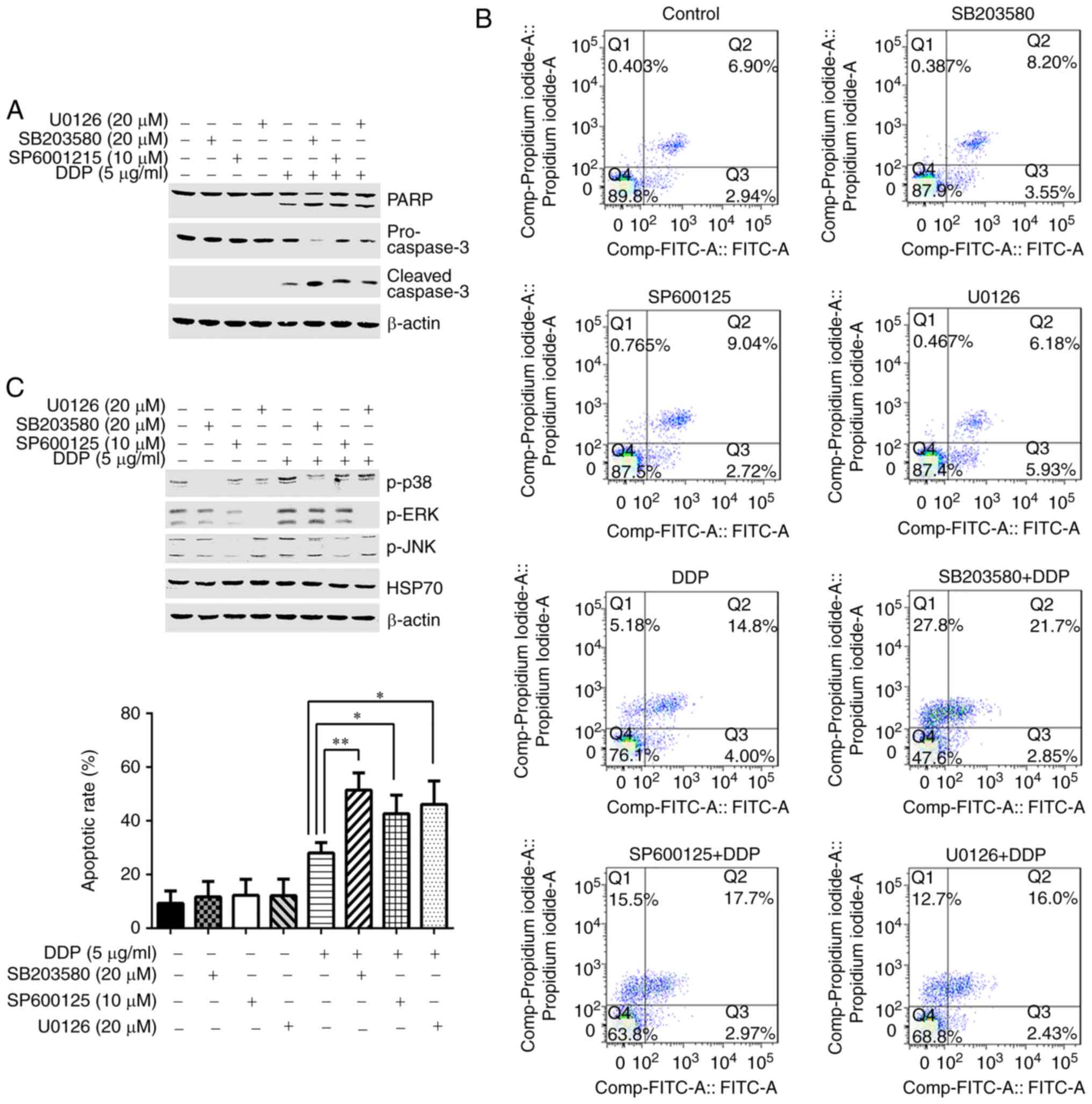 | Figure 5MAPK pathway inhibition enhances
cisplatin-induced HGC-27 cell apoptosis. (A) HGC-27 cells were
pretreated with specific inhibitors for p38, ERK or JNK for 2 h and
then treated with cisplatin for 24 h. Expression levels of PARP,
cleaved caspase-3 and pro-caspase-3 were detected by western
blotting. (B) Apoptotic rate was determined by flow cytometry
(representative plots and quantification is shown). (C) HGC-27
cells were pretreated with specific inhibitors for p38, ERK or JNK
for 2 h and then treated with cisplatin for 6 h. Phosphorylation of
p38, ERK, JNK and the levels of HSP70 were detected by western
blotting. *P<0.05 and **P<0.01, with
comparisons indicated by lines. MAPK, mitogen-activated protein
kinase; ERK, extracellular signal-regulated kinase; JNK, c-Jun
N-terminal kinase; PARP, poly-ADP-ribose-polymerase; HSP70, heat
shock protein 70; DDP, cisplatin; p-, phosphorylated. |
Discussion
HSPs are chaperone proteins, which are induced by a
variety of different stresses. As a member of this family, HSP70
has been reported to be highly expressed in different types of
cancer (6) and is considered to
be a negative prognostic factor in breast cancer, osteosarcoma and
bladder cancer. By contrast, it has been reported as a positive
prognostic factor in esophageal, pancreatic and renal cancers
(14-16). Recently, multiple studies have
demonstrated that HSP70 is involved in resistance to anticancer
agents in tumors (14,17,18); however, the molecular mechanism of
its action in resistance to chemotherapy remains unclear.
Several signaling pathways are reported to be
involved in the regulation of tumor cells apoptosis, such as the
MAPK, Akt and NF-κB signaling pathways (11–13). Ding et al (19) demonstrated that HSP70 expression
decreases the sensitivity of osteosarcoma cells to baicalein via
activation of the PI3K/Akt and MAPK/ERK signaling pathways. Other
studies have also demonstrated that HSP70 regulates tumor cell
apoptosis by inhibiting the activation of JNK and NF-κB signaling
(20,21). Cisplatin, a commonly used
chemotherapeutic drug, is used clinically to treat cancer, and
HSP70 expression decreases the sensitivity of osteosarcoma cells to
cisplatin (14). However, the
question of whether HSP70 regulates the sensitivity of gastric
cancer cells to cisplatin and its underlying molecular mechanism
still remain to be explored.
Cisplatin induces cell death by activating the
apoptosis pathway (22,23); however, HSP70 expression has been
demonstrated to exert a strong anti-apoptotic effect (15). In the present study, the effect of
HSP70 on cisplatin-induced HGC-27 gastric cancer cell apoptosis was
examined. Western blotting was used to detect the expression levels
of cleaved PARP, cleaved caspase-3 and pro-caspase-3, an Annexin
V/PI assay was used to determine apoptotic rates, and DAPI staining
was used to monitor the morphology of apoptotic nuclei. As
illustrated in Figs. 1Figure 2–3, HSP70 overexpression decreased
cisplatin-induced HGC-27 cell apoptosis, while inhibition of HSP70
using a specific inhibitor or by transfection of specific shRNA
enhanced the cellular apoptosis induced by cisplatin. The present
results are consistent with the study of Mori et al
(14). In addition, Liu et
al (2) reported that an HSP70
inhibitor combined with cisplatin suppresses cervical cancer cell
proliferation and transplanted tumor growth. Collectively, these
results suggest that inhibition of HSP70 is beneficial to the
antitumor activity of cisplatin.
The present study also investigated the potential
molecular mechanism of HSP70 antagonism of cisplatin-induced
apoptosis. HGC-27 cells were treated with cisplatin for different
amounts of time, and the phosphorylation states of p38 MAPK, ERK,
JNK, Src, Akt and IκB were monitored by western blotting. The
phosphorylation of p38, ERK and JNK were enhanced following
cisplatin treatment, but Src, Akt and IκB were unaffected. This
finding suggested that the MAPK signaling pathway is involved in
cisplatin-induced apoptosis in gastric cancer cells. Therefore, to
determine whether HSP70 regulates the activation of MAPK signaling,
HSP70 was over-expressed in HGC-27 cells, and the results revealed
no effect on the cisplatin-induced phosphorylation of p38, ERK and
JNK compared with the empty plasmid-only transfected cells.
However, knockdown of HSP70 by HSP70 shRNA transfection attenuated
the phosphorylation of p38, ERK and JNK, similar to the HSP70
inhibition by PES treatment (Fig.
4). Taken together, these results demonstrated that HSP70
facilitates the activation of MAPK signaling in response to
cisplatin in HGC-27 cells. Notably, overexpression of HSP70 in
HGC-27 cell did not affect the phosphorylation of p38, ERK and JNK
induced by cisplatin. A possible reason for this phenomenon might
be the high expression levels of HSP70 in gastric cancer cells; the
phosphorylation of p38, ERK and JNK induced by cisplatin may
already be at a maximum level in these cells and therefore
additional HSP70 expression may have no further effect on the
activation of MAPK signaling pathway.
To establish that the MAPK pathway is involved in
cisplatin-induced gastric cell apoptosis, specific inhibitors were
used to suppress the activation of p38, ERK and JNK, and then
cisplatin-induced apoptosis was examined. As expected, suppression
of p38, ERK and JNK phosphorylation enhanced cisplatin-induced
apoptosis (Fig. 5). It has been
reported that ERK regulates cell apoptosis by promoting cell
proliferation, while p38 and JNK are typically described as
stress-activated kinases that mediate apoptotic signals (24–26). However, there are also some
reports demonstrating that JNK and p38 act as anti-apoptotic
signals (27,28). The present study tested the
hypothesis that the MAPK signaling pathway acts as an
anti-apoptotic signal in cisplatin-induced gastric cell
apoptosis.
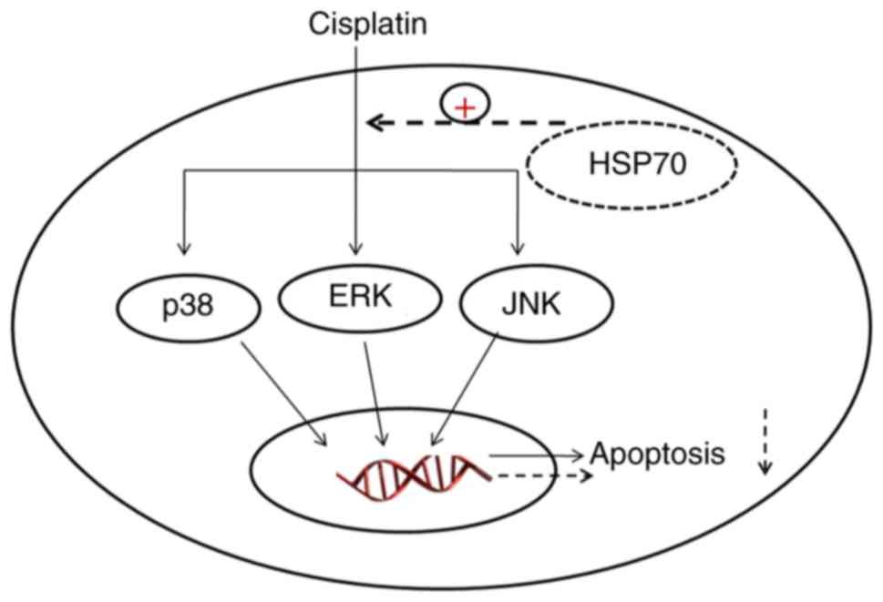
Collectively, the current results demonstrated that
HSP70 protected cisplatin-induced gastric cancer cells from
apoptosis by regulating the MAPK signaling pathway (Fig. 6). These findings present a novel
insight into the anti-apoptotic mechanism of HSP70 and provide a
theoretical basis for targeting HSP70 in clinical therapy of cancer
drug resistance.
Acknowledgments
The authors thank teacher Deyu Dou for her help with
the flow cytometry experiments. We appreciate the department of
Parasitology for assisting with the experiments.
Funding
This work was supported by the Anhui Province
Natural Science Foundation (grant no. 1708085MH231), National
Natural Science Foundation of China (grant no. 81601380), Natural
Science Research Project of Anhui Colleges and Universities (grant
no. KJ2016SD59), Outstanding Young Talent Support Program Key
Projects in Anhui Colleges and Universities (grant no.
gxyqZD2016173), research funding project for college students of
Wannan Medical College (grant no. WK2016S24), College Students'
Innovation and Entrepreneurship Training Program project (grant
nos. 201710368002 and 201710368166) and Anhui Province Key
Laboratory of Active Biological Macro-molecules (grant no.
1306C083008).
Availability of data and materials
The analyzed datasets generated during the study are
available from the corresponding author on reasonable request.
Authors' contributions
ZQ and YZ conceived and designed the experiments.
LS, TT, HT and YM performed the experiments. ZW, YL analyzed the
data. LS and ZQ wrote the paper. All authors read and approved the
final manuscript.
Ethics approval and consent to
participate
Not applicable.
Patient consent for publication
Not applicable.
Competing interests
The authors declare that they have no competing
interests.
References
|
1
|
Dasari S and Tchounwou PB: Cisplatin in
cancer therapy: Molecular mechanisms of action. Eur J Pharmacol.
740:364–378. 2014. View Article : Google Scholar : PubMed/NCBI
|
|
2
|
Liu J, Liu J, Guo SY, Liu HL and Li SZ:
HSP70 inhibitor combined with cisplatin suppresses the cervical
cancer proliferation in vitro and transplanted tumor growth: An
experimental study. Asian Pac J Trop Med. 10:184–188. 2017.
View Article : Google Scholar : PubMed/NCBI
|
|
3
|
Budina-Kolomets A, Webster MR, Leu JI,
Jennis M, Krepler C, Guerrini A, Kossenkov AV, Xu W, Karakousis G,
Schuchter L, et al: HSP70 inhibition limits FAK-dependent invasion
and enhances the response to melanoma treatment with BRAF
inhibitors. Cancer Res. 76:2720–2730. 2016. View Article : Google Scholar : PubMed/NCBI
|
|
4
|
Yoshidomi K, Murakami A, Yakabe K, Sueoka
K, Nawata S and Sugino N: Heat shock protein 70 is involved in
malignant behaviors and chemosensitivities to cisplatin in cervical
squamous cell carcinoma cells. J Obstet Gynaecol Res. 40:1188–1196.
2014. View Article : Google Scholar : PubMed/NCBI
|
|
5
|
Daugaard M, Rohde M and Jäättelä M: The
heat shock protein 70 family: Highly homologous proteins with
overlapping and distinct functions. FEBS Lett. 581:3702–3710. 2007.
View Article : Google Scholar : PubMed/NCBI
|
|
6
|
Kumar S, Stokes J III, Singh UP, Scissum
Gunn K, Acharya A, Manne U and Mishra M: Targeting Hsp70: A
possible therapy for cancer. Cancer Lett. 374:156–166. 2016.
View Article : Google Scholar : PubMed/NCBI
|
|
7
|
Radons J: The human HSP70 family of
chaperones: Where do we stand? Cell Stress Chaperones. 21:379–404.
2016. View Article : Google Scholar : PubMed/NCBI
|
|
8
|
Assimon VA, Gillies AT, Rauch JN and
Gestwicki JE: Hsp70 protein complexes as drug targets. Curr Pharm
Des. 19:404–417. 2013. View Article : Google Scholar :
|
|
9
|
Granato M, Lacconi V, Peddis M, Lotti LV,
Di Renzo L, Gonnella R, Santarelli R, Trivedi P, Frati L, D'Orazi
G, et al: HSP70 inhibition by 2-phenylethynesulfonamide induces
lysosomal cathepsin D release and immunogenic cell death in primary
effusion lymphoma. Cell Death Dis. 4:e7302013. View Article : Google Scholar : PubMed/NCBI
|
|
10
|
Leu JI, Pimkina J, Frank A, Murphy ME and
George DL: A small molecule inhibitor of inducible heat shock
protein 70. Mol Cell. 36:15–27. 2009. View Article : Google Scholar : PubMed/NCBI
|
|
11
|
Cao J, Tong C, Liu Y, Wang J, Ni X and
Xiong MM: Ginkgetin inhibits growth of breast carcinoma via
regulating MAPKs pathway. Biomed Pharmacother. 96:450–458. 2017.
View Article : Google Scholar : PubMed/NCBI
|
|
12
|
Mou S, Zhou Z, He Y, Liu F and Gong L:
Curcumin inhibits cell proliferation and promotes apoptosis of
laryngeal cancer cells through Bcl-2 and PI3K/Akt, and by
upregulating miR-15a. Oncol Lett. 14:4937–4942. 2017. View Article : Google Scholar : PubMed/NCBI
|
|
13
|
Yang L, Zhou Y, Li Y, Zhou J, Wu Y, Cui Y,
Yang G and Hong Y: Mutations of p53 and KRAS activate NF-κB to
promote chemo-resistance and tumorigenesis via dysregulation of
cell cycle and suppression of apoptosis in lung cancer cells.
Cancer Lett. 357:520–526. 2015. View Article : Google Scholar
|
|
14
|
Mori Y, Terauchi R, Shirai T, Tsuchida S,
Mizoshiri N, Arai Y, Kishida T, Fujiwara H, Mazda O and Kubo T:
Suppression of heat shock protein 70 by siRNA enhances the
antitumor effects of cisplatin in cultured human osteosarcoma
cells. Cell Stress Chaperones. 22:699–706. 2017. View Article : Google Scholar : PubMed/NCBI
|
|
15
|
Goloudina AR, Demidov ON and Garrido C:
Inhibition of HSP70: A challenging anti-cancer strategy. Cancer
Lett. 325:117–124. 2012. View Article : Google Scholar : PubMed/NCBI
|
|
16
|
Daniel R: Heat shock proteins in cancer:
Diagnostic, prognostic, predictive, and treatment implications.
Cell Stress Chaperones. 10:86–103. 2005. View Article : Google Scholar
|
|
17
|
Yang X, Wang J, Zhou Y, Wang Y, Wang S and
Zhang W: Hsp70 promotes chemoresistance by blocking Bax
mitochondrial trans-location in ovarian cancer cells. Cancer Lett.
321:137–143. 2012. View Article : Google Scholar : PubMed/NCBI
|
|
18
|
Behnsawy HM, Miyake H, Kusuda Y and
Fujisawa M: Small interfering RNA targeting heat shock protein 70
enhances chemosensitivity in human bladder cancer cells. Urol
Oncol. 31:843–848. 2013. View Article : Google Scholar
|
|
19
|
Ding L, He S and Sun X: HSP70 desensitizes
osteosarcoma cells to baicalein and protects cells from undergoing
apoptosis. Apoptosis. 19:1269–1280. 2014. View Article : Google Scholar : PubMed/NCBI
|
|
20
|
Li H, Liu L, Xing D and Chen WR:
Inhibition of the JNK/Bim pathway by Hsp70 prevents Bax activation
in UV-induced apoptosis. FEBS Lett. 584:4672–4678. 2010. View Article : Google Scholar : PubMed/NCBI
|
|
21
|
Chen CD, Chen SU, Chou CH, Chen MJ, Wen
WF, Wu SY, Yang YS and Yang JH: High estradiol concentrations
induce heat shock protein 70 expression and suppress nuclear factor
kappa B activation in human endometrial epithelial cells. Biol
Reprod. 95:872016. View Article : Google Scholar : PubMed/NCBI
|
|
22
|
Zhang Z, Shao Z, Xiong L and Yang S:
Inhibition of autophagy enhances cisplatin-induced apoptosis in the
MG63 human osteosarcoma cell line. Oncol Lett. 10:2941–2946. 2015.
View Article : Google Scholar
|
|
23
|
Seki K, Yoshikawa H, Shiiki K, Hamada Y,
Akamatsu N and Tasaka K: Cisplatin (CDDP) specifically induces
apoptosis via sequential activation of caspase-8, -3 and -6 in
osteosarcoma. Cancer Chemother Pharmacol. 45:199–206. 2000.
View Article : Google Scholar : PubMed/NCBI
|
|
24
|
Qi S, Kou X, Lv J, Qi Z and Yan L:
Ampelopsin induces apoptosis in HepG2 human hepatoma cell line
through extrinsic and intrinsic pathways: Involvement of P38 and
ERK. Environ Toxicol Pharmacol. 40:847–854. 2015. View Article : Google Scholar : PubMed/NCBI
|
|
25
|
Dyari HRE, Rawling T, Chen Y, Sudarmana W,
Bourget K, Dwyer JM, Allison SE and Murray M: A novel synthetic
analogue of ω-3 17,18-epoxyeicosatetraenoic acid activates TNF
receptor-1/ASK1/JNK signaling to promote apoptosis in human breast
cancer cells. FASEB J. 31:5246–5257. 2017. View Article : Google Scholar : PubMed/NCBI
|
|
26
|
Vethakanraj HS, Sesurajan BP, Padmanaban
VP, Jayaprakasam M, Murali S and Sekar AK: Anticancer effect of
acid ceramidase inhibitor ceranib-2 in human breast cancer cell
lines MCF-7, MDA MB-231 by the activation of SAPK/JNK, p38 MAPK
apoptotic pathways, inhibition of the Akt pathway, downregulation
of ERα. Anticancer Drugs. 29:50–60. 2018. View Article : Google Scholar
|
|
27
|
Ruan J, Qi Z, Shen L, Jiang Y, Xu Y, Lan
L, Luo L and Yin Z: Crosstalk between JNK and NF-κB signaling
pathways via HSP27 phosphorylation in HepG2 cells. Biochem Biophys
Res Commun. 456:122–128. 2015. View Article : Google Scholar
|
|
28
|
Qi Z, Shen L, Zhou H, Jiang Y, Lan L, Luo
L and Yin Z: Phosphorylation of heat shock protein 27 antagonizes
TNF-α induced HeLa cell apoptosis via regulating TAK1
ubiquitination and activation of p38 and ERK signaling. Cell
Signal. 26:1616–1625. 2014. View Article : Google Scholar : PubMed/NCBI
|