Introduction
Osteoarthritis (OA) is a common disease in elderly
individuals worldwide, and is characterized by articular cartilage
degradation, which leads to joint pain, particularly in the knee
joint (1). Although OA is
primarily associated with aging, diverse etiologies also contribute
to the progression of this disease. Various reasons, including
congenital factors, biomechanical imbalance, variation of gene
expression in chondrocytes and secondary acquired chondral or
osteochondral injuries, are involved in its pathological changes
(2). During the development of
OA, imbalanced anabolism and catabolism of the extracellular matrix
(ECM) facilitates its degradation and aggravates the lesion of
cartilage overlying the bone at the joint surface (3). During the progression of ECM
degradation, catabolic factors such as matrix metalloproteinase 13
(MMP-13) and ADAM metallopeptidase with thrombospondin type 1 motif
5 (ADAMTS5)-mediated loss of type II collagen/aggrecan contribute
significantly to this process (4). Verma and Dalal (5) revealed that ADAMTS-5 was responsible
for aggrecan degradation in a human model of OA. Chen et al
(6) reported that the degradation
and destruction of type II collagen caused by MMP-13 was the core
factor in the occurrence and development of OA. Therefore,
identifying the factors that can simultaneously regulate MMP-13 and
ADAMTS5 is of particular importance.
Long non-coding RNAs (lncRNAs) are
non-protein-coding transcripts that are longer than 200 nucleotides
and are involved in the regulation of various molecular and
cellular functions (7,8). As an lncRNA, the X-inactive-specific
transcript (XIST) is widely reported to be an oncogene in numerous
types of cancer, including colon, breast, bladder and prostate
cancer (9-12). Fu et al (13) analyzed the differentially
expressed lncRNAs in 6 pairs of OA tissue specimens using an lncRNA
array assay and demonstrated that XIST was upregulated in OA tissue
compared with normal cartilage (fold-change, 32.71). To date, the
role that XIST serves in OA and its detailed molecular mechanism
remains unclear.
Generally, lncRNAs function through chromatin
regulation, histone modification, chromatin remodeling, genomic
imprinting and as competing endogenous RNAs (ceRNAs) in
post-transcriptional regulation (14-19). The ceRNA theory was first proposed
by Salmena et al (20) in
2011 and hypothesized that all types of RNA transcripts could
communicate with each other through a new 'language' mediated by
microRNA (miR/miRNA) response elements. Gu et al (21) reported that XIST contributed to
neuronal apoptosis by functioning as a ceRNA of the phosphatase and
tensin homolog gene in rat spinal cord injury. Zhang and Xia
(22) found that XIST inhibited
the proliferation and mobility of osteosarcoma cells by
competitively binding to miR-21-5p and leading to the upregulation
of the programmed cell death 4 gene.
The present study focused on the facilitative role
of XIST in ECM degradation. Furthermore, XIST was revealed to
promote MMP-13/ADAMTS5 expression by functioning as a ceRNA of
miR-1277-5p.
Materials and methods
Patients and tissue samples
The present study and the experimental protocols
within were performed in compliance with ethical guidelines and
approved by the Institute Research Medical Ethics Committee of the
Central Hospital Affiliated to Shenyang Medical College, Shenyang,
China (approval no. 2016JULY15-7). Written informed consent was
obtained from the patients whose tissues were used in this study.
Samples of cartilage from 40 patients with OA undergoing total knee
arthroplasty (10 males and 30 females with an average age of 63
years), and non-OA cartilage from 20 patients undergoing total hip
arthroplasty (diagnosis, femoral neck fracture; average age, 62
years) were collected at the Central Hospital Affiliated to
Shenyang Medical College between January 2017 and January 2018. All
the samples were obtained at the time of surgery and immediately
sent to the central laboratory of Dalian Medical University,
(Dalian, China) for further testing.
Articular chondrocyte (AC) isolation,
culture and IL-1β treatment
Primary ACs were isolated from the collected
cartilage tissues as described previously (23,24). The isolated ACs were cultured in
Dulbecco's modified Eagle's medium (Gibco; Thermo Fisher
Scientific, Inc.) supplemented with 10% fetal bovine serum (Gibco;
Thermo Fisher Scientific, Inc.), 100 IU/ml penicillin and 100 mg/ml
streptomycin in an incubator at 37°C and a humidified atmosphere
containing 5% CO2. ACs isolated from non-OA and OA
tissues were labeled AC and AC/OA, respectively. Interleukin-1β
(IL-1β) treatment was used to simulate an OA chondrocyte model,
according to a previous study (25). ACs treated with 1.0 and 5.0 ng/ml
ectogenic recombinant human IL-1β (R&D Systems, Inc.) were
labeled AC/IL-1β-1.0 and AC/IL-1β-5.0, respectively.
Reverse transcription-quantitative
polymerase chain reaction (RT-qPCR)
Total RNA was isolated from the tissue specimens and
the ACs using TRIzol reagent (Invitrogen; Thermo Fisher Scientific,
Inc.), and RT reactions were performed at 44°C for 1 h then 92°C
for 10 min using a Takara RNA PCR kit (Takara Biotechnology Co.,
Ltd.) according to the manufacturer's protocol. PCR reactions
containing SYBR Premix Ex Taq II (Takara Biotechnology Co., Ltd.)
were performed according to the manufacturer's protocol. GAPDH and
U6 were used as the reference genes. PCR amplification conditions
are as follows: 95°C for 5 min, 38 cycles of 95°C for 5 sec and
61°C for 30 sec. The primers used in this study (Guangzhou RiboBio,
Co., Ltd.) are listed in Table I.
The results were quantified using the 2-∆∆Cq method
(26).
 | Table IOligonucleotide and primer sequences
used in this research. |
Table I
Oligonucleotide and primer sequences
used in this research.
| A, Primers |
|
| Target | Sequences
(5′-3′) |
|
| XIST forward
primer |
AGGGAGCAGTTTGCCCTACT |
| XIST reverse
primer |
CACATGCAGCGTGGTATCTT |
| MMP-13 forward
primer |
TAAGGAGCATGGCGACTTCT |
| MMP-13 reverse
primer |
GGTCCTTGGAGTGGTCAAGA |
| ADAMTS5 forward
primer |
GTGTCACATGAATGATGCCC |
| ADAMTS5 reverse
primer |
CGACCCTCAAGAACTTTTGC |
| GAPDH forward
primer |
CTCTGCTCCTCCTGTTCGAC |
| GAPDH reverse
primer |
GCGCCCAATACGACCAAATC |
| miR-1277-5p forward
primer |
ACACTCCAGCTGGGAAATATATATATATATGT |
| miR-1277-5p reverse
primer |
TGGTGTCGTGGAGTCG |
| U6 forward
primer |
CTCGCTTCGGCAGCACA |
| U6 reverse
primer |
AACGCTTCACGAATTTGCGT |
|
| B,
Oligonucleotides | |
|
| Target | Sequences
(5′-3′) |
|
| XIST shRNA |
GGCAATTTTTAATATTTAA |
| NC shRNA |
GCTAGCGGTATTGGCCAA |
| miR-1277-5p
mimics |
AAATATATATATATATGTACGTAT |
| Mimic control |
ATATGCATGCATTAGAATTGATTA |
| miR-1277-5p
inhibitor |
ATACGTACATATATATATATTTT |
| Inhibitor
control |
TATACGTACGTATATTGATTAAT |
Western blot analysis
Total protein was extracted using a
radioimmunoprecipitation assay lysis buffer (Sigma-Aldrich; Merck
KGaA) and quantified using a bicinchoninic acid protein assay kit
(Santa Cruz Biotechnology, Inc.). The samples (100 µg) were
separated by 10% SDS-PAGE, transferred onto a polyvinylidene
difluoride membrane, and then blocked for 1 h at room temperature
by using of 5% bovine serum albumin (MP Biomedicals, LLC). The
membranes were incubated with primary antibodies at 4°C overnight
and subsequently with secondary antibodies [goat anti-rabbit
immunoglobulin G horseradish peroxidase (HRP)-conjugated; cat. no.
ab205718; dilution, 1:2,000; Abcam) at room temperature for 1 h.
The primary antibodies used in the present study were the
following: Anti-MMP-13 (cat. no. ab39012; 1:3,000 dilution),
anti-ADAMTS5 (cat. no. ab231595; 1:250 dilution) and anti-GAPDH
(cat. no. ab9485; 1:500 dilution; all Abcam). Following washing
with 0.1% TBST 3 times, an ECL Western Blotting Substrate kit (cat.
no. ab65623; Abcam) was used for chemiluminescence imaging and
bands were analyzed with ImageJ software version 2 (National
Institutes of Health).
Oligonucleotide transfection
XIST smart silencer short hairpin RNA (shRNA),
negative control (NC) shRNA, miR-1277-5p mimics, mimic control,
miR-1277-5p inhibitor and inhibitor control were chemically
synthesized by Guangzhou RiboBio Co., Ltd. The plasmids
wt-pcDNA-XIST and mut-pcDNA-XIST were purchased from Shanghai
GenePharma Co., Ltd. When the cells reached 80% confluence, the
oligonucleotides (100 nM) were transfected into the cells using
Lipofectamine 2000 (Invitrogen; Thermo Fisher Scientific, Inc.),
according to the manufacturer's protocol. The ACs were harvested 48
h after transfection for further experiments. The sequences of the
above oligonucleotides were also listed in Table I.
Immunofluorescence analysis
An immunofluorescence assay was performed as
reported previously (24).
Briefly, AC/OA and AC/IL-1β-5.0 cells (5×10⁵/ml) were
seeded onto glass coverslips (0.8×0.8 cm) and cultured until they
reached 50-60% confluence. The culture medium was discarded, and
the coverslips were washed twice with PBS, fixed with 4%
paraformaldehyde for 15 min at room temperature, blocked with 5%
bovine serum albumin (Sigma-Aldrich; Merck KGaA) for 1 h at room
temperature and incubated with primary antibodies (anti-MMP13:
Dilution 1:500; Abcam; cat. no. ab39012; and anti-ADAMTS5: Dilution
1:100; Abcam; cat. no. ab231595) at 4°C overnight. The next day,
following incubation with fluorescent Alexa Fluor® 555
secondary antibody (dilution 1:500; cat. no. A-21428; Invitrogen;
Thermo Fisher Scientific, Inc.) for 1 h and DAPI (Cell Signaling
Technology, Inc.) for 5 min at room temperature, the coverslips
were observed, and images were captured using a fluorescent
microscope (Leica Microsystems GmbH). The images were analyzed
using Image-Pro Plus version 6.0 software (Media Cybernetics,
Inc.).
Bioinformatic analysis
The online software TargetScan (version 7.1;
www.targetscan.org/vert_71/) was
utilized to predict the miR-1277-5p binding sites on MMP-13 and
ADAMTS5 3′-untranslated regions (3′UTRs). In addition, another
online software LncBase (version 2; carolina.imis.
athena-innovation.gr) was applied to predict the miR-1277-5p
binding sites on XIST.
Dual luciferase reporter assay
XIST, MMP-13 and ADAMTS5 reporter plasmids wt-XIST,
mut-XIST, wt-MMP-13, mut-MMP-13, wt-ADAMTS5 and mut-ADAMTS5 were
purchased from Shanghai GenePharma Co., Ltd. When the cells reached
60-80% confluence, the aforementioned reporter plasmids as well as
the miR-1277-5p mimics and mimic control were co-transfected into
the cells using Lipofectamine 2000™. The luciferase activity was
measured using the Dual-Luciferase Reporter Assay System (Promega
Corporation) 48 h after co-transfection, according to the
manufacturer's protocol.
RNA immunoprecipitation (RIP) assay
An RIP assay was conducted as previously described,
using the Magna RNA-binding protein immunoprecipitation kit (Merck
KGaA) (27). Briefly, whole-cell
lysate was incubated with RIP buffer containing magnetic beads
conjugated with human anti-argonaute-2 antibody (cat. no. ab156870;
dilution 1:200; Abcam), or normal mouse immunoglobulin G (cat. no.
ab188776; dilution 1:200; Abcam) as a negative control. The samples
were incubated with proteinase K and the immunoprecipitated RNA was
then isolated. The RNA concentration was measured by
spectrophotometry (wavelength, A260 nm) and the RNA quality was
assessed using a BioAnalyzer (Agilent Technologies, Inc.).
Furthermore, the purified RNA was analyzed by qPCR to confirm the
presence of the binding targets.
Establishment of an OA rat model
Lentivirus-mediated XIST silencing plasmids
(lenti-siXIST) and the corresponding negative control plasmids
(lenti-vector) were chemically synthesized by Hanbio (Hanbio
Biotechnology Co., Ltd.). A total of 30 male rats (~8 weeks old)
with bodyweight of 300-325 g were purchased from Shanghai SLAC
Laboratory Animal Co., Ltd and housed at 24°C with 40-50% humidity,
a 12-h light/dark cycle and free access to food and water. The
Institute Research Medical Ethics Committee of the Central Hospital
Affiliated to Shenyang Medical College approved the animal
experiments. A destabilization of the medial meniscus (DMM) method
was used to simulate OA in rats, as previously reported (28). Briefly, 30 rats were randomly
divided into 3 groups: Sham surgery group (n=10, non-DMM and
intra-articular injection of sterile saline), lenti-vector group
(n=10, DMM and intra-articular injection of lenti-vector) and
lenti-siXIST group (n=10, DMM and intra-articular injection of
lenti-siXIST). A 3-mm longitudinal incision was made over the
distal patella to the proximal tibial plateau on the right knee of
each rat, the fat pad over the inter-condylar area was bluntly
resected, and the medial meniscus was transected using a #15
surgical blade. The medial retinaculum was repaired, and the
capsule and skin were layer-sutured. The rats were injected with
1×10⁸ plaque-forming units of lenti-vector, lenti-siXIST
or the equivalent volume of sterile saline into the operated knee
joint cavities fortnightly. All the rats were housed in groups of
five mice per cage and fed with free water and food in room
temperature normal atmosphere. After 8 weeks, the rats were
sacrificed for further experimentation.
Sample collection and
immunohistochemistry (IHC)
Samples were collected from the rats and IHC was
performed as reported previously (24). Briefly, cartilage samples of the
tibial plateau containing 2-cm thick subchondral bone were
collected and sliced following 4% paraformaldehyde fixation for 4 h
at room temperature, 10% EDTA (pH 7.3) decalcification, dehydration
and paraffin embedding. For Safranin-O staining, the slices
(5-µm thickness) were stained with 1% Safranin-O for 1 min
at room temperature using a Safranin-O staining kit (Nanjing KeyGen
Biotech Co., Ltd.) according to the manufacturer's protocol. The
expression of MMP-13/ADAMTS5 proteins were detected by IHC staining
as previously described (29).
Briefly, slices were incubated with primary antibodies anti-MMP-13
(cat. no. ab39012; dilution 1:200), anti-ADAMTS5 (cat. no.
ab231595; dilution 1:200; both Abcam) at 4°C overnight, and
secondary antibodies at 37°C for 30 min consecutively. The samples
were then incubated with streptavidin horseradish peroxidase,
stained with 3, 3-diaminobenzidine for 1 min at room temperature,
counterstained with hematoxylin for 30 sec at room temperature,
dehydrated in a graded ethanol series (absolute ethyl alcohol, 95%
ethanol and 85% ethanol, for 3 min each), and finally mounted.
Statistical analysis
All experiments were repeated in triplicate and all
data from three independent experiments are expressed as the mean ±
standard deviation. GraphPad Prism version 5.0 (GraphPad Software,
Inc.) software and SPSS version 19.0 (IBM Corp.) were used for the
statistical analysis. The association between miR-1277-5p and XIST
was assessed using a Pearson's correlation analysis. Means of
different groups were compared using a one-way analysis of variance
test and a followed Bonferroni's test. P<0.05 was considered to
indicate a statistically significant difference.
Results
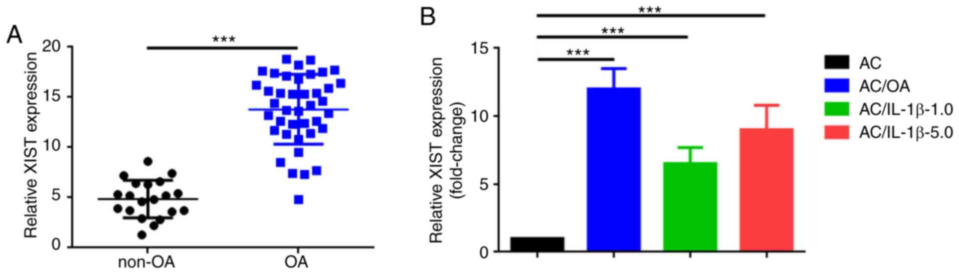
Expression of XIST in OA tissues, AC/OA
cells and IL-1β-treated ACs
The expression of XIST in the 40 clinical OA and 20
non-OA cartilage specimens was detected by RT-qPCR. The levels of
this lncRNA were significantly elevated in the OA samples compared
with those measured in the non-OA samples (P<0.001; Fig. 1A). Furthermore, the expression of
XIST was determined in the cultured AC/OA, AC/IL-1β-1.0,
AC/IL-1β-5.0 and normal ACs. As observed in Fig. 1B, compared with the normal ACs,
the XIST expression was significantly increased in AC/OA,
AC/IL-1β-1.0 and AC/IL-1β-5.0 cells (P<0.001).
Downregulation of XIST inhibits ECM
degradation in AC/OA and AC/IL-1β-5.0 cells
The findings that XIST was overex-pressed in OA
tissues and in OA chondrocytes (AC/OA and AC/IL-1β-5.0) indicated
that XIST may contribute to the pathological mechanism of OA.
Therefore, the function of XIST in AC/OA cells was explored. First,
the expression of XIST was significantly downregulated using XIST
shRNA in AC/OA and AC/IL-1β-5.0 cells, which was verified by
RT-qPCR (P<0.01; Fig. 2A).
Secondly, the expression of MMP-13, a marker of ECM degradation,
was separately measured by RT-qPCR, western blot analysis and
immunofluorescence staining. The downregulation of XIST led to the
inhibition of MMP-13 expression in the AC/OA and in AC/IL-1β-5.0
cells (Fig. 2B-D). Thirdly,
another marker of ECM degradation, ADAMTS5, was also investigated.
Similar to the results of MMP-13, the knockdown of XIST resulted in
a decrease in the expression of ADAMTS5 (Fig. 2E-G). Overall, these findings imply
that the downregulation of XIST decreases ECM degradation in AC/OA
and AC/IL-1β-5.0 cells.
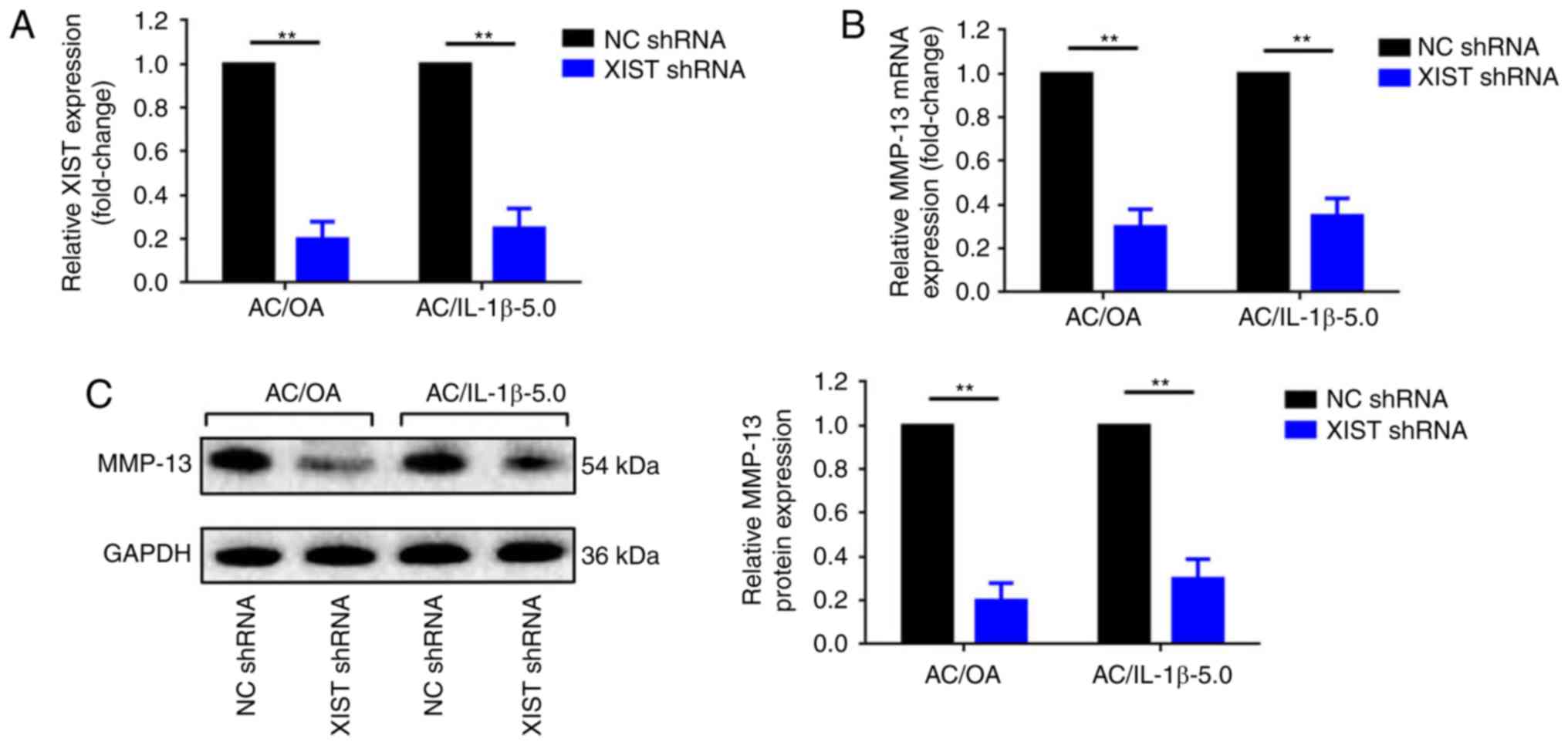 | Figure 2Downregulation of XIST decreases
extracellular matrix degradation in AC/OA and AC/IL-1β-5.0 cells.
(A) Downregulation of XIST by an RNA interference assay was
confirmed by RT-qPCR. MMP-13 expression decreased following
knockdown of XIST, as determined by (B) RT-qPCR, (C) western blot
analysis (lysates were analyzed by immunoblotting with MMP-13 and
GAPDH antibodies; the left panel presents data from 3 independent
experiments) and (D) immunofluorescence analysis. The knockdown of
XIST also downregulated the expression of ADAMTS5 as determined by
(E) RT-qPCR, (F) western blot analysis (lysates were analyzed by
immunoblotting with ADAMTS5 and GAPDH antibodies; the left panel
presents data from 3 independent experiments) and (G)
immunofluorescence analysis. All data were normalized to the
negative control shRNA group. Magnification, ×100; scale bar, 100
µm. The error bars represent standard deviation (n=3).
**P<0.01. XIST, X-inactive-specific transcript; AC,
articular chondrocyte; OA, osteoarthritis; IL-1β, interleukin-1β;
RT-qPCR, reverse transcription-quantitative polymerase chain
reaction; MMP-13, matrix metalloproteinase 13; ADAMTS5, ADAM
metallopeptidase with thrombospondin type 1 motif 5; NC, negative
control; sh, short hairpin. |
miR-1277-5p attenuates ECM degradation by
directly targeting MMP-13 and ADAMTS5 in AC/OA and AC/IL-1β-5.0
cells
The predominant theory of how lncRNAs work is that
they function as ceRNAs. The present study questioned whether XIST
exerts its function through certain miRNAs. Through an online
bioinformatic prediction via TargetScan and LncBase, miR-1277-5p
was selected due to its potential binding for MMP-13, ADAMTS and
XIST (Fig. 3A). The expression
levels of miR-1277-5p were confirmed to be significantly decreased
in OA tissues (P<0.01) and in AC/OA, AC/IL-1β-1.0 and
AC/IL-1β-5.0 cells compared with non-OA tissue samples and ACs,
respectively (Fig. 3B).
Furthermore, the upregulation of miR-1277-5p led to the attenuation
of the expression of MMP-13 and ADAMTS5, two markers of ECM
degradation, in AC/OA and AC/IL-1β-5.0 cells (Fig. 3C and D). In addition, wild-type
and mutant luciferase plasmids of MMP-13 (wt-MMP-13 and mut-MMP-13)
and ADAMTS5 (wt-ADAMTS5 and mut-ADAMTS5) were constructed,
containing wild-type and mutant miR-1277-5p binding sites,
respectively (Fig. 3E). Finally,
the luciferase reporter assay confirmed that miR-1277-5p directly
targets MMP-13 and ADAMTS5 via the same binding site (Fig. 3F and G).
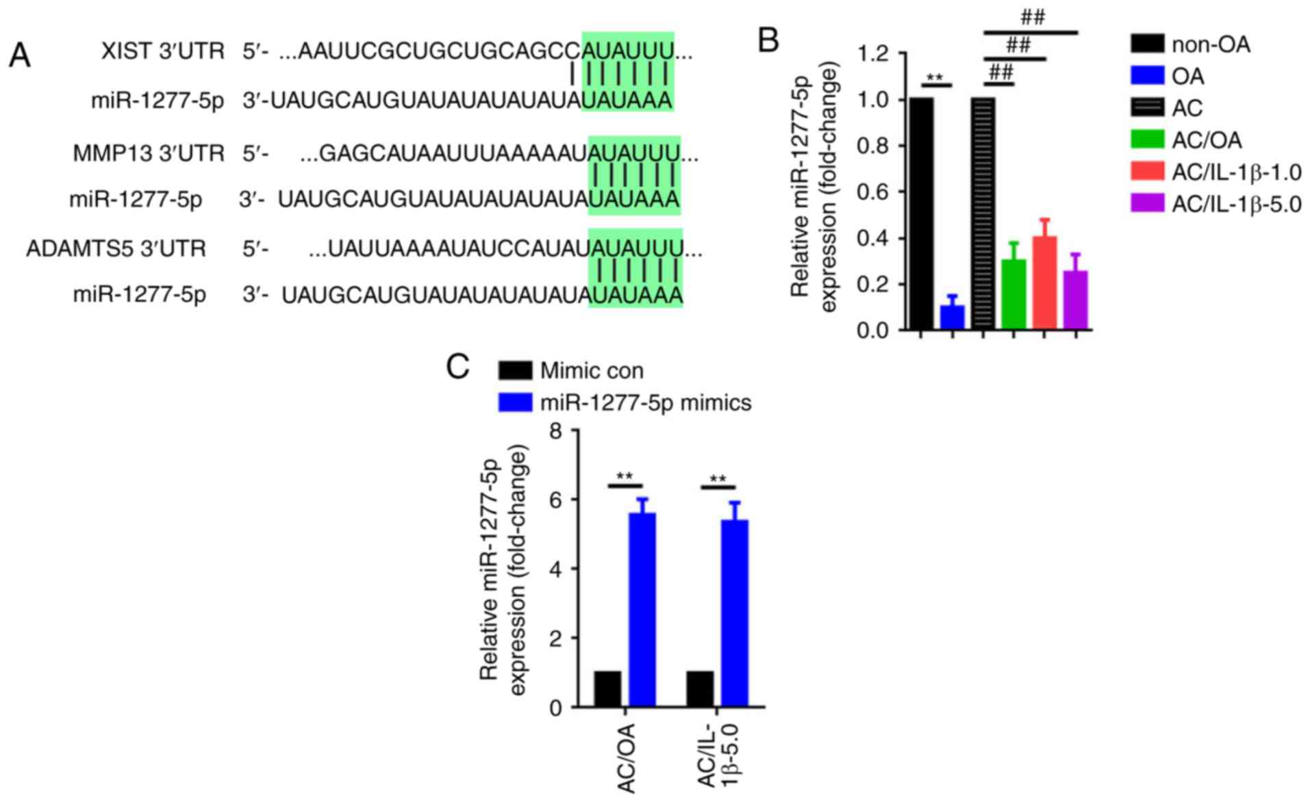 | Figure 3miR-1277-5p attenuates extracellular
matrix degradation by directly targeting MMP-13 and ADAMTS5 in
AC/OA and AC/IL-1β-5.0 cells. (A) XIST, MMP-13 and ADAMTS5 share
the same miR-1277-5p binding site within their 3′UTRs. (B)
Downregulation of miR-1277-5p in OA tissue specimens and in AC/OA,
AC/IL-1β-1.0 and AC/IL-1β-5.0 cells, as confirmed by reverse
transcription-quantitative polymerase chain reaction.
**P<0.01 vs. non-OA group; ##P<0.01 vs.
AC group. (C) miR-1277-5p was upregulated by transfection of
miR-1277-5p mimics as determined by reverse
transcription-quantitative polymerase chain reaction.
**P<0.01 vs. mimic con group. (D) Upregulation of
miR-1277-5p (transfection of miR-1277-5p mimics) led to a notable
decrease in MMP-13 and ADAMTS5 expression, as measured by
immunofluorescence analysis. **P<0.01 vs. mimic
control group; magnification, ×100; scale bar, 100 µm. (E)
Diagram of the luciferase reporter plasmids with the wt- or
mut-MMP-13 and -ADAMTS5 3′UTRs. (F) Compared with the effect of the
mimic con, co-transfection of miR-1277-5p mimic and wt-MMP-13 led
to a distinct decrease in fluorescence, but the suppressive effect
was rescued by a mutation in the putative miR-1277-5p binding sites
in the MMP-13 3′UTR (co-transfection of mut-MMP-13 and miR-1277-5p
mimic), as verified by dual luciferase assays.
**P<0.01 and &P>0.05 vs. mimic con
group. (G) The same effects were observed during the verification
of the interaction between miR-1277-5p and ADAMTS5 3′UTR.
**P<0.01 and &P>0.05 vs. mimic con
group. All error bars represented standard deviation (n=3). miR,
microRNA; MMP-13, matrix metallopro-teinase 13; ADAMTS5, ADAM
metallopeptidase with thrombospondin type 1 motif 5; AC, articular
chondrocyte; OA, osteoarthritis; IL-1β, interleukin-1β; UTR,
untranslated region; wt, wild type; mut, mutant; con, control;
LUC+, luciferase. |
XIST and miR-1277-5p exhibit reciprocal
repression and XIST is a target of miR-1277-5p
The downregulation of XIST and the upregulation of
miR-1277-5p were revealed to present a similar protective effect on
ECM degradation. This finding highlighted the question of the
nature of the association between the two RNAs. Initially, a
negative correlation was demonstrated (Fig. 4A). Subsequently, the upregulation
and downregulation of XIST were revealed to significantly inversely
regulate miR-1277-5p expression (P<0.01; Fig. 4B and C). An increase or a decrease
of miR-1277-5p also affected XIST expression in the opposite manner
(Fig. 4D and E). These results
indicate that XIST and miR-1277-5p repress each other in a
reciprocal manner. As predicted, a binding site was confirmed
between XIST 3′-UTR and miR-1277-5p. Furthermore, a luciferase
reporter assay was used to investigate whether miR-1277-5p targets
XIST. The luciferase reporting plasmids containing a wild-type and
mutant miR-1277-5p binding sites (wt-XIST and mut-XIST; Fig. 4F) were constructed as follows:
Wt-XIST or mut-XIST and miR-1277-5p mimics were co-transfected into
293 cells while the mimic control was used as a negative control,
and luciferase activity changes were measured. Co-transfection of
wt-XIST and miR-1277-5p mimics led to a significant decrease in
luminescence, compared with that detected in the mimic control
(P<0.01; Fig. 4G). However,
the luminescence signal was significantly recovered in the
co-transfection of mut-XIST and miR-1277-5p mimics (P<0.05).
Ultimately, an RNA pull-down assay was conducted to confirm the
interaction between XIST and miR-1277-5p. As demonstrated in
Fig. 4H and I, wt-XIST, but not
mut-XIST, could pull down miR-1277-5p. Together, these findings
reveal that miR-1277-5p directly targets XIST.
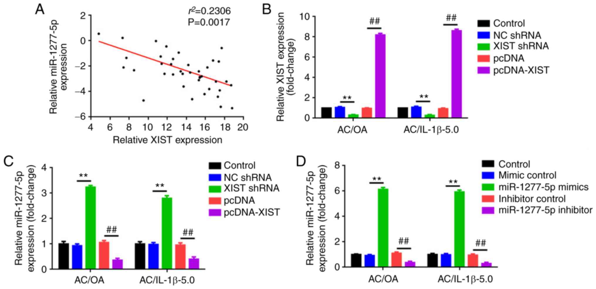 | Figure 4XIST and miR-1277-5p repress each
other and XIST is a target of miR-1277-5p. (A) The expression of
XIST was inversely correlated with that of miR-1277-5p. The
overexpression and downregulation of (B) XIST negatively regulated
(C) miR-1277-5p expression, as determined by RT-qPCR.
**P<0.01 and ##P<0.01 vs. the
corresponding control group (n=3). Increasing and decreasing (D)
miR-1277-5p levels negatively affected (E) XIST expression, as
indicated by RT-qPCR. **P<0.01 and
##P<0.01 vs. control group (n=3). (F) Diagram of the
luciferase reporter plasmids with the wt- or mut-XIST 3′UTR. (G)
The targeted binding between XIST 3′UTR and miR-1277-5p was
verified by a dual luciferase assay. **P<0.01 and
&P>0.05 vs. mimic control group. (H) wt-XIST, but
not mut-XIST, could pull down miR-1277-5p, as determined by an RNA
pull-down assay in AC/OA and (I) AC/IL-1b-5.0.
***P<0.001 vs. the wt-XIST group (normalized to the
beads group). The error bars represent standard deviation. (n=3).
XIST, X-inactive-specific transcript; miR, microRNA; RT-qPCR,
reverse transcription-quantitative polymerase chain reaction; wt,
wild type; mut, mutant; UTR, untranslated region. |
XIST promotes ECM degradation by acting
as a ceRNA of miR-1277-5p in AC/OA and AC/IL-1β-5.0 cells
To confirm whether the effect of XIST on ECM
degradation is achieved through the miR-1277-5p pathway with a
ceRNA mechanism, the expression of MMP-13 and ADAMTS5 was
determined following a dual intervention with XIST and miR-1277-5p.
Wild-type and mutant XIST overexpression plasmids wt-pcDNA-XIST and
mut-pcDNA-XIST were co-transfected with an miR-1277-5p mimic into
AC/OA and AC/IL-1β-5.0 cells. The expression of MMP-13 was
determined by immunofluorescence staining, RT-qPCR and western
blotting. As observed in Fig.
5A-C, the transfection of wt-pcDNA-XIST led to a notable
elevation of MMP-13 mRNA and protein expression. On the other hand,
when the putative miR-1277-5p binding site in XIST was mutated, the
facilitative effect was not present. Even more convincingly, when
the miR-1277-5p mimic was co-transfected, the facilitative effect
of wt-pcDNA-XIST on the MMP-13 expression was significantly
weakened (P<0.01). The expression of ADAMTS5 presented a similar
tendency to MMP-13 (Fig. 5D-F).
Combined with the reciprocally repressing association between XIST
and miR-1277-5p, XIST was confirmed to promote ECM degradation by
acting as a ceRNA of miR-1277-5p in AC/OA and AC/IL-1β-5.0
cells.
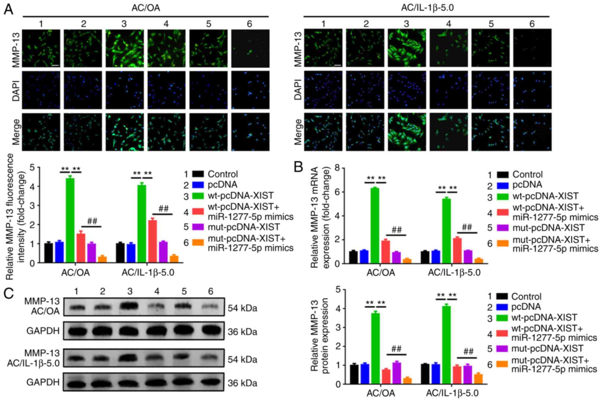 | Figure 5XIST promotes extracellular matrix
degradation by acting as a competing endogenous RNA of miR-1277-5p
in AC/OA and AC/IL-1β-5.0 cells. Normal XIST overexpression
plasmids (wt-pcDNA-XIST) led to a notable upregulation of MMP-13,
but the effect was attenuated by the overexpression of miR-1277-5p
(co-transfection of wt-pcDNA-XIST and miR-1277-5p mimics). When the
putative miR-1277-5p binding site in XIST was mutated (transfection
of mut-pcDNA-XIST), the facilitative effect was eliminated. The
phenomenon was detected by (A) immunofluorescence analysis, (B)
RT-qPCR and (C) western blot analysis (lysates were analyzed by
immunoblotting with MMP-13 and GAPDH antibodies; the left panel
presents data from 3 independent experiments).
**P<0.01 vs. wt-pcDNA-XIST; ##P<0.01
vs. wt-pcDNA-XIST + miR-1277-5p mimic group. wt-pcDNA-XIST, but not
mut-pcDNA-XIST, promoted ADAMTS5 expression, and the effect was
repressed when the putative miR-1277-5p binding site in XIST was
mutated (transfection of mut-pcDNA-XIST). As with MMP-13, elevation
of miR-1277-5p also reversed the facilitative effect of
wt-pcDNA-XIST on ADAMTS5 expression, as determined by (D)
immunofluorescence analysis, (E) RT-qPCR and (F) western blot
analysis (lysates were analyzed by immunoblotting with ADAMTS5 and
GAPDH antibodies; the left panel presents data from 5 independent
experiments). **P<0.01 vs. wt-pcDNA-XIST;
##P<0.01 vs. wt-pcDNA-XIST + miR-1277-5p mimic group.
All data were normalized to the control group, and all error bars
represent standard deviation (n=3). XIST, X-inactive-specific
transcript; miR, microRNA; AC, articular chondrocyte; OA,
osteoarthritis; IL-1β, interleukin-1β; wt, wild type; mut, mutant;
MMP-13, matrix metalloproteinase 13; ADAMTS5, ADAM metallopeptidase
with thrombospondin type 1 motif 5; RT-qPCR, reverse
transcription-quantitative polymerase chain reaction. |
Depression of XIST inhibits ECM
degradation in vivo
A DMM mouse model was used to further investigate
the function of XIST in ECM degradation in vivo, according
to a previous study (27).
Lenti-vector and lenti-siXIST were injected into the joint cavities
of the animals. The results of Safranin-O staining revealed that,
compared with the results of the sham surgery group, the resection
of the medial meniscus led to an elevated Safranin-O staining, a
rougher articular surface and a significantly increased summed
score (P<0.001; Fig. 6A). On
the other hand, compared with the lenti-vector group, the group
with silenced XIST expression presented with decreased Safranin-O
staining, smoother articular surface and a significantly decreased
summed score (P<0.001), indicating that the knockdown of XIST
led to a protective effect on the development of OA. In each animal
group, the expression levels of XIST and miR-1277-5p were measured
by RT-qPCR (Fig. 6B-C), and the
level of ECM degradation (expression of MMP-13 and ADAMTS5) was
determined by IHC, RT-qPCR and western blot analysis. DMM resulted
in a significant elevation of MMP-13 and ADAMTS5, compared with
that in the sham surgery group (P<0.001; Fig. 6D and E), whereas the knockdown of
XIST led to a decrease in the expression of MMP-13 and ADAMTS5 (ECM
degradation), compared with that observed in the lenti-vector
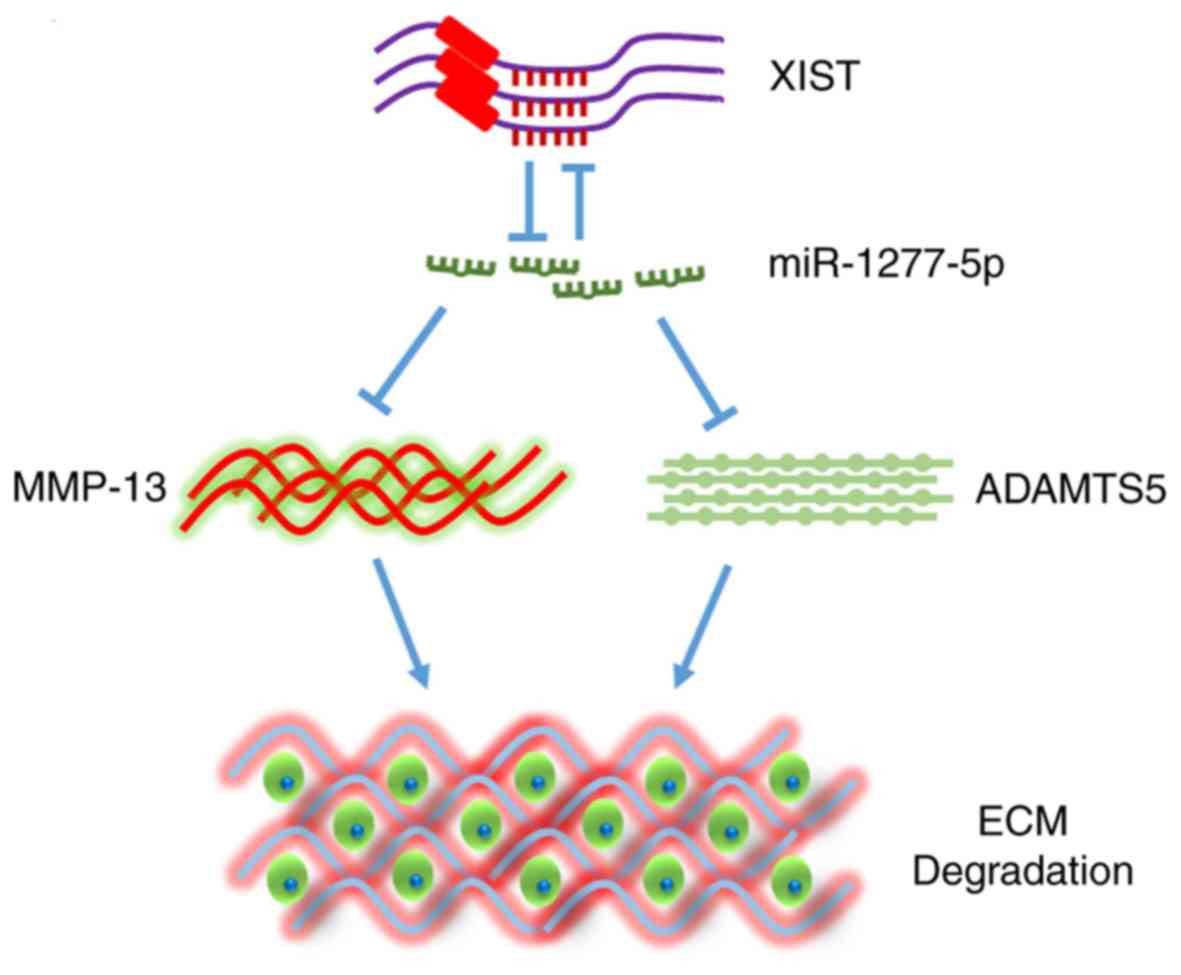
group. In conclusion, as presented in the schematic of the
mechanism in Fig. 7, the present
results suggest that the downregulation of XIST inhibits ECM
degradation during the formation of OA.
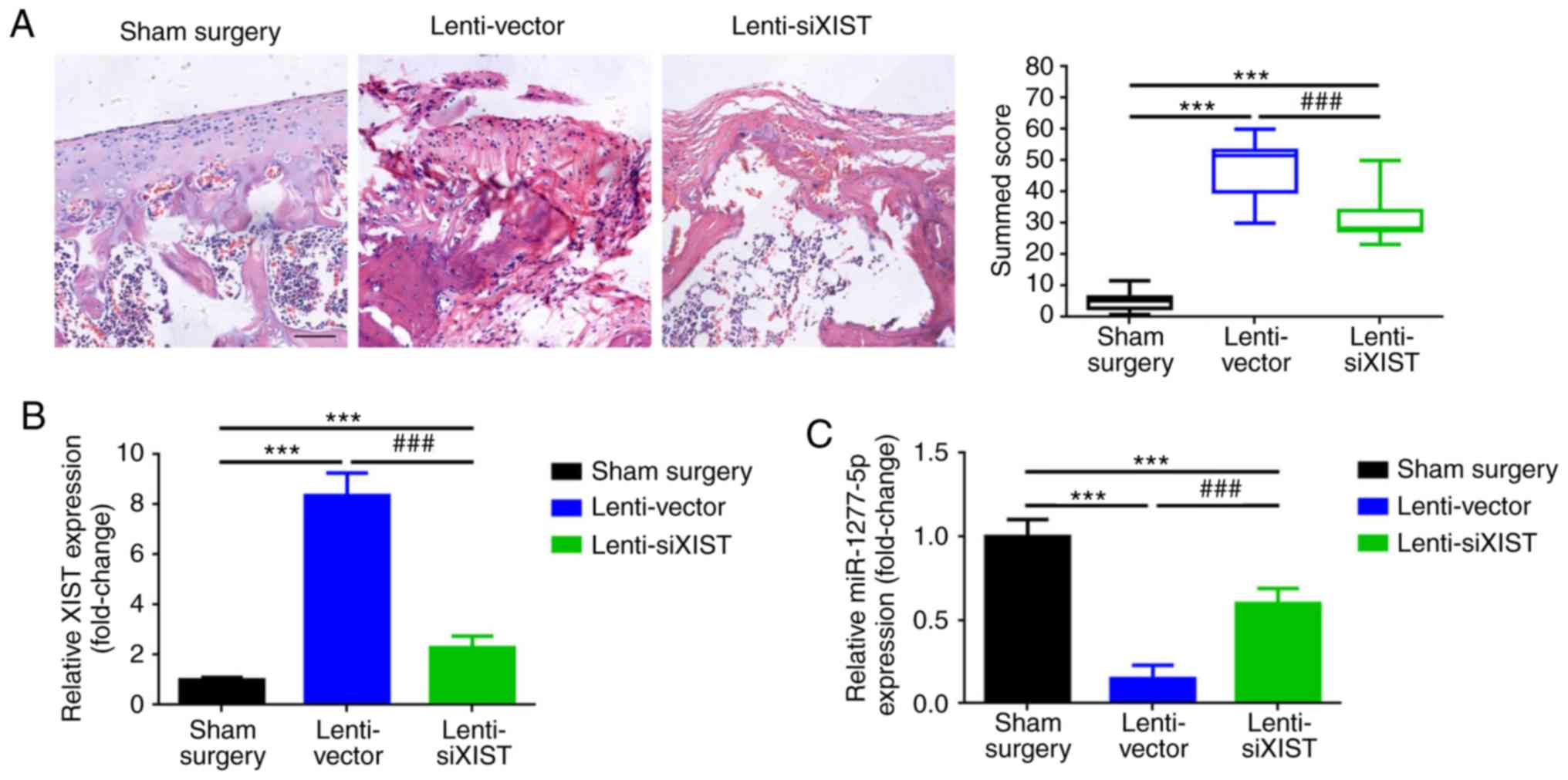 | Figure 6Depression of XIST inhibited
extracellular matrix degradation in vivo. (A) DMM
(lenti-vector and lenti-siXIST group) resulted in increased
Safranin-O staining and an elevated summed score compared with the
sham surgery group, whereas a certain cartilage-protective effect
was presented in the lenti-siXIST group compared with the
lenti-vector group. (B) Elevated XIST and (C) depressed miR-1277-5p
levels were attributed to DMM, and the injection of lenti-siXIST
led to the inhibition of XIST and the promotion of miR-1277-5p
compared with the lenti-vector group, as determined by RT-qPCR. (D)
In contrast with the sham surgery group, increased MMP-13
expression was detected in the DMM groups (lenti-vector and
lenti-siXIST group). In addition, downregulation of XIST (injection
of lenti-siXIST) inhibited MMP-13 expression, as detected by IHC,
RT-qPCR and western blot analysis (lysates were analyzed by
immunoblotting with MMP-13 and GAPDH antibodies; the left panel
presents data from 5 independent experiments). (E) Upregulated
ADAMTS5 expression was also observed in the DMM groups
(lenti-vector and lenti-siXIST group) compared with the surgery
group, whereas, compared with the lenti-vector group, the injection
of lenti-siXIST led to a decrease in ADAMTS5 expression, as
confirmed by IHC, RT-qPCR and western blot analysis (lysates were
analyzed by immunoblotting with ADAMTS5 and GAPDH antibodies; the
left panel presents data from 5 independent experiments).
***P<0.001 vs. the sham surgery group;
###P<0.001 vs. the lenti-vector group. All data were
normalized to the control group, and all error bars represent
standard deviation (n=3). XIST, X-inactive-specific transcript;
DMM, destabilization of the medial meniscus; siXIST, XIST
silencing; RT-qPCR, reverse transcription-quantitative polymerase
chain reaction; ADAMTS5, ADAM metallopeptidase with thrombospondin
type 1 motif 5; MMP-13, matrix metalloproteinase 13; IHC,
immu-nohistochemistry. |
Discussion
OA is a common degenerative disease of the joints,
characterized by cartilage loss, subchondral bone remodeling and
osteophyte development. A number of factors contribute to the
composition and structural changes of the ECM during the
development and progression of OA. As the only cell type in
cartilage, chondrocytes participate in the catabolic activities
that ultimately lead to the degradation of cartilaginous ECM
(30). During ECM degradation,
catabolic factors including MMP-13 and ADAMTS5 mediating the loss
of type II collagen/aggrecan, contribute significantly to the
process (4). As an ECM-degrading
enzyme, MMP-13 is structurally characterized among the
zinc-dependent endopeptidases that degrade various components of
the ECM (31). Numerous studies
have demonstrated that MMP-13 is a key regulator in the pathology
of OA (32-34). ADAMTS5, a member of the ADAMTS
family, contains a domain arrangement that consists of a
pro-domain, a catalytic metalloproteinase domain, a
disintegrin-like domain, a cysteine-rich domain and a spacer domain
(5). ADAMTS5, which can degrade
the cartilage proteoglycan aggrecan, is widely reported as the most
efficient aggrecanase and has been generally considered to be the
most likely candidate for its role in the pathological mechanisms
of OA (35-38). In the present study, MMP-13 and
ADAMTS5 were used as markers of ECM degradation, as previously
reported.
miR-1277-5p is a miRNA encoded by a site located on
chromosome Xq24. The function of miR-1277-5p is presently not well
understood. Budak et al (39) reported that miR-1277-5p was
downregulated in chronic brucellosis, but its function in this
infectious disease remains unknown. In a study of colorectal
cancer, Motieghader et al (40) revealed that miR-1277-5p was
downregulated in adenocarcinoma compared with high-grade
intraepithelial neoplasia. Through deep sequencing and northern
blot analyses, Crowe et al (41) demonstrated that miR-1277-5p was a
novel miRNA which was downregulated in OA. The results of the
present study revealed that miR-1277-5p expression was
downregulated in OA specimens and in AC/OA, AC/IL-1β-1.0 and
AC/IL-1β-5.0 cells. Furthermore, the detection of markers MMP-13
and ADAMTS5 demonstrated that the upregulation of miR-1277-5p could
reverse ECM degradation. It is well known that miRNAs exert their
functions by directly or indirectly regulating their targets
(42-44). In the present study, the results
of the constructed luciferase assay verified that MMP-13 and
ADAMTS5 are targets of miR-1277-5p by sharing the same binding
sites within their 3′UTRs. These outcomes indicate that miR-1277-5p
may function as a protective factor in OA development.
It is widely accepted that non-coding RNAs lncRNAs,
circular RNAs and miRNAs are involved in numerous diseases,
including OA (45-48). The lncRNA XIST has been
extensively studied in a number of types of cancer, including
colorectal cancer, pancreatic cancer, osteosarcoma, non-small cell
lung cancer and bladder cancer (9,49-52). To date, studies on XIST and OA are
rare. In the present study, XIST was revealed to be upregulated in
OA in clinical tissue samples as well as cell lines. Subsequently,
a loss-of-function experiment demonstrated that the knockdown of
XIST suppressed ECM degradation in AC/OA and AC/IL-1β-5.0 cells, as
determined by measuring the levels of markers MMP-13 and ADAMTS5,
indicating that XIST promotes OA development. Now a prevalent
theory in the field of non-coding RNAs, the mechanism of ceRNAs
between lncRNAs and miRNAs was first proposed by Salmena et
al (20) in 2011. Li et
al (25) reported that lncRNA
CIR promoted chondrocyte ECM degradation in OA by acting as a ceRNA
of miR-27b. Li et al (53)
revealed that lncRNA plasmacytoma variant translocation 1 regulated
the apoptosis of chondrocytes by acting as a sponge for miR-488-3p
in OA. In the present study, an online bioinformatic analysis
predicted that XIST contains a similar binding site for miR-1277-5p
as MMP-13 and ADAMTS5, implying that XIST may be a ceRNA of
miR-1277-5p. The ensuing overexpression and downregulation
experiments revealed a reciprocal suppression effect between XIST
and miR-1277-5p. The subsequent co-localization and luciferase
assays verified that XIST is a direct target of miR-1277-5p.
Furthermore, through a series of antisense experiments, XIST was
confirmed to exert its effect on ECM degradation (increased
expression of MMP-13 and ADAMTS5) by acting as a ceRNA of
miR-1277-5p. Finally, the constructed OA mouse model confirmed the
facilitative effect of XIST on ECM degradation in vivo.
Based on the present outcomes, it was concluded that XIST promotes
ECM degradation by functioning as a ceRNA of miR-1277-5p in OA.
The development of OA is a complicated process
covering broad mechanisms and a number of molecules. The results of
the present study suggest that XIST promotes ECM degradation by
working as a ceRNA of miR-1277-5p. These findings may provide a
novel molecular target in treating OA.
Abbreviations:
|
OA
|
osteoarthritis
|
|
ECM
|
extracellular matrix
|
|
lncRNA
|
long non-coding RNA
|
|
XIST
|
X-inactive-specific transcript
|
|
miR/miRNA
|
microRNA
|
|
AC
|
articular chondrocyte
|
|
MMP-13
|
matrix metalloproteinase 13
|
|
ADAMTS5
|
ADAM metallopeptidase with
thrombospondin type 1 motif 5
|
|
ceRNA
|
competing endogenous RNA
|
|
IL-1β
|
interleukin-1β
|
|
RIP
|
RNA immunoprecipitation
|
|
DMM
|
destabilization of the medial
meniscus
|
Acknowledgments
Not applicable.
Funding
The present study was supported by the Foundation
for Innovative Talent in Higher Education of Liaoning (grant no.
LR2017056) and the China Postdoctoral Science Foundation (grant no.
2016M591437).
Availability of data and materials
The datasets used during the present study are
available from the corresponding author upon reasonable
request.
Authors' contributions
YW and ZZ conceived the experiments; TW, YL and XH
performed the experiments; YW and WZ analyzed the data; YW wrote
the manuscript. All authors read and approved the final
manuscript.
Ethics approval and consent to
participate
The present study and the associated experimental
protocols (both human and animal experiments) were performed in
compliance with ethical guidelines and approved by the Institute
Research Medical Ethics Committee of the Central Hospital
Affiliated to Shenyang Medical College, Shenyang, China (approval
no. 2016JULY15-7). All OA tissues and non-OA tissues were also used
in accordance with the Helsinki declaration. Written informed
consent was signed by each patient before the study. All patients
agreed that the data from their samples could be used for
experimental studies and paper presentations.
Patient consent for publication
Not applicable.
Competing interests
The authors declare that they have no competing
interests.
References
|
1
|
van der Kraan PM: Osteoarthritis year 2012
in review: Biology. Osteoarthritis Cartilage. 20:1447–1450. 2012.
View Article : Google Scholar : PubMed/NCBI
|
|
2
|
Mobasheri A: Osteoarthritis year 2012 in
review: Biomarkers. Osteoarthritis Cartilage. 20:1451–1464. 2012.
View Article : Google Scholar : PubMed/NCBI
|
|
3
|
Lee AS, Ellman MB, Yan D, Kroin JS, Cole
BJ, van Wijnen AJ and Im HJ: A current review of molecular
mechanisms regarding osteoarthritis and pain. Gene. 527:440–447.
2013. View Article : Google Scholar : PubMed/NCBI
|
|
4
|
Sofat N: Analysing the role of endogenous
matrix molecules in the development of osteoarthritis. Int J Exp
Pathol. 90:463–479. 2009. View Article : Google Scholar : PubMed/NCBI
|
|
5
|
Verma P and Dalal K: ADAMTS-4 and
ADAMTS-5: Key enzymes in osteoarthritis. J Cell Biochem.
112:3507–3514. 2011. View Article : Google Scholar : PubMed/NCBI
|
|
6
|
Chen WX, Shan FJ, Jin HT, Wang PE, Xiao LW
and Tong PJ: Research on application of determination of MMP-13 in
osteoarthritis. Zhongguo Gu Shang. 27:617–620. 2014.In Chinese.
PubMed/NCBI
|
|
7
|
Huynh NP, Anderson BA, Guilak F and
McAlinden A: Emerging roles for long noncoding RNAs in skeletal
biology and disease. Connect Tissue Res. 58:116–141. 2017.
View Article : Google Scholar :
|
|
8
|
Jiang SD, Lu J, Deng ZH, Li YS and Lei GH:
Long noncoding RNAs in osteoarthritis. Joint Bone Spine.
84:553–556. 2017. View Article : Google Scholar
|
|
9
|
Hu Y, Deng C, Zhang H, Zhang J, Peng B and
Hu C: Long non-coding RNA XIST promotes cell growth and metastasis
through regulating miR-139-5p mediated Wnt/β-catenin signaling
pathway in bladder cancer. Oncotarget. 8:94554–94568.
2017.PubMed/NCBI
|
|
10
|
Sun N, Zhang G and Liu Y: Long non-coding
RNA XIST sponges miR-34a to promotes colon cancer progression via
Wnt/β-catenin signaling pathway. Gene. 665:141–148. 2018.
View Article : Google Scholar : PubMed/NCBI
|
|
11
|
Zheng R, Lin S, Guan L, Yuan H, Liu K, Liu
C, Ye W, Liao Y, Jia J and Zhang R: Long non-coding RNA XIST
inhibited breast cancer cell growth, migration, and invasion via
miR-155/CDX1 axis. Biochem Biophys Res Commun. 498:1002–1008. 2018.
View Article : Google Scholar : PubMed/NCBI
|
|
12
|
Zhou Q, Hu W, Zhu W, Zhang F, Lin-Lin L,
Liu C, Songyang YY, Sun CC and Li D: Long non coding RNA XIST as a
prognostic cancer marker - A meta-analysis. Clin Chim Acta.
482:1–7. 2018. View Article : Google Scholar : PubMed/NCBI
|
|
13
|
Fu M, Huang G, Zhang Z, Liu J, Zhang Z,
Huang Z, Yu B and Meng F: Expression profile of long noncoding RNAs
in cartilage from knee osteoarthritis patients. Osteoarthritis
Cartilage. 23:423–432. 2015. View Article : Google Scholar
|
|
14
|
Denzler R, Agarwal V, Stefano J, Bartel DP
and Stoffel M: Assessing the ceRNA hypothesis with quantitative
measurements of miRNA and target abundance. Mol Cell. 54:766–776.
2014. View Article : Google Scholar : PubMed/NCBI
|
|
15
|
Kanduri C: Long noncoding RNAs: Lessons
from genomic imprinting. Biochim Biophys Acta. 1859:102–111. 2016.
View Article : Google Scholar
|
|
16
|
Mondal T, Subhash S, Vaid R, Enroth S,
Uday S, Reinius B, Mitra S, Mohammed A, James AR, Hoberg E, et al:
MEG3 long noncoding RNA regulates the TGF-β pathway genes through
formation of RNA-DNA triplex structures. Nat Commun. 6:77432015.
View Article : Google Scholar
|
|
17
|
Wang C, Wang L, Ding Y, Lu X, Zhang G,
Yang J, Zheng H, Wang H, Jiang Y and Xu L: LncRNA structural
characteristics in epigenetic regulation. Int J Mol Sci. 18:2017.
View Article : Google Scholar
|
|
18
|
Yuan SX, Zhang J, Xu QG, Yang Y and Zhou
WP: Long noncoding RNA, the methylation of genomic elements and
their emerging crosstalk in hepatocellular carcinoma. Cancer Lett.
379:239–244. 2016. View Article : Google Scholar
|
|
19
|
Zampetaki A and Mayr M: Long noncoding
RNAs and angiogenesis: Regulatory information for chromatin
remodeling. Circulation. 136:80–82. 2017. View Article : Google Scholar : PubMed/NCBI
|
|
20
|
Salmena L, Poliseno L, Tay Y, Kats L and
Pandolfi PP: A ceRNA hypothesis: The Rosetta Stone of a hidden RNA
language? Cell. 146:353–358. 2011. View Article : Google Scholar : PubMed/NCBI
|
|
21
|
Gu S, Xie R, Liu X, Shou J, Gu W and Che
X: Long coding RNA XIST contributes to neuronal apoptosis through
the downregulation of AKT phosphorylation and is negatively
regulated by miR-494 in rat spinal cord injury. Int J Mol Sci.
18:2017. View Article : Google Scholar
|
|
22
|
Zhang R and Xia T: Long non-coding RNA
XIST regulates PDCD4 expression by interacting with miR-21-5p and
inhibits osteosarcoma cell growth and metastasis. Int J Oncol.
51:1460–1470. 2017. View Article : Google Scholar : PubMed/NCBI
|
|
23
|
Gosset M, Berenbaum F, Thirion S and
Jacques C: Primary culture and phenotyping of murine chondrocytes.
Nat Protoc. 3:1253–1260. 2008. View Article : Google Scholar : PubMed/NCBI
|
|
24
|
Wang Y, Yang T, Liu Y, Zhao W, Zhang Z, Lu
M and Zhang W: Decrease of miR-195 promotes chondrocytes
proliferation and maintenance of chondrogenic phenotype via
targeting FGF-18 pathway. Int J Mol Sci. 18:2017.
|
|
25
|
Li YF, Li SH, Liu Y and Luo YT: Long
noncoding RNA CIR promotes chondrocyte extracellular matrix
degradation in osteoarthritis by acting as a sponge for Mir-27b.
Cell Physiol Biochem. 43:602–610. 2017. View Article : Google Scholar : PubMed/NCBI
|
|
26
|
Livak KJ and Schmittgen TD: Analysis of
relative gene expression data using real-time quantitative PCR and
the 2(−Delta Delta C(T)) Method. Methods. 25:402–408. 2001.
View Article : Google Scholar
|
|
27
|
Cai H, Liu X, Zheng J, Xue Y, Ma J, Li Z,
Xi Z, Li Z, Bao M and Liu Y: Long non-coding RNA taurine
upregulated 1 enhances tumor-induced angiogenesis through
inhibiting microRNA-299 in human glioblastoma. Oncogene.
36:318–331. 2017. View Article : Google Scholar
|
|
28
|
Glasson SS, Blanchet TJ and Morris EA: The
surgical destabilization of the medial meniscus (DMM) model of
osteoarthritis in the 129/SvEv mouse. Osteoarthritis Cartilage.
15:1061–1069. 2007. View Article : Google Scholar : PubMed/NCBI
|
|
29
|
Wang Y, Zhang Y, Yang T, Zhao W, Wang N,
Li P, Zeng X and Zhang W: Long non-coding RNA MALAT1 for promoting
metastasis and proliferation by acting as a ceRNA of miR-144-3p in
osteosarcoma cells. Oncotarget. 8:59417–59434. 2017.PubMed/NCBI
|
|
30
|
Hoff P, Buttgereit F, Burmester GR,
Jakstadt M, Gaber T, Andreas K, Matziolis G, Perka C and Röhner E:
Osteoarthritis synovial fluid activates pro-inflammatory cytokines
in primary human chondrocytes. Int Orthop. 37:145–151. 2013.
View Article : Google Scholar :
|
|
31
|
Chowdhury TT, Schulz RM, Rai SS, Thuemmler
CB, Wuestneck N, Bader A and Homandberg GA: Biomechanical
modulation of collagen fragment-induced anabolic and catabolic
activities in chondrocyte/agarose constructs. Arthritis Res Ther.
12:R822010. View
Article : Google Scholar : PubMed/NCBI
|
|
32
|
Di Pizio A, Agamennone M and Tortorella P:
Non-Zinc-binding inhibitors of MMP-13: GRID-based approaches to
rationalize the binding process. Curr Top Med Chem. 16:449–459.
2016. View Article : Google Scholar
|
|
33
|
Li H, Wang D, Yuan Y and Min J: New
insights on the MMP-13 regulatory network in the pathogenesis of
early osteoarthritis. Arthritis Res Ther. 19:2482017. View Article : Google Scholar : PubMed/NCBI
|
|
34
|
Xie XW, Wan RZ and Liu ZP: Recent research
advances in selective matrix metalloproteinase-13 inhibitors as
anti-osteoarthritis agents. ChemMedChem. 12:1157–1168. 2017.
View Article : Google Scholar : PubMed/NCBI
|
|
35
|
Apte SS: Anti-ADAMTS5 monoclonal
antibodies: Implications for aggrecanase inhibition in
osteoarthritis. Biochem J. 473:e1–e4. 2016. View Article : Google Scholar
|
|
36
|
Bondeson J, Wainwright S, Hughes C and
Caterson B: The regulation of the ADAMTS4 and ADAMTS5 aggrecanases
in osteoarthritis: A review. Clin Exp Rheumatol. 26:139–145.
2008.PubMed/NCBI
|
|
37
|
Tortorella MD, Burn TC, Pratta MA,
Abbaszade I, Hollis JM, Liu R, Rosenfeld SA, Copeland RA, Decicco
CP, Wynn R, et al: Purification and cloning of aggrecanase-1: A
member of the ADAMTS family of proteins. Science. 284:1664–1666.
1999. View Article : Google Scholar : PubMed/NCBI
|
|
38
|
Zeng W, Corcoran C, Collins-Racie LA,
Lavallie ER, Morris EA and Flannery CR: Glycosaminoglycan-binding
properties and aggrecanase activities of truncated ADAMTSs:
Comparative analyses with ADAMTS-5, -9, -16 and -18. Biochim
Biophys Acta. 1760:517–524. 2006. View Article : Google Scholar : PubMed/NCBI
|
|
39
|
Budak F, Bal SH, Tezcan G, Akalın H, Goral
G and Oral HB: Altered expressions of miR-1238-3p miR-494,
miR-6069, and miR-139-3p in the formation of chronic brucellosis. J
Immunol Res. 2016:45914682016. View Article : Google Scholar
|
|
40
|
Motieghader H, Kouhsar M, Najafi A,
Sadeghi B and Masoudi-Nejad A: mRNA-miRNA bipartite network
reconstruction to predict prognostic module biomarkers in
colorectal cancer stage differentiation. Mol Biosyst. 13:2168–2180.
2017. View Article : Google Scholar : PubMed/NCBI
|
|
41
|
Crowe N, Swingler TE, Le LT, Barter MJ,
Wheeler G, Pais H, Donell ST, Young DA, Dalmay T and Clark IM:
Detecting new microRNAs in human osteoarthritic chondrocytes
identifies miR-3085 as a human, chondrocyte-selective, microRNA.
Osteoarthritis Cartilage. 24:534–543. 2016. View Article : Google Scholar :
|
|
42
|
Nugent M: MicroRNAs: Exploring new
horizons in osteoarthritis. Osteoarthritis Cartilage. 24:573–580.
2016. View Article : Google Scholar
|
|
43
|
Sondag GR and Haqqi TM: The role of
MicroRNAs and their targets in osteoarthritis. Curr Rheumatol Rep.
18:562016. View Article : Google Scholar : PubMed/NCBI
|
|
44
|
Zhang M, Lygrisse K and Wang J: Role of
MicroRNA in osteoarthritis. J Arthritis. 6:2017. View Article : Google Scholar : PubMed/NCBI
|
|
45
|
Kopańska M, Szala D, Czech J, Gabło N,
Gargasz K, Trzeciak M, Zawlik I and Snela S: MiRNA expression in
the cartilage of patients with osteoarthritis. J Orthop Surg Res.
12:512017. View Article : Google Scholar
|
|
46
|
Peffers MJ, Balaskas P and Smagul A:
Osteoarthritis year in review 2017: Genetics and epigenetics.
Osteoarthritis Cartilage. 26:304–311. 2018. View Article : Google Scholar
|
|
47
|
Zhang HD, Jiang LH, Sun DW, Hou JC and Ji
ZL: CircRNA: A novel type of biomarker for cancer. Breast Cancer.
25:1–7. 2018. View Article : Google Scholar
|
|
48
|
Zhao C, Wang Y, Jin H and Yu T: Knockdown
of microRNA-203 alleviates LPS-induced injury by targeting MCL-1 in
C28/I2 chondrocytes. Exp Cell Res. 359:171–178. 2017. View Article : Google Scholar : PubMed/NCBI
|
|
49
|
Li C, Wan L, Liu Z, Xu G, Wang S, Su Z,
Zhang Y, Zhang C, Liu X, Lei Z and Zhang HT: Long non-coding RNA
XIST promotes TGF-β-induced epithelial-mesenchymal transition by
regulating miR-367/141-ZEB2 axis in non-small-cell lung cancer.
Cancer Lett. 418:185–195. 2018. View Article : Google Scholar : PubMed/NCBI
|
|
50
|
Liu X, Cui L and Hua D: Long non-coding
RNA XIST regulates miR-137-EZH2 axis to promote tumor metastasis in
colorectal cancer. Oncol Res. 27:99–106. 2018. View Article : Google Scholar : PubMed/NCBI
|
|
51
|
Sun Z, Zhang B and Cui T: Long non-coding
RNA XIST exerts oncogenic functions in pancreatic cancer via
miR-34a-5p. Oncol Rep. 39:1591–1600. 2018.PubMed/NCBI
|
|
52
|
Yang C, Wu K, Wang S and Wei G: Long
non-coding RNA XIST promotes osteosarcoma progression by targeting
YAP via miR-195-5p. J Cell Biochem. 119:5646–5656. 2018. View Article : Google Scholar : PubMed/NCBI
|
|
53
|
Li Y, Li S, Luo Y, Liu Y and Yu N: LncRNA
PVT1 regulates chondrocyte apoptosis in osteoarthritis by acting as
a sponge for miR-488-3p. DNA Cell Biol. 36:571–580. 2017.
View Article : Google Scholar : PubMed/NCBI
|