Introduction
Bladder cancer (BCa) ranks ninth among the most
common solid tumors worldwide (1). Approximately 75% of newly diagnosed
BCa cases are non-muscle invasive, and the majority are
histologically low-grade cancer (2). Routine surveillance to monitor BCa
recurrence includes cystoscopic examination due to a high risk of
recurrence after initial resection. The repeat transurethral
resection of bladder tumor (TURBT) remains the first-line treatment
for BCa recurrence (3). However,
all these invasive procedures result in a high cost of care, and
are often associated with significant morbidity. Therefore, more
effective interventions to prevent BCa recurrence are urgently
needed.
Submucosal injection of antitumor drugs
(pirarubicin) after standard TURBT was proven to be an effective
approach to reducing superficial tumor recurrence (4). Gemcitabine is a pivotal
chemotherapeutic agent widely used for BCa due to its low toxicity
in general (5) and as an
intravesical instillation (3).
Data from our experimental and clinical studies also demonstrated
that submucosal injection of gemcitabine prior to TURBT
significantly reduced BCa recurrence (6). However, the underlying mechanisms
are largely unknown.
Metabolomics is a newly emerging technology, which
enables the identification of endogenous compounds and potentially
novel mechanisms associated with disease processes (7). Metabolomics has been used to profile
metabolites in various biological samples, such as serum (8), urine (9-15)
and tissue (16,17), which are the results of the
metabolic response of living systems to drug toxicity or disease
(10). Potential biomarkers
identified from metabo-lomic profiling studies on BCa may be of
diagnostic value and act as indicators of cancer recurrence
(18). Currently, a number of
analytical platforms, such as high-performance liquid
chromatography/mass spectrometry (HPLC/MS) (10), ultra-performance LC-MS (UPLC-MS)
(12), and UPLC time-of-flight MS
(UPLC-TOF-MS) (15), have been
employed to study the metabolomics of BCa by using urine samples.
However, metabolomic studies on BCa tissues is relatively scarce
(16,17). Notably, different metabolomic
platforms with their unique analytical approaches provide
complementary insights into metabolome changes (9,11-15). Therefore, there is a need to study
the tissue-based metabolic signatures of BCa using a new
metabolomics platform.
Metabolomics has also been applied to cancer
treatment and drug target discovery. Eidelman et al reported
using metabolomics to screen the potential therapeutic pathways in
prostate cancer (PCa) (19).
Metabolomics has also been proven to be a promising approach to
developing reliable therapeutic targets for PCa treatment (20). The present study employed liquid
chromatography (LC)-Q-Exactive MS-based metabo-lomic technology to
study the metabolic changes in BCa tissues before and after
treatment with gemcitabine. Identification of the key metabolites
may reveal new metabolic changes associated with BCa and uncover
the changes that mediate the effect of gemcitabine in the treatment
of BCa.
Materials and methods
Clinical samples
A total of 12 patients (9 men and 3 women; age
range, 55-85 years) who had undergone TURBT at the Affiliated
Huai'an No. 1 People's Hospital of Nanjing Medical University were
recruited between December 2016 and September 2017. Bladder tissue
samples were collected from the same patient immediately prior to
and 30 min after submucosal injection of gemcitabine (50 mg,
dissolved in 20 ml normal saline). The bladder tissues included BCa
as well as adjacent non-cancerous bladder tissues. Therefore, a
total of 48 samples were collected in four groups: Pre-gemcitabine
normal, pre-gemcitabine BCa, post-gemcitabine normal, and
post-gemcitabine BCa. Histopathological diagnosis was conducted by
two independent pathologists according to the classification
criteria of the World Health Organization/International Society of
Urological Pathology (21).
Written informed consent was obtained from each participant prior
to recruitment. The Ethics Committee of The Affiliated Huai'an No.
1 People's Hospital of Nanjing Medical University reviewed and
approved the study protocol (serial no. YL-P-2013-21-01). The
sample size of the present study was similar to that of a previous
study on PCa tissues, which produced valuable findings (22). In addition, the experimental
design of self-control and complete collection of samples in our
study may improve statistical power by avoiding confounding
factors.
Tissue preparation for metabolomic
analysis
Following harvesting, all tissues were snap-frozen
in liquid nitrogen, and kept in a -80°C freezer until further
analysis. The sample preparation was conducted as described
previously (23). Briefly, the
tissues were fragmented, ultrasonicated for 5 min (power: 60%,
pulses: 6/4) in distilled water, and then 150 μl homogenate
and 450 μl methanol (Merck KGaA) were mixed in a 1.5-ml
Eppendorf tube for protein precipitation. The mixture was
centrifuged at 16,000 x g for 15 min at 4°C, and the supernatant
was collected and dried in a vacuum centrifugal concentrator. The
dry residue was reconstituted in ultra-pure water and used for
metabolomic analysis.
Metabolomic analysis
The metabolomic analysis was conducted as previously
reported (23). LC-HRMS analysis
was performed on an UPLC Ultimate 3000 system (Dionex), coupled
with a Q-Exactive mass spectrometer (Thermo Fisher Scientific,
Inc.). The instrument operated at a 70,000 resolution with a
full-scan acquisition ranging from 70 to 1,500 m/z. The
chromatographic separation of metabolites associated with the
metabolomic profiling used a multistep gradient containing
ultra-pure water (mobile phase A) and acetonitrile (ACN; mobile
phase B), both acidified with 0.1% formic acid. The gradient
operated at a flow rate of 0.4 ml/min over a 15-min period. The
metabolites were identified based on the accurate mass and the
retention time compared with the commercial standards. The
metabolite standards were purchased from Sigma-Aldrich; Merck KGaA,
Damas-beta Co., Ltd., Aladdin Reagent Company and Adamas Reagent
Co., Ltd.
Statistical analysis
Data collected from the mass spectrometer were
processed for pattern recognition analysis (principal component
analysis, PCA). Normalized MS data were exported to SIMCA-P+
software (V14.0, Umetrics AB) to perform PCA where grouping trends
could be observed. The difference in metabolites between two groups
was compared by paired t-test. According to previous reports
(24,25), the correlation between metabolite
changes and cancer stage was analyzed based on the comparison
between two groups using paired t-test based on the cancer stage
classification. A P-value of <0.05 was considered as the
threshold for statistically significant differences.
To further characterize the metabolic changes and
the metabolic pathways involved, the differentiated metabolites
were first annotated with Kyoto Encyclopedia of Genes and Genomes
(KEGG, http://www.genome.jp/kegg/) and Human
Metabolome Database (HMDB, http://www.hmdb.ca/) (date of access for databases:
April 19, 2018). Data were then processed and analyzed using
MetaboAnalyst 4.0 (http://www.metaboanalyst.ca/MetaboAnalyst/) by R
software (v3.4.3, GitHub). Two modules of MetaboAnalyst were used,
namely pathway analysis and enrichment analysis, which are based on
the KEGG database and Small Molecule Pathway Database (SMPDB,
http://smpdb.ca/), respectively (26). The metabolic network of the
differential metabolites and altered metabolic pathways in KEGG
general metabolic pathway was visualized by iPath 3.0 (http://pathways.embl.de/).
Results
Clinical characteristics of 12
subjects
The mean age of the subjects, including 9 men and 3
women, was 67 years (range, 55-85 years). In all 12 patients, the
diagnosis of urothelial carcinoma was confirmed by
histopathological examination. A total of 2 patients had
Ta, 1 patient had T1, and 9 patients had
T2 disease. Two patients were diagnosed with high-grade
T2 tumors with squamous metaplasia. The clinical
characteristics of the participants are shown in Table I, and are considered to be
representative according to the general population of BCa in China
(2,27).
 | Table IClinical characteristics of 12 BCa
patients. |
Table I
Clinical characteristics of 12 BCa
patients.
| Number | Sex | Age (years) | Tumor type | Histopathology |
|---|
| Stage | Grade |
|---|
| 01 | Male | 55 | MIUC | T2 | High |
| 02 | Male | 56 | MIUC | T2 | High |
| 03 | Male | 59 | MIUC | T2 | High |
| 04 | Female | 82 | MIUC | T1 | High |
| 05 | Male | 67 | MIUC | T2 | High |
| 06 | Male | 76 | MIUC | T2b | Squamous
metaplasia |
| 07 | Male | 76 | MIUC | T2 | High |
| 08 | Female | 60 | MIUC | T2 | High |
| 09 | Male | 67 | NMIUC | Ta | High |
| 10 | Male | 58 | NMIUC | Ta | High |
| 11 | Male | 85 | MIUC | T2a | High |
| 12 | Female | 61 | MIUC | T2 | Squamous
metaplasia |
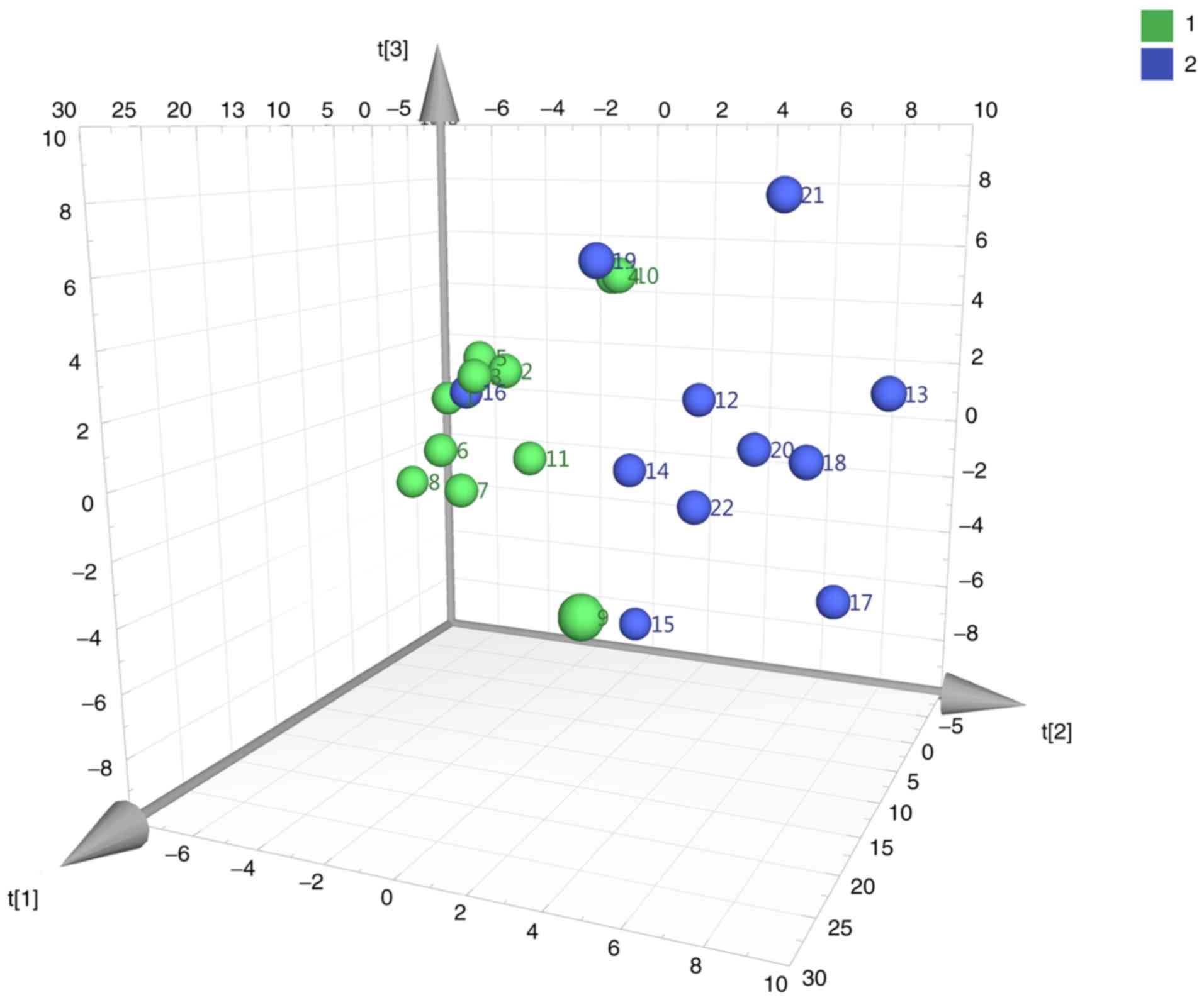
PCA
A total of 165 metabolites were detected. PCA was
performed to process the metabolite data based on a mean
center-scaling model, which is an unsupervised projection method
employed to visually display the intrinsic similarities and
differences in the dataset. As shown in Fig. 1, PCA (pre-gemcitabine normal vs.
pre-gemcitabine BCa tissues) revealed a well-differentiated and
clustered pattern in score plots, indicating the significant
metabolome changes between these two groups.
Altered metabolites and cancer-associated
metabolic pathways in BCa
UPLC-Q-Exactive analysis identified 34
differentially expressed metabolites annotated in the KEGG and HMDB
databases in pre-gemcitabine BCa tissues compared with
pre-gemcitabine adjacent normal tissues (Table II).
| Table IIList of the altered metabolites
identified in BCa and their changes in the comparison between BCa
tissues with or without gemcitabine pretreatment and
pre-gemcitabine normal tissues. |
Table II
List of the altered metabolites
identified in BCa and their changes in the comparison between BCa
tissues with or without gemcitabine pretreatment and
pre-gemcitabine normal tissues.
| Metabolites | KEGG | HMDB | Pre-gemcitabine BCa
vs. normal tissuesa | Post-gemcitabine
BCa vs. pre-gemcitabine normal tissuesa |
|---|
| Fold changea | P-value | Fold changea | P-value |
|---|
| Deoxycytidine | C00881 | HMDB0000014 | 4.03 | 1.22E-02 | 10.10 | 4.15E-05 |
|
5′-Methylthioadenosine | C00170 | HMDB0001173 | 15.16 | 2.38E-04 | 12.22 | 4.16E-04 |
| 3′-AMP | C01367 | HMDB0003540 | 4.95 | 3.02E-02 | 6.36 | 3.33E-02 |
|
Androstenedione | C00280 | HMDB0000053 | 0.16 | 4.09E-02 | 0.10 | 5.76E-03 |
|
Bilirubin | C00486 |
HMDB0000054 | 0.4 |
3.97E-02 | 1.26 |
2.04E-01 |
| Cholic acid | C00695 | HMDB0000619 | 0.29 | 3.14E-02 | 0.24 | 2.00E-03 |
| Cytidine | C00475 | HMDB0000089 | 3.83 | 2.43E-03 | 3.05 | 6.77E-03 |
|
5-Hydroxylysine | C16741 | HMDB0000450 | 0.64 | 4.40E-02 | 0.48 | 7.49E-03 |
| Deoxyinosine | C05512 | HMDB0000071 | 2.44 | 3.87E-02 | 4.12 | 1.70E-02 |
| Glucosamine
6-phosphate | C00352 | HMDB0001254 | 6.04 | 6.26E-03 | 6.87 | 3.02E-05 |
| Glyceraldehyde | C02154 | HMDB0001051 | 0.29 | 5.86E-04 | 0.32 | 1.65E-03 |
| Sphingosine | C00319 | HMDB0000252 | 2.03 | 3.30E-02 | 3.34 | 2.18E-05 |
|
Glycerophosphocholine | C00670 | HMDB0000086 | 56.88 | 1.93E-03 | 51.03 | 3.44E-03 |
| Glycine | C00037 | HMDB0000123 | 0.55 | 2.22E-02 | 0.47 | 4.68E-03 |
| Guanidine | C17349 | HMDB0001842 | 0.47 | 5.25E-03 | 0.01 | 3.79E-04 |
| Hexadecanedioic
acid | C19615 | HMDB0000672 | 0.27 | 1.34E-02 | 0.23 | 3.44E-03 |
| Histamine | C00388 | HMDB0000870 | 0.58 | 2.49E-02 | 0.35 | 7.49E-03 |
| Hypotaurine | C00519 | HMDB0000965 | 0.07 | 4.19E-04 | 0.08 | 5.33E-04 |
| Inosinic acid | C00130 | HMDB0000175 | 92.72 | 1.73E-05 | 150.57 | 1.43E-03 |
| L-Carnitine | C00318 | HMDB0000062 | 2.24 | 2.39E-03 | 2.70 | 3.97E-03 |
| L-Cystine | C00491 | HMDB0000192 | 0.49 | 2.70E-02 | 0.13 | 1.17E-03 |
|
L-Phenylalanine | C00079 | HMDB0000159 | 0.51 | 2.80E-02 | 0.31 | 4.70E-03 |
| N-Acetylneuraminic
acid | C19910 | HMDB0000230 | 2.68 | 2.13E-02 | 2.90 | 3.38E-02 |
| Oxidized
glutathione | C00127 | HMDB0003337 | 13.97 | 4.36E-03 | 16.05 | 2.77E-03 |
|
L-Palmitoylcarnitine | C02990 | HMDB0000222 | 3.59 | 1.88E-02 | 5.09 | 2.77E-04 |
| Pantothenol | C05944 | HMDB0004231 | 0.44 | 3.12E-02 | 0.18 | 8.44E-05 |
| Pyroglutamic
acid | C01879 | HMDB0000267 | 0.29 | 1.85E-03 | 0.16 | 5.30E-04 |
| Quinic acid | C06746 | HMDB0003072 | 0.09 | 4.05E-02 | 0.25 | 4.76E-02 |
| Retinal | C00376 |
HMDB0001358 | 0.13 |
4.98E-02 | 0.43 |
3.81E-01 |
| Rhamnose | C00507 | HMDB0000849 | 0.22 | 2.90E-03 | 0.17 | 2.40E-04 |
| Deoxycholic acid
glycine conjugate | C05464 | HMDB0000631 | 0.19 | 6.50E-03 | 0.15 | 4.52E-04 |
| Sorbitol | C00794 | HMDB0000247 | 0.29 | 9.19E-03 | 0.13 | 1.44E-03 |
| Tetradecanedioic
acid | C11002 | HMDB0000872 | 0.44 | 4.46E-02 | 0.36 | 4.47E-02 |
| Thiamine | C00378 | HMDB0000235 | 0.34 | 6.95E-05 | 0.18 | 2.47E-05 |
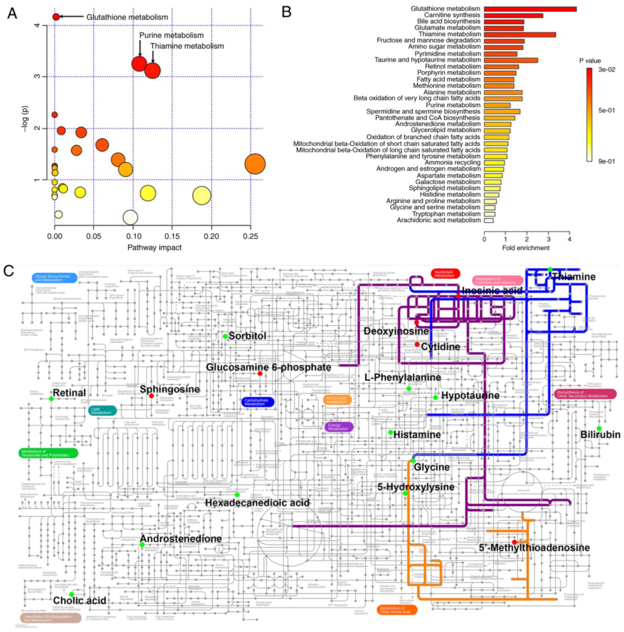
The 34 BCa-associated metabolites were then
submitted to MetaboAnalyst for analysis. Table II lists all metabolites found to
be altered in BCa. Table III
and Fig. 2A show the metabolic
pathways connected with these 34 metabolites, among which three
pathways, namely glutathione, purine and thiamine metabolism, were
significantly associated with BCa. Furthermore, in order to expand
our understanding of metabolic pathways related to BCa, the module
of enrichment analysis of MetaboAnalyst was used, which verified
that glutathione metabolism was significantly associated with BCa
(Table IV and Fig. 2B). The metabolic network of the
differential metabolites and altered metabolic pathways in the KEGG
general metabolic pathway map is shown in Fig. 2C.
| Table IIIPathway analysis of metabolite
changes in BCaa. |
Table III
Pathway analysis of metabolite
changes in BCaa.
| KEGG pathway | Total | Hits | P-value |
|---|
| Glutathione
metabolism | 38 | 3 | 1.55E-02 |
| Purine
metabolism | 92 | 4 | 3.86E-02 |
| Thiamine
metabolism | 24 | 2 | 4.40E-02 |
| Nitrogen
metabolism | 39 | 2 | 1.04E-01 |
| Primary bile acid
biosynthesis | 47 | 2 | 1.41E-01 |
| Fructose and
mannose metabolism | 48 | 2 | 1.46E-01 |
| Cysteine and
methionine metabolism | 56 | 2 | 1.86E-01 |
| Cyanoamino acid
metabolism | 16 | 1 | 2.04E-01 |
| Pyrimidine
metabolism | 60 | 2 | 2.07E-01 |
| Taurine and
hypotaurine metabolism | 20 | 1 | 2.48E-01 |
| Retinol
metabolism | 22 | 1 | 2.70E-01 |
| Ether lipid
metabolism | 23 | 1 | 2.80E-01 |
| Aminoacyl-tRNA
biosynthesis | 75 | 2 | 2.86E-01 |
| Alanine, aspartate
and glutamate metabolism | 24 | 1 | 2.90E-01 |
| Sphingolipid
metabolism | 25 | 1 | 3.01E-01 |
| Pantothenate and
CoA biosynthesis | 27 | 1 | 3.20E-01 |
| Phenylalanine,
tyrosine and tryptophan biosynthesis | 27 | 1 | 3.20E-01 |
| Methane
metabolism | 34 | 1 | 3.86E-01 |
| Glycerophospholipid
metabolism | 39 | 1 | 4.28E-01 |
| Porphyrin and
chlorophyll metabolism | 104 | 2 | 4.36E-01 |
| Galactose
metabolism | 41 | 1 | 4.45E-01 |
| Histidine
metabolism | 44 | 1 | 4.68E-01 |
| Phenylalanine
metabolism | 45 | 1 | 4.76E-01 |
| Lysine
degradation | 47 | 1 | 4.91E-01 |
| Glycine, serine and
threonine metabolism | 48 | 1 | 4.98E-01 |
| Fatty acid
metabolism | 50 | 1 | 5.13E-01 |
| Amino sugar and
nucleotide sugar metabolism | 88 | 1 | 7.21E-01 |
| Steroid hormone
biosynthesis | 99 | 1 | 7.63E-01 |
| Table IVEnrichment analysis of metabolite
changes in BCaa. |
Table IV
Enrichment analysis of metabolite
changes in BCaa.
| Pathway from
SMPDB | Total | Hits | P-value |
|---|
| Glutathione
metabolism | 21 | 3 | 2.96E-02 |
| Carnitine
synthesis | 22 | 2 | 1.64E-01 |
| Bile acid
biosynthesis | 65 | 4 | 1.64E-01 |
| Glutamate
metabolism | 49 | 3 | 2.19E-01 |
| Thiamine
metabolism | 9 | 1 | 2.63E-01 |
| Fructose and
mannose degradation | 32 | 2 | 2.88E-01 |
| Amino Sugar
metabolism | 33 | 2 | 3.00E-01 |
| Pyrimidine
metabolism | 59 | 3 | 3.11E-01 |
| Taurine and
hypotaurine metabolism | 12 | 1 | 3.35E-01 |
| Retinol
metabolism | 37 | 2 | 3.50E-01 |
| Porphyrin
metabolism | 40 | 2 | 3.87E-01 |
| Fatty acid
metabolism | 43 | 2 | 4.23E-01 |
| Methionine
metabolism | 43 | 2 | 4.23E-01 |
| Alanine
metabolism | 17 | 1 | 4.39E-01 |
| Beta oxidation of
very long-chain fatty acids | 17 | 1 | 4.39E-01 |
| Purine
metabolism | 74 | 3 | 4.51E-01 |
| Spermidine and
spermine biosynthesis | 18 | 1 | 4.58E-01 |
| Pantothenate and
CoA biosynthesis | 21 | 1 | 5.11E-01 |
| Androstenedione
metabolism | 24 | 1 | 5.59E-01 |
| Glycerolipid
metabolism | 25 | 1 | 5.74E-01 |
| Oxidation of
branched chain fatty acids | 26 | 1 | 5.89E-01 |
| Mitochondrial
beta-oxidation of short-chain saturated fatty acids | 27 | 1 | 6.03E-01 |
| Mitochondrial
beta-oxidation of long-chain saturated fatty acids | 28 | 1 | 6.16E-01 |
| Phenylalanine and
tyrosine metabolism | 28 | 1 | 6.16E-01 |
| Ammonia
recycling | 32 | 1 | 6.66E-01 |
| Androgen and
estrogen metabolism | 33 | 1 | 6.78E-01 |
| Aspartate
metabolism | 35 | 1 | 6.99E-01 |
| Galactose
metabolism | 38 | 1 | 7.29E-01 |
| Sphingolipid
metabolism | 40 | 1 | 7.48E-01 |
| Histidine
metabolism | 43 | 1 | 7.73E-01 |
| Arginine and
proline metabolism | 53 | 1 | 8.41E-01 |
| Glycine and serine
metabolism | 59 | 1 | 8.72E-01 |
| Tryptophan
metabolism | 60 | 1 | 8.76E-01 |
| Arachidonic acid
metabolism | 69 | 1 | 9.10E-01 |
Candidate targets of submucosal injection
of gemcitabine
Based on targeted metabolomic analysis, we next
analyzed these 34 differential metabolite changes in
post-gemcitabine BCa tissues, and compared their levels with those
in pre-gemcitabine normal tissues. A total of 32 metabolites
maintained significant changes with identical trends in the
comparison between pre-gemcitabine normal vs. pre-gemcitabine BCa
tissues, indicating that the findings for differential metabolites
in BCa are reliable. Importantly, the significant decrease in the
levels of two metabolites associated with BCa recovered to
insignificant levels following submucosal injection of gemcitabine
(Table II and Fig. 3A, B and E-F). These were bilirubin
and retinal, which may be the candidate targets of gemcitabine for
the prevention of recurrence of urothelial BCa. We further analyzed
the changes in the two metabolites in association with cancer
stage. In Ta/T1 stage disease, when comparing pre-gemcitabine
normal vs. pre-gemcitabine BCa tissues, bilirubin was decreased
significantly in the BCa tissues (P=4.15E-2) (Table SI and Fig. 3C), while this decrease recovered
to an insignificant level following submucosal injection of
gemcitabine (P=5.40E-1) (Table SI and Fig. 3D); however, at this stage, the
decrease of retinal in the tumor was still not statistically
significant (P=4.76E-1) (Table SI). In T2 stage disease, when
comparing pre-gemcitabine normal vs. pre-gemcitabine BCa tissues,
retinal was decreased significantly in the BCa tissues (P=4.98E-2)
(Table SI and Fig. 3G), while its
change became statistically insignificant following submucosal
injection of gemcitabine (P=3.22E-1) (Table SI and Fig. 3H); however, at this stage, the
decrease of retinal in tumor was not statistically significant
(P=1.68E-1) (Table SI). These results indicate that bilirubin and
retinal changes were correlated with cancer stage, and gemcitabine
may exert its effect through metabolic pathways associated with
cancer stage.
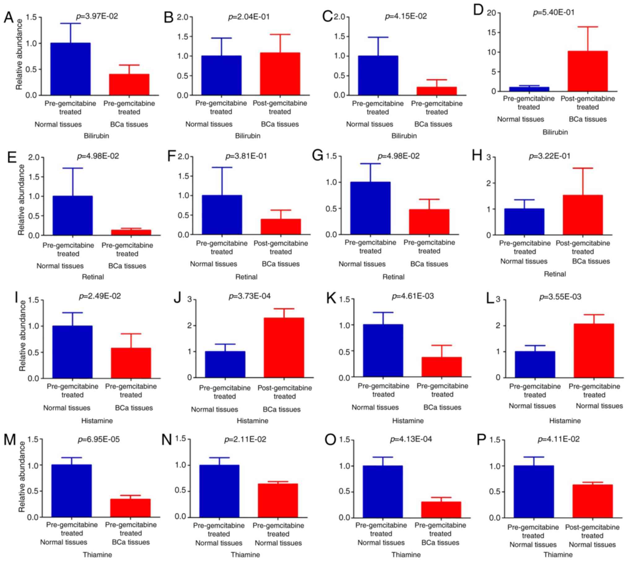 | Figure 3Relative levels of potential
candidate targets of submucosal injection of gemcitabine (A, B, E,
F, I, J, M and N). All samples; (C and D) Ta/T1 stage; (G, H, K, L,
O and P) T2 stage. Values are presented as mean ± standard error of
the mean. The P-values of paired t-test are indicated. |
Effects of submucosal injection of
gemcitabine on normal tissues
To identify the effects of submucosal injection of
gemcitabine on normal bladder tissues, the metabolomes of normal
tissues were compared pre- and post-gemcitabine treatment. A total
of 10 metabolites were found to be significantly altered in the
normal tissues following submucosal injection of gemcitabine,
whereas only 2 metabolites were associated with BCa (Table V). Histamine was significantly
decreased in BCa, and its level was significantly increased in
normal tissues after submucosal injection of gemcitabine (Fig. 3I and J). However, thiamine was
significantly decreased in BCa, and its level was significantly
decreased in normal tissues after submucosal injection of
gemcitabine (Fig. 3M and N). We
further analyzed the changes in the two metabolite in relation to
cancer stage. In Ta/T1 stage disease, when comparing
pre-gemcitabine normal vs. pre-gemcitabine BCa tissues, the changes
in histamine (P=2.60E-1) and thiamine (P=8.39E-2) in the tumor were
insignificant (Table SI). However, in T2 stage disease, when
comparing pre-gemcitabine normal vs. pre-gemcitabine BCa tissues,
the decrease in histamine (P=4.61E-3) (Table SI and Fig. 3K) and thiamine (P=4.13E-4) (Table
SI and Fig. 3O) in the BCa
tissues was significant. In T2 stage disease, the histamine level
was significantly increased in normal tissues after submucosal
injection of gemcitabine (P=3.55E-3) (Table SI and Fig. 3L), while thiamine was
significantly decreased (P=4.11E-2) (Table SI and Fig. 3P). These results indicate that
these metabolite changes were correlated with cancer stage, and
gemcitabine may exert its effects mainly through these metabolic
pathways in T2 stage disease.
| Table VChanges in normal tissues after
gemcitabine treatment. |
Table V
Changes in normal tissues after
gemcitabine treatment.
| Metabolites | Pre-gemcitabine BCa
vs. normal tissuesa | Post-gemcitabine
vs. pre-gemcitabine normal tissuesa |
|---|
| Fold-changea | P-value | Fold-changea | P-value |
|---|
|
3-Methyladenine | 1.54 | 3.78E-01 | 0.17 | 2.33E-02 |
| Ascorbic acid | 22.26 | 1.85E-01 | 0.28 | 5.46E-03 |
| Creatinine | 0.48 | 3.44E-01 | 0.13 | 1.16E-02 |
| D-Glyceraldehyde
3-phosphate | 0.72 | 3.33E-01 | 0.15 | 2.16E-02 |
| Histamine | 0.58 | 2.49E-02 | 2.26 | 3.73E-04 |
| N-Acetylglutamic
acid | 0.52 | 8.58E-02 | 0.57 | 4.95E-02 |
|
N-Acetylglutamine | 0.68 | 5.45E-01 | 0.11 | 4.25E-02 |
| Thiamine | 0.34 | 6.95E-05 | 0.65 | 2.11E-02 |
| Tryptamine | 0.10 | 7.66E-02 | 0.11 | 3.23E-02 |
| Uridine | 1.41 | 6.74E-02 | 1.51 | 3.65E-02 |
Discussion
In the present study, UPLC-Q-Exactive-based
metabolomic analysis was utilized to profile metabolites in BCa
tissues. The major findings may be summarized as follows: i) A
total of 34 key metabolites associated with BCa were identified
(Table II); ii) three metabolic
pathways, namely glutathione, purine and thiamine metabolism, were
altered in BCa, and glutathione metabolism was the consistently
altered pathway in enrichment and pathway analyses (Tables III and IV, and Fig. 2A and B); iii) among the 34
cancer-associated metabolites, the levels of bilirubin and retinal
recovered after gemcitabine injection, suggesting that these two
are likely the targets of gemcitabine treatment (Table II, Fig. 3A-D and E-H); iv) the effects of
gemcitabine on normal bladder tissues were also investigated, and
it was deduced that histamine may have the ability to protect
against disease recurrence, whereas thiamine may be involved in the
side effects of treatment (Table
V, Fig. 3I-L and M-P), which
requires further confirmation in future studies.
The identification of the three metabolic pathways
significantly altered in BCa may be pathophysiologically important.
Glycine is an important amino acid that participates in all three
metabolic pathways. A decrease in glycine was reported as a
biomarker in BCa (14). As a
precursor of purine synthesis, reduced glycine in BCa indicates
that a critical metabolic process associated with cell
proliferation is altered in BCa (14). Notably, glutathione metabolism is
the consistently altered pathway in enrichment and pathway
analysis. Glutathione is the most abundant low-molecular-weight
peptide present in eukaryotic cells (28). Glutathione is a primary cellular
antioxidant that effectively scavenges free radicals and other
reactive oxygen species (29)
and, therefore, plays an important role in protecting cells from
oxidative injury (30).
Glutathione is also involved in cellular detoxification, and is
required in several aspects of the immune response (31). Ke et al reported altered
oxidized glutathione in BCa; they discovered four single-nucleotide
polymorphisms in the glutathione synthetase gene, and these changes
were associated with BCa recurrence after TUR and Bacillus Calmette
Guerin treatment (31). Our
findings, together with others, suggest that oxidative stress in
BCa cells is at least partially due to the disrupted glutathione
metabolism.
By using LC-Q-Exactive MS, this study is, to the
best of our knowledge, the first to investigate the metabolite
changes in BCa treated with gemcitabine followed by TURBT. Among
the 34 cancer-associated metabolites, the levels of bilirubin and
retinal recovered after gemcitabine treatment, indicating that they
may be the potential targets of gemcitabine for reducing BCa
recurrence. Bilirubin, a degradation product of free heme groups,
protected LLC-PK1 cells against cisplatin-induced death (32). A large population-based study
demonstrated that patients with primary biliary cirrhosis (PBC)
have a nine-fold increased risk of developing urinary BCa, and BCa
and PBC share a number of etiological factors (33). Uridine
5'-diphospho-glucuronosyltransferases (UGTs) are enzymes that
participate in several biological processes involving bilirubin
conjugation. UGTs catabolize carcinogens and, therefore, protect
bladder cells from the harmful effects of toxic chemicals
accumulated in the bladder. Targeting UGT1A may serve as a novel
therapeutic intervention against uroepithelial carcinomas (34). Retinal (retinaldehyde; Rald) is
derived from retinol (vitamin A) oxidized by alcohol dehydrogenases
(35). Retinal plays an essential
role in molecular signaling in vision, and serves mainly as a
retinoic acid (an active form of vitamin A) precursor outside the
eye (36). The serum levels of
vitamin A were found to be decreased in patients with BCa (37). A high dietary intake of vitamin A
reduces the incidence of BCa (38). Ziouzenkova et al reported
retinal as a distinct biological regulator involved in suppressing
adipo-genesis, diet-induced obesity and insulin resistance
(39). The potential effect of
bilirubin and retinal on the clinical outcome of patients with BCa
identified in the present study is worthy of further investigation
in the future.
The effects of gemcitabine on the metabolism of
adjacent normal tissues were also examined. Most
gemcitabine-induced metabolites do not overlap with those
identified in BCa, indicating that gemcitabine did not exert
notable adverse effects on normal bladder tissues. It was observed
that histamine change may be associated with the prevention of
relapse. Histamine is derived from the decarboxylation of histidine
by histidine decarboxylase in mammals (8). Histamine is primarily released in
inflammatory processes by mast cells (8), which are closely associated with BCa
(40). Histamine H1 receptor
(HRH1) expression was identified in BCa and found to be associated
with the prognosis (41). It was
observed that thiamine change may be involved in treatment-related
side effects. Thiamine, or vitamin B-1, is a water-soluble vitamin
(42). An early report by Pamukcu
et al demonstrated that the incidence of urinary bladder
carcinomas in rats fed bracken fern and additionally s.c. injected
once weekly with 2 mg of thiamine hydrochloride was significantly
higher compared with that in rats fed bracken fern but receiving no
thiamine supplements, as thiamine may interfere with the
absorption, distribution, metabolism, or excretion of the bracken
fern (43). However, a
case-control study from New Hampshire investigated the effect of
minerals and vitamins on the risk of BCa, and found that a higher
total intake of thiamine was inversely correlated with BCa risk in
older participants (44).
There is currently a lack of effective biomarkers
for BCa diagnosis and prognosis. Metabolomic profiles from tissue
have the potential to be used, along with other current
diagnostics, to help guide the clinical management of patients with
BCa. The changed metabolites identified in the present study, such
as glycine, may be used as potential biomarkers for BCa. In
addition, those metabolites discovered in BC gemcitabine treatment
may reveal new metabolic pathways that mediate the anti-recurrence
effect of gemcitabine, which currently remain elusive.
In summary, the present study employed
UPLC-HRMS-based metabolomic analysis to investigate metabolite
changes in bladder tissues from BCa and BCa treated with
gemcitabine. The findings may provide new insights into metabolic
changes in BCa and the biomolecular basis of submucosal injection
of gemcitabine for BCa. In addition, the study demonstrated that
the UPLC-HRMS-based metabolomic analysis provides comprehensive
metabolite profiling data that may pave the way to a novel approach
to BCa research.
Supplementary Data
Acknowledgments
Not applicable.
Abbreviations:
|
TURBT
|
transurethral resection of bladder
tumor
|
|
PCA
|
principal component analysis
|
|
ACN
|
acetonitrile
|
|
KEGG
|
Kyoto Encyclopedia of Genes and
Genomes
|
|
HMDB
|
human metabolome database
|
|
SMPDB
|
Small Molecule Pathway Database
|
|
HPLC/MS
|
high-performance liquid
chromatography/ mass spectrometry
|
|
UPLC-MS
|
ultra-performance LC-MS
|
|
UPLC-TOF-MS
|
UPLC time-of-flight MS
|
|
UPLC-HRMS
|
UPLC high-resolution MS
|
|
BCa
|
bladder cancer
|
Funding
The present study was supported by grants from the
National Natural Science Foundation (81872650 and 81573182); the
Natural Science Foundation of the Jiangsu Higher Education
Institutions of China (18KJA320003 and 18KJB320001); the Key
Research & Development Plan of Jiangsu Province (BE2017628);
the Southeast University & Nanjing Medical University
Collaborative Research Project (2242018K3DN25); the Priority
Academic Program Development of Jiangsu Higher Education
Institutions (PAPD); the 333 Project of Jiangsu Province
(BRA2017241); the Natural Science Foundation of Huai'an
(HAB201801); the Huai'an Promotion Project for Science and
Technology International Cooperation (HAC201708); and the
Innovation Fund Project of the State Key Laboratory of Reproductive
Medicine (SKLRM-GC201901).
Availability of data and materials
The datasets generated and/or analyzed during the
present study are available from the corresponding author on
reasonable request.
Authors' contributions
CY, XS, KW, YG, RS and MC designed and performed the
experiments. TL and MC analyzed and interpreted the raw data. SZ,
XJ, BZ and HW collected the bladder tissue samples. ZG and SS
performed the histopathological examination of BCa tissues. JL and
JT preserved the samples and collected the basic information of the
participants. CY, TL and MC wrote the manuscript. XW, HJ, XN, XW,
MC and GF critically revised the manuscript for important
intellectual content. MC and GF supervised the project. All authors
have read and approved the final version of the manuscript for
publication.
Ethics approval and consent to
participate
The Ethics Committee of The Affiliated Huai'an No. 1
People's Hospital of Nanjing Medical University reviewed and
approved the study protocol (serial no. YL-P-2013-21-01).
Patient consent for publication
Not applicable.
Competing interests
The authors declare that they have no competing
interests.
Authors' information
Chao Yang: 15949177271@163.com; Xian Sun:
1019949991@qq.com; Hengbing
Wang: wanghengbing2004@163.com;
Ting Lu: 552095014@qq.com;
Keqing Wu: wkwly1993@qq.com;
Yusheng Guan: guanys@njmu.edu.cn; Jing Tang:
tjing19681222@sina.com;
Jian Liang: lj3936@126.com;
Rongli Sun: 101012172@seu.edu.cn; Zhongying
Guo: guozhongying407@163.com;
Sinian Zheng: zhengsinian@163.com; Xiaoli Wu:
wuxiaoli2233@126.com;
Hesong Jiang: njjhs2007@163.com; Xi Jiang:
piscesjxi@163.com; Bing
Zhong: 15152569186@163.com; Xiaobing Niu:
bingke2008@sina.com; Suan
Sun: hayyssa@163.com; Xinru
Wang: xrwang@njmu.edu.cn
References
|
1
|
Ferlay J, Soerjomataram I, Dikshit R, Eser
S, Mathers C, Rebelo M, Parkin DM, Forman D and Bray F: Cancer
incidence and mortality worldwide: Sources methods and major
patterns in GLOBOCAN 2012. Int J Cancer. 136:E359–E386. 2015.
View Article : Google Scholar
|
|
2
|
Messing EM, Madeb R, Young T, Gilchrist
KW, Bram L, Greenberg EB, Wegenke JD, Stephenson L, Gee J and Feng
C: Long-term outcome of hematuria home screening for bladder cancer
in men. Cancer. 107:2173–2179. 2006. View Article : Google Scholar : PubMed/NCBI
|
|
3
|
Messing EM, Tangen CM, Lerner SP,
Sahasrabudhe DM, Koppie TM, Wood DP Jr, Mack PC, Svatek RS, Evans
CP, Hafez KS, et al: Effect of intravesical instillation of
gemcitabine vs saline immediately following resection of suspected
low-grade non-muscle-invasive bladder cancer on tumor recurrence:
SWOG S0337 randomized clinical trial. JAMA. 319:1880–1888. 2018.
View Article : Google Scholar : PubMed/NCBI
|
|
4
|
Chen X, Wang B, Tian HZ and Gao JZ:
Submucosal injection of anti-tumor drug on the prevention of
Post-TUR-Bt recurrence. Zhonghua Wai Ke Za Zhi. 42:580–582. 2004.In
Chinese. PubMed/NCBI
|
|
5
|
Caffo O, Thompson C, De Santis M, Kragelj
B, Hamstra DA, Azria D, Fellin G, Pappagallo GL, Galligioni E and
Choudhury A: Concurrent gemcitabine and radiotherapy for the
treatment of muscle-invasive bladder cancer: A pooled individual
data analysis of eight phase I-II trials. Radiother Oncol.
121:193–198. 2016. View Article : Google Scholar : PubMed/NCBI
|
|
6
|
Sternberg CN, Bellmunt J, Sonpavde G,
Siefker-Radtke AO, Stadler WM, Bajorin DF, Dreicer R, George DJ,
Milowsky MI, Theodorescu D, et al: ICUD-EAU international
consultation on bladder cancer 2012: Chemotherapy for urothelial
carcinoma-neoadjuvant and adjuvant settings. Eur Urol. 63:58–66.
2013. View Article : Google Scholar
|
|
7
|
Van QN and Veenstra TD: How close is the
bench to the bedside? Metabolic profiling in cancer research.
Genome Med. 1:52009. View
Article : Google Scholar : PubMed/NCBI
|
|
8
|
Bansal N, Gupta A, Mitash N, Shakya PS,
Mandhani A, Mahdi AA, Sankhwar SN and Mandal SK: Low- and
high-grade bladder cancer determination via human serum-based
metabolo-mics approach. J Proteome Res. 12:5839–5850. 2013.
View Article : Google Scholar : PubMed/NCBI
|
|
9
|
Jin X, Yun SJ, Jeong P, Kim IY, Kim WJ and
Park S: Diagnosis of bladder cancer and prediction of survival by
urinary metabo-lomics. Oncotarget. 5:1635–1645. 2014. View Article : Google Scholar : PubMed/NCBI
|
|
10
|
Issaq HJ, Nativ O, Waybright T, Luke B,
Veenstra TD, Issaq EJ, Kravstov A and Mullerad M: Detection of
bladder cancer in human urine by metabolomic profiling using high
performance liquid chromatography/mass spectrometry. J Urol.
179:2422–2426. 2008. View Article : Google Scholar : PubMed/NCBI
|
|
11
|
Pasikanti KK, Esuvaranathan K, Hong Y, Ho
PC, Mahendran R, Raman Nee Mani L, Chiong E and Chan EC: Urinary
metabotyping of bladder cancer using two-dimensional gas
chromatography time-of-flight mass spectrometry. J Proteome Res.
12:3865–3873. 2013. View Article : Google Scholar : PubMed/NCBI
|
|
12
|
Liu X, Cheng X, Liu X, He L, Zhang W, Wang
Y, Sun W and Ji Z: Investigation of the urinary metabolic
variations and the application in bladder cancer biomarker
discovery. Int J Cancer. 143:408–418. 2018. View Article : Google Scholar : PubMed/NCBI
|
|
13
|
Alberice JV, Amaral AF, Armitage EG,
Lorente JA, Algaba F, Carrilho E, Márquez M, García A, Malats N and
Barbas C: Searching for urine biomarkers of bladder cancer
recurrence using a liquid chromatography-mass spectrometry and
capillary electrophoresis-mass spectrometry metabolomics approach.
J Chromatogr A. 1318:163–170. 2013. View Article : Google Scholar : PubMed/NCBI
|
|
14
|
Zhou Y, Song R, Ma C, Zhou L, Liu X, Yin
P, Zhang Z, Sun Y, Xu C, Lu X and Xu G: Discovery and validation of
potential urinary biomarkers for bladder cancer diagnosis using a
pseudotargeted GC-MS metabolomics method. Oncotarget.
8:20719–20728. 2017.PubMed/NCBI
|
|
15
|
Shao CH, Chen CL, Lin JY, Chen CJ, Fu SH,
Chen YT, Chang YS, Yu JS, Tsui KH, Juo CG and Wu KP: Metabolite
marker discovery for the detection of bladder cancer by comparative
metabolomics. Oncotarget. 8:38802–38810. 2017. View Article : Google Scholar : PubMed/NCBI
|
|
16
|
von Rundstedt FC, Rajapakshe K, Ma J,
Arnold JM, Gohlke J, Putluri V, Krishnapuram R, Piyarathna DB,
Lotan Y, Gödde D, et al: Integrative pathway analysis of metabolic
signature in bladder cancer: A linkage to the cancer genome atlas
project and prediction of survival. J Urol. 195:1911–1919. 2016.
View Article : Google Scholar : PubMed/NCBI
|
|
17
|
Putluri N, Shojaie A, Vasu VT, Vareed SK,
Nalluri S, Putluri V, Thangjam GS, Panzitt K, Tallman CT, Butler C,
et al: Metabolomic profiling reveals potential markers and
biopro-cesses altered in bladder cancer progression. Cancer Res.
71:7376–7386. 2011. View Article : Google Scholar : PubMed/NCBI
|
|
18
|
Cheng Y, Yang X, Deng X, Zhang X, Li P,
Tao J, Qin C, Wei J and Lu Q: Metabolomics in bladder cancer: A
systematic review. Int J Clin Exp Med. 8:11052–11063.
2015.PubMed/NCBI
|
|
19
|
Eidelman E, Tripathi H, Fu DX and Siddiqui
MM: Linking cellular metabolism and metabolomics to
risk-stratification of prostate cancer clinical aggressiveness and
potential therapeutic pathways. Transl Androl Urol. 7(Suppl 4):
S490–S497. 2018. View Article : Google Scholar : PubMed/NCBI
|
|
20
|
Lima AR, Bastos Mde L, Carvalho M and
Guedes de Pinho P: Biomarker discovery in human prostate cancer: An
update in metabolomics studies. Transl Oncol. 9:357–370. 2016.
View Article : Google Scholar : PubMed/NCBI
|
|
21
|
Epstein JI, Amin MB, Reuter VR and Mostofi
FK: The world health organization/international society of
urological pathology consensus classification of urothelial
(transitional cell) neoplasms of the urinary bladder. Bladder
consensus conference committee Am J Surg Pathol. 22:1435–1448.
1998.
|
|
22
|
Sreekumar A, Poisson LM, Rajendiran TM,
Khan AP, Cao Q, Yu J, Laxman B, Mehra R, Lonigro RJ, Li Y, et al:
Metabolomic profiles delineate potential role for sarcosine in
prostate cancer progression. Nature. 457:910–914. 2009. View Article : Google Scholar : PubMed/NCBI
|
|
23
|
Hu W, Dong T, Wang L, Guan Q, Song L, Chen
D, Zhou Z, Chen M, Xia Y and Wang X: Obesity aggravates toxic
effect of BPA on spermatogenesis. Environ Int. 105:56–65. 2017.
View Article : Google Scholar : PubMed/NCBI
|
|
24
|
Christen S, Lorendeau D, Schmieder R,
Broekaert D, Metzger K, Veys K, Elia I, Buescher JM, Orth MF,
Davidson SM, et al: Breast cancer-derived lung metastases show
increased pyruvate carboxylase-dependent anaplerosis. Cell Rep.
17:837–848. 2016. View Article : Google Scholar : PubMed/NCBI
|
|
25
|
Davidson SM, Papagiannakopoulos T,
Olenchock BA, Heyman JE, Keibler MA, Luengo A, Bauer MR, Jha AK,
O'Brien JP, Pierce KA, et al: Environment impacts the metabolic
dependencies of Ras-driven non-small cell lung cancer. Cell Metab.
23:517–528. 2016. View Article : Google Scholar : PubMed/NCBI
|
|
26
|
Frolkis A, Knox C, Lim E, Jewison T, Law
V, Hau DD, Liu P, Gautam B, Ly S, Guo AC, et al: SMPDB: The small
molecule pathway database. Nucleic Acids Res. 38:Database Issue.
D480–D487. 2010. View Article : Google Scholar :
|
|
27
|
He YT, Li DJ, Liang D, Zheng RS, Zhang SW,
Zeng HM, Chen WQ and He J: Incidence and mortality of bladder
cancer in China, 2014. Zhonghua Zhong Liu Za Zhi. 40:647–652.
2018.In Chinese. PubMed/NCBI
|
|
28
|
Estrela JM, Ortega A and Obrador E:
Glutathione in cancer biology and therapy. Crit Rev Clin Lab Sci.
43:143–181. 2006. View Article : Google Scholar : PubMed/NCBI
|
|
29
|
Balendiran GK, Dabur R and Fraser D: The
role of glutathione in cancer. Cell Biochem Funct. 22:343–352.
2004. View Article : Google Scholar : PubMed/NCBI
|
|
30
|
Amelio I, Cutruzzolá F, Antonov A,
Agostini M and Melino G: Serine and glycine metabolism in cancer.
Trends Biochem Sci. 39:191–198. 2014. View Article : Google Scholar : PubMed/NCBI
|
|
31
|
Ke HL, Lin J, Ye Y, Wu WJ, Lin HH, Wei H,
Huang M, Chang DW, Dinney CP and Wu X: Genetic variations in
glutathione pathway genes predict cancer recurrence in patients
treated with transurethral resection and bacillus calmetteguerin
instillation for non-muscle invasive bladder cancer. Ann Surg
Oncol. 22:4104–4110. 2015. View Article : Google Scholar : PubMed/NCBI
|
|
32
|
Patricia Moreno-Londoño A, Bello-Alvarez C
and Pedraza- Chaverri J: Isoliquiritigenin pretreatment attenuates
cisplatin induced proximal tubular cells (LLC-PK1) death and
enhances the toxicity induced by this drug in bladder cancer T24
cell line. Food Chem Toxicol. 109:143–154. 2017. View Article : Google Scholar
|
|
33
|
Boonstra K, Bokelaar R, Stadhouders PH,
Tuynman HA, Poen AC, van Nieuwkerk KM, Witteman EM, Hamann D,
Witteman BJ, Beuers U and Ponsioen CY: Increased cancer risk in a
large population-based cohort of patients with primary biliary
cirrhosis: Follow-up for up to 36 years. Hepatol Int. 8:266–274.
2014.PubMed/NCBI
|
|
34
|
Sundararaghavan VL, Sindhwani P and Hinds
TD Jr: Glucuronidation and UGT isozymes in bladder: New targets for
the treatment of uroepithelial carcinomas? Oncotarget. 8:3640–3648.
2017. View Article : Google Scholar :
|
|
35
|
Duester G, Mic FA and Molotkov A:
Cytosolic retinoid dehydro-genases govern ubiquitous metabolism of
retinol to retinaldehyde followed by tissue-specific metabolism to
retinoic acid. Chem Biol Interact. 143-144:201–210. 2003.
View Article : Google Scholar : PubMed/NCBI
|
|
36
|
Napoli JL: Retinoic acid: Its biosynthesis
and metabolism. Prog Nucleic Acid Res Mol Biol. 63:139–188. 1999.
View Article : Google Scholar : PubMed/NCBI
|
|
37
|
Mahmoud LA and Robinson WA: Vitamin A
levels in human bladder cancer. Int J Cancer. 30:143–145. 1982.
View Article : Google Scholar : PubMed/NCBI
|
|
38
|
Mettlin C and Graham S: Dietary risk
factors in human bladder cancer. Am J Epidemiol. 110:255–263. 1979.
View Article : Google Scholar : PubMed/NCBI
|
|
39
|
Ziouzenkova O, Orasanu G, Sharlach M,
Akiyama TE, Berger JP, Viereck J, Hamilton JA, Tang G, Dolnikowski
GG, Vogel S, et al: Retinaldehyde represses adipogenesis and
diet-induced obesity. Nat Med. 13:695–702. 2007. View Article : Google Scholar : PubMed/NCBI
|
|
40
|
Serel TA, Soyupek S and Çandir Ö:
Association between mast cells and bladder carcinoma. Urol Int.
72:299–302. 2004. View Article : Google Scholar : PubMed/NCBI
|
|
41
|
Wang M, Wei X, Shi L, Chen B, Zhao G and
Yang H: Integrative genomic analyses of the histamine H1 receptor
and its role in cancer prediction. Int J Mol Med. 33:1019–1026.
2014. View Article : Google Scholar : PubMed/NCBI
|
|
42
|
Kerns JC and Gutierrez JL: Thiamin Adv
Nutr. 8:395–397. 2017. View Article : Google Scholar
|
|
43
|
Pamukcu AM, Yalciner S, Price JM and Bryan
GT: Effects of the coadministration of thiamine on the incidence of
urinary bladder carcinomas in rats fed bracken fern. Cancer Res.
30:2671–2674. 1970.PubMed/NCBI
|
|
44
|
Brinkman MT, Karagas MR, Zens MS, Schned
A, Reulen RC and Zeegers MP: Minerals and vitamins and the risk of
bladder cancer: Results from the new hampshire study. Cancer Causes
Control. 21:609–619. 2010. View Article : Google Scholar : PubMed/NCBI
|