Introduction
Liver cancer is a serious pathology with a global
impact (1), and liver
cancer-associated morbidity ranks third worldwide, after lung and
gastric cancer (1,2). In China, the morbidity rate of liver
cancer exceeds that of gastric cancer, and it ranks second among
all cancer-related deaths (3-5).
There is currently little indication that this condition will
improve in the future, and the number of liver cancer-associated
deaths is predicted to increase (5). As liver cancer exhibits no obvious
or specific early stage symptoms, the majority of patients are
diagnosed in the advanced stages of disease when metastasis has
already occurred, resulting in poor prognosis and a high mortality
rate (1,6,7).
Despite the emergence of tumor immunotherapy
(8,9) and molecular targeted drug therapy
(10,11), due to low efficiency and side
effects, these methods are not widely used in clinical practice for
the treatment of liver cancer. At present, the first-line treatment
for early liver cancer is surgical resection with adjuvant
radiotherapy and chemotherapy (6,12),
while patients with advanced disease and metastasis often miss the
opportunity to benefit from surgery (13). Additionally, the recurrence of
liver cancer is associated with metastasis (13,14). Previously, it was believed that
tumor metastasis was the result of tumor cell infiltration from the
tumor boundary when the tumor had exceeded a certain volume
(14). However, it has been
confirmed that metastasis occurs at various disease stages and is
closely associated with the tumor microenvironment, including
immune, metabolic and inflammatory reactions (15-17). The process of tumor metastasis
involves numerous factors, and there are currently no available
treatments that collectively target these factors. Therefore, the
development of effective therapeutic agents is critical.
The stromal cell-derived factor 1 (SDF1)/C-X-C
chemo-kine receptor type 4 (CXCR4) pathway is implicated in liver
cancer metastasis (18,19), and includes the elevated
expression of CXCR4. The most common metastatic regions in patients
with liver cancer are the lungs and bone marrow, which are both
abundant in SDF1 (19-21). Mechanistically, the overexpression
of CXCR4 was found to promote the migration and invasion of liver
cancer cells due to the increased chemotaxis to SDF1 and activation
of downstream signaling pathways. Conversely, silencing CXCR4
reduced the metastatic ability of liver cancer cells (22-24). Xia et al (25) demonstrated that the natural
extract hesperidin not only induced apoptosis, but also decreased
the expression of CXCR4 by regulating the activation of the
PI3K/Akt signaling pathway in lung cancer cells (26). Overwhelming evidence has suggested
that natural extracts can significantly inhibit the migration and
invasion of liver cancer cells by regulating CXCR4 expression
(27-29); this suggest that these natural
extracts may have potential for preventing liver cancer metastasis
by blocking the SDF1/CXCR4 pathway.
Cordycepin, extracted from Cordyceps
militaris, has anti-inflammatory, antioxidant and anticancerous
properties (30-32). Previous studies have shown that
cordycepin inhibits the cell cycle and induces apoptosis in
non-small cell lung cancer by activating AMP-activated protein
kinase signaling, without affecting the proliferation of normal
lung epithelial cells (33).
Additionally, cordycepin significantly inhibited the proliferation
and migration of gastric cancer cells in a dose-dependent manner,
and induced apoptosis (34).
Furthermore, cordycepin inhibited the migration and invasion
abilities of human gastric cancer cells by activating the PI3K/Akt
signaling pathway, and ultimately upregulated the expression of the
antimetastatic factor C-type lectin-like receptor 2 (34). However, it is unclear whether
cordycepin affects the expression of CXCR4 in liver cancer cells.
In a previous study, a range of natural reagents were screened to
highlight monomeric compounds that downregulated CXCR4, wherein
cordycepin was identified (34).
The aim of the present study was to investigate the role and
underlying mechanisms of cordycepin-induced downregulation of CXCR4
in liver cancer cells. These findings may promote its clinical use,
alone or in combination with other compounds, for the treatment of
liver cancer.
Materials and methods
Reagents
DMEM and RPMI-1640 medium, as well as fetal bovine
serum (FBS), were purchased from Gibco (Thermo Fisher Scientific,
Inc.). PrimeScript™ RT reagent kit (cat. no. RR037A) and One Step
TB Green® PrimeScript™ RT-PCR kit (cat. no. RR066A) were
obtained from Takara Bio, Inc. Transwell chambers and Matrigel were
purchased from BD Biosciences. Rabbit anti-human CXCR4 (cat. no.
ab124824), rabbit anti-human total (t-)P65 (cat. no. ab16502),
rabbit anti-human phosphorylated (p-)P65 (cat. no. ab86299) and
rabbit anti-human β-actin (cat. no. ab179467) primary antibodies,
as well as horseradish-peroxidase (HRP)-conjugated goat anti-rabbit
(cat. no. ab6721) and HRP-conjugated goat anti-mouse (cat. no.
ab6789) secondary antibodies, recombinant human SDF1 protein (cat.
no. ab9798) and goat anti-rabbit IgG H&L (Alexa
Fluor® 555; cat. no. ab150078) were purchased from
Abcam. The primary antibodies targeting IκBα (cat. no. 9242),
p-IκBα (Ser32/36; cat. no. 9246) and histone H3 (cat. no. 9715)
were obtained from Cell Signaling Technology, Inc. PCR primers were
synthesized by Shanghai Sangon Biotech Co., Ltd. TRIzol®
reagent (cat. no. 15596026) and NE-PER™ Nuclear and Cytoplasmic
Extraction Reagents (cat. no. 78833) were purchased from Thermo
Fisher Scientific, Inc. The NF-κB activation inhibitor JSH-23 (cat.
no. 481408-M) was obtained from Sigma-Aldrich (Merck KGaA).
Cell lines and culture
The HepG2 and Huh7 human liver cancer cell lines
were purchased from the Cell Bank of Shanghai Institutes for
Biological Sciences and maintained within our laboratory. HepG2
cells were cultured in DMEM containing 10% FBS and 1% streptomycin.
Huh7 cells were cultured in RPMI-1640 containing 10% FBS and 1%
streptomycin. Both cell lines were maintained in a humidified
incubator at 37°C with 5% CO2, and passaged using 0.25%
trypsin EDTA at a confluence of 80-90%. Tumor cells were treated
with either cordycepin alone or in combination with the NF-κB
pathway inhibitor, JSH-23. Untreated cells were used as the
control.
Cordycepin and JSH-23 preparation
Cordycepin powder (Santa Cruz Biotechnology, Inc.)
was dissolved in DMSO to prepare a 10 mM stock solution, which was
then diluted with medium to 1, 5 and 10 µM working
solutions. JSH-23 was dissolved in DMSO to produce a 1 mM stock
solution, and diluted to a working concentration of 1 µM
using the appropriate medium for each cell type.
Migration assay
HepG2 and Huh7 cells were seeded into a 6-well plate
at a density of 8×10⁵ cells per well. At ~50%
confluency, 1, 5 or 10 µM cordycepin and/or 1 µM
JSH-23 was added and the cells were further incubated for 72 h. The
cells were then digested using trypsin, and resuspended in
serum-free medium; 1×10⁴ cells per well were added to
the upper chamber of the Transwell insert, and 500 µl medium
(with 10% FBS) was added to the lower chamber. Following a further
8 h incubation period, the upper chamber was removed and the
unmigrated cells were wiped away with a cotton swab. Finally, the
cells on the lower chamber were stained with 0.1% crystal violet at
37°C for 15 min, washed, air-dried and counted under an inverted
light microscope prior to being photographed. All experiments were
repeated three times.
Invasion assay
Matrigel working solution (100 µl; 500 ng/ml)
prepared using serum-free medium was used to pre-coat the
polycarbonate membrane of the upper chamber of a Transwell insert.
After a 2 h incubation at 37°C, the residual basal medium was
removed. The subsequent steps were performed similar to the
aforementioned migration assay. Briefly, HepG2 and Huh7 cells
(8×10⁵ cells per well) were seeded into a 6-well plate
and treated with cordycepin or JSH-23 for 72 h. The cells were
subsequently added to the upper chamber at a density of
1×10⁴ cells per well, and 500 µl medium (10% FBS)
was added to the lower chamber. The cells were incubated for 24 h,
stained and photographed as aforementioned.
Chemotaxis assay
As described in the migration assay, cells were
seeded into the upper chamber of a Transwell insert at a density of
1×10⁴ cells per well, and incubated for 12 h; 500
µl medium (serum-free) containing recombinant human SDF1
(500 ng/ml) was added to the lower chamber. After staining with
0.1% crystal violet, the cells were visualized under an inverted
light microscope as aforementioned.
Reverse transcription-quantitative (RT-q)
PCR
Total RNA was extracted from HepG2 and Huh7 cells
treated with cordycepin and/or JSH-23 using TRIzol®
reagent, according to the manufacturer's instructions. The RNA
concentration and purity were measured using an ultraviolet
spectrophotometer, and A260/A280 in the range of 1.8-2 was regarded
as acceptable. Total RNA (1 µg) was reverse transcribed into
cDNA using the PrimeScriptTM RT reagent according to the
manufacturer's instructions. The reverse transcription conditions
were 37°C for 15 min, followed by 85°C for 5 sec, and storage at
4°C. Subsequently, qPCR was conducted using a Bio-Rad qPCR
instrument (Bio-Rad Laboratories, Inc.) with β-actin as the
internal reference. The reaction procedure was as follows:
pre-denaturation at 95°C for 2 min, denaturation at 95°C for 15
sec, and annealing at 60°C for 15 sec (repeated for 40 cycles). The
relative expression levels of the target gene were calculated using
the 2−ΔΔCq method (35). Each sample was run in triplicate
and all experiments were repeated three independent times. The
primer sequences were as follows: CXCR4 forward,
5′-CGGAATTCCAGCAGGTAGCAAAGTGACG-3′ and reverse,
5′-GACGCCAACATAGACCACCT-3′; and β-actin forward,
5′-CCTCGCCTTTGCCGATCC-3′ and reverse,
5′-GGATCTTCATGAGGTAGTCAGTC-3′.
Western blot analysis
HepG2 and Huh7 cells treated with cordycepin or
JSH-23 were harvested for total protein extraction using cell lysis
buffer for western and IP (Beyotime Institute of Biotechnology),
and the protein concentration was determined using the
bicinchoninic acid assay method. The protein samples (30 µg
per lane) were separated by 10% SDS-PAGE, transferred onto a PVDF
membrane, and blocked with 5% skim milk at room temperature for 2
h. The membranes were incubated with the primary antibodies
overnight at 4°C (β-actin dilution 1:4,000; all other primary
antibodies diluted 1:1,000). The membrane was then incubated with
the secondary antibody at room temperature for 2-4 h (1:5,000).
After washing with TBS/0.1% Tween-20, the bands were visualized
using enhanced chemiluminescence solution and images were captured
using a gel imager (Bio-Rad Laboratories, Inc.). The gray values
were analyzed using Bio-Rad Image Lab Software 5.1 (Bio-Rad
Laboratories, Inc.).
Nuclear protein extraction using NE-PER™
nuclear and cytoplasmic extraction reagents
HepG2 and Huh7 cells (5-10×106) treated
with cordycepin and/or JSH-23 were collected and washed with cold
PBS via centrifugation at 8,000 × g for 10 min (4°C). After
discarding the supernatant, the cell pellets were resuspended in
pre-cooled Cytoplasmic Extraction Reagent (CER) I, followed by a 10
min incubation on ice. The cells were then vortexed for 5 sec with
pre-cooled CER II reagent, and incubated on ice for 1 min.
Following centrifugation at 15,000 × g for 10 min, the supernatants
(cytoplasmic fraction) were immediately transferred to a
pre-chilled EP tube. The pellets were resuspended in pre-cooled
Nuclear Extraction Reagent for 40 min on ice, with vortexing at 15
min intervals. Finally, the solution was centrifuged for 10 min at
15,000 × g (4°C), and the supernatants (nuclear fraction) were
transferred to a clean pre-cooled EP tube. The nuclear extracts
were subsequently analyzed by western blotting.
Immunofluorescence
HepG2 and Huh7 cells were cultured on sterile slides
and treated with cordycepin and/or JSH-23. The slides were washed
with PBS three times for 5 min each, and then fixed with 4%
paraformaldehyde for 30 min at room temperature. The slides were
washed for a further three times for 5 min each with PBS, followed
by permeabilization with PBS/0.25% Triton for 10 min. The slides
were then washed once more (PBS, three times for 5 min each) and
blocked with 5% bovine serum albumin (BSA) solution for 1 h. Each
sample was incubated with rabbit anti-human P65 primary antibody
(1:300, diluted in 5% BSA) at 4°C for 12-24 h, washed with PBS
three times, and then incubated with goat anti-rabbit Alexa
Fluor® 555 secondary antibody (1:300, diluted in 5% BSA)
at room temperature for 2-3 h. Following further washing with PBS,
the slides were stained with DAPI solution (1:10,000) for 5-10 min.
Following a final round of washing with PBS, 50% glycerol were
added onto each coverslip and the cells were observed under a
fluorescence microscope.
Statistical analysis
Data analysis was conducted using SPSS 21.0 (IBM
Corp.) and GraphPad Prism 6.0 (GraphPad Software, Inc.). Mean
comparisons among multiple groups were performed using one-way
analysis of variance with Dunnett's post hoc test. P<0.05 was
considered to indicate a statistically significant difference.
Results
Cordycepin inhibits the migration and
invasion abilities of HepG2 and Huh7 cells, and downregulates the
expression of CXCR4
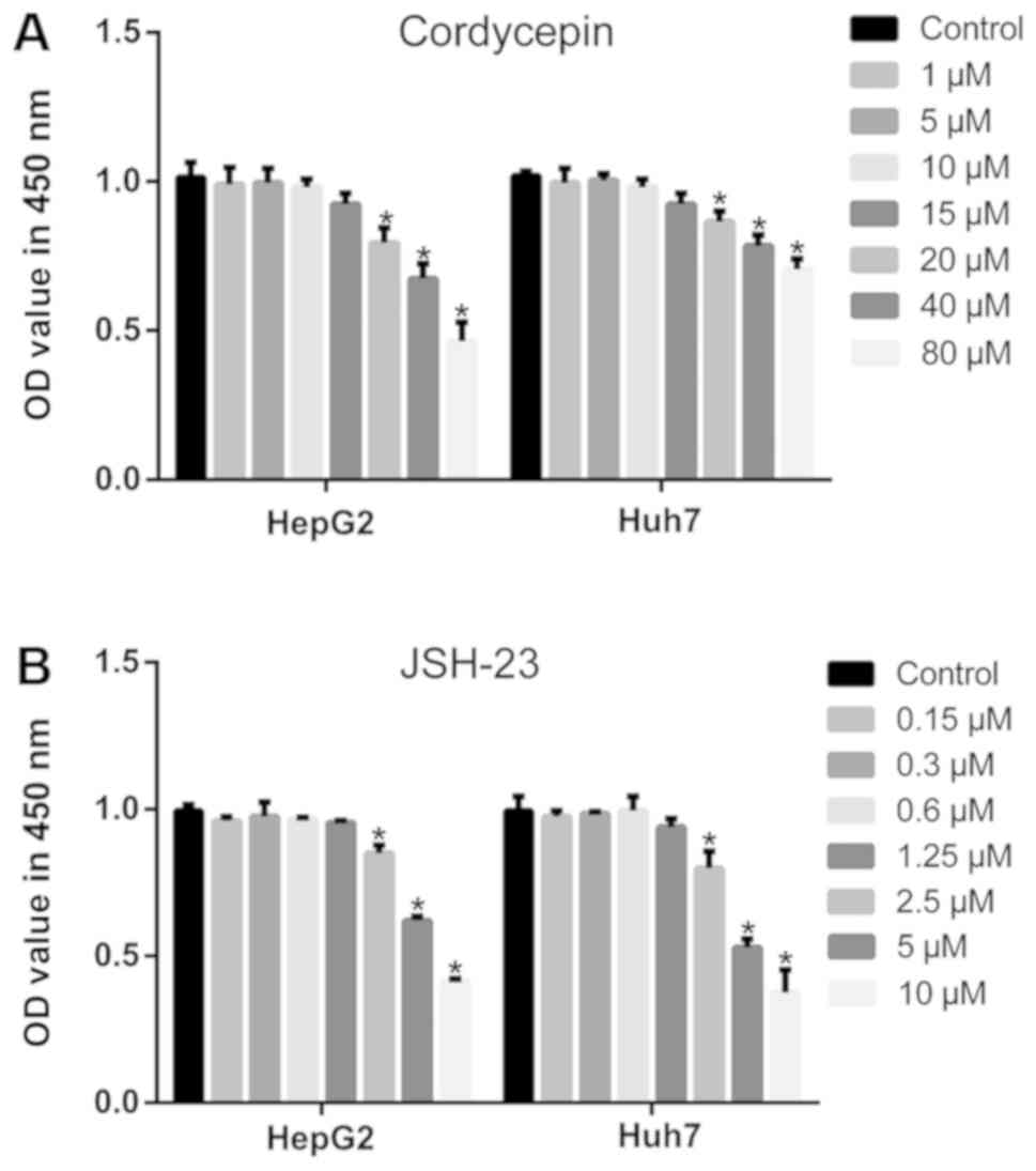
As described previously (36), relatively high concentrations of
cordycepin induce apoptosis and inhibit proliferation of HepG2
cells. To determine an appropriate concentration that has no
significant effect on the proliferation of HepG2 and Huh7 cells, a
dose-curve experiment was performed in the present study. When the
concentration of cordycepin was 1, 5 and 10 µM, no
significant difference was observed in the proliferation of HepG2
and Huh7 cells by CCK-8 assay, compared with the untreated cells
(Fig. 1A). Additionally, as
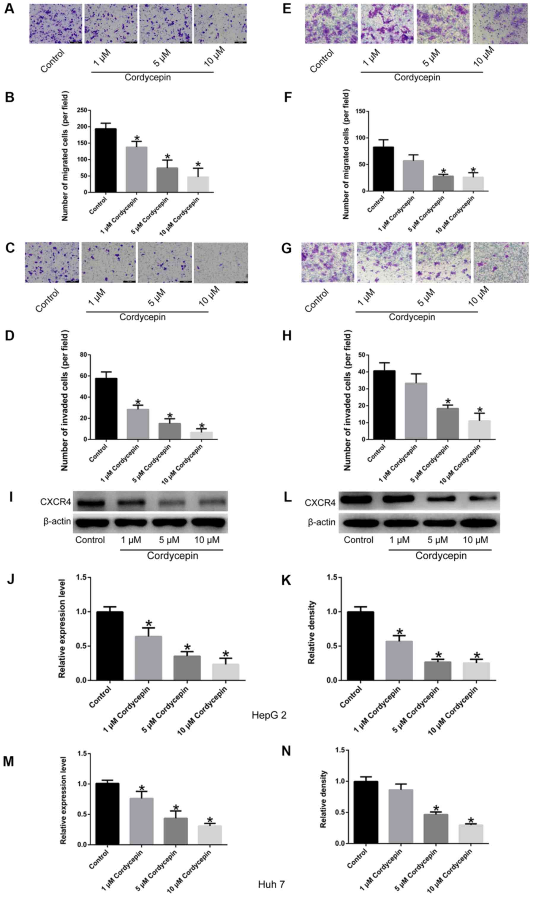
presented in Fig. 2, the
migratory and invasive abilities of HepG2 (Fig. 2A-D) and Huh7 cells (Fig. 2E-H) treated with these
concentrations of cordycepin (1, 5 and 10 µM) were
significantly inhibited (P<0.05) in a dose-dependent manner
(with the exception of Huh7 cells treated with 1 µM
cordycepin; P>0.05), compared with the untreated controls.
Finally, compared with untreated cells, a significant
dose-dependent downregulation of CXCR4 expression was observed by
RT-qPCR and western blot assays (P<0.05; Fig. 2I-N). Therefore, three different
concentrations of cordycepin (1, 5 and 10 µM) were selected
for subsequent experiments, which suppressed the cell migratory and
invasive capabilities, but had no significant effect on cell
proliferation.
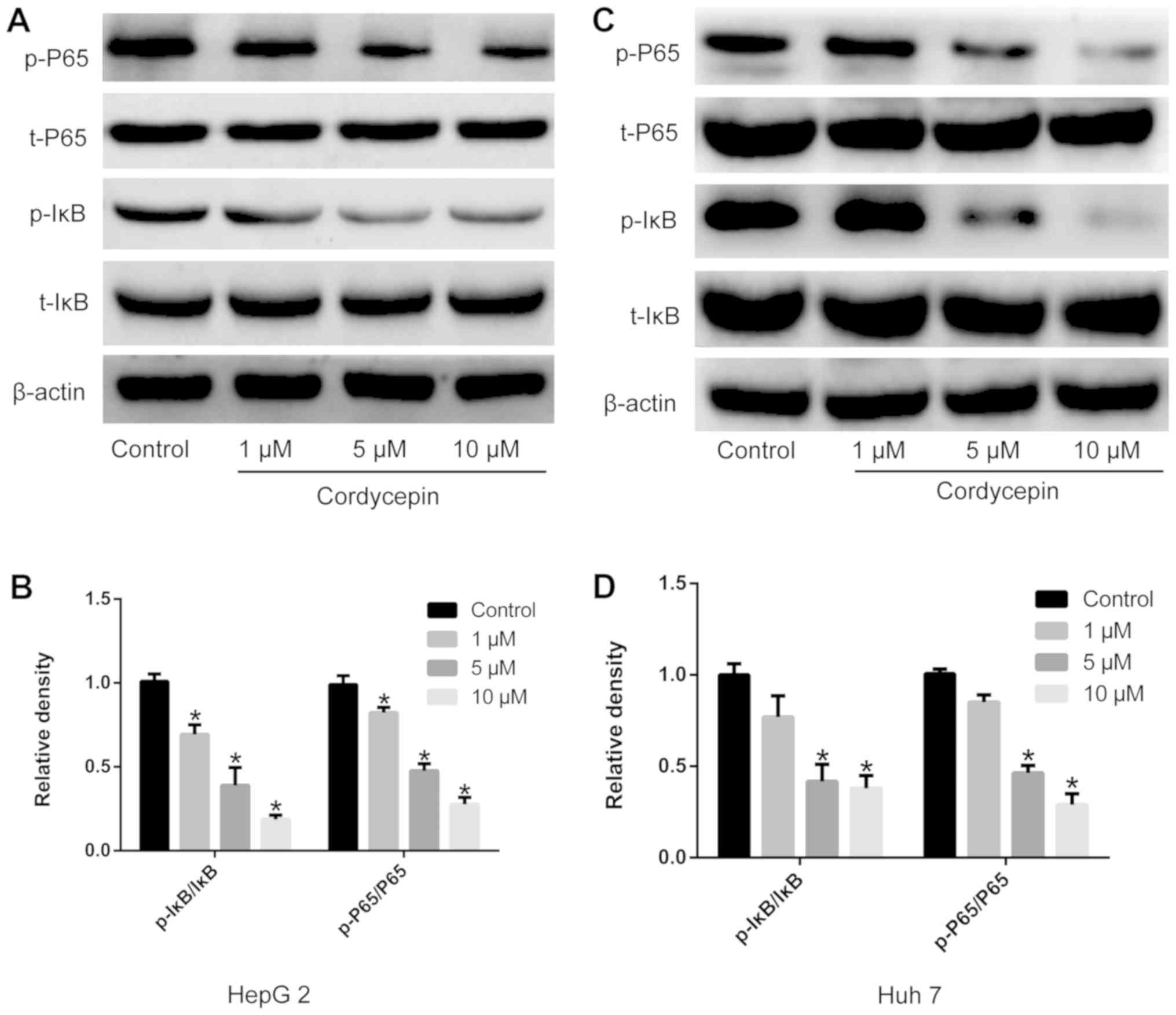
Cordycepin inhibits the activation of the
NF-κB signaling pathway
Human liver cancer cells were treated with 1, 5 and
10 µM cordycepin. The expression and activation of NF-κB
signaling pathway-related proteins, including t-IκBα, p-IκBα, t-P65
and p-P65, were evaluated by western blotting. As demonstrated in
Fig. 3, different concentrations
of cordycepin significantly downregulated the activation of IκBα
and P65, and the expression levels of p-IκBα and p-P65 (P<0.05).
No significant differences were observed in the expression levels
of t-IκBα and t-P65.
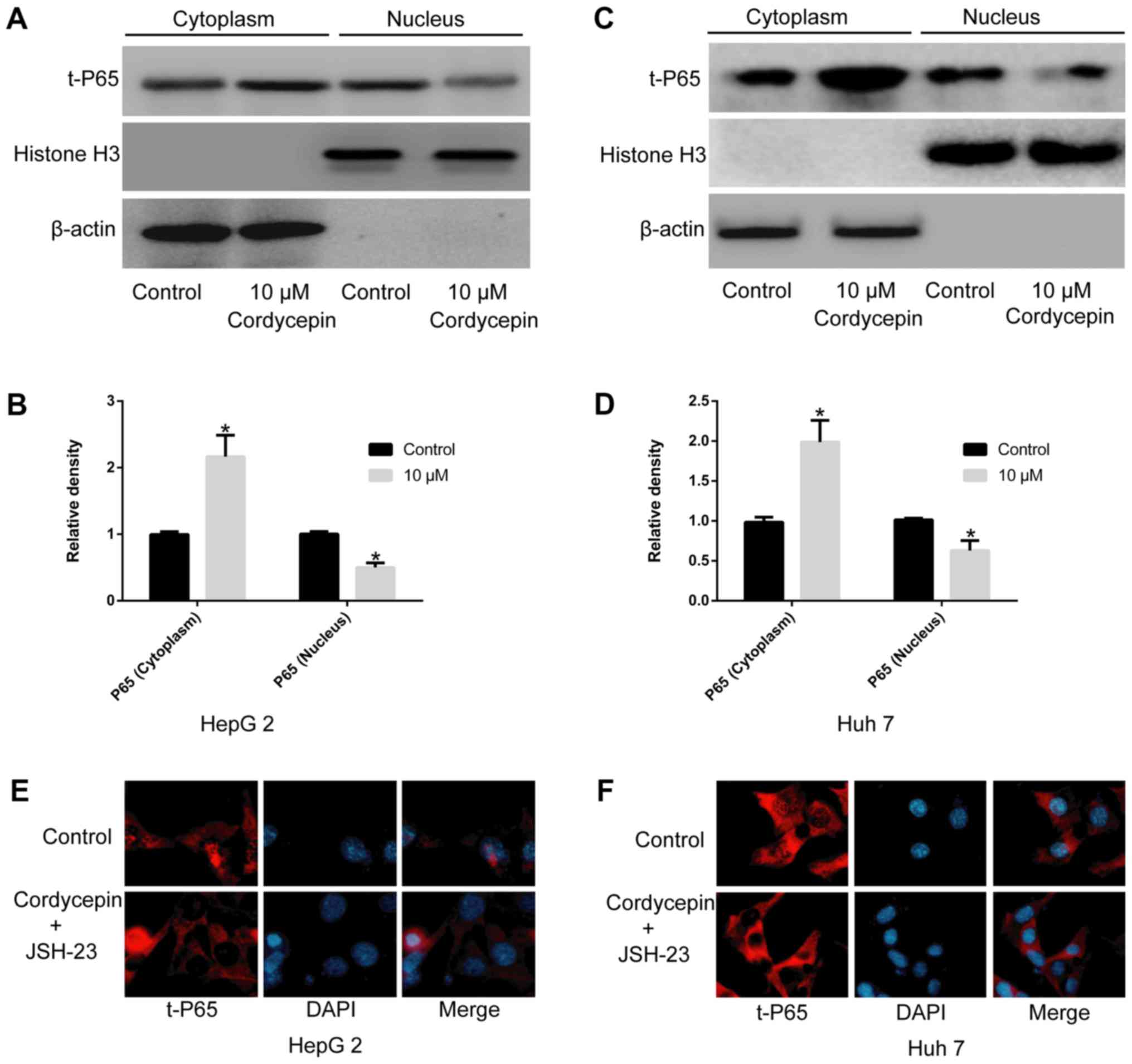
Cordycepin alone, or in combination with
JSH-23, restricts the nuclear translocation of P65
To exclude the possibility of an effect of JSH-23 on
the viability of HepG2 and Huh7 cells and to select a suitable drug
concentration (37), a CCK-8
assay was performed. The results indicated that concentrations up
to 1 µM of JSH-23 did not significantly alter cell
proliferation compared with the untreated cells (Fig. 1B). As presented in Fig. 4, a significant increase in t-P65
was observed in the cytoplasm of HepG2 (Fig. 4A and B) and Huh7 (Fig. 4C and D) cells following treatment
with 10 µM cordycepin (P<0.05), as well as a
corresponding reduction in the nuclear fraction (P<0.05),
compared with the untreated-control groups. Immunofluorescence
analysis indicated that the co-administration of 1 µM JSH-23
and 10 µM cordycepin markedly inhibited the nuclear
translocation of P65 in HepG2 (Fig.
4E) and Huh7 cells (Fig.
4F).
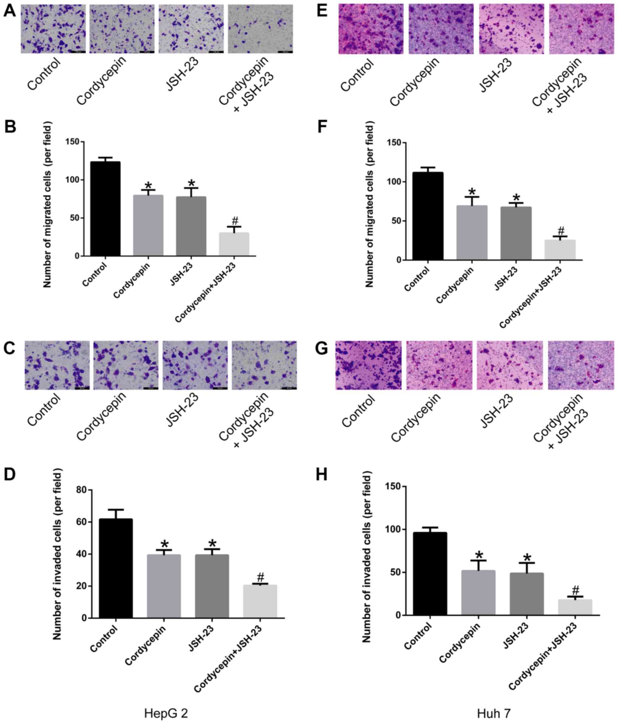
Synergistic effects of cordycepin and
JSH-23 in suppressing the migration and invasion abilities of HepG2
and Huh7 human liver cancer cells
The results of the present study preliminarily
suggested that cordycepin inhibited the phosphorylation of IκB, as
well as the activation and nuclear localization of P65, altering
the expression of its downstream gene, CXCR4. As demonstrated in
Fig. 5, treatment with 10
µM cordycepin and/or 1 µM JSH-23 suppressed the
migration (Fig. 5A, B, E and F)
and invasion (Fig. 5C, D, G and
H) capacities of liver cancer cells compared with untreated
controls (P<0.05). Furthermore, the combination treatment
revealed a synergistic inhibitory effect (P<0.05; Fig. 5) compared with cordycepin
treatment alone.
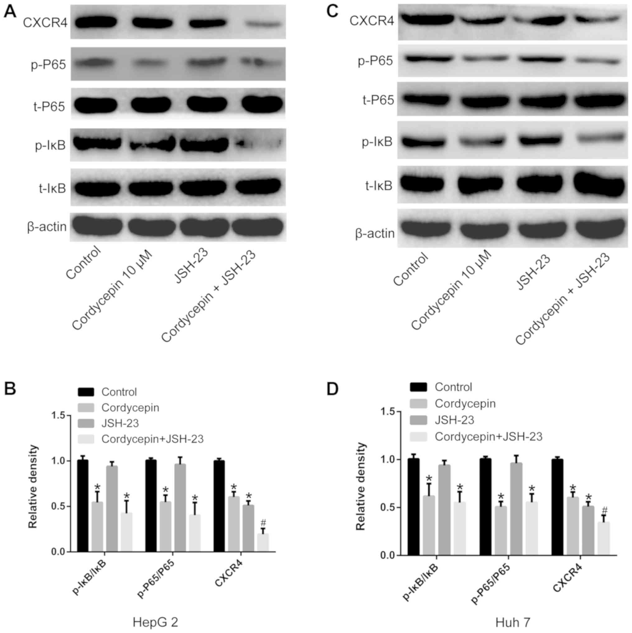
Co-treatment with cordycepin and JSH-23
inhibits the activation of the NF-κB signaling pathway, decreasing
the expression of CXCR4 in human liver cancer cells
In order to understand the underlying synergistic
mechanism between cordycepin and JSH-23, the expression of NF-κB
signaling pathway-associated proteins (t-IκBα, p-IκBα, t-P65 and
p-P65) was detected using RT-qPCR and western blotting. As shown in
Fig. 6, treatment with cordycepin
(10 µM) alone reduced IκBα and P65 phosphorylation in HepG2
and Huh7 human liver cancer cells (P<0.05). The co-treatment
with cordycepin (10 µM) and JSH-23 (1 µM)
significantly inhibited the expression of CXCR4 (P<0.05;
Fig. 6), compared with cordycepin
treatment alone. No significant effect on the expression levels of
t-IκBα and t-P65 was observed by any of the indicated treatments
(Fig. 6).
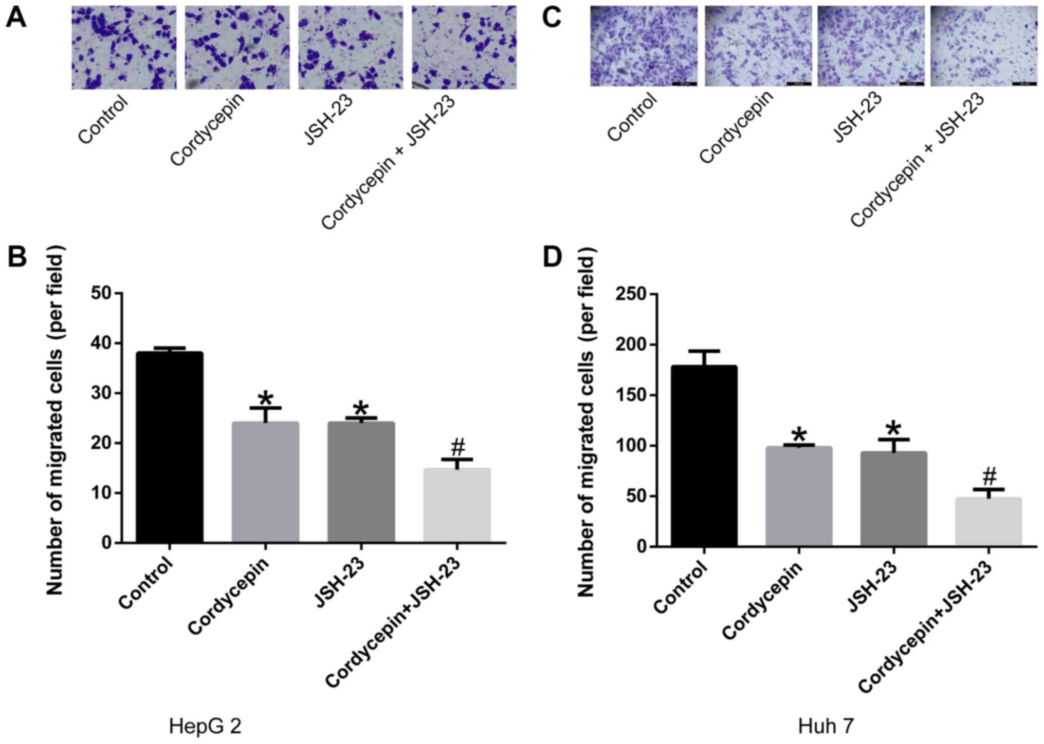
Cordycepin and JSH-23 treatment inhibits
the chemotactic migration ability of HepG2 and Huh7 human liver
cancer cells to SDF1
SDF1, a specific ligand for CXCR4, was used to
detect the chemotactic ability of human liver cancer cells treated
with 10 µM cordycepin and/or 1 µM JSH-23 via
Transwell assays. Fig. 7
illustrates that cordycepin and JSH-23 monotherapy significantly
reduced the chemotactic sensitivity of the cells to SDF1
(P<0.05), and that combination treatment further enhanced this
effect compared with the control cells (P<0.05).
Discussion
Worldwide, liver cancer results in almost one
million deaths annually, most of which are associated with
metastasis (1-3,12).
The treatment of liver cancer remains predominantly based on
surgical resection combined with radio-, chemo- and targeted
therapy, which inhibit the malignant proliferation of liver cancer
cells, but do not inhibit tumor metastasis (6,12).
For example, 5-fluorouracil and cisplatin primarily inhibit liver
cancer cell proliferation and induce apoptosis (38-40). Sorafenib, a first-line liver
cancer drug, inhibits angiogenesis of liver cancer cells, thereby
cutting off the blood supply to tumors and inhibiting the
proliferation of tumor cells (41,42). However, there are few reports
concerning the application of targeted inhibitors of liver cancer
metastasis. Cordycepin, a natural fungal compound, has been
implicated in a variety of pathophysiological processes (31,32), and has been approved for clinical
trials due its significant antiviral and anticancerous activities
(43-46).
Using several functional experiments, the present
study demonstrated that cordycepin inhibited the migration and
invasion of liver cancer cells in vitro and significantly
down-regulated the expression of CXCR4. Although the Huh7 cell line
is derived from well-differentiated liver cancer, and the HepG2
cell line is commonly used in metabolism-related studies, these two
types of cells are widely used in migration and invasion
experiments (47-50). In the present study, the results
suggested that cordycepin had positive effect on two different
liver cancer cell lines, which suggests that it might be a valuable
drug for cancer treatment. Additionally, the synergistic effect of
cordycepin in combination with the NF-κB pathway inhibitor JSH-23
was demonstrated. Previous studies have also shown that CXCR4 is
highly expressed in liver cancer tissues (19), and that liver cancer cells
preferentially metastasize to organs and tissues with abundant
expression of the CXCR4 ligand SDF1, such as the lungs (51,52) and bone marrow (53-55). In vitro studies have
demonstrated that overexpression or silencing of CXCR4 in tumor
cells promoted or inhibited, respectively, their migratory and
invasive ability (19,26). Therefore, the present study
preliminarily concluded that cordycepin inhibited the in
vitro migration and invasion of liver cancer cells by
down-regulating the expression of CXCR4.
Mechanistically, the present study revealed that the
phosphorylation levels of P65 and IκBα in liver cancer cells
changed significantly following cordycepin treatment. A previous
study suggested that IκB-molecules form a conjugated complex with
the P65/P50 heterodimer, inactivating P65 (56). When phosphorylated, IκB
dissociated from the P65/P50 complex. Free p-IκBs undergo
ubiquitination, while P65 is phosphory-lated and activated.
Subsequently, p-P65 enters to nucleus, activating specific
transcription factors and increasing the expression of a series of
genes (57,58). In the present study, cordycepin
treatment was found to significantly inhibit IκBα phosphorylation,
restricting the nuclear translocation of P65 and preventing
transcription factor activation, thereby regulating the expression
of CXCR4. These results suggested that cordycepin may regulate the
NF-κB pathway by inhibiting IκBα phosphorylation, thereby
modulating its downstream targets, which include CXCR4.
JSH-23, an NF-κB pathway inhibitor (59), has similar effects to cordycepin,
but does not significantly influence the phosphorylation status of
IκBα. When JSH-23 was combined with cordycepin in the present
study, a synergistic inhibitory effect on the migration and
invasion capacities of liver cancer cells was observed.
Collectively, the results of the present study
demonstrated that cordycepin inhibited the activation and nuclear
translocation of P65 by inhibiting the phosphorylation of IκBα,
thereby downregulating CXCR4. Furthermore, the inhibitory effects
of cordycepin were significantly enhanced following combination
with JSH-23, which suggested that cordycepin may have the potential
to prevent liver cancer metastasis when used in combination with
other therapeutic compounds.
Acknowledgments
We thank Rongmu Xia (School of Medicine, Xiamen
University, Xiamen, Fujian, China) for repeating part of the
experiments and his technical guidance on our experiments.
Funding
No funding was received.
Availability of data and materials
The datasets used and/or analyzed during the current
study are available from the corresponding author on reasonable
request.
Authors' contributions
YH conceived and designed the study. ZG, WC and GD
performed experiments, data acquisition and analysis. YH wrote the
manuscript. All authors read and approved the final manuscript.
Ethics approval and consent to
participate
Not applicable.
Patient consent for publication
Not applicable.
Competing interests
The authors declare that they have no competing
interests.
References
|
1
|
Allemani C, Matsuda T, Di Carlo V,
Harewood R, Matz M, Nikšić M, Bonaventure A, Valkov M, Johnson CJ,
Estève J, et al CONCORD Working Group: Global surveillance of
trends in cancer survival 2000-14 (CONCORD 3): analysis of
individual records for 37,513,025 patients diagnosed with one of 18
cancers from 322 population based registries in 71 countries.
Lancet. 391:1023–1075. 2018. View Article : Google Scholar : PubMed/NCBI
|
|
2
|
Siegel RL, Miller KD and Jemal A: Cancer
statistics, 2018. CA Cancer J Clin. 68:7–30. 2018. View Article : Google Scholar : PubMed/NCBI
|
|
3
|
Chen W, Zheng R, Baade PD, Zhang S, Zeng
H, Bray F, Jemal A, Yu XQ and He J: Cancer statistics in China,
2015. CA Cancer J Clin. 66:115–132. 2016. View Article : Google Scholar : PubMed/NCBI
|
|
4
|
Chen W, Zheng R, Zhang S, Zeng H, Zuo T,
Xia C, Yang Z and He J: Cancer incidence and mortality in China in
2013: an analysis based on urbanization level. Chin J Cancer Res.
29:1–10. 2017. View Article : Google Scholar : PubMed/NCBI
|
|
5
|
Zheng R, Qu C, Zhang S, Zeng H, Sun K, Gu
X, Xia C, Yang Z, Li H, Wei W, et al: Liver cancer incidence and
mortality in China: Temporal trends and projections to 2030. Chin J
Cancer Res. 30:571–579. 2018. View Article : Google Scholar
|
|
6
|
Liu CY, Chen KF and Chen PJ: Treatment of
Liver Cancer. Cold Spring Harb Perspect Med. 5:a0215352015.
View Article : Google Scholar : PubMed/NCBI
|
|
7
|
Shiani A, Narayanan S, Pena L and Friedman
M: The Role of Diagnosis and Treatment of Underlying Liver Disease
for the Prognosis of Primary Liver Cancer. Cancer control.
24:10732748177292402017. View Article : Google Scholar : PubMed/NCBI
|
|
8
|
Li S, Yang F and Ren X: Immunotherapy for
hepatocellular carcinoma. Drug Discov Ther. 9:363–371. 2015.
View Article : Google Scholar : PubMed/NCBI
|
|
9
|
Yu S, Wang Y, Jing L, Claret FX, Li Q,
Tian T, Liang X, Ruan Z, Jiang L, Yao Y, et al: Autophagy in the
'inflammation-carcinogenesis' pathway of liver and HCC
immunotherapy. Cancer Lett. 411:82–89. 2017. View Article : Google Scholar : PubMed/NCBI
|
|
10
|
Klungboonkrong V, Das D and McLennan G:
Molecular Mechanisms and Targets of Therapy for Hepatocellular
Carcinoma. J Vasc Interv Radiol. 28:949–955. 2017. View Article : Google Scholar : PubMed/NCBI
|
|
11
|
Kudo M and Arizumi T: Transarterial
Chemoembolization in Combination with a Molecular Targeted Agent:
Lessons Learned from Negative Trials (Post-TACE, BRISK-TA, SPACE,
ORIENTAL, and TACE-2). Oncology. 93(Suppl 1): 127–134. 2017.
View Article : Google Scholar : PubMed/NCBI
|
|
12
|
Tohme S, Simmons RL and Tsung A: Surgery
for Cancer: A Trigger for Metastases. Cancer Res. 77:1548–1552.
2017. View Article : Google Scholar : PubMed/NCBI
|
|
13
|
Zeeshan R and Mutahir Z: Cancer metastasis
- tricks of the trade. Bosn J Basic Med Sci. 17:172–182.
2017.PubMed/NCBI
|
|
14
|
Chaffer CL and Weinberg RA: A perspective
on cancer cell metastasis. Science. 331:1559–1564. 2011. View Article : Google Scholar : PubMed/NCBI
|
|
15
|
Ai J, Tang Q, Wu Y, Xu Y, Feng T, Zhou R,
Chen Y, Gao X, Zhu Q, Yue X, et al: The role of polymeric
immunoglobulin receptor in inflammation-induced tumor metastasis of
human hepatocellular carcinoma. J Natl Cancer Inst. 103:1696–1712.
2011. View Article : Google Scholar : PubMed/NCBI
|
|
16
|
Budhu A, Forgues M, Ye QH, Jia HL, He P,
Zanetti KA, Kammula US, Chen Y, Qin LX, Tang ZY, et al: Prediction
of venous metastases, recurrence, and prognosis in hepatocellular
carcinoma based on a unique immune response signature of the liver
microenvironment. Cancer Cell. 10:99–111. 2006. View Article : Google Scholar : PubMed/NCBI
|
|
17
|
Ma C, Han M, Heinrich B, Fu Q, Zhang Q,
Sandhu M, Agdashian D, Terabe M, Berzofsky JA, Fako V, et al: Gut
microbiome-mediated bile acid metabolism regulates liver cancer via
NKT cells. Science. 360:eaan59312018. View Article : Google Scholar : PubMed/NCBI
|
|
18
|
Song T, Dou C, Jia Y, Tu K and Zheng X:
TIMP-1 activated carcinoma-associated fibroblasts inhibit tumor
apoptosis by activating SDF1/CXCR4 signaling in hepatocellular
carcinoma. Oncotarget. 6:12061–12079. 2015. View Article : Google Scholar : PubMed/NCBI
|
|
19
|
Wang J, Huang Y, Zhang J, Xing B, Xuan W,
Wang H, Huang H, Yang J and Tang J: High co-expression of the
SDF1/CXCR4 axis in hepatocarcinoma cells is regulated by AnnexinA7
in vitro and in vivo. Cell Commun Signal. 16:222018. View Article : Google Scholar : PubMed/NCBI
|
|
20
|
Gao PT, Ding GY, Yang X, Dong RZ, Hu B,
Zhu XD, Cai JB, Ji Y, Shi GM, Shen YH, et al: Invasive potential of
hepatocellular carcinoma is enhanced by loss of selenium-binding
protein 1 and subsequent upregulation of CXCR4. Am J Cancer Res.
8:1040–1049. 2018.PubMed/NCBI
|
|
21
|
Li M, Lu Y, Xu Y, Wang J, Zhang C, Du Y,
Wang L, Li L, Wang B, Shen J, et al: Horizontal transfer of
exosomal CXCR4 promotes murine hepatocarcinoma cell migration,
invasion and lymphangiogenesis. Gene. 676:101–109. 2018. View Article : Google Scholar : PubMed/NCBI
|
|
22
|
Chen Y, Liu YC, Sung YC, Ramjiawan RR, Lin
TT, Chang CC, Jeng KS, Chang CF, Liu CH, Gao DY, et al: Overcoming
sorafenib evasion in hepatocellular carcinoma using CXCR4-targeted
nanoparticles to co-deliver MEK-inhibitors. Sci Rep. 7:441232017.
View Article : Google Scholar : PubMed/NCBI
|
|
23
|
Li X, Li P, Chang Y, Xu Q, Wu Z, Ma Q and
Wang Z: The SDF-1/CXCR4 axis induces epithelial-mesenchymal
transition in hepatocellular carcinoma. Mol Cell Biochem.
392:77–84. 2014. View Article : Google Scholar : PubMed/NCBI
|
|
24
|
Wang X, Zhang W, Ding Y, Guo X, Yuan Y and
Li D: CRISPR/Cas9-mediated genome engineering of CXCR4 decreases
the malignancy of hepatocellular carcinoma cells in vitro and in
vivo. Oncol Rep. 37:3565–3571. 2017. View Article : Google Scholar : PubMed/NCBI
|
|
25
|
Xia R, Sheng X, Xu X, Yu C and Lu H:
Hesperidin induces apoptosis and G0/G1 arrest in human non-small
cell lung cancer A549 cells. Int J Mol Med. 41:464–472. 2018.
|
|
26
|
Xia R, Xu G, Huang Y, Sheng X, Xu X and Lu
H: Hesperidin suppresses the migration and invasion of non-small
cell lung cancer cells by inhibiting the SDF-1/CXCR-4 pathway. Life
Sci. 201:111–120. 2018. View Article : Google Scholar : PubMed/NCBI
|
|
27
|
Fontanella R, Pelagalli A, Nardelli A,
D'Alterio C, Ieranò C, Cerchia L, Lucarelli E, Scala S and Zannetti
A: A novel antagonist of CXCR4 prevents bone marrow-derived
mesenchymal stem cell-mediated osteosarcoma and hepatocellular
carcinoma cell migration and invasion. Cancer Lett. 370:100–107.
2016. View Article : Google Scholar
|
|
28
|
Kaemmerer D, Schindler R, Mußbach F,
Dahmen U, Altendorf-Hofmann A, Dirsch O, Sänger J, Schulz S and
Lupp A: Somatostatin and CXCR4 chemokine receptor expression in
hepatocellular and cholangiocellular carcinomas: Tumor capillaries
as promising targets. BMC Cancer. 17:8962017. View Article : Google Scholar : PubMed/NCBI
|
|
29
|
Mardomi A, Sabzichi M, Hussein Somi M,
Shanehbandi D, Rahbarghazi R, Taj Sanjarani O and Samadi N:
Trafficking mechanism of bone marrow-derived mesenchymal stem cells
toward hepatocellular carcinoma HepG2 cells by modulating Endoglin,
CXCR4 and TGF-β. Cell Mol Biol (Noisy-le-grand). 62:81–86.
2016.
|
|
30
|
Chen YC, Chen YH, Pan BS, Chang MM and
Huang BM: Functional study of Cordyceps sinensis and cordycepin in
male reproduction: A review. Yao Wu Shi Pin Fen Xi. 25:197–205.
2017.
|
|
31
|
Tuli HS, Sharma AK, Sandhu SS and Kashyap
D: Cordycepin: A bioactive metabolite with therapeutic potential.
Life Sci. 93:863–869. 2013. View Article : Google Scholar : PubMed/NCBI
|
|
32
|
Yoon SY, Park SJ and Park YJ: The
Anticancer Properties of Cordycepin and Their Underlying
Mechanisms. Int J Mol Sci. 19:E30272018. View Article : Google Scholar : PubMed/NCBI
|
|
33
|
Wei C, Yao X, Jiang Z, Wang Y, Zhang D,
Chen X, Fan X, Xie C, Cheng J, Fu J, et al: Cordycepin Inhibits
Drug-resistance Non-small Cell Lung Cancer Progression by
Activating AMPK Signaling Pathway. Pharmacol Res. 144:79–89. 2019.
View Article : Google Scholar : PubMed/NCBI
|
|
34
|
Wang Y, Lv Y, Liu TS, Yan WD, Chen LY, Li
ZH, Piao YS, An RB, Lin ZH and Ren XS: Cordycepin suppresses cell
proliferation and migration by targeting CLEC2 in human gastric
cancer cells via Akt signaling pathway. Life Sci. 223:110–119.
2019. View Article : Google Scholar : PubMed/NCBI
|
|
35
|
Livak KJ and Schmittgen TD: Analysis of
relative gene expression data using real-time quantitative PCR and
the 2(-Delta Delta C(T)) Method. Methods. 25:402–408. 2001.
View Article : Google Scholar
|
|
36
|
Shao LW, Huang LH, Yan S, Jin JD and Ren
SY: Cordycepin induces apoptosis in human liver cancer HepG2 cells
through extrinsic and intrinsic signaling pathways. Oncol Lett.
12:995–1000. 2016. View Article : Google Scholar : PubMed/NCBI
|
|
37
|
Lai Y, Fan L, Zhao Y, Ge H, Feng X, Wang
Q, Zhang X, Peng Y, Wang X and Tao L: Cx32 suppresses extrinsic
apoptosis in human cervical cancer cells via the NF-κB signalling
pathway. Int J Oncol. 51:1159–1168. 2017. View Article : Google Scholar : PubMed/NCBI
|
|
38
|
Ikeda M, Okusaka T, Sato Y, Furuse J,
Mitsunaga S, Ueno H, Morizane C, Inaba Y, Kobayashi T and Arai Y: A
Phase I/II trial of continuous hepatic intra-arterial infusion of
5-fluoro-uracil, mitoxantrone and cisplatin for advanced
hepatocellular carcinoma. Jpn J Clin Oncol. 47:512–519. 2017.
View Article : Google Scholar : PubMed/NCBI
|
|
39
|
Kumamoto T, Tanaka K, Matsuo K, Takeda K,
Nojiri K, Mori R, Taniguchi K, Matsuyama R, Ueda M, Akiyama H, et
al: Adjuvant hepatic arterial infusion chemotherapy with
5-Fluorouracil and interferon after curative resection of
hepatocellular carcinoma: A preliminary report. Anticancer Res.
33:5585–5590. 2013.PubMed/NCBI
|
|
40
|
Zhang W, Zhong Y, Cui H, Wang L, Yang R,
Su Z, Xiang B and Wei Q: Combination of calcineurin B subunit (CnB)
and 5-fluoro-uracil reverses 5-fluorouracil-induced
immunosuppressive effect and enhances the antitumor activity in
hepatocellular carcinoma. Oncol Lett. 14:6135–6142. 2017.PubMed/NCBI
|
|
41
|
Sun T, Liu H and Ming L: Multiple Roles of
Autophagy in the Sorafenib Resistance of Hepatocellular Carcinoma.
Cell Physiol Biochem. 44:716–727. 2017. View Article : Google Scholar : PubMed/NCBI
|
|
42
|
Zhu YJ, Zheng B, Wang HY and Chen L: New
knowledge of the mechanisms of sorafenib resistance in liver
cancer. Acta Pharmacol Sin. 38:614–622. 2017. View Article : Google Scholar : PubMed/NCBI
|
|
43
|
De Clercq E: Curious (Old and New)
Antiviral Nucleoside Analogues with Intriguing Therapeutic
Potential. Curr Med Chem. 22:3866–3880. 2015. View Article : Google Scholar : PubMed/NCBI
|
|
44
|
Du Y, Yu J, Du L, Tang J and Feng WH:
Cordycepin enhances Epstein-Barr virus lytic infection and
Epstein-Barr virus-positive tumor treatment efficacy by
doxorubicin. Cancer Lett. 376:240–248. 2016. View Article : Google Scholar : PubMed/NCBI
|
|
45
|
Lee JB, Adrower C, Qin C, Fischer PM, de
Moor CH and Gershkovich P: Development of Cordycepin Formulations
for Preclinical and Clinical Studies. AAPS PharmSciTech.
18:3219–3226. 2017. View Article : Google Scholar : PubMed/NCBI
|
|
46
|
Ryu E, Son M, Lee M, Lee K, Cho JY, Cho S,
Lee SK, Lee YM, Cho H, Sung GH, et al: Cordycepin is a novel
chemical suppressor of Epstein-Barr virus replication. Oncoscience.
1:866–881. 2014. View Article : Google Scholar
|
|
47
|
Bai H, Weng Y, Bai S, Jiang Y, Li B, He F,
Zhang R, Yan S, Deng F, Wang J, et al: CCL5 secreted from bone
marrow stromal cells stimulates the migration and invasion of Huh7
hepatocellular carcinoma cells via the PI3K-Akt pathway. Int J
Oncol. 45:333–343. 2014. View Article : Google Scholar : PubMed/NCBI
|
|
48
|
Lee JH, Hur W, Hong SW, Kim JH, Kim SM,
Lee EB and Yoon SK: ELK3 promotes the migration and invasion of
liver cancer stem cells by targeting HIF-1α. Oncol Rep. 37:813–822.
2017. View Article : Google Scholar
|
|
49
|
Lin XL, Liu M, Liu Y, Hu H, Pan Y, Zou W,
Fan X and Hu X: Transforming growth factor β1 promotes migration
and invasion in HepG2 cells: Epithelial-to-mesenchymal transition
via JAK/STAT3 signaling. Int J Mol Med. 41:129–136. 2018.
|
|
50
|
Xie X, Zhu H, Zhang J, Wang M, Zhu L, Guo
Z, Shen W and Wang D: Solamargine inhibits the migration and
invasion of HepG2 cells by blocking epithelial-to-mesenchymal
transition. Oncol Lett. 14:447–452. 2017. View Article : Google Scholar : PubMed/NCBI
|
|
51
|
Pectasides E, Miksad R, Pyatibrat S,
Srivastava A and Bullock A: Spontaneous Regression of
Hepatocellular Carcinoma with Multiple Lung Metastases: A Case
Report and Review of the Literature. Dig Dis Sci. 61:2749–2754.
2016. View Article : Google Scholar : PubMed/NCBI
|
|
52
|
Yang T, Lu JH, Lin C, Shi S, Chen TH, Zhao
RH, Wang Y and Wu MC: Concomitant lung metastasis in patients with
advanced hepa-tocellular carcinoma. World J Gastroenterol.
18:2533–2539. 2012. View Article : Google Scholar : PubMed/NCBI
|
|
53
|
Bhatia R, Ravulapati S, Befeler A,
Dombrowski J, Gadani S and Poddar N: Hepatocellular carcinoma with
bone metastases: Incidence, prognostic significance, and
management-single-center experience. J Gastrointest Cancer.
48:321–325. 2017. View Article : Google Scholar : PubMed/NCBI
|
|
54
|
Hong YM, Yoon KT, Cho M, Kang DH, Kim HW,
Choi CW, Park SB, Heo J, Woo HY, Lim W, et al: Bone marrow
metastasis presenting as bicytopenia originating from
hepatocellular carcinoma. Clin Mol Hepatol. 22:267–271. 2016.
View Article : Google Scholar : PubMed/NCBI
|
|
55
|
Di Stadio CS, Altieri F, Minopoli G,
Miselli G, Rippa E and Arcari P: Role of human GKN1 on APP
processing in gastric cancer. Biochimie. 135:149–153. 2017.
View Article : Google Scholar : PubMed/NCBI
|
|
56
|
Hoesel B and Schmid JA: The complexity of
NF-κB signaling in inflammation and cancer. Mol Cancer. 12:862013.
View Article : Google Scholar
|
|
57
|
DiDonato JA, Mercurio F and Karin M: NF-κB
and the link between inflammation and cancer. Immunol Rev.
246:379–400. 2012. View Article : Google Scholar : PubMed/NCBI
|
|
58
|
Sokolova O and Naumann M: NF-κB Signaling
in gastric cancer. Toxins (Basel). 9:E1192017. View Article : Google Scholar
|
|
59
|
Thiel G, Ulrich M, Mukaida N and Rössler
OG: Resveratrol stimulation induces interleukin-8 gene
transcription via NF-κB. Pharmacol Res. 134:238–245. 2018.
View Article : Google Scholar : PubMed/NCBI
|