Introduction
Cardiovascular disease is the disease with the
highest associated mortality rate worldwide (1,2).
According to data from the World Health Organization, 17.9 million
individuals succumb to cardiovascular disease each year, which is
estimated to account for 31% of global mortality. Heart failure is
the leading cause of cardiovascular-associated mortality, and
myocardial hypertrophy is a common pathological stage in numerous
cardiovascular diseases, which is widely recognized as a risk
factor leading to cardiac dysfunction or eventual heart failure
(3). The increased area of
myocardial cells is the major characteristic of cardiac
hypertrophy, which is divided into physiological cardiac
hypertrophy and pathological hypertrophy. The former is an adaptive
response to preserve cardiac function and is initially reversible,
while it may develop into irreversible pathological hypertrophy as
the disease progresses (4,5).
Thus, if it could be effectively reversed to normal conditions
without delay before the condition develops to an irreversible one,
this may effectively prevent myocardial infarction.
The development of multiple diseases is accompanied
by alterations in the levels of biomarkers. For cardiac hypertrophy
and heart failure, the biomarkers are atrial natriuretic peptide
(ANP), brain natriuretic peptide (BNP) and β-myosin heavy chain
(β-MHC), whose levels increase with the progression of cardiac
hypertrophy (6). Isoproterenol
hydrochloride (Iso) is an agonist of the β-adrenergic receptor, as
well as a model widely used to mimic persistent adrenergic
stimulation that can trigger maladaptive cardiac hypertrophy
accompanied by the reactivation of ANP, BNP and β-MHC. These
alterations are in accordance with the development of myocardial
hypertrophy (7).
Pyrroloquinoline quinone (PQQ) is an anionic
water-soluble complex that was originally identified as a methanol
dehydrogenase coenzyme (8) and
can be isolated from methylotrophic bacteria. PQQ has the functions
of protecting neurons, stimulating immunity, and has
anti-inflammatory, antioxidant, anticancer and anti-aging
properties (9-14), which are mediated by a decrease in
the release of inflammatory factors and an attenuation of the
activation of nuclear (NF)-κB phosphorylation, thus attenuating
inflammatory diseases (12). In
addition, PQQ has been shown to exert effective cardioprotective
effects, and it has been shown to be more effective in protecting
the mitochondria from ischemia/reperfusion oxidative damage than
metoprolol, an effective cardioprotective drug (15). A previous study demonstrated that
PQQ can modulate the number and function of the mitochondria in
mice, and can directly react with reactive oxygen species (ROS) in
the mitochondria to maintain the dynamic balance of ROS (16). However, to date, at least to the
best of our knowledge, there are only few reports on the effects of
PQQ on cardiac hypertrophy (17-21). Based on previous studies (17-21), it was hypothesized that PQQ may be
protective against Iso-induced cardiac hypertrophy.
There are numerous studies on the pathogenesis of
cardiac hypertrophy focusing on myocardial pressure overload,
myocardial apoptosis, vascular remodeling, oxidative stress and the
inflammatory response (4,22-26); however, the pathogenesis of
cardiac hypertrophy has not yet been fully clarified. The
inflammatory response is one of the research hotspots in the
pathogenesis of cardiac hypertrophy (26). Previous studies have demonstrated
that the activation of the NF-κB signaling pathway is closely
associated with the inflammatory response, and it has been reported
that NF-κB is involved in vascular remodeling, apoptosis and
oxidative stress (27-30). In addition, previous studies have
confirmed that Iso can cause cardiac hypertrophy by activating the
NF-κB signaling pathway, while the inhibition of NF-κB activation
can counteract Iso-induced hypertrophy (7,31).
The activation of the NF-κB signaling pathway is marked by NF-κB
degradation and entry into the nucleus (32). The present study thus aimed to
examine the expression of NF-κB in Iso-induced cardiac hypertrophy
following pre-treatment PQQ in order to confirm the activation of
the NF-κB signaling pathway and to detect ROS levels in
cardiomyocytes. This study also aimed to explore the potential role
of PQQ in Iso-induced cardiac hypertrophy.
Materials and methods
Chemicals, reagents and kits
The human myocardial cell line, AC16 (33), and the rat myocardial cell line,
H9c2 (CRL-1446), were obtained from the American Type Culture
Collection (ATCC). Dulbecco's modified Eagle's medium (DMEM;
C11995500BT) and fetal bovine serum (FBS; 10270-106) were purchased
from Gibco (Thermo Fisher Scientific, Inc.).
Penicillin-streptomycin solution (PSS) (100X; E607011-0100) was
purchased from Sangon Biotech (BBI Co., Ltd.). Iso (I5627) and PQQ
(D7783) were purchased from Sigma-Aldrich (Merck KGaA). Anti-ANP
antibody (GTX109255) was acquired from GeneTex, Inc. Anti-GAPDH
antibody (10900R) was obtained from BIOSS. Anti-p65 (10745)
antibody, which is an antibody against NF-κB, and anti-NF-κBIA
(10268) were purchased from ProteinTech Group, Inc. Anti-histone-3
(H3; ab1791) antibody was obtained from Abcam.
Horseradish-peroxidase (HRP)-conjugated secondary antibody (BA1054)
was purchased from Wuhan Boster Biological Technology, Ltd. RIPA
lysis buffer (P0013) was purchased from the Beyotime Institute of
Biotechnology. The Cell Counting Kit-8 (CCK-8) kit (40203ES76) was
purchased from Yeasen (Yeasen Biotech Co., Ltd.). Reverse
transcription-quantitative (RT-q) PCR primers were synthesized by
Thermo Fisher Scientific, Inc., while the PrimeScript™ RT Reagent
kit (Perfect Real Time) (RR037Q) and TB Green™
Advantage® qPCR Premix (639676) were acquired from
Takara Bio, Inc. A bicinchoninic acid (BCA) protein quantitation
kit (P0010), a reactive oxygen species assay kit (S0033) and the
Actin-Tracker Green kit (C1033) were purchased from the Beyotime
Institute of Biotechnology. All other chemicals used in the
experiments were of analytical grade.
Experimental animals
The animal procedures were approved by the Animal
Care and Use Committee of Shanghai University of Medicine and
Health Sciences (Shanghai, China; permit no.
210105197306201429-kjc) and were in accordance with the Guide for
the Care and Use of Laboratory Animals published by the US National
Institutes of Health (NIH) (34).
Eighteen C57BL/6 male mice (8 weeks old) were obtained from the
Animal Resource Center of Shanghai University of Medicine and
Health Sciences. All mice were housed in a constant environment
with 55±10% humidity, a temperature of 20±5°C and a 12 h light/dark
cycle. Adequate food and water were provided ad libitum. The
health and behavior of the mice were monitored in real-time.
Animal grouping
In this study, SPF grade C57 male mice (weighing
22-25 g) were used to establish the experimental model (35-38). First, 18 mice were randomly
divided into 3 groups (6 mice per group). The treatment groups were
as follows: The sham group in which mice received an
intraperito-neal injection of normal saline (NS) 100
µl/time, and 3 h later, an intraperitoneal injection of
physiological saline 100 µl/time; the Iso (30 mg/kg) group
[the doses of Iso were in accordance with those of a previous study
(39)] in which mice received an
intraperitoneal injection of NS 100 µl/time, and 3 h later,
an intraperitoneal injection of ISO dissolved in NS 100
µl/time; the PQQ (40 mg/kg) [the doses of PQQ were in
accordance with those of a previous study (40)] + Iso (30 mg/kg) group in which PQQ
dissolved in NS was intraperitoneally injected into the mice 100
µl/time, and 3 h later, ISO dissolved in NS was
intraperitoneally injected into the mice 100 µl/time. The
above-mentioned managements were performed once every 4 days, and
the duration of the experiment was approximately 5 weeks. Following
anesthesia by an intraperitoneal injection of 3% sodium
pentobarbital at a dose of 40 mg/kg, the mice were examined to
confirm that they were in a state of deep anesthesia; the adequacy
of the anesthesia was detected by pinching the hindfoot to ensure
there was no reflex, and the heart was then harvested.
Hematoxylin and eosin (H&E)
staining
The recovered heart tissue was rinsed with NS, fixed
with 4% paraformaldehyde for 1 h at room temperature, and
subsequently, H&E staining was performed at room temperature
(hematoxylin, 7 min; eosin, 5 min) to observe the pathological
changes of myocardial tissue and the images were observed using a
digital pathological scanner (Precice 500; UNIC Technologies, Inc.)
at ×200 magnification.
Cell culture and grouping
Either the AC16 cells or H9c2 cells were cultured in
a 6-well plate with 2 ml DMEM containing 1 g/l glucose, 10% FBS and
1% PSS, and incubated at a constant temperature of 37°C in a humid
atmosphere of 95% air and 5% CO2. Once the cells had
adhered to the wall, they were grouped as follows: i) The control
group, which were treated with 2 ml DMEM without FBS (DMEMNF, DMEM
NO FBS) as a blank control for 48 h; ii) the Iso group, which were
treated with 2 ml DMEMNF for 24 h and then with 20 µM Iso in
2 ml DMEMNF for 24 h; iii) the low-dose PQQ group, which, after
being cultured for 21 h in 2 ml DMEMNF, were treated with 1
µM PQQ in 2 ml DMEMNF for 3 h and then with 20 µM Iso
in 2 ml DMEMNF for 24 h; iv) the mid-dose group, which, following
21 h of culture in 2 ml DMEMNF, were treated with 2.5 µM PQQ
in 2 ml DMEMNF for 3 h and then with 20 µM Iso in 2 ml
DMEMNF for 24 h; and v) the high-dose group, which, after 21 h of
culture in 2 ml DMEMNF, were treated with 5 µM PQQ in 2 ml
DMEMNF for 3 h and then with 20 µM Iso in 2 ml DMEMNF for 24
h (Fig. S1).
Cell viability measurements
To explore the cytotoxicity of Iso and PQQ on the
AC16 cells, cell viability was determined using a rapid and
sensitive CCK-8 assay kit according to the manufacturer's
instructions. The AC16 cells were seeded into 96-well plates at a
density of 5×10³ cells per well. After 24 h, Iso was
added to the 96-well plates at increasing concentrations (5, 10,
20, 40, 80 and 160 µM) and each concentration had 6 set
parallel groups, and cultured in an incubator at 37°C with a humid
atmosphere of 95% air and 5% CO2 for 24 h. Moreover, PQQ
was added to other 96-well plates at a concentration of 1, 5, 10,
25, 50, 100, 150 and 200 µM setting 6 parallel groups as
well, and cultured for 12 h. The medium was then aspirated from the
plates, which were rinsed gently with PBS before being filled with
CCK-8 reagent and DMEM at a ratio of 1:10 in 100 ml. The plates
were incubated for 2 h in an incubator at 37°C and the optical
density (OD) value at a wavelength of 450 nm was measured using a
microplate reader (SpectraMax M5; Molecular Devices, LLC).
Cell morphometric analysis
To examine the effect of PQQ against Iso-induced
hypertrophy, visual experimentation was carried out. First, either
the AC16 cells or H9c2 cells were plated in 6-well plates, and
cultured in a humid atmosphere of 95% air and 5% CO2 at
a constant temperature of 37°C. The cells were then treated as
described above in the section entitled 'Cell culture and
grouping'. The cells were then washed with PBS thrice (5 min each
wash) before being fixed in a 4% paraformaldehyde solution. After
10 min, the 4% paraformaldehyde solution was aspirated, and 0.1%
Triton X-100 (v/v in PBS) was used to wash the plates thrice for 5
min to flush any residual 4% paraformaldehyde. Subsequently,
Actin-Tracker Green was diluted in PBS in a ratio of 1:40 as the
working solution, which containing 5% FBS (v/v) and 1% Triton X-100
(v/v). Each well was supplemented with 300 µl working
solution, and incubated at 37°C without light for 45 min. The
working solution was then poured out and the excess solution
containing PBS and 0.1% Triton X-100 (v/v) was removed by washing
thrice (5 min each). Subsequently, either the AC16 cells or H9c2
cells were observed under a Nikon inverted microscope (Nikon Corp.)
equipped with a Polaroid digital camera and photographed at ×200
magnification. A total of 5 random images from each well were
captured, and 10 individual cell surface areas per photography were
measured by ImageJ software (National Institutes of Health).
RT-qPCR
Total RNA was isolated from the AC16 cells using
TRIzol reagent (Invitrogen; Thermo Fisher Scientific, Inc.)
according to the manufacturer's instructions. The concentration and
purity of the total RNA were determined using a NanoDrop 2000c
Spectrophotometer (Thermo Fisher Scientific, Inc.) and absorbance
readings at 260 nm and 260/280 nm, respectively, which were used as
indicators of RNA concentration and purity. First-strand
complementary DNA (cDNA) synthesis was performed with the
PrimeScript™ RT Reagent kit (Perfect Real Time) in the reaction
system presented in Table I, and
the reaction temperature was set as indicated in the manufacturer's
protocol. Subsequently, using TB Green™ Advantage® qPCR
Premix, the obtained cDNA and the homologous primers [as presented
in Table II (41)] were mixed at the ratio indicated
in Table III) in 96-well
optical reaction plates (04729692001, Roche) and analyzed on a
LightCycler 96 system (05815916001, Roche Diagnostics) according to
the manufacturer's protocol. The reaction conditions were set as
presented in Table IV. Each gene
was analyzed in 3 parallel wells in each experiment, and the
difference in incorporation data (Cq values) from parallel wells
should be <1. The results of ANP, BNP and β-MHC were normalized
to those of GAPDH within the same sample and quantified by the
formula 2−ΔΔCq (42)
relative to the control group.
 | Table IReaction system of first-strand cDNA
synthesis. |
Table I
Reaction system of first-strand cDNA
synthesis.
| Reagent | Volume
(µl) |
|---|
| 5X PrimeScript
Buffer (for real-time) | 2 |
| PrimeScriptRT
Enzyme Mix I | 0.5 |
| Oligo dT Primer (50
µM) | 0.5 |
| Random 6 mers (100
µM) | 0.5 |
| Total RNA | x |
| RNase Free
dH2O | 6.5-x |
| Total volume per
rxn | 10 |
| Table IIPrimer sequences used for RT-qPCR
assays (41). |
Table II
Primer sequences used for RT-qPCR
assays (41).
| Gene | Primers
|
|---|
| Forward (5′ to
3′) | Reverse (5′ to
3′) |
|---|
| ANP |
CAGCAAGCAGTGGATTGCTCCT |
TCTGCGTTGGACACGGCATTGT |
| BNP |
TGGAAACGTCCGGGTTACAGGA |
TCCGGTCCATCTTCCTCCCAAA |
| β-MHC |
GGGCAAAGGCAAGGCCAAGAAA |
ATGGGTGGAGCGCAAGTTGGTCA |
| GAPDH |
GGAGCGAGATCCCTCCAAAAT |
GGCTGTTGTCATACTTCTCATGG |
| Table IIIReaction system used for qPCR. |
Table III
Reaction system used for qPCR.
| Reagent | Volume
(µl) |
|---|
| TB Green Advantage
qPCR Premix | 10 |
| Forward primer (10
µM) | 0.4 |
| Reverse primer (10
µM) | 0.4 |
| Rnase Free
dH2O | 7.2 |
| cDNA sample | 2 |
| Total volume per
rxn | 20 |
| Table IVReaction conditions for RT-qPCR
assays. |
Table IV
Reaction conditions for RT-qPCR
assays.
| Procedure | Temperature
(°C) | Duration (sec) | Cycle |
|---|
| Initial
denaturation | 95 | 600 | 1 |
| Denaturation | 95 | 15 | 45 |
| Annealing | 60 | 34 | |
Western blot analysis
Either the AC16 cells or H9c2 cells were lysed with
RIPA lysis buffer and the total protein content was analyzed with a
BCA kit. Western blot analysis was performed as previously
described (43). Briefly, equal
amounts of total protein were separated by 10-15% SDS-PAGE and then
transferred onto PVDF membranes (EMD Millipore). The membranes were
blocked with 5% non-fat milk solution for 1 h at room temperature
and incubated overnight at 4°C with primary antibodies against
GAPDH (1:3,000), ANP (1:2,000), H3 (1:3,000), NF-κBIA (1:2,000), or
p65 (1:2,000). The membranes were then washed 3 times with
TBS-Tween-20 and further incubated with HRP-conjugated secondary
antibodies (1:5,000) at room temperature for 1 h. The membranes
with immune complexes were visualized by a chemiluminescence
imaging system (ImageQuant LAS 4000 Mini; GE Healthcare Life
Sciences). The intensity of the bands was measured by ImageJ
software (National Institutes of Health) and analyzed with IBM SPSS
Statistics 23.0 (IBM Corp.).
ROS detection and quantification
Using the Reactive Oxygen Species Assay kit, the
intracellular ROS levels in the PQQ-pre-treated and/or Iso-treated
groups were detected, and the changes in the mean fluorescence
intensity were quantified by ImageJ software. The experimental
procedure was performed according to the manufacturer's
instructions. In brief, the cells were loaded with
dichlorodihydro-fluorescein diacetate probes at a ratio of 1:1,000
(v/v) in 10 ml serum-free medium and were incubated at room
temperature for 30 min. The groups were set as described above in
the section entitled 'Cell culture and grouping'. The cells were
then washed thrice for 15 min with serum-free medium. Images were
obtained at ×200 magnification with a Nikon inverted microscope
(Nikon Corp.) equipped with a charge-coupled device camera
(Hamamatsu). Each well was photographed in 5 random fields and
analyzed by ImageJ software as described above in the section
entitled 'Cell morphometric analysis'. Moreover, the ROS
levels were also analyzed by analytical flow cytometry (BD
Biosciences) at an excitation wavelength of 488 nm and an emission
wavelength of 525 nm, respectively. Flowjo software (Flowjo, LLC)
was used to analyze the results of flow cytometry.
Mitochondrial membrane potential (MMP)
detection
As JC-1 is an ideal fluorescent probe for detecting
MMP, the change in fluorescent color by the JC-1 probe was obtained
to detect the change in MMP (44). The JC-1 probe (Mitochondrial
membrane potential assay kit with JC-1, C2006, Beyotime Institute
of Biotechnology) was used to detect changes in MMP in the AC16
cells treated with Iso and/or PQQ pre-treatment. The experimental
procedure was performed as previously described (45,46). The fluorescence images were
obtained using a fluorescent microscope (Nikon Corp.).
Statistical analysis
IBM SPSS Statistics 23.0 (IBM Corp.) was used for
statistical analysis. All data are presented as the means ±
standard deviation. Differences between 2 groups were analyzed with
an unpaired Student's t-test. Statistical analysis among various
groups was conducted by one-way analysis of variance with Tukey's
post hoc test. P<0.05 was considered to indicate a statistically
significant difference.
Results
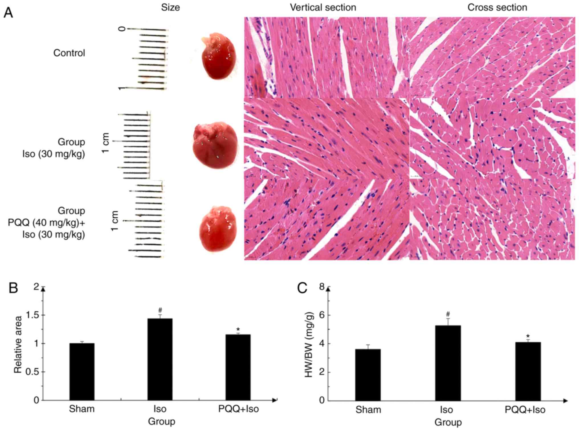
PQQ prevents Iso-induced hypertrophy in
mice
The results obtained are presented in Fig. 1, which illustrates that the cell
morphological changes in the mouse cardiac muscle in the
Iso-treated C57 mice. The surface area increased significantly,
while following pre-treatment with PQQ, the increase in the surface
area was reduced (Fig. 1A and B).
Moreover, the ratio of heart weight/body weight in the Iso group
was higher than that in the control group. In the PQQ + Iso group,
a decrease in the ratio of heart weight/body weight was observed
compared to the Iso group (Fig.
1C). These results indicated that PQQ exerted an inhibitory
effect on ISO-induced cardiac hypertrophy in vivo.
Effects of Iso and PQQ on the viability
of AC16 cells
To examine the effects of Iso and PQQ on AC16 cells,
cell viability was determined with a rapid and sensitive CCK-8
assay kit according to the manufacturer's instructions. The OD
values, which directly reflect the survival status and viability of
the AC16 cells, were 0.2-0.4 for the Iso and PQQ groups of AC16
cells (Fig. 2A and C). AC16 cell
viability for the control well was set at 100%, and relatively, the
other well percentages were calculated. The results of the CCK-8
assay demonstrated that concentrations of Iso from 5 to 160
µM and those of PQQ from 1 to 200 µM had no
significant effect on AC16 cell viability (Fig. 2B and D). Thus, subsequent studies
that could identify alterations in gene expression would not lead
to changes in cell viability or cytotoxicity. Based on the above
findings and those of previous studies (22,47), the concentration of 20 µM
was selected as the therapeutic concentration of Iso.
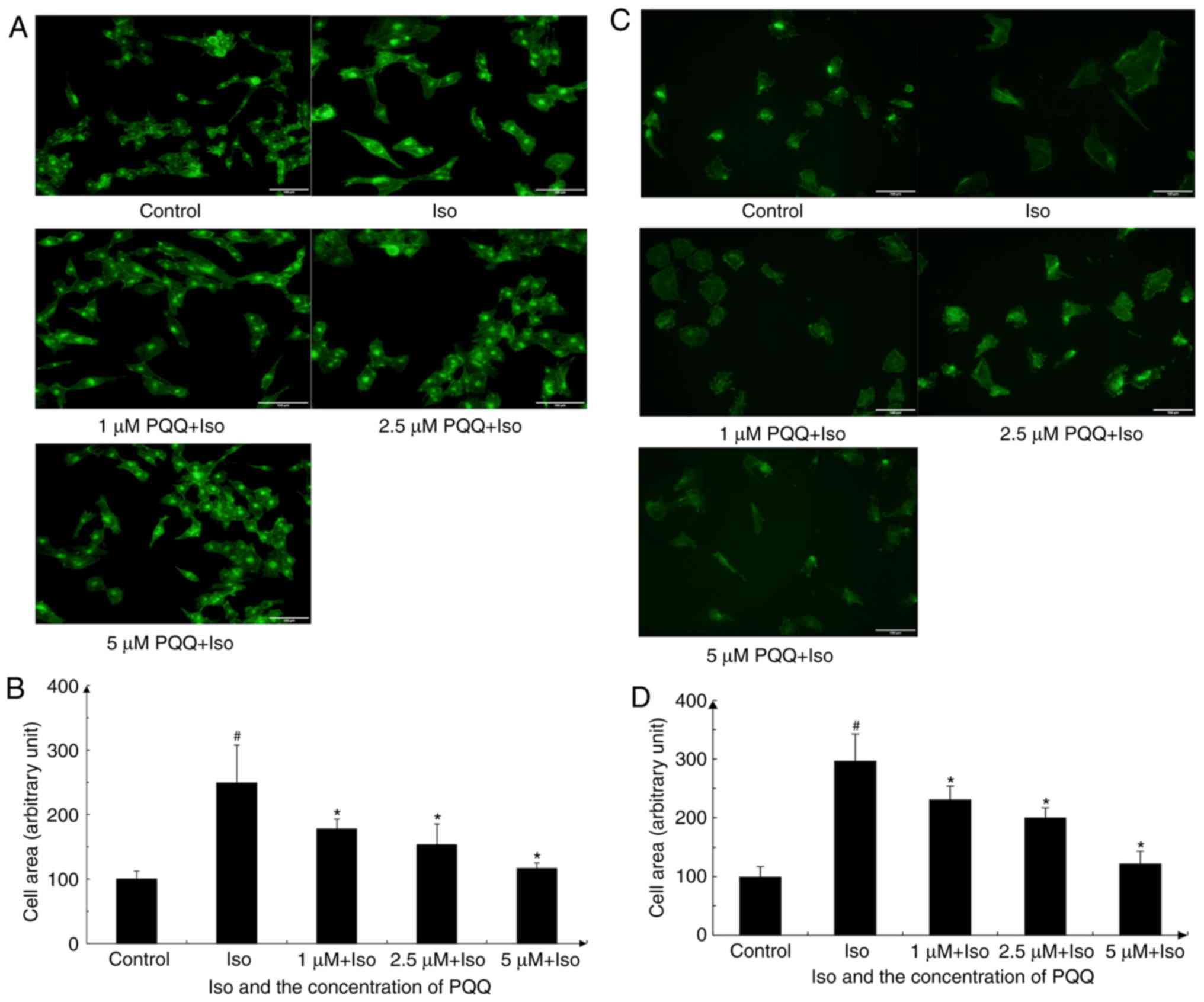
PQQ acts against Iso-induced
hypertrophy
The increase in AC16 cell size can directly reflect
the progression of cardiac hypertrophy. This study found that the
Iso-treated AC16 cells exhibited a marked change in morphology and
their surface area increased significantly, while the increase in
the surface area was reduced following pre-treatment of the AC16
cells with PQQ (Fig. 3A and B).
With 1 µM PQQ pre-treatment, the surface area of Iso-induced
cardiomyocyte hypertrophy was significantly reduced compared with
that of the Iso group without PQQ pre-treatment, and the increased
surface area was reduced by 29%. The 2.5 µM PQQ pre-treated
group exhibited a 38% decrease in cell surface area compared with
that of the Iso group, while the 5 µM PQQ pre-treated group
exhibited a decrease of 53% in cell size compared with that of the
Iso group, and the cell surface area was almost unaltered
macroscopically compared with that of the control group (Fig. 3A and B). To further investigate
the protective effects of PQQ on the heart, the effects of PQQ on
H9c2 cells with Iso-induced hypertrophy were also detected
(Fig. 3C and D), and the results
revealed a consistent trend with what was observed with the AC16
cells.
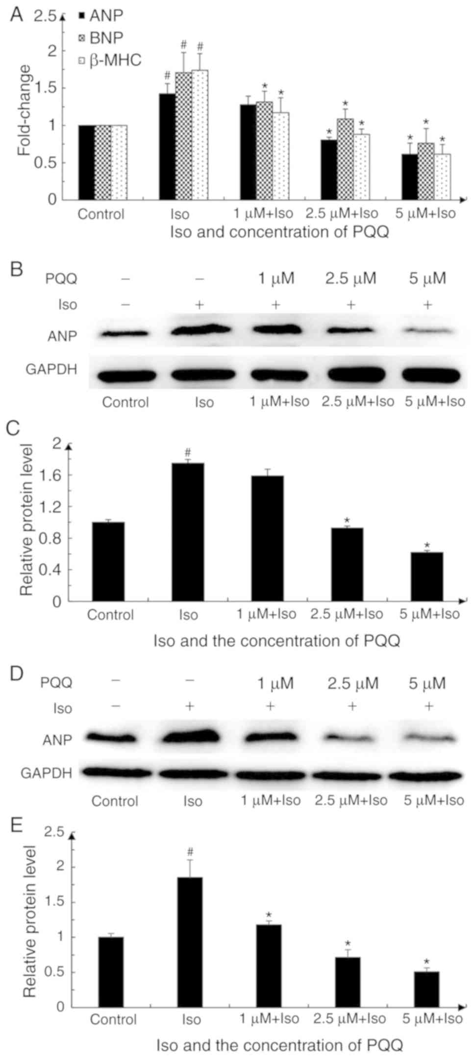
The present study examined the mRNA expression
levels of the hypertrophic markers, ANP, BNA and β-MHC, in AC16
cardiomyocytes relative to those of the blank control group in
order to verify whether PQQ can protect against Iso-induced cardiac
hypertrophy. The results (Cq value) of ANP, BNP and β-MHC were
normalized to those of GAPDH in the same sample and were quantified
by the formula 2−ΔΔCq. The expression of ANP, BNP and
β-MHC in the Iso-treated group increased by 1.42-, 1.71- and
1.74-fold vs. the control group at 24 h. Upon pre-treatment with
PQQ, the expression of hypertrophic markers decreased. Following
pre-treatment with 1 µM PQQ, the expression levels of ANP,
BNP and β-MHC were downregulated, and the downregulation of BNP and
β-MHC was statistically significant, while the difference in ANP
was not statistically significant. Pre-treatment with a higher
concentration of PQQ (i.e., 2.5 and 5 µM) led to a
downregulation in the expression levels of ANP, BNP and β-MHC,
which was more significant with increasing concentrations. The
levels of these markers in the 5 µM PQQ-pre-treated group
were similar to those of the control group (Fig. 4A).
To further examine the effect of PQQ on cardiac
hypertrophy, the present study also detected the changes in ANP
protein expression by western blot analysis. ANP expression in the
Iso-treated group was significantly upregulated. Upon pre-treatment
with various concentrations of PQQ (1, 2.5 and 5 µM), ANP
protein expression was decreased compared with that of the
Iso-treated group. Although the difference in the ANP protein level
between the Iso-treated group and the 1 µM PQQ-pre-treated
group was not statistically significant, the trend in the
downregulation of ANP protein with the increasing PQQ concentration
was consistent with the trend observed for ANP mRNA expression
(Fig. 4B and C). Overall, these
findings indicated that PQQ pre-treatment attenuated Iso-induced
hypertrophy, and the protective effects of PQQ were more effective
as the concentration increased from 1 to 5 µM. Furthermore,
ANP expression was also detected in the H9c2 cells. The results
revealed that ANP protein expression in the Iso-treated group was
significantly upregulated, and the protective effects of PQQ were
enhanced as the concentration increased, which was in accordance
with the results obtained for the AC16 cells (Fig. 4D and E).
PQQ prevents the Iso-induced hypertrophy
of AC16 cells via the inhibition of the NF-κB signaling
pathway
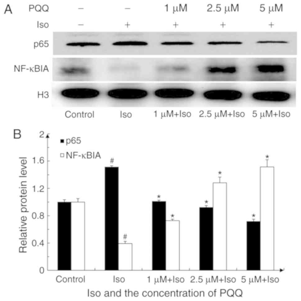
Under normal physiological conditions, NF-κB in the
cytoplasm and NF-κB inhibitor (IκB) are combined and remain in an
inactive state. However, under inflammatory or other stimulating
conditions, the IκB kinase complex composed of IκB kinase α/β/γ is
activated to promote serine phosphorylation at positions 32 and 36
of the IκBα protein, leading to the degradation of IκBα by
ubiquitination and the release of free NF-κB. This free NF-κB can
enter the nucleus binding with the specific site, and can
subsequently mediate a series of cellular responses, such as
inflammation through transcriptional activation of its target gene
(48). NF-κBIA is one of the
proteins of IκB that can inactivate NF-κB signaling by combining
with NF-κB in the cytoplasm. The present study thus analyzed the
expression of NF-κBIA and nuclear p65 by extracting nuclear
proteins to determine the mechanism through which the NF-κB
signaling pathway participates in cardiac hypertrophy induced by
Iso or by treatment with PQQ.
As shown in Fig.
5, nuclear p65 expression in the Iso pre-treated group
increased by 1.5-fold vs. the control group, while the expression
of NF-κBIA decreased by 60%. Following pre-treatment with PQQ, the
expression of nuclear p65 in the 1 µM PQQ group was
decreased by 33% compared with that of the Iso pre-treated group,
and it was significantly decreased with the increasing
concentrations of PQQ. By contrast, NF-κBIA expression
significantly increased with the increasing concentrations of PQQ
(Fig. 5B). Following treatment
with Iso, the NF-κB translocation to the nucleus significantly
increased, while with the increasing concentrations of PQQ, the
protective effects of PQQ were enhanced, as indicated by the
significant proportionate decrease of NF-κB translocation to the
nucleus (Fig. 5). The
above-mentoined results indicated that, by inhibiting the NF-κB
signaling pathway, PQQ may inhibit NF-κB from entering the nucleus,
thus acting against Iso-induced hypertrophy in AC16 cells.
PQQ reduces ROS levels in AC16 cells with
Iso-induced hypertrophy
It has been reported that ROS accumulation is
closely associated with inflammation (49), and PQQ effectively scavenges the
production of intracellular ROS, as demonstrated in previous
studies (50-52). The changes in ROS levels in AC16
cells with Iso or PQQ treatment were thus detected and photographed
and quantified with ImageJ software (Fig. 6). As observed, the fluorescence
intensity of the Iso pre-treated group was markedly stronger than
that of the control and PQQ-treated groups (Fig. 6A). Upon treatment with Iso, the
ROS levels were significantly increased by 2.2-fold relative to the
control group, whereas the ROS levels of the group pre-treated with
1, 2.5 and 5 µM PQQ decreased by 18, 27 and 50% compared
with those of the Iso pre-treated group (Fig. 6B). Moreover, the changes in the
intracellular fluorescence intensity of the AC16 cells were
detected by flow cytometry and the results revealed a consistent
trend with the results of the images (Fig. 6C and D).
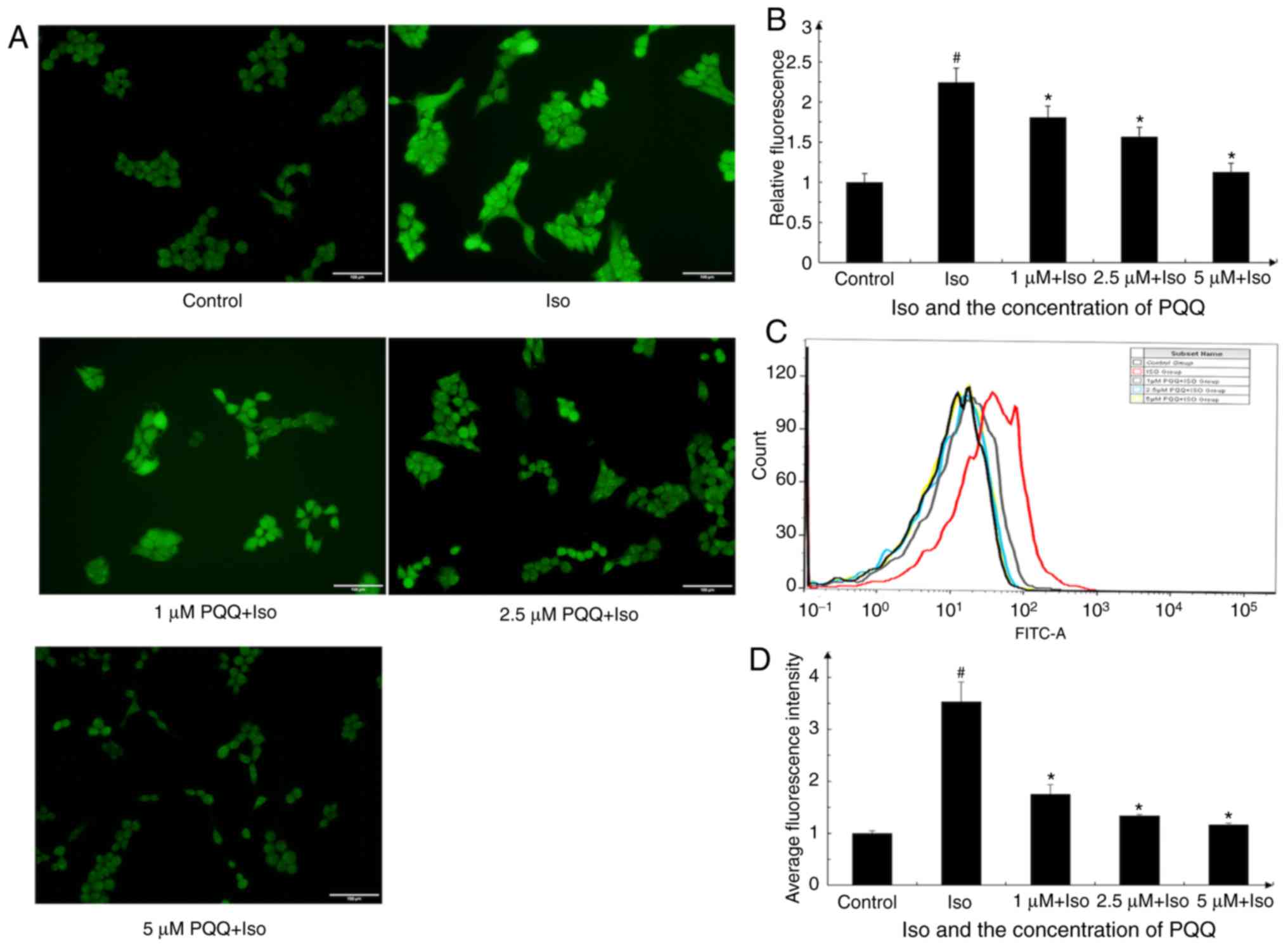 | Figure 6PQQ alters the levels of ROS in AC16
cells. (A) Effect of PQQ on Iso-induced ROS levels in AC16 cells
was detected with the Reactive Oxygen Species Assay kit and then
photographed (groups were set as described in the 'Materials and
methods'). (B) The levels of ROS were quantified by ImageJ
software, with the mean fluorescence intensity of the control group
set as 1. (C) DCFH-DA fluorescent dye was used to detect ROS
production in the AC16 cells with a flow cytometer. PQQ attenuated
Iso-induced ROS levels in AC16 cells. Black line, control group;
red line, Iso group; gray line, 1 µM PQQ + Iso group; blue
line, 2.5 µM PQQ + Iso group; yellow line, 5 µM PQQ +
Iso group. (D) Statistical data are presented based on experiments
performed in triplicate. Data are presented as the means ± SD
(n=3). #P<0.05 vs. the control group,
*P<0.05 vs. the Iso group. PQQ, pyrroloquinoline
quinone; Iso, isoproterenol hydrochloride; ROS, reactive oxygen
species. |
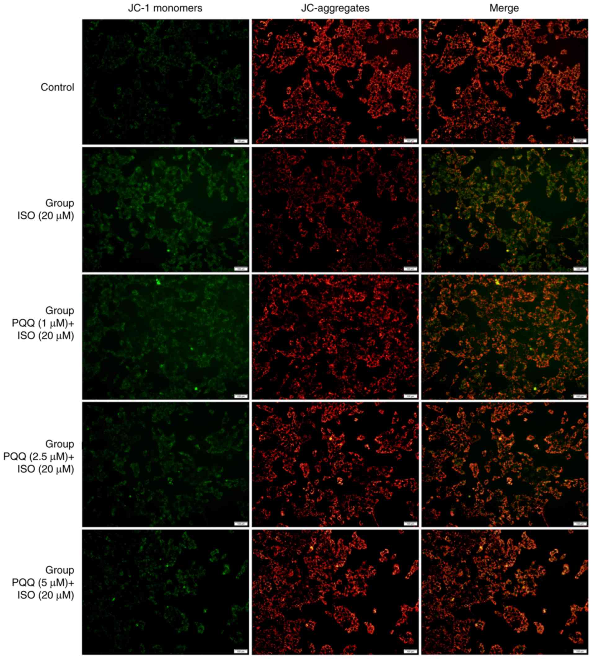
PQQ increases MMP in AC16 cells with
Iso-induced hypertrophy
During the process of respiratory oxidation, the
mitochondria store the energy generated by electrochemical
potential energy in the mitochondrial inner membrane, causing
asymmetric distribution of proton and other ion concentrations on
both sides of the inner membrane to form MMP (53), which is a key parameter for
evaluating mitochondrial function (54,55). The results obtained in this study
for MMP are presented in Fig. 7.
The red fluorescence intensity of JC-aggregates in the control
group was significantly higher than that of JC-1monomers and the
'Merge' image synthesized by the two panels is dominated by the red
fluorescence of JC-aggregates. The green fluorescence intensity of
JC-1 monomers in the AC16 cells in the Iso group was significantly
higher than that of the control group; the synthesized 'Merge'
image is mainly compiled of green fluorescence. The red
fluorescence of JC-aggregates in the group pre-treated with 1
µM PQQ was higher than that in the Iso group, and the
'Merge' image red fluorescence and the green fluorescence are
almost the same, and the 'Merge' image is colored orange. The green
fluorescence in the group pre-treated with 2.5 µM PQQ was
weakened, and the red fluorescence in the synthesized 'Merge' image
is greater than that in the Iso group. The green fluorescence in
the group pre-treated with 5 µM PQQ was significantly weaker
than that in the Iso group, and the red fluorescence in the 'Merge'
image is greater than that in the Iso group, but slightly weaker
than that in the control group (Fig.
7).
Discussion
Cardiomyocytes of the adult human heart almost lose
their proliferative capacity; thus, the heart does not regenerate
significant amounts of lost tissue following injury, and is not
able to create new, functional muscle following scarring and
hypertrophy, which indicates that myocardial damage may be
irreversible (56,57). Therefore, protective measures are
often ineffective in the end stages of cardiovascular disease.
Cardiac hypertrophy occurs at the initial stage of heart failure
development. If this could be prevented, and the compensatory
period of cardiac hypertrophy could be protected to restore the
function of cardiomyocytes, heart failure could be effectively
prevented. Therefore, the present study focused on the preventive
effects of PQQ against cardiac hypertrophy.
PQQ is present in mammalian tissues and human milk,
and has excellent antioxidant properties. It is similar to B
vitamins, which are currently proposed to be classified as vitamins
in Japan (58,59). PQQ is widely used in industrial,
agricultural and medical treatments (14). In clinical practice, PQQ has
demonstrated protective effects in the liver and brain (9,11),
and it has been studied in relation to cardiovascular diseases.
Previous studies have demonstrated that PQQ can attenuate the
expression of matrix metalloproteinase-1 (MMP-1), matrix
metalloproteinase-3 (MMP)-3, tumor necrosis factor-α (TNF-α) and
interleukin (IL)-6 in IL-1β-treated SW982 cells by attenuating the
activation of NF-κB, and can also significantly improve the
clinical symptoms of collagen-induced arthritis in mice (12). Previous in vitro studies
have indicated that PQQ exerts significant anti-neuroinflammatory
effects in microglial cells by regulating the NF-κB and p38
mitogen-activated protein kinase (MAPK) signaling pathways
(9,60).
In rats, high doses of PQQ (15 mg/kg) have been
shown to reduce the myocardial infarct size and attenuate
myocardial dysfunction and the levels of
malondialdehyde/thiobarbituric acid reactive substances in
myocardial tissue (17). These
substances are often used as a measure of free radical-induced
lipid peroxidation and oxidative stress. Moreover, the
administration of low doses of PQQ (3 mg/kg) or metoprolol at the
beginning of reperfusion has been shown to be effective in reducing
the myocardial infarct size, improving cardiac function and
preventing mitochondrial dysfunction. At non-toxic doses, PQQ is
superior to metoprolol in protecting mitochondria from oxidative
damage and reducing lipid peroxidation (15). The above-mentioned results
indicate that the effects of PQQ on protecting the heart from
ischemia/reperfusion injury may be accomplished by its ability to
scavenge free radicals to protect the mitochondria from oxidative
stress. In addition, it has been reported that the nanocurcumin-PQQ
formulation prevents hypertrophy-induced pathological damage by
relieving mitochondrial stress in cardiomyocytes under hypoxic
conditions, while under these conditions, PQQ treatment alone can
improve cellular viability (19).
As previously reported, Iso can promote the
degradation and nuclear translocation of NF-κB, thereby activating
the NF-κB signaling pathway (61). With the activation of NF-κB,
intracellular ROS levels are elevated, and the adaptive response of
the heart to this involves a series of corresponding compensatory
processes such as changes in gene expression, protein synthesis and
the myocardial cell area, which ultimately leads to compensatory
hypertrophy. The results of this study revealed that the ROS levels
in the AC16 cells following PQQ pre-treatment were significantly
lower than those in the Iso-treated group, and that PQQ decreased
the expression of NF-κB and inhibited its entry into the nucleus,
suggesting that PQQ pre-treatment attenuated Iso-induced cardiac
hypertrophy.
Previous studies have reported that the change in
MMP is closely related to the accumulation of ROS, that a variety
of cells with the accumulation of ROS under the action of different
factors are accompanied by a decrease in MMP (62-64). Normal MMP is a prerequisite for
maintaining oxidative phosphorylation of mitochondria and producing
adenosine triphosphate. The stabilization of MMP is beneficial to
maintain the normal physiological function of cells. The findings
of this study demonstrated that PQQ pre-treatment increased the
level of MMP to mitigate Iso-induced cardiac hypertrophy, which
suggested that PQQ can retain the stability of MMP to protect
cardiomyocytes.
In conclusion, this study, to the best of our
knowledge, demonstrated for the first time that PQQ can act against
Iso-induced cardiac hypertrophy by attenuating the activation of
NF-κB phosphorylation in vitro. In addition, this study
verified the effects of PQQ on preventing myocardial hypertrophy
in vivo. PQQ also significantly reduced the Iso-induced
accumulation of ROS and increased the level of MMP in cardiac
hypertrophic AC16 cells. These findings suggest that PQQ has great
potential for use as a novel therapeutic agent that may aid in the
treatment of cardiac hypertrophy caused by inflammatory
reactions.
Despite this, there are still some limitations to
this study. In the present study, the authors did not supplement
the other two western blot analysis results of BNP and β-MHC in
addition to ANP, as these two indicators should be routinely
tested. The mRNA expression levels of hypertrophic markers,
including ANP, BNP and β-MHC were examined and changes in the
protein expression in combination of ANP (Fig. 4), as well as changes in cell
characterization (observed by Actin-Green staining experiments;
Fig. 2) were examined, which
proved that Iso can cause AC16 cardiomyocyte hypertrophy, and that
PQQ exerts a protective effect against Iso-induced cardiac
hypertrophy.
Although AC16 cells are broadly used in
cardiovascular diseases, such as cardiomyocyte hypertrophy and
myocardial injury (65,66), it would have greatly increased the
clinical value of this study if the role of PQQ was compared
between cardiomyocyte cell lines and primary human cardiac
fibroblasts; however, the authors failed to obtain primary human
cardiac fibroblasts for some inevitable reasons. Thus, future
studies to resolve this issue and aiming to verify the results in
original human heart fibroblasts are warranted.
Furthermore, this study only validated the effects
of pre-treatment with PQQ; thus, thereby further studies exploring
the therapeutic and restorative effects of PQQ in later stages and
time points are warranted to confirm any long-term beneficial
effects of PQQ.
Supplementary Data
Funding
The present study was supported by grants from the
Shanghai Municipal Education Commission-Plateau Disciplinary
Program for Medical Technology of Shanghai University of Medicine
and Health Sciences (SUMHS) (grant no. 2018-2020) and the Special
Program for Collaborative Innovation of SUMHS (grant no.
SPCI-17-17-001).
Availability of data and materials
The datasets used and/or analyzed during the current
study are available from the corresponding author on reasonable
request.
Authors' contributions
YJ and JS conceived and designed the study. JW
performed the experiments, analyzed the data and wrote the
manuscript. ZD conducted research and prepared critical reagents.
YZ performed the experiments and edited the manuscript. XZ
participated in the work of animal experiment, collected and
analyzed the data. All the authors have read and approved the final
manuscript.
Ethics approval and consent to
participate
The animal procedures were approved by the Animal
Care and Use Committee of Shanghai University of Medicine and
Health Sciences (Shanghai, China; permit no.
210105197306201429-kjc) and were in accordance with the Guide for
the Care and Use of Laboratory Animals published by the US National
Institutes of Health (NIH) (34).
Patient consent for publication
Not applicable.
Competing interests
All authors declare that they have no competing
interests.
Acknowledgments
The authors would like to thank members of the State
Key Laboratory of arrhythmia (Dr Li Jue), and Dr Yanfei Li and Miss
Yuan Ma for providing guidance with fluorescence microscopy, as
well as Miss Jiawen Wu and Miss Yongjia Hu for providing assistance
with the statistical analysis.
References
|
1
|
GBD 2013 Mortality and Causes of Death
Collaborators: Global, regional, and national age-sex specific
all-cause and cause-specific mortality for 240 causes of death,
1990-2013: A systematic analysis for the global burden of disease
study 2013. Lancet. 385:117–171. 2015. View Article : Google Scholar :
|
|
2
|
GBD 2017 Causes of Death Collaborators:
Global, regional, and national age-sex-specific mortality for 282
causes of death in 195 countries and territories, 1980-2017: A
systematic analysis for the global burden of disease study 2017.
Lancet. 392:1736–1788. 2018. View Article : Google Scholar : PubMed/NCBI
|
|
3
|
Braunwald E: Heart failure. JACC Heart
Fail. 1:1–20. 2013. View Article : Google Scholar
|
|
4
|
Shimizu I and Minamino T: Physiological
and pathological cardiac hypertrophy. J Mol Cell Cardiol.
97:245–262. 2016. View Article : Google Scholar : PubMed/NCBI
|
|
5
|
Zhang QJ, Tran TAT, Wang M, Ranek MJ,
Kokkonen-Simon KM, Gao J, Luo X, Tan W, Kyrychenko V, Liao L, et
al: Histone lysine dimethyl-demethylase KDM3A controls pathological
cardiac hypertrophy and fibrosis. Nat Commun. 9:52302018.
View Article : Google Scholar : PubMed/NCBI
|
|
6
|
Savic-Radojevic A, Pljesa-Ercegovac M,
Matic M, Simic D, Radovanovic S and Simic T: Novel biomarkers of
heart failure. Adv Clin Chem. 79:93–152. 2017. View Article : Google Scholar : PubMed/NCBI
|
|
7
|
Zhang S, Yin Z, Dai FF, Wang H, Zhou MJ,
Yang MH, Zhang SF, Fu ZF, Mei YW, Zang MX and Xue L: miR-29a
attenuates cardiac hypertrophy through inhibition of PPARδ
expression. J Cell Physiol. 234:13252–13262. 2019. View Article : Google Scholar
|
|
8
|
Salisbury SA, Forrest HS, Cruse WB and
Kennard O: A novel coenzyme from bacterial primary alcohol
dehydrogenases. Nature. 280:843–844. 1979. View Article : Google Scholar : PubMed/NCBI
|
|
9
|
Lu J, Chen S, Shen M, He Q, Zhang Y, Shi
Y, Ding F and Zhang Q: Mitochondrial regulation by pyrroloquinoline
quinone prevents rotenone-induced neurotoxicity in Parkinson's
disease models. Neurosci Lett. 687:104–110. 2018. View Article : Google Scholar : PubMed/NCBI
|
|
10
|
Steinberg FM, Gershwin ME and Rucker RB:
Dietary pyrroloquinoline quinone: Growth and immune response in
BALB/c mice. J Nutr. 124:744–753. 1994. View Article : Google Scholar : PubMed/NCBI
|
|
11
|
Jonscher KR, Stewart MS, Alfonso-Garcia A,
DeFelice BC, Wang XX, Luo Y, Levi M, Heerwagen MJ, Janssen RC, de
la Houssaye BA, et al: Early PQQ supplementation has persistent
long-term protective effects on developmental programming of
hepatic lipotoxicity and inflammation in obese mice. FASEB J.
31:1434–1448. 2017. View Article : Google Scholar :
|
|
12
|
Liu Z, Sun C, Tao R, Xu X, Xu L, Cheng H,
Wang Y and Zhang D: Pyrroloquinoline quinone decelerates rheumatoid
arthritis progression by inhibiting inflammatory responses and
joint destruction via modulating NF-κB and MAPK pathways.
Inflammation. 39:248–256. 2016. View Article : Google Scholar
|
|
13
|
Wu R, Pan J, Shen M and Xing C: Apoptotic
effect of pyrrolo-quinoline quinone on chondrosarcoma cells through
activation of the mitochondrial caspase-dependent and
caspase-independent pathways. Oncol Rep. 40:1614–1620.
2018.PubMed/NCBI
|
|
14
|
Misra HS, Rajpurohit YS and Khairnar NP:
Pyrroloquinoline-quinone and its versatile roles in biological
processes. J Biosci. 37:313–325. 2012. View Article : Google Scholar : PubMed/NCBI
|
|
15
|
Zhu BQ, Simonis U, Cecchini G, Zhou HZ, Li
L, Teerlink JR and Karliner JS: Comparison of pyrroloquinoline
quinone and/or metoprolol on myocardial infarct size and
mitochondrial damage in a rat model of ischemia/reperfusion injury.
J Cardiovasc Pharmacol Ther. 11:119–128. 2006. View Article : Google Scholar : PubMed/NCBI
|
|
16
|
Stites T, Storms D, Bauerly K, Mah J,
Harris C, Fascetti A, Rogers Q, Tchaparian E, Satre M and Rucker
RB: Pyrroloquinoline quinone modulates mitochondrial quantity and
function in mice. J Nutr. 136:390–396. 2006. View Article : Google Scholar : PubMed/NCBI
|
|
17
|
Zhu B, Zhou H, Teerlink JR and Karliner
JS: Pyrroloquinoline quinone (PQQ) decreases myocardial infarct
size and improves cardiac function in rat models of ischemia and
ischemia/reper-fusion. Cardiovasc Drugs Ther. 18:421–431. 2004.
View Article : Google Scholar
|
|
18
|
Bauerly K, Harris C, Chowanadisai W,
Graham J, Havel PJ, Tchaparian E, Satre M, Karliner JS and Rucker
RB: Altering pyrroloquinoline quinone nutritional status modulates
mitochondrial, lipid, and energy metabolism in rats. PLoS One.
6:e217792011. View Article : Google Scholar : PubMed/NCBI
|
|
19
|
Nehra S, Bhardwaj V, Bansal A,
Chattopadhyay P and Saraswat D: Nanocurcumin-pyrroloquinoline
formulation prevents hypertrophy-induced pathological damage by
relieving mitochondrial stress in cardiomyocytes under hypoxic
conditions. Exp Mol Med. 49:e4042017. View Article : Google Scholar
|
|
20
|
Nehra S, Bhardwaj V, Bansal A and Saraswat
D: Combinatorial therapy of exercise-preconditioning and
nanocurcumin formulation supplementation improves cardiac
adaptation under hypobaric hypoxia. J Basic Clin Physiol Pharmacol.
28:443–453. 2017. View Article : Google Scholar : PubMed/NCBI
|
|
21
|
Xu F, Yu H, Liu J and Cheng L:
Pyrroloquinoline quinone inhibits oxygen/glucose
deprivation-induced apoptosis by activating the PI3K/AKT pathway in
cardiomyocytes. Mol Cell Biochem. 386:107–115. 2014. View Article : Google Scholar
|
|
22
|
Gong W, Duan Q, Cai Z, Chen C, Ni L, Yan
M, Wang X, Cianflone K and Wang DW: Chronic inhibition of
cGMP-specific phosphodiesterase 5 suppresses endoplasmic reticulum
stress in heart failure. Br J Pharmacol. 170:1396–1409. 2013.
View Article : Google Scholar : PubMed/NCBI
|
|
23
|
Facundo HDTF, Brainard RE, Caldas FRL and
Lucas AMB: Mitochondria and cardiac hypertrophy. Adv Exp Med Biol.
982:203–226. 2017. View Article : Google Scholar : PubMed/NCBI
|
|
24
|
Lyon RC, Zanella F, Omens JH and Sheikh F:
Mechanotransduction in cardiac hypertrophy and failure. Circ Res.
116:1462–1476. 2015. View Article : Google Scholar : PubMed/NCBI
|
|
25
|
Garcia-Redondo AB, Aguado A, Briones AM
and Salaices M: NADPH oxidases and vascular remodeling in
cardiovascular diseases. Pharmacol Res. 114:110–120. 2016.
View Article : Google Scholar : PubMed/NCBI
|
|
26
|
Chen F, Wang H, Zhao J, Yan J, Meng H,
Zhan H, Chen L and Yuan L: Grape seed proanthocyanidin inhibits
monocro-taline-induced pulmonary arterial hypertension via
attenuating inflammation: In vivo and in vitro studies. J Nutr
Biochem. 67:72–77. 2019. View Article : Google Scholar : PubMed/NCBI
|
|
27
|
Han Q, Liu Q, Zhang H, Lu M, Wang H, Tang
F and Zhang Y: Simvastatin improves cardiac hypertrophy in diabetic
rats by attenuation of oxidative stress and inflammation induced by
calpain-1-mediated activation of nuclear factor-κB (NF-κB). Med Sci
Monit. 25:1232–1241. 2019. View Article : Google Scholar : PubMed/NCBI
|
|
28
|
Shi R, Wei Z, Zhu D, Fu N, Wang C, Yin S,
Liang Y, Xing J, Wang X and Wang Y: Baicalein attenuates
monocrotaline-induced pulmonary arterial hypertension by inhibiting
vascular remodeling in rats. Pulm Pharmacol Ther. 48:124–135. 2018.
View Article : Google Scholar
|
|
29
|
Zhang C, Wang F, Zhang Y, Kang Y, Wang H,
Si M, Su L, Xin X, Xue F, Hao F, et al: Celecoxib prevents pressure
overload-induced cardiac hypertrophy and dysfunction by inhibiting
inflammation, apoptosis and oxidative stress. J Cell Mol Med.
20:116–127. 2016. View Article : Google Scholar
|
|
30
|
Li Y, Xia J, Jiang N, Xian Y, Ju H, Wei Y
and Zhang X: Corin protects H2O2-induced
apoptosis through PI3K/AKT and NF-κB pathway in cardiomyocytes.
Biomed Pharmacother. 97:594–599. 2018. View Article : Google Scholar
|
|
31
|
Guo Z, Lu J, Li J, Wang P, Li Z, Zhong Y,
Guo K, Wang J, Ye J and Liu P: JMJD3 inhibition protects against
isoproterenol-induced cardiac hypertrophy by suppressing β-MHC
expression. Mol Cell Endocrinol. 477:1–14. 2018. View Article : Google Scholar : PubMed/NCBI
|
|
32
|
Sasaki CY, Barberi TJ, Ghosh P and Longo
DL: Phosphorylation of RelA/p65 on serine 536 defines an
I{kappa}B{alpha}-independent NF-{kappa}B pathway. J Biol Chem.
280:34538–34547. 2005. View Article : Google Scholar : PubMed/NCBI
|
|
33
|
Davidson M, Nesti C, Palenzuela L, Walker
WF, Hernandez E, Protas L, Hirano M and Isaac ND: Novel cell lines
derived from adult human ventricular cardiomyocytes. J Mol Cell
Cardiol. 39:133–147. 2005. View Article : Google Scholar : PubMed/NCBI
|
|
34
|
National Institutes of Health: Guide for
the care and use of laboratory animals. 8th eidition. National
research council (US) committee for the update of the guide for the
care and use of laboratory animals Washington (DC): National
Academies Press (US); 2011
|
|
35
|
Yoshida T, Yamashita M, Horimai C and
Hayashi M: Kruppel-like factor 4 protein regulates
isoproterenol-induced cardiac hypertrophy by modulating myocardin
expression and activity. J Biol Chem. 289:26107–26118. 2014.
View Article : Google Scholar : PubMed/NCBI
|
|
36
|
Li C, Huang D, Tang J, Chen M, Lu Q, Li H,
Zhang M, Xu B and Mao J: ClC-3 chloride channel is involved in
isoprenaline-induced cardiac hypertrophy. Gene. 642:335–342. 2018.
View Article : Google Scholar
|
|
37
|
Ren J, Zhang N, Liao H, Chen S, Xu L, Li
J, Yang Z, Deng W and Tang Q: Caffeic acid phenethyl ester
attenuates pathological cardiac hypertrophy by regulation of
MEK/ERK signaling pathway in vivo and vitro. Life Sci. 181:53–61.
2017. View Article : Google Scholar : PubMed/NCBI
|
|
38
|
Sato H, Suzuki JI, Aoyama N, Watanabe R,
Kaneko M, Shiheido Y, Yoshida A, Wakayama K, Kumagai H, Ikeda Y, et
al: A periodontal pathogen porphyromonas gingivalis deteriorates
isoproterenol-induced myocardial remodeling in mice. Hypertens Res.
40:35–40. 2017. View Article : Google Scholar
|
|
39
|
Lucas AMB, de Lacerda Alexandre JV, Araújo
MTS, David CEB, Viana YIP, Coelho BN, Caldas FRL, Varela ALN,
Kowaltowski AJ and Facundo HT: Diazoxide modulates cardiac
hypertrophy by targeting H2O2 generation and mitochondrial
superoxide dismutase activity. Curr Mol Pharmacol. Jul 23–2019.Epub
ahead of print. PubMed/NCBI
|
|
40
|
Gong D, Geng C, Jiang L, Aoki Y, Nakano M
and Zhong L: Effect of pyrroloquinoline quinone on neuropathic pain
following chronic constriction injury of the sciatic nerve in rats.
Eur J Pharmacol. 697:53–58. 2012. View Article : Google Scholar : PubMed/NCBI
|
|
41
|
Xiao Y, Yang Z, Wu QQ, Jiang XH, Yuan Y,
Chang W, Bian ZY, Zhu JX and Tang QZ: Cucurbitacin B protects
against pressure overload induced cardiac hypertrophy. J Cell
Biochem. 118:3899–3910. 2017. View Article : Google Scholar : PubMed/NCBI
|
|
42
|
Livak KJ and Schmittgen TD: Analysis of
relative gene expression data using real-time quantitative PCR and
the 2(-Delta Delta C(T)) method. Methods. 25:402–408. 2001.
View Article : Google Scholar
|
|
43
|
Kim B: Western blot techniques. Methods
Mol Biol. 1606:133–139. 2017. View Article : Google Scholar : PubMed/NCBI
|
|
44
|
Elefantova K, Lakatos B, Kubickova J,
Sulova Z and Breier A: Detection of the mitochondrial membrane
potential by the cationic dye JC-1 in L1210 cells with massive
overexpression of the plasma membrane ABCB1 drug transporter. Int J
Mol Sci. 19:pii: E1985. 2018. View Article : Google Scholar : PubMed/NCBI
|
|
45
|
Li D, Ye Y, Lin S, Deng L, Fan X, Zhang Y,
Deng X, Li Y, Yan H and Ma Y: Evaluation of deoxynivalenol-induced
toxic effects on DF-1 cells in vitro: Cell-cycle arrest, oxidative
stress, and apoptosis. Environ Toxicol Pharmacol. 37:141–149. 2014.
View Article : Google Scholar
|
|
46
|
Xia W, Zhuang L and Hou M: Role of
lincRNA-p21 in the protective effect of macrophage inhibition
factor against hypoxia/serum deprivation-induced apoptosis in
mesenchymal stem cells. Int J Mol Med. 42:2175–2184.
2018.PubMed/NCBI
|
|
47
|
Althurwi HN, Tse MM, Abdelhamid G, Zordoky
BN, Hammock BD and El-Kadi AO: Soluble epoxide hydrolase inhibitor,
TUPS, protects against isoprenaline-induced cardiac hypertrophy. Br
J Pharmacol. 168:1794–1807. 2013. View Article : Google Scholar :
|
|
48
|
Rothschild DE, McDaniel DK, Ringel-Scaia
VM and Allen IC: Modulating inflammation through the negative
regulation of NF-κB signaling. J Leukoc Biol. Feb 1–2018.Epub ahead
of print. View Article : Google Scholar
|
|
49
|
Zeng MY, Miralda I, Armstrong CL, Uriarte
SM and Bagaitkar J: The roles of NADPH oxidase in modulating
neutrophil effector responses. Mol Oral Microbiol. 34:27–38. 2019.
View Article : Google Scholar : PubMed/NCBI
|
|
50
|
Singh AK, Pandey SK and Naresh Kumar G:
Pyrroloquinoline quinone-secreting probiotic Escherichia coli
Nissle 1917 ameliorates ethanol-induced oxidative damage and
hyperlip-idemia in rats. Alcohol Clin Exp Res. 38:2127–2137. 2014.
View Article : Google Scholar : PubMed/NCBI
|
|
51
|
Lu H, Shen J, Song X, Ge J, Cai R, Dai A
and Jiang Z: Protective effect of pyrroloquinoline quinone (PQQ) in
rat model of intra-cerebral hemorrhage. Cell Mol Neurobiol.
35:921–930. 2015. View Article : Google Scholar : PubMed/NCBI
|
|
52
|
Zhou XQ, Yao ZW, Peng Y, Mao SS, Xu D, Qin
XF and Zhang RJ: PQQ ameliorates D-galactose induced cognitive
impairments by reducing glutamate neurotoxicity via the GSK-3β/Akt
signaling pathway in mouse. Sci Rep. 8:88942018. View Article : Google Scholar
|
|
53
|
Mitchell P: Coupling of phosphorylation to
electron and hydrogen transfer by a chemi-osmotic type of
mechanism. Nature. 191:144–148. 1961. View Article : Google Scholar : PubMed/NCBI
|
|
54
|
Chen LB: Mitochondrial membrane potential
in living cells. Annu Rev Cell Biol. 4:155–181. 1988. View Article : Google Scholar : PubMed/NCBI
|
|
55
|
Sakamuru S, Attene-Ramos MS and Xia M:
Mitochondrial membrane potential assay. Methods Mol Biol.
1473:17–22. 2016. View Article : Google Scholar : PubMed/NCBI
|
|
56
|
Mohamed TMA, Ang YS, Radzinsky E, Zhou P,
Huang Y, Elfenbein A, Foley A, Magnitsky S and Srivastava D:
Regulation of cell cycle to stimulate adult cardiomyocyte
proliferation and cardiac regeneration. Cell. 173:104–116.e12.
2018. View Article : Google Scholar : PubMed/NCBI
|
|
57
|
Foglia MJ and Poss KD: Building and
re-building the heart by cardiomyocyte proliferation. Development.
143:729–740. 2016. View Article : Google Scholar : PubMed/NCBI
|
|
58
|
Kasahara T and Kato T: Nutritional
biochemistry: A new redox-cofactor vitamin for mammals. Nature.
422:8322003. View Article : Google Scholar : PubMed/NCBI
|
|
59
|
Bishop A, Gallop PM and Karnovsky ML:
Pyrroloquinoline quinone: A novel vitamin? Nutr Rev. 56:287–293.
1998. View Article : Google Scholar : PubMed/NCBI
|
|
60
|
Yang C, Yu L, Kong L, Ma R, Zhang J, Zhu
Q, Zhu J and Hao D: Pyrroloquinoline quinone (PQQ) inhibits
lipopolysaccharide induced inflammation in part via downregulated
NF-κB and p38/JNK activation in microglial and attenuates microglia
activation in lipopolysaccharide treatment mice. PLoS One.
9:e1095022014. View Article : Google Scholar
|
|
61
|
Hong HQ, Lu J, Fang XL, Zhang YH, Cai Y,
Yuan J, Liu PQ and Ye JT: G3BP2 is involved in
isoproterenol-induced cardiac hypertrophy through activating the
NF-κB signaling pathway. Acta Pharmacol Sin. 39:184–194. 2018.
View Article : Google Scholar
|
|
62
|
Song SB, Jang SY, Kang HT, Wei B, Jeoun
UW, Yoon GS and Hwang ES: Modulation of mitochondrial membrane
potential and ROS generation by nicotinamide in a manner
independent of SIRT1 and mitophagy. Mol Cells. 40:503–514.
2017.PubMed/NCBI
|
|
63
|
Ortiz-Avila O, Esquivel-Martinez M,
Olmos-Orizaba BE, Saavedra-Molina A, Rodriguez-Orozco AR and
Cortés-Rojo C: Avocado oil improves mitochondrial function and
decreases oxidative stress in brain of diabetic rats. J Diabetes
Res. 2015:4857592015. View Article : Google Scholar : PubMed/NCBI
|
|
64
|
Chikando A, Boyman L, Khairallah R,
Williams GSB, Kettlewell S, Ward CW, Smith G, Kao J and Lderer WJ:
ROS and mitochondrial membrane potential dependent modulation of
calcium signaling in the heart. Biophys J. 104:3612013. View Article : Google Scholar
|
|
65
|
Yan B, Sun Y and Wang J: Depletion of ubiA
prenyltransferase domain containing 1 expression promotes
angiotensin II-induced hypertrophic response in AC16 human
myocardial cells via modulating the expression levels of coenzyme
Q10 and endothelial nitric oxide synthase. Mol Med Rep.
16:6910–6915. 2017. View Article : Google Scholar : PubMed/NCBI
|
|
66
|
Zhang W, Zhang Y, Ding K, Zhang H, Zhao Q,
Liu Z and Xu Y: Involvement of JNK1/2-NF-κBp65 in the regulation of
HMGB2 in myocardial ischemia/reperfusion-induced apoptosis in human
AC16 cardiomyocytes. Biomed Pharmacother. 106:1063–1071. 2018.
View Article : Google Scholar : PubMed/NCBI
|