Introduction
With the aging of the global population,
osteoarthritis (OA) is having an increasing impact on the quality
of life and is becoming an increasing burden on society. At
present, 50% of the elderly population (≥75 years) are diagnosed
with OA (1). In the US
population, the age- and body mass index (BMI)-adjusted prevalence
of knee pain and symptomatic knee OA (KOA) has increased ~2-fold in
women and 3-fold in men over the past 20 years (2). Therefore, the prevention and
treatment of KOA is an important topic requiring urgent attention
from researchers and practitioners.
Lethal-7 (let-7) gene is one of the earliest
discovered microRNAs (miRNAs) (3). To date, 10 mature let-7 family
members have been discovered in the human body, formed by 13
precursors, and let-7e is a member of this family (4). Altered levels of let-7e are
associated with a number of diseases. As a tumor suppressor
microRNA, the plasma levels of let-7e are downregulated in
malignant germ cell tumors, retinoblastoma, and esophageal
adenocarcinoma (5–7). Let-7e is also associated with
cardiovascular diseases; let-7e expression levels were
significantly upregulated in patients with hypertension (8) and ischemic stroke during the acute
stage (9), and downregulated in
patients with ventricular septal defect (10). In addition, let-7e is associated
with the number of metabolic syndrome traits (11), and the expression levels of let-7e
are significantly increased in patients with Hashimoto’s disease
(12).
In 2014, a large-sample prospective study performed
in Germany revealed that the levels of let-7e in circulation were
significantly lower in patients with OA, independent of age, sex
and BMI factors (13). miRNA
let-7e has been suggested as a potential serum marker for the
diagnosis of OA and predicting the risk and severity of the disease
(13). However, additional
studies are required to support the link between let-7e and KOA,
and to uncover the underlying mechanisms between let-7e and
KOA.
The pathology of KOA is complex and involves
numerous factors, including genetic predisposition (14,15), altered mechanical loading
(16) and inflammation (17). Although the key processes leading
to OA remain unclear, the death of chondrocytes and the loss of
extracellular matrix are considered to be important features of the
degeneration of articular cartilage in OA (18). Articular chondrocytes synthesize
and secrete extracellular matrix, and their apoptosis serves a key
role in the pathogenesis of OA (19,20). Under certain circumstances, cells
adapt to environmental pressures and survive through autophagy, and
the apoptotic effect is attenuated (21,22). Studies have demonstrated that
chondrocyte autophagy exerts a protective effect on normal
chondrocytes, and increasing the levels of autophagy in
chondrocytes can alleviate OA (23,24). In view of the pivotal role of
apoptosis and autophagy in the pathogenesis of KOA, let-7e may be
involved in KOA by dysregulating apoptosis and autophagy of
chondrocytes.
Si-Miao-San (SMS) is a classic treatment prescribed
for the treatment of KOA as a traditional Chinese medicine, which
was initially recorded in the ‘Cheng Fang Bian Du’ written by Zhang
Bingcheng >100 years ago. SMS is composed of
Atractylodes, Phellodendron, Achyranthes and
Coix seed; these herbs exhibit anti-inflammatory activity,
thus, KOA may be treated with SMS (25). SMS is a safe and effective
treatment that has been demonstrated to alleviate KOA by inhibiting
cartilage matrix degradation (26,27).
The present study aimed to determine the possibility
of using miRNA let-7e as a serum marker for the diagnosis of KOA,
explore the underlying mechanism of let-7e function and determine
whether SMS alleviated KOA through the regulation of apoptosis and
autophagy.
Materials and methods
Collection of serum samples from
patients
The involvement of patients in the present study was
approved by The Ethics Committee of The First Affiliated Hospital
of Zhejiang Chinese Medicine University (Hangzhou, China) and
patients provided informed written consent prior to participation.
A total of 10 patients underwent artificial knee joint replacement
due to KOA at The First Affiliated Hospital of Zhejiang Chinese
Medicine University between September 2014 and February 2015, and
were included in this study as the KOA group. The diagnosis of KOA
was based on the symptoms, physical examination and X-ray or
magnetic resonance image (MRI) examinations. For the control group,
10 age-matched patients suffering from trauma without KOA during
the same period were recruited. X-ray or MRI was performed to
exclude the presence of KOA. The median age was 69 (64–74) years in
the KOA group and 66.5 (60–77) years in the control group. Age, sex
and body mass index (BMI) of the patients were comparable between
the two groups (Table I). Blood
samples were obtained from all patients at the time of admittance.
The blood was clotted and centrifuged (4°C and 1,000 × g for 10
min), and the liquid component (serum) was transferred to a clean
tube and stored at −80°C until further use.
 | Table ISummary of patient
characteristics. |
Table I
Summary of patient
characteristics.
| Characteristic | Control (n=10) | KOA (n=10) | P-value |
|---|
| Age, years | 67.50±1.80 | 69.20±0.94 | 0.41 |
| Sex (male), n
(%) | 5 (50%) | 3 (30%) | 0.24 |
| Height, cm | 162.20±2.57 | 160.30±2.20 | 0.58 |
| Weight, kg | 68.22±1.96 | 66.68±2.24 | 0.61 |
| BMI,
kg/m2 | 25.97±0.67 | 26.02±0.91 | 0.97 |
| Tobacco
consumption, n (%) | 4 (40%) | 3 (30%) | 0.32 |
Establishment of an animal model of
KOA
The animal protocols were reviewed and approved by
The Animal Care and Use Committee of Zhejiang Chinese Medicine
University. Male Sprague-Dawley rats (8 weeks old) were obtained
from the Experimental Animal Center, Medical Science Academy of
Zhejiang Province (Hangzhou, China) and were divided into three
groups: Control, KOA and SMS (n=9 per group). Rats were
anaesthetized by intraperitoneal injection of 10% chloral hydrate
(400 mg/kg). For the KOA and SMS groups, the knee joint cavity of
right hind limb was exposed following a medial longitudinal
capsular incision. The anterior cruciate ligament was exposed and
transected, and the medial meniscus was removed. The patella was
relocated, the medial capsular incision was sutured, and the skin
was closed (28–30).
After 6 weeks, histopathological examination was
performed on one rat in each group to evaluate the KOA model.
Subsequently, rats in the SMS group were intragastrically
administered 1 ml SMS concentrated solution (4.3 g/kg body weight),
and rats in the control and KOA groups were administered 1 ml
normal saline. After daily gavage for 4 weeks, the rats were
sacrificed by cervical dislocation after intraperitoneal injection
of 10% chloral hydrate (400 mg/kg). Whole blood specimens were
collected from the retro-orbital sinus, and serum was separated as
described above and stored at −80°C. Whole knee joints were
dissected into three parts; one part each was used for routine
histology and cell culture, and the other part of was washed in PBS
and stored at −80°C.
Histopathology of the knee joint
Dissected knee joints were fixed in 10%
paraformaldehyde (pH 7.4) at room temperature for 24 h, flushed
with water for 30 min and decalcified in EDTA at room temperature
for 4–6 weeks. The decalcification solution was replaced weekly.
Subsequently, the joints were rinsed with water for 30 min,
dehydrated with different concentrations of ethanol (50–100%),
transparentized with xylene, embedded in paraffin and sectioned at
5 μm. The histological sections were stained with hematoxylin for
10 min and eosin for 3–5 min at room temperature and assessed by
light microscopy at ×50–400 magnification.
Reverse transcription-quantitative (RT-q)
PCR
For cartilage, PureLink® miRNA Isolation
kit (cat. no. K1570-01; Thermo Fisher Scientific, Inc.) was used to
purify total RNA, including miRNA, from cartilage tissue. Reverse
transcription was performed using SuperScript™ III Reverse
Transcriptase (cat. no. 18080085; Thermo Fisher Scientific, Inc.)
and hsa-let-7e Real-Time RT-PCR Detection and U6 Calibration kit
(cat. no. orb220270; Biorbyt, Ltd.) in a 20 μl volume containing
100 ng miRNA, 1 μl dNTPs (10 mM), 1 μl miRNA let-7e or U6 stem-loop
primer (2 μM), 1 μl 0.1 M DTT, 4 μl 5X First-Strand Buffer, 1 μl
RNase Inhibitor (40 U/μl), 1 μl SuperScipt III RTase (200 U/μl) and
RNase-Free Water. The reverse transcription temperature protocol
was 25°C for 5 min, 50°C for 15 min and 85°C for 5 min.
For serum, a miRNeasy Serum/Plasma kit (cat. no.
217184; Qiagen, Inc.) and a miRNeasy Serum/Plasma Spike-In Control
kit (cat. no. 219610; Qiagen, Inc.) were used to purify cell-free
total RNA, including miRNA, from the serum. Reverse transcription
was performed using SuperScript™ III Reverse Transcriptase and
hsa-let-7e Real-Time RT-PCR Detection and cel-mir-39-3p Calibration
kit in a 20 μl system containing 100 ng miRNA, 1 μl dNTPs (10 mM),
1 μl miRNA let-7e or cel-mir-39 stem-loop primer (2 μM), 1 μl 0.1 M
DTT, 4 μl 5X First-Strand Buffer, 1 μl RNase Inhibitor (40 U/μl), 1
μl SuperScipt III RTase (200 U/μl) and RNase-Free Water. The
reverse transcription temperature protocol was 25°C for 5 min, 50°C
for 15 min and 85°C for 5 min.
PowerUp™ SYBR® Green Master mix (Applied
Biosystems; Thermo Fisher Scientific, Inc.) was used to detect the
expression of miRNA let-7e. The master mix for one reaction
included 8 μl sterile distilled water, 10 μl PowerUp
SYBR® Green Master mix, 0.5 μl miR-let-7e (10 μM)/U6
specific primer set or miR-let-7e (10 μM)/Cel-miR-39 specific
primer set (10 μM) included in the above kit; 1 μl sample cDNA was
added to the respective wells. CFX384 Touch Real-Time PCR Detection
System (Bio-Rad Laboratories, Inc.) was used to run the PCR with
one cycle of 95°C for 1 min and 40 cycles of 95°C for 15 sec and
60°C for 25 sec. The experiment was repeated three times. The
relative mRNA expression levels were calculated using the
2−ΔΔCq method (31).
Bioinformatics analysis of miRNA-mRNA
networks associated with OA
To evaluate the general expression profile of genes
in OA, previously published gene expression datasets were analyzed
by array and high throughput sequencing. The gene expression data
of GSE114007 (32) and GSE117999
(33) were downloaded from the
Gene Expression Omnibus (GEO) database (https://www.ncbi.nlm.nih.gov/geo). GSE114007 compared
the genome-wide molecular profile of 20 patients with OA and 18
controls based on the GPL11154 (Illumina HiSeq 2000) and GPL18573
(Illumina NextSeq 500) platforms. GSE117999 compared the
transcriptomes of articular cartilage of 10 patients with OA and 10
controls based on the platform of GPL20844 (Agilent-072363
SurePrint G3 Human GE version 3 8×60K Microarray). NetworkAnalyst
(http://www.networkanalyst.ca) and
Cytoscape version 3.5.1 (https://cytoscape.org/download.php) were used to
identify significantly differentially expressed genes (DEGs),
construct miRNA-mRNA networks and visualize the results.
Unsupervised hierarchical clustering using Ward’s
method was performed to cluster the genes and samples (34). The significantly DEGs were mapped
to miRNA-gene interaction data obtained from TarBase (http://microrna.gr/tarbase) and miRTarBase (mirtarbase.mbc.nctu.edu.tw) (35,36). Enriched Kyoto Encyclopedia of
Genes and Genomes (KEGG) pathways were identified in target genes
with P≤0.05 using the ClueGo version 2.3.5 plug-in in Cytoscape.
Pan-Cancer analysis of starBase (starbase.sysu.edu.cn/panCancer.php) was used to
predict the co-expression between let-7e, apoptosis-related
proteins and autophagy-related proteins with regression
analysis.
Western blotting
Total proteins were extracted from knee tissues
using T-PER Tissue Protein Extraction Reagent (cat. no. 78510;
Thermo Fisher Scientific, Inc.) and Halt Protease and Phosphatase
Inhibitor Cocktail (cat. no. 78440; Thermo Fisher Scientific,
Inc.). The protein concentration in the supernatant was determined
using a Bicinchoninic Acid Protein Assay kit (Beyotime Institute of
Biotechnology). A total of 50 μg proteins were loaded per lane onto
a 10% SDS-gel and resolved using SDS-PAGE. The separated samples
were transferred to nitrocellulose membranes, blocked with TBS +
Tween-20 (TBST) containing 5% skimmed milk powder at room
temperature for 1 h and incubated with primary antibodies (target
proteins and internal reference are listed in Table II) overnight at 4°C. Following
washing with TBST for 10 min twice and TBS for 10 min, the
membranes were incubated with the secondary antibody (Table II) for 1 h at room temperature
and visualized using SuperSignal® West Dura Extended
Duration Substrate (Thermo Fisher Scientific, Inc.). The experiment
was repeated three times and densitometry analysis was performed
with ImageJ software version 1.52 (National Institutes of Health)
relative to the respective GAPDH bands in each sample.
| Table IIAntibodies used for western
blotting. |
Table II
Antibodies used for western
blotting.
| Antibody | Supplier | Cat. no. | Dilution | Molecular weight,
kDa |
|---|
| LC3B | CST | 4108 | 1:1,000 | 14; 16 |
| Beclin 1 | Abcam | ab62557 | 1:1,000 | 52 |
| p-FADD | Sigma | SAB4504752 | 1:500 | 23 |
| FADD | Abcam | ab24533 | 1:500 | 28 |
| Cleaved caspase
3 | Abcam | ab2302 | 1:200 | 17 |
| Pro-caspase 3 | Abcam | ab90437 | 1:1,000 | 32 |
| GAPDH (internal
reference) | Abcam | ab181602 | 1:10,000 | 36 |
| Goat anti-Mouse IgG
(H+L) Secondary antibody | Thermo | 31160 | 1:5,000 | |
| Goat anti-Rabbit
IgG (H+L) Secondary antibody | Thermo | 31210 | 1:5,000 | |
Primary culture of chondrocytes
Cartilage from rats was removed and rinsed twice
with PBS, cut into 1 mm3 pieces, rinsed twice with PBS
and digested with 0.25% trypsin solution for 30 min at 37°C with 5%
CO2. The trypsin solution was aspirated, and 0.2%
collagenase II solution was added and incubated at 37°C for 3 h.
The suspension was collected, centrifuged and resuspended three
times. Finally, DMEM (Gibco; Thermo Fisher Scientific, Inc.)
containing 20% FBS (Sigma-Aldrich; Merck KGaA) and 1% penicillin
and streptomycin (Beijing Solarbio Science & Technology Co.,
Ltd.) was added, and the cells were plated in a culture flask.
Apoptosis assay
Apoptosis analysis of chondrocytes was performed
using an Annexin V-FITC/propidium iodide (PI) apoptosis kit (cat.
no. AP101; Multi Sciences, Inc.), and apoptosis was detected using
a BD Accuri C6 flow cytometer (BD Biosciences) with a FITC
detection channel (FL1) for Annexin V-FITC and a PE detection
channel (FL2) for PI. A total of 1–5×10⁵ cells were
transferred to a centrifuge tube, centrifuged at 110 × g at room
temperature for 5 min and resuspended in 5-fold Binding Buffer
diluted with deionized water. A total of 5 μl Annexin V-FITC and 10
μl PI were added to each centrifuge tube, gently vortexed,
incubated at room temperature for 5 min in the dark and analyzed by
the flow cytometer. FITC and PI staining were low (FITC-/PI-) in
normal living cells and distributed in the lower left area (LL) of
the flow cytometry dot plots; early apoptotic cells were strongly
stained with FITC but not PI (FITC+/PI-), and were
located in the lower right region of the plots (RL); in late
apoptotic cells, FITC and PI staining were strong
(FITC+/PI+) and were distributed in the upper
right region (RU) of the plots. The sum of the percentages of early
and late apoptotic cells represented the total apoptotic rate
(%).
Autophagy assays
Autophagy of chondrocytes was detected using a
CYTO-ID® Autophagy Detection kit 2.0 (cat. no.
ENZ-51031-K200; Enzo Life Sciences). A total of 1–5×10⁵
chondrocytes in the exponential phase were transferred to a
centrifuge tube, centrifuged at room temperature at 110 × g for 5
min, washed with 1X assay buffer included in the kit and
resuspended in 0.25 ml 1X assay buffer included in the kit; 0.25 ml
CYTO-ID green staining solution diluted with 1X assay buffer
supplemented with 5% FBS was added and incubated at room
temperature in the dark for 30 min with gentle pipetting to
disperse the cells and ensure equal staining. Following
centrifugation and washing with the 1X assay buffer, the cells were
resuspended in the 1X assay buffer and immediately analyzed using
flow cytometry with a FITC detection channel (FL1). A drop of cell
suspension was added to a glass microscope slide and a coverslip
was placed on top. The cells on the slide were visualized using a
confocal microscope at ×400 magnification. Green fluorescence
represented the autophagosome and blue fluorescence represented the
nucleus, which was stained with DAPI.
Statistical analysis
Statistical analysis was performed using SPSS
version 19.0 (IBM Corp.). A two-tailed Student’s t-test and one-way
ANOVA were used to compare the quantified data. Tukey’s post-hoc
pairwise comparisons were conducted when significant terms were
present. Fisher’s exact test was performed to compare the
categorical data. Data are presented as the mean ± standard
deviation. P<0.05 was considered to indicate a statistically
significant difference.
Results
Let-7e expression is decreased in the
serum of patients with KOA
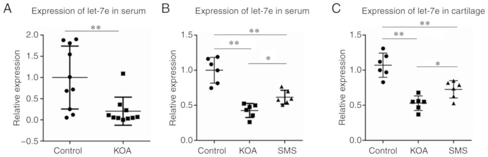
The relative expression levels of let-7e were
calculated using 2−ΔΔCq method with spike-in control
cel-miR-39 as the normalization control. As presented in Fig. 1A, the expression levels of let-7e
in the peripheral serum of the KOA group was ~20% of that in the
control group (P<0.01).
Establishment of a KOA rat model
To further examine the role of let-7e in OA and the
underlying mechanism, a KOA rat model was constructed by surgery.
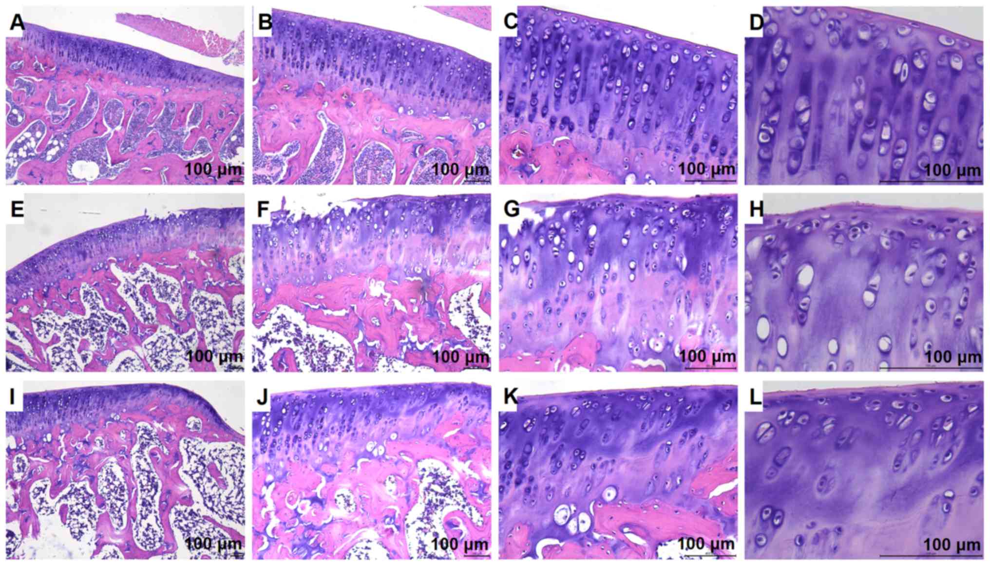
The microscopic morphological images of the cartilage tissue of the
knee joints of different treatment groups were observed (Fig. 2). In the control group, the
surface of the cartilage was smooth and intact with no noticeable
cracks, the chondrocytes were normal in shape and arranged neatly,
the cartilage lacunae were oval and regular with 2–8 cells
distributed inside, and the tide line was clear and intact
(Fig. 2A–D). In the KOA model
group, the surface of the cartilage was less smooth, and edge
defects were present in some of the visual fields, the tide line
was blurred, irregular or missing, cartilage cells were arranged in
a disordered manner, and cartilage lacunae were sparsely
distributed (Fig. 2E–H). Compared
with the KOA group, the morphology of the cartilage tissue in the
SMS group was notably improved; the smoothness of the cartilage
surface was increased, the tide line was clearer, and the cartilage
lacunae were more regularly arranged (Fig. 2I–L).
Let-7e expression is decreased in
cartilage and peripheral serum in the KOA rat model
The relative expression levels of let-7e were
calculated using the 2−ΔΔCq method with U6 used as the
normalization control. As presented in Fig. 1B, in peripheral serum, the
expression of let-7e in the KOA group was significantly decreased
compared with the control group (P<0.01), and the decrease in
let-7e expression was partially reversed in the animals treated
with SMS (P<0.05), although it was still lower compared with the
control group (P<0.01). The levels of let-7e in the cartilage
tissue were examined, and similar results were observed (Fig. 1C); the expression of let-7e in the
cartilage of the KOA group was significantly decreased compared
with the control group (P<0.01), and the reduction was partially
reversed in the SMS group (P<0.05), but was still lower compared
with the control group (P<0.01).
Dysregulated mRNA and miRNA expression in
OA
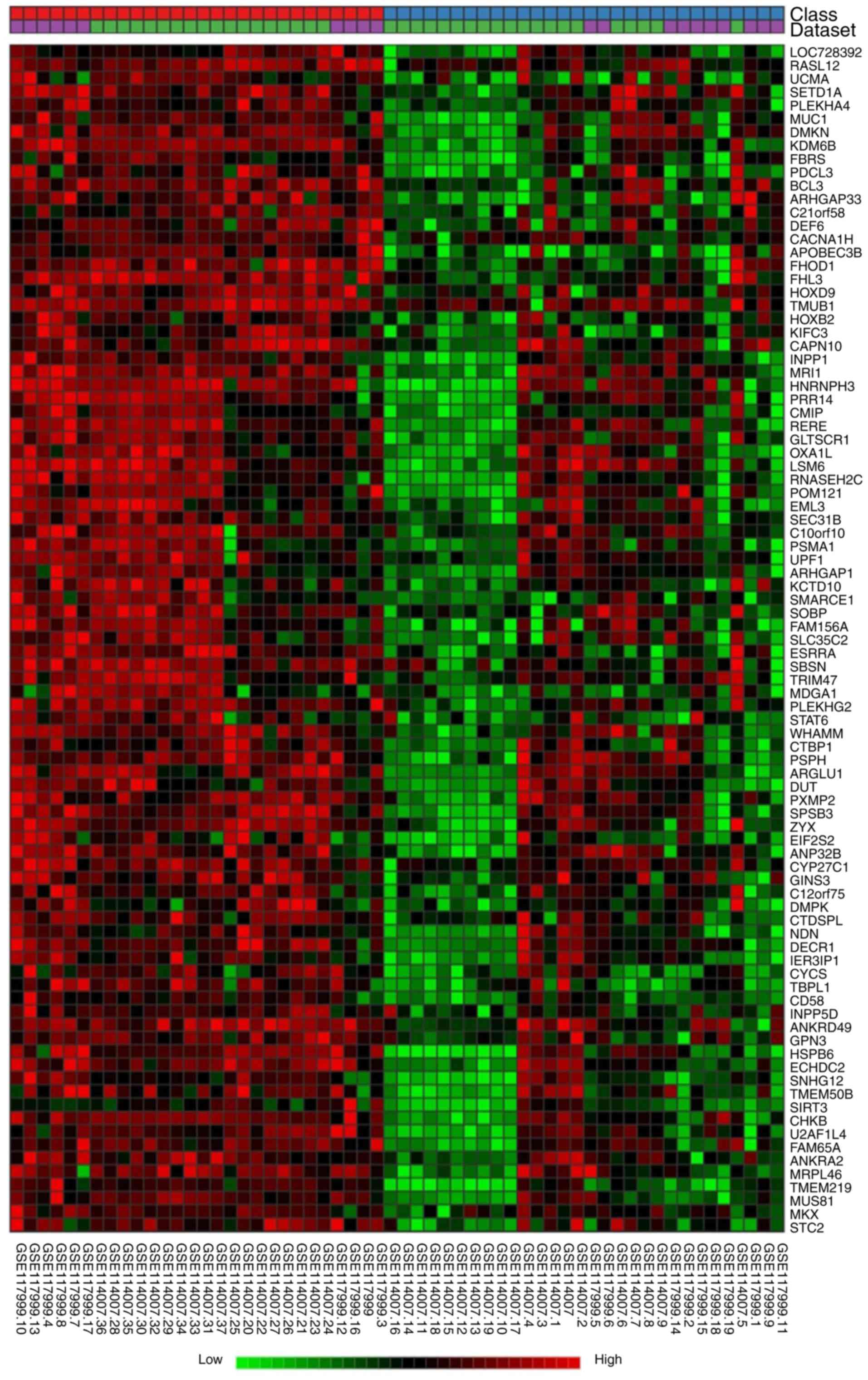
A total of 30 OA and 28 non-OA cases were included
in the bioinformatics analysis, which identified 592 DEGs by a
random effect model with a significance level of P=0.01.
Unsupervised hierarchical clustering using Ward’s method was
performed to cluster the genes and samples. As presented in
Fig. 3, OA samples and controls
were classified separately. The whole heatmap including all 592
DEGs is included in Fig. S1. The
significantly DEGs were mapped to miRNA-gene interaction data
obtained from the TarBase and miRTarBase. A sub-network including
425 seeds, 2,604 nodes and 9,265 edges was constructed, and let-7e
was included in this network, supporting its involvement in the
onset of OA.
KEGG pathway analysis was performed to identify the
significant pathways of the identified DEGs. Enriched KEGG pathways
included ‘Small cell lung cancer’, ‘p53 signaling pathway’,
‘Apoptosis’ and ‘Amyotrophic lateral sclerosis (ALS)’. Considering
that previous studies had demonstrated the role of apoptosis in OA
(19,20), the focus of the present study was
on apoptosis-associated pathways.
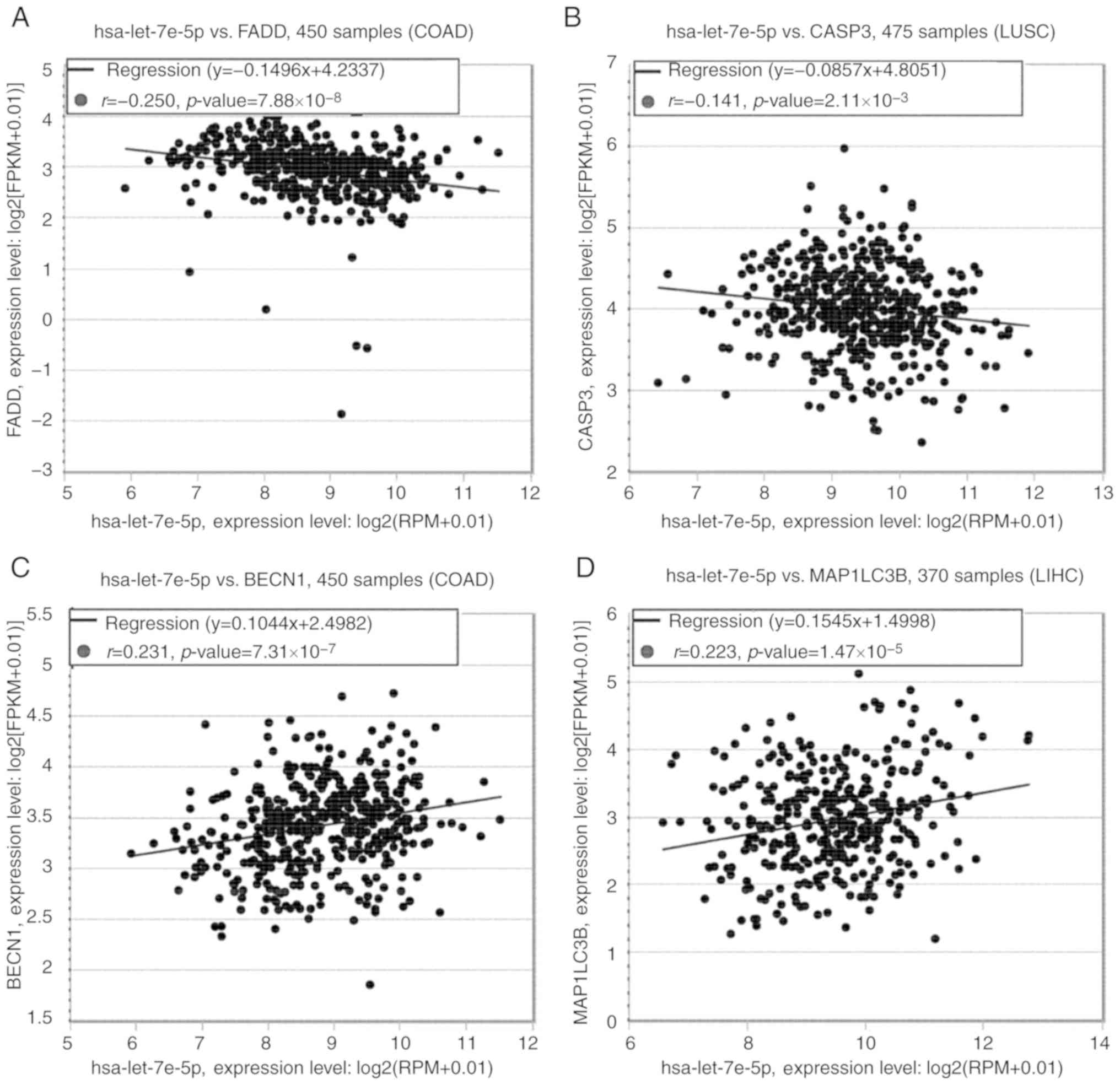
Based on previous studies (19,20,23,24) and bioinformatic analysis, let-7e
may function through regulating apoptotic and autophagic activity
of articular chondrocytes. Pan-Cancer analysis of starBase
(starbase.sysu.edu.cn/panCancer.php) was used to
predict the co-expression among let-7e, apoptosis- and
autophagy-related proteins. As no data regarding chondrocytes were
available, the cell lines with the highest significance in the
Pan-Cancer analysis were used instead. Fig. 4 demonstrates the associations
between let-7e and FAS-associated death domain (FADD), caspase-3
(CASP3), beclin 1 (BECN1) and microtubule-associated protein 1
light chain 3 β (LC3B) in different types of cancer, including
colon adenocarcinoma, lung squamous cell carcinoma and liver
hepatocellular carcinoma.
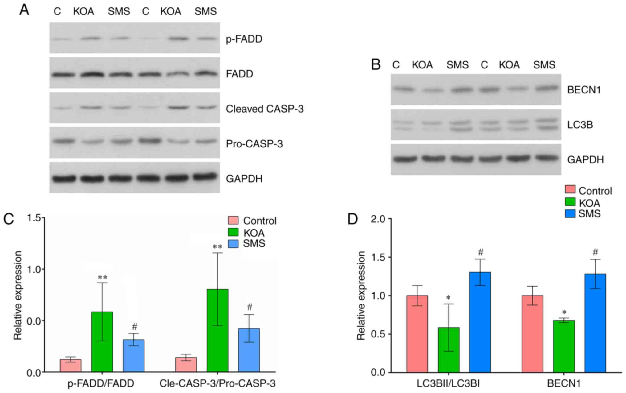
Expression of apoptotic proteins is
increased, whereas expression of autophagy-related proteins is
decreased in the KOA rat model
The expression of apoptotic proteins FADD and CASP3
in the cartilage were examined using western blotting. As presented
in Fig. 5B, the ratio of
p-FADD/FADD was significantly increased in the KOA group compared
with the control group (P<0.01), and treatment with SMS
significantly reduced the ratio of p-FADD/FADD compared with the
KOA group (P<0.05). The ratio of cleaved-CASP3/pro-CASP3 was
significantly increased in the KOA group compared with the control
group (P<0.01), and treatment with SMS significantly reduced the
ratio of cleaved-CASP3/pro-CASP3 compared with the KOA group
(P<0.05).
The expression of autophagy-associated proteins LC3B
and BECN1 are presented in Fig.
5D. The ratio of LC3BII/LC3BI was reduced in the KOA group
compared with the control group (P<0.01). In rats treated with
SMS, the LC3BII/LC3BI ratio was significantly increased compared
with the KOA group (P<0.01). Compared with the control group,
BECN1 expression was significantly reduced in the KOA group
(P<0.01) and increased in rats treated with SMS (P<0.01).
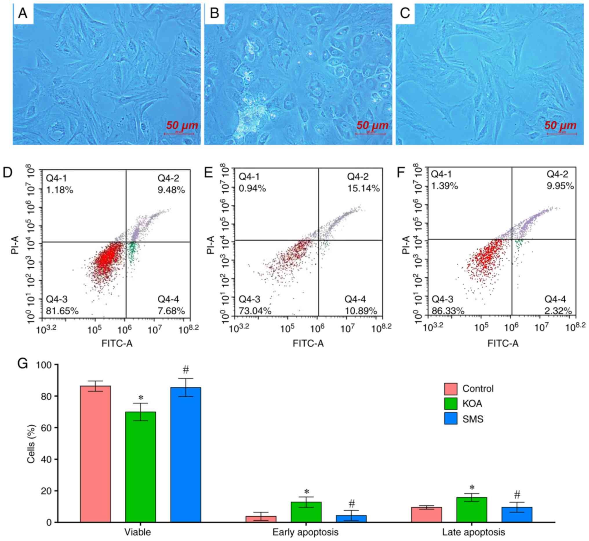
Apoptotic activity is increased in the
chondrocytes of KOA rats
Primary culture of chondrocytes was performed using
chondrocytes isolated from the rat knee cartilage, as presented in
Fig. 6. In the control group, the
chondrocytes were fusiform or spindle-shaped, with a small
proportion exhibiting an oval-shaped morphology (Fig. 6A). In the KOA group, the
chondrocytes were typically round or elliptical, with a small
proportion of fusiform or spindle-shaped cells. The nuclei were
large, a number of cells possessed vacuoles, and some cells died
during cell culture. (Fig. 6B).
Compared with the KOA group, the overall morphology was
significantly improved in the chondrocytes obtained from
SMS-treated rats, where the majority of the cells were
spindle-shaped, although the chondrocytes of the SMS group still
exhibited reduced activity and a lower number of cells compared
with the control group (Fig.
6C).
The chondrocytes from the control, KOA and SMS
groups were stained with Annexin-V-FITC/PI and apoptosis was
assessed using flow cytometry. Compared with the control group, the
proportion of viable cells in the KOA group was significantly
lower, and the proportion of early and late apoptotic cells was
increased (P<0.01; Fig. 6D, E and
G). In the chondrocytes obtained from rats treated with SMS,
the proportion of live cells was significantly increased compared
with the KOA group, and the proportion of early and late apoptotic
cells was reduced (P<0.01; Fig. 6F
and G).
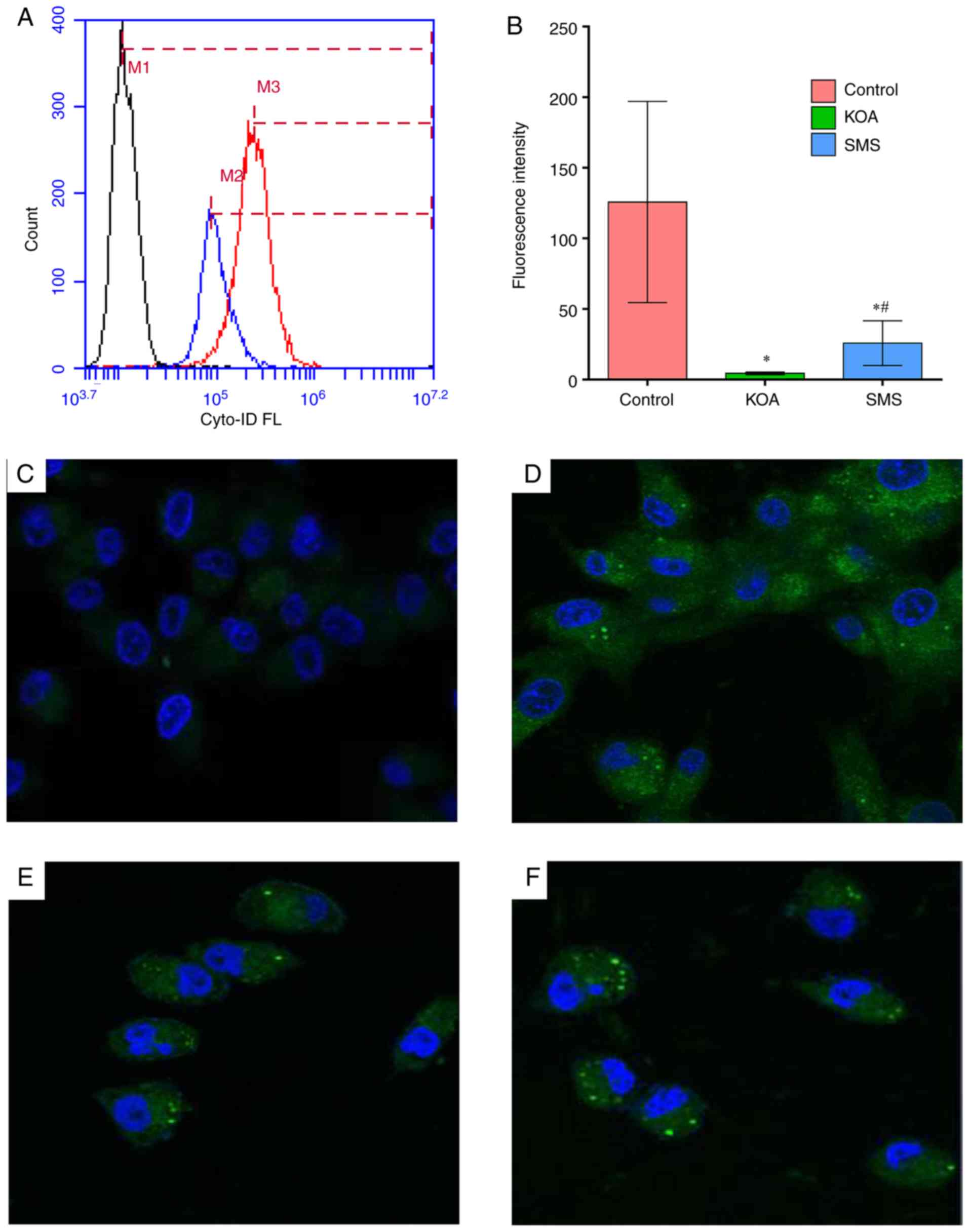
Autophagic activity is decreased in the
chondrocytes from the KOA rat model
The chondrocytes from the three groups of rats were
stained with an ENZO kit and analyzed using flow cytometry and
confocal microscopy. The autophagosomes exhibited green
fluorescence; the chondrocytes in the control group exhibited
medium levels of fluorescence intensity (Fig. 7A, B and D). In the KOA group,
fluorescence intensity was significantly decreased compared with
the control group (P<0.01), suggesting that autophagic activity
was decreased (Fig. 7E). Compared
with the KOA group, the fluorescence intensity of the SMS group
significantly increased, suggesting that autophagic activity was
increased, although it was still significantly lower compared with
the control group (Fig. 7F). No
green fluorescence was observed in the blank control group
(Fig. 7C).
Discussion
At present, the diagnosis of KOA is based on
clinical and imaging findings. The identification of effective
serum markers for diagnosis may potentially improve the rapidity
and accuracy of diagnoses. Therefore, studies in the past decade
have focused on the levels of circulating miRNAs. In 2014,
circulating let-7e was reported to be decreased in patients with
OA, and lower let-7e levels were associated with an increased risk
of arthroplasty due to severe OA (13). Subsequently, a series of miRNAs
have been demonstrated to be abnormally expressed in OA, such as
miR-140-30, mir-33b-3p, miR-671-3p, let-7e, miR-9, miR-27, miR-34a,
miR-140, miR-146a, miR-558, and miR-602 (37–39). However, the results of these
studies vary widely. In the present study, in patients with KOA,
let-7e levels in the peripheral blood were downregulated. In
addition, the expression levels of let-7e in the serum and the
cartilage of the knee in the KOA rat model were significantly
decreased compared with the control group.
Beyer et al (13) demonstrated that let-7e levels were
a negative predictor for total joint arthroplasty. The expression
of let-7e in severe osteoarthritis cases was 0.66 of that in the
control group; unadjusted Cox regression analysis revealed that
let-7e was inversely associated with severe knee and hip
osteoarthritis (13). After
adjustment for sex, age and BMI, the results remained consistent
with an adjusted HR of 0.75 (95% CI, 0.58–0.95; P=0.019) (13). In the present study, serum let-7e
level in the KOA group decreased to ~20% of that in control group.
Generally, the two studies achieved consistent results, although in
the study by Beyer et al (13), patients with severe OA of the knee
and the hip joints were included, whereas in present study only
included patients with KOA. Based on the previous and the present
study, miRNA let-7e may serve as a promising candidate for the
diagnosis of KOA. However, as the sample size of the present study
was small, future studies are required with larger sample sizes
from multiple centers to explore the clinical value of this
indicator.
The role of let-7e in the occurrence of KOA remains
unclear. Let-7e regulates key molecules involved in apoptotic
signaling pathways in neurons (40,41), breast cells (42) and hemocytes (43). In a neuron-like cell line PC12,
transfection of let-7e decreased Casp3 expression and apoptosis,
whereas co-transfection of anti-let-7e significantly alleviated the
effects of let-7e, indicating that let-7e may protect neurons
against apoptosis by negatively regulating the expression of CASP3
(40). However, the effect of
let-7e on chondrocyte apoptosis is unclear. In the present study,
the expression levels of let-7e were decreased in the cartilage and
peripheral blood of patients with KOA, which was accompanied by an
increase in apoptotic activity and decreased autophagic activity,
suggesting that let-7e may be involved in the pathology of KOA
through the regulation of apoptosis and autophagy.
In the present study, expression levels of various
members of apoptotic signaling pathways were increased in the
cartilage of KOA rats, as was the rate of apoptosis. Treatment with
SMS reduced apoptotic activity and alleviated KOA. It has been
reported that the incidence of apoptosis in chondrocytes in
patients with OA is significantly higher compared with normal
articular chondrocytes (44), and
the degree of apoptosis is associated with the degree of articular
cartilage destruction (45).
Pro-inflammatory cytokines tumor necrosis factor-α (TNF-α) and
interleukin 1β (IL-1β) reduce the anabolic metabolism of
chondrocytes, inducing apoptosis and increasing the occurrence of
OA (46). A large sample
microarray study performed in the Netherlands revealed that the
expression of molecules involved in a series of apoptotic signaling
pathways in hemocytes of patients with OA were increased, including
FADD and CASP3 (47). A study
based on a rabbit knee arthritis model demonstrated that SMS
inhibited the levels of IL-1β and TNF-α and effectively inhibited
the inflammatory response (48).
Modified SMS reduced the expression of IL-6 and increased the
expression of basic fibroblast growth factor, thus reducing the
degradation of cartilage matrix (49). In our previous study, SMS
treatment significantly reduced mRNA expression levels of FADD and
CASP3 in the articular cartilage of KOA rats (50). The present study confirmed the
importance of chondrocyte apoptosis in the pathogenesis of OA, and
SMS inhibited chondrocyte apoptosis.
The results of the present study demonstrated that
the expression of autophagy-related proteins in the articular
cartilage of KOA rats was lower compared with that in the controls,
and autophagic activity was decreased; SMS treatment reversed the
alterations in autophagic activity. At present, it is hypothesized
that cell autophagy is different from apoptosis and necrosis of
cells and inhibits the apoptosis of damaged chondrocytes, which may
be an important mechanism for chondrocyte self-protection (51). A previous study has reported that
the expression of Beclin1 and LC3 in OA cartilage and chondrocytes
is reduced, and the expression levels of the associated apoptotic
signaling factors are increased (23). Rapamycin enhances the autophagic
activity of articular chondrocytes, thus preventing the death of
chondrocytes in a mouse model of KOA and reducing the severity of
OA (24). Studies using rat
models of OA have demonstrated that the activity of autophagy
pathways is inhibited in the cartilage from the OA group compared
with a non-OA control and may be involved in the pathogenesis of OA
cartilage (50,52,53). These studies suggested that the
activation of chondrocyte autophagy may inhibit apoptosis, which
may be a novel mechanism for delaying and controlling the
progression of KOA. SMS may exert its therapeutic role through this
mechanism.
The present study has several limitations. Firstly,
as the sample size of this study was small, the significant
reduction of let-7e in the KOA group was not robust enough;
additional studies with larger sample sizes are required to
validate this conclusion. Secondly, to ascertain whether let-7e is
involved in the pathogenesis of KOA, further rescue experiments are
required to discern whether let-7e induces the pathogenesis or its
levels are increased following the onset of KOA. Thirdly, the
present study was designed to explore the mechanisms underlying the
involvement of let-7e; further experiments are required to confirm
the association between let-7e and elevated apoptotic and decreased
autophagic activity of the cartilage tissue. In addition, the
present study did not provide direct evidence that SMS reversed KOA
by regulating let-7e and apoptosis-related proteins. Further
experiments are required to confirm this cause-effect
relationship.
In conclusion, in the peripheral blood of patients
with KOA, the expression of let-7e was significantly decreased, as
well as in peripheral serum and cartilage of the knee in a rat KOA
model. The expression of apoptotic pathway proteins and apoptotic
activity was increased in the cartilage of KOA rats, whereas the
expression of autophagy-related proteins and autophagic activity
was decreased in the articular cartilage of KOA rats compared with
the control group. SMS treatment reversed the changes in the
apoptotic and autophagic activity. Therefore, the present study
supported the hypothesis that circulating let-7e may serve as a
potential serum biomarker for the diagnosis and treatment of KOA.
Elevated apoptotic activity and decreased autophagic activity of
cartilage tissues may be involved in KOA, and treatment with SMS
may reverse these effects.
Supplementary Information
Acknowledgements
Not applicable.
Funding
The present study was supported by grants from the
Zhejiang Provincial Science and Technology Plan of Traditional
Chinese Medicine (grant no. 2017ZQ013), the Medical and Health
Science and Technology Plan of Zhejiang Province (grant no.
2017KY078), the Zhejiang Provincial Natural Science Foundation
(grant no. LY17H040005) and the National Natural Science Foundation
of China (grant nos. 81871176 and 81701461).
Availability of data and materials
The datasets used and/or analysed during the current
study are available from the corresponding author on reasonable
request.
Authors’ contributions
JMS conceived and designed the study. LF, CF, CXW,
DYX, JJC, JFH and PLT performed the experiments. LF and CF analyzed
the data. JMS and CF organized and wrote the manuscript. All
authors read and approved the final manuscript.
Ethics approval and consent to
participate
All human studies were approved by the Ethics
Committee of The First Affiliated Hospital of Zhejiang Chinese
Medicine University, and written informed consent was obtained from
each participant. All animal studies were approved by the Animal
Care and Use Committee of Zhejiang Chinese Medicine University.
Patient consent for publication
Not applicable.
Competing interests
The authors declare that they have no competing
interests.
Abbreviations:
|
KOA
|
knee osteoarthritis
|
|
SMS
|
Si-Miao-San
|
|
OA
|
osteoarthritis
|
|
let-7
|
lethal-7
|
|
BMI
|
body mass index
|
|
DEG
|
differentially expressed gene
|
|
KEGG
|
Kyoto Encyclopedia of Genes and
Genomes
|
|
FADD
|
FAS-associated death domain
|
|
CASP3
|
caspase-3
|
|
LC3B
|
microtubule-associated protein 1 light
chain 3 β
|
References
|
1
|
Jordan JM, Helmick CG, Renner JB, Luta G,
Dragomir AD, Woodard J, Fang F, Schwartz TA, Abbate LM, Callahan
LF, et al: Prevalence of knee symptoms and radiographic and
symptomatic knee osteoarthritis in African Americans and
Caucasians: The Johnston County Osteoarthritis Project. J
Rheumatol. 34:172–180. 2007.PubMed/NCBI
|
|
2
|
Nguyen US, Zhang Y, Zhu Y, Niu J, Zhang B
and Felson DT: Increasing prevalence of knee pain and symptomatic
knee osteoarthritis: Survey and cohort data. Ann Intern Med.
155:725–732. 2011. View Article : Google Scholar : PubMed/NCBI
|
|
3
|
Zhao BW, Zhou LF, Liu YL, Wan SM and Gao
ZX: Evolution of fish Let-7 MicroRNAs and their expression
correlated to growth development in blunt snout bream. Int J Mol
Sci. 18:2017. View Article : Google Scholar
|
|
4
|
Roush S and Slack FJ: The let-7 family of
microRNAs. Trends Cell Biol. 18:505–516. 2008. View Article : Google Scholar : PubMed/NCBI
|
|
5
|
Liu SS, Wang YS, Sun YF, Miao LX, Wang J,
Li YS, Liu HY and Liu QL: Plasma microRNA-320, microRNA-let-7e and
microRNA-21 as novel potential biomarkers for the detection of
retinoblastoma. Biomed Rep. 2:424–428. 2014. View Article : Google Scholar : PubMed/NCBI
|
|
6
|
Murray MJ, Saini HK, Siegler CA, Hanning
JE, Barker EM, van Dongen S, Ward DM, Raby KL, Groves IJ, Scarpini
CG, et al: LIN28 expression in malignant germ cell tumors
downregulates let-7 and increases oncogene levels. Cancer Res.
73:4872–4884. 2013. View Article : Google Scholar : PubMed/NCBI
|
|
7
|
Chiam K, Wang T, Watson DI, Mayne GC,
Irvine TS, Bright T, Smith L, White IA, Bowen JM, Keefe D, et al:
Circulating serum exosomal miRNAs as potential biomarkers for
esophageal adenocarcinoma. J Gastrointest Surg. 19:1208–1215. 2015.
View Article : Google Scholar : PubMed/NCBI
|
|
8
|
Cengiz M, Karatas OF, Koparir E, Yavuzer
S, Ali C, Yavuzer H, Kirat E, Karter Y and Ozen M: Differential
expression of hypertension-associated microRNAs in the plasma of
patients with white coat hypertension. Medicine (Baltimore).
94:e6932015. View Article : Google Scholar
|
|
9
|
Peng G, Yuan Y, Wu S, He F, Hu Y and Luo
B: MicroRNA let-7e is a potential circulating biomarker of acute
stage ischemic stroke. Transl Stroke Res. 6:437–445. 2015.
View Article : Google Scholar : PubMed/NCBI
|
|
10
|
Li D, Ji L, Liu L, Liu Y, Hou H, Yu K, Sun
Q and Zhao Z: Characterization of circulating microRNA expression
in patients with a ventricular septal defect. PLoS One.
9:e1063182014. View Article : Google Scholar : PubMed/NCBI
|
|
11
|
Krause BJ, Carrasco-Wong I, Dominguez A,
Arnaiz P, Farías M, Barja S, Mardones F and Casanello P: Micro-RNAs
Let7e and 126 in plasma as markers of metabolic dysfunction in 10
to 12 years old children. PLoS One. 10:e01281402015. View Article : Google Scholar : PubMed/NCBI
|
|
12
|
Kagawa T, Watanabe M, Inoue N, Otsu H,
Saeki M, Katsumata Y, Takuse Y and Iwatani Y: Increases of microRNA
let-7e in peripheral blood mononuclear cells in Hashimoto’s
disease. Endocr J. 63:375–380. 2016. View Article : Google Scholar : PubMed/NCBI
|
|
13
|
Beyer C, Zampetaki A, Lin NY, Kleyer A,
Perricone C, Iagnocco A, Distler A, Langley SR, Gelse K, Sesselmann
S, et al: Signature of circulating microRNAs in osteoarthritis. Ann
Rheum Dis. 74:e852015. View Article : Google Scholar
|
|
14
|
Weidauer L, Beare T, Binkley T, Minett M
and Specker B: Longitudinal growth and pQCT measures in hutterite
children and grandchildren are associated with prevalence of hip or
knee replacement resulting from osteoarthritis in parents and
grandparents. Clin Orthop Relat Res. 476:1093–1103. 2018.
View Article : Google Scholar : PubMed/NCBI
|
|
15
|
den Hollander W, Boer CG, Hart DJ, Yau MS,
Ramos YFM, Metrustry S, Broer L, Deelen J, Cupples LA, Rivadeneira
F, et al: Genome-wide association and functional studies identify a
role for matrix Gla protein in osteoarthritis of the hand. Ann
Rheum Dis. 76:2046–2053. 2017. View Article : Google Scholar : PubMed/NCBI
|
|
16
|
Wellsandt E, Khandha A, Manal K, Axe MJ,
Buchanan TS and Snyder-Mackler L: Predictors of knee joint loading
after anterior cruciate ligament reconstruction. J Orthop Res.
35:651–656. 2017. View Article : Google Scholar
|
|
17
|
Kim JR, Yoo JJ and Kim HA: Therapeutics in
osteoarthritis based on an understanding of its molecular
pathogenesis. Int J Mol Sci. 19:2018.
|
|
18
|
Funato S, Yasuhara R, Yoshimura K,
Miyamoto Y, Kaneko K, Suzawa T, Chikazu D, Mishima K, Baba K and
Kamijo R: Extracellular matrix loss in chondrocytes after exposure
to interleukin-1β in NADPH oxidase-dependent manner. Cell Tissue
Res. 368:135–144. 2017. View Article : Google Scholar : PubMed/NCBI
|
|
19
|
Rose J, Soder S, Skhirtladze C, Schmitz N,
Gebhard PM, Sesselmann S and Aigner T: DNA damage, discoordinated
gene expression and cellular senescence in osteoarthritic
chondrocytes. Osteoarthritis Cartilage. 20:1020–1028. 2012.
View Article : Google Scholar : PubMed/NCBI
|
|
20
|
Hosseinzadeh A, Kamrava SK, Joghataei MT,
Darabi R, Shakeri-Zadeh A, Shahriari M, Reiter RJ, Ghaznavi H and
Mehrzadi S: Apoptosis signaling pathways in osteoarthritis and
possible protective role of melatonin. J Pineal Res. 61:411–425.
2016. View Article : Google Scholar : PubMed/NCBI
|
|
21
|
Maiuri MC, Zalckvar E, Kimchi A and
Kroemer G: Self-eating and self-killing: Crosstalk between
autophagy and apoptosis. Nat Rev Mol Cell Biol. 8:741–752. 2007.
View Article : Google Scholar : PubMed/NCBI
|
|
22
|
Kroemer G, Mariño G and Levine B:
Autophagy and the integrated stress response. Mol Cell. 40:280–293.
2010. View Article : Google Scholar : PubMed/NCBI
|
|
23
|
Caramés B, Taniguchi N, Otsuki S, Blanco
FJ and Lotz M: Autophagy is a protective mechanism in normal
cartilage, and its aging-related loss is linked with cell death and
osteoarthritis. Arthritis Rheum. 62:791–801. 2010. View Article : Google Scholar : PubMed/NCBI
|
|
24
|
Caramés B, Hasegawa A, Taniguchi N, Miyaki
S, Blanco FJ and Lotz M: Autophagy activation by rapamycin reduces
severity of experimental osteoarthritis. Ann Rheum Dis. 71:575–581.
2012. View Article : Google Scholar :
|
|
25
|
Fougere B: Si-Miao-San for chronic
inflammatory conditions. In: Proceedings of the 40th Annual IVAS
and 15th Annual ItVAS International Congress on Veterinary
Acupuncture; Florence, Italy. pp. 2572014
|
|
26
|
Xu Y, Liu Q, Liu ZL, Lim L, Chen WH and
Lin N: Treatment with SiMiaoFang, an anti-arthritis chinese herbal
formula, inhibits cartilage matrix degradation in osteoarthritis
rat model. Rejuvenation Res. 16:364–376. 2013. View Article : Google Scholar : PubMed/NCBI
|
|
27
|
Chen B, Zhan H, Marszalek J, Chung M, Lin
X, Zhang M, Pang J and Wang C: Traditional Chinese medications for
knee osteoarthritis pain: A meta-analysis of randomized controlled
trials. Am J Chin Med. 44:677–703. 2016. View Article : Google Scholar : PubMed/NCBI
|
|
28
|
Hayami T, Pickarski M, Zhuo Y, Wesolowski
GA, Rodan GA and Duong LT: Characterization of articular cartilage
and subchondral bone changes in the rat anterior cruciate ligament
transection and meniscectomized models of osteoarthritis. Bone.
38:234–243. 2006. View Article : Google Scholar
|
|
29
|
Rogart JN, Barrach HJ and Chichester CO:
Articular collagen degradation in the Hulth-Telhag model of
osteoarthritis. Osteoarthritis Cartilage. 7:539–547. 1999.
View Article : Google Scholar : PubMed/NCBI
|
|
30
|
Hulth A, Lindberg L and Telhag H:
Experimental osteoarthritis in rabbits. Preliminary report. Acta
Orthop Scand. 41:522–530. 1970. View Article : Google Scholar : PubMed/NCBI
|
|
31
|
Livak KJ and Schmittgen TD: Analysis of
relative gene expression data using real-time quantitative PCR and
the 2(−Delta Delta C(T)) method. Methods. 25:402–408. 2001.
View Article : Google Scholar
|
|
32
|
Fisch KM, Gamini R, Alvarez-Garcia O,
Akagi R, Saito M, Muramatsu Y, Sasho T, Koziol JA, Su AI and Lotz
MK: Identification of transcription factors responsible for
dysregulated networks in human osteoarthritis cartilage by global
gene expression analysis. Osteoarthritis Cartilage. 26:1531–1538.
2018. View Article : Google Scholar : PubMed/NCBI
|
|
33
|
Brophy RH, Zhang B, Cai L, Wright RW,
Sandell LJ and Rai MF: Transcriptome comparison of meniscus from
patients with and without osteoarthritis. Osteoarthritis Cartilage.
26:422–432. 2018. View Article : Google Scholar :
|
|
34
|
Scian MJ, Maluf DG, David KG, Archer KJ,
Suh JL, Wolen AR, Mba MU, Massey HD, King AL, Gehr T, et al:
MicroRNA profiles in allograft tissues and paired urines associate
with chronic allograft dysfunction with IF/TA. Am J Transplant.
11:2110–2122. 2011. View Article : Google Scholar : PubMed/NCBI
|
|
35
|
Papadopoulos GL, Reczko M, Simossis VA,
Sethupathy P and Hatzigeorgiou AG: The database of experimentally
supported targets: A functional update of TarBase. Nucleic Acids
Res. 37:D155–D158. 2009. View Article : Google Scholar :
|
|
36
|
Chou CH, Shrestha S, Yang CD, Chang NW,
Lin YL, Liao KW, Huang WC, Sun TH, Tu SJ, Lee WH, et al: miRTarBase
update 2018: A resource for experimentally validated
microRNA-target interactions. Nucleic Acids Res. 46:D296–D302.
2018. View Article : Google Scholar :
|
|
37
|
Nugent M: MicroRNAs: Exploring new
horizons in osteoarthritis. Osteoarthritis Cartilage. 24:573–580.
2016. View Article : Google Scholar
|
|
38
|
Ntoumou E, Tzetis M, Braoudaki M, Lambrou
G, Poulou M, Malizos K, Stefanou N, Anastasopoulou L and Tsezou A:
Serum microRNA array analysis identifies miR-140-3p, miR-33b-3p and
miR-671-3p as potential osteoarthritis biomarkers involved in
metabolic processes. Clin Epigenetics. 9:1272017. View Article : Google Scholar : PubMed/NCBI
|
|
39
|
Seeliger C, Balmayor ER and van Griensven
M: miRNAs related to skeletal diseases. Stem Cells Dev.
25:1261–1281. 2016. View Article : Google Scholar : PubMed/NCBI
|
|
40
|
Peng G, Yuan Y, He Q, Wu W and Luo BY:
MicroRNA let-7e regulates the expression of caspase-3 during
apoptosis of PC12 cells following anoxia/reoxygenation injury.
Brain Res Bull. 86:272–276. 2011. View Article : Google Scholar : PubMed/NCBI
|
|
41
|
Gong W, Zheng J, Liu X, Ma J, Liu Y and
Xue Y: Knockdown of NEAT1 restrained the malignant progression of
glioma stem cells by activating microRNA let-7e. Oncotarget.
7:62208–62223. 2016. View Article : Google Scholar : PubMed/NCBI
|
|
42
|
Aure MR, Leivonen SK, Fleischer T, Zhu Q,
Overgaard J, Alsner J, Tramm T, Louhimo R, Alnæs GI, Perälä M, et
al: Individual and combined effects of DNA methylation and copy
number alterations on miRNA expression in breast tumors. Genome
Biol. 14:R1262013. View Article : Google Scholar : PubMed/NCBI
|
|
43
|
Ferreira AF, Moura LG, Tojal I, Ambrósio
L, Pinto-Simões B, Hamerschlak N, Calin GA, Ivan C, Covas DT,
Kashima S and Castro FA: ApoptomiRs expression modulated by BCR-ABL
is linked to CML progression and imatinib resistance. Blood Cells
Mol Dis. 53:47–55. 2014. View Article : Google Scholar : PubMed/NCBI
|
|
44
|
Héraud F, Héraud A and Harmand MF:
Apoptosis in normal and osteoarthritic human articular cartilage.
Ann Rheum Dis. 59:959–965. 2000. View Article : Google Scholar : PubMed/NCBI
|
|
45
|
Thomas CM, Fuller CJ, Whittles CE and
Sharif M: Chondrocyte death by apoptosis is associated with the
initiation and severity of articular cartilage degradation. Int J
Rheum Dis. 14:191–198. 2011. View Article : Google Scholar : PubMed/NCBI
|
|
46
|
Rahmati M, Mobasheri A and Mozafari M:
Inflammatory mediators in osteoarthritis: A critical review of the
state-of-the-art, current prospects, and future challenges. Bone.
85:81–90. 2016. View Article : Google Scholar : PubMed/NCBI
|
|
47
|
Ramos YF, Bos SD, Lakenberg N, Böhringer
S, den Hollander WJ, Kloppenburg M, Slagboom PE and Meulenbelt I:
Genes expressed in blood link osteoarthritis with apoptotic
pathways. Ann Rheum Dis. 73:1844–1853. 2014. View Article : Google Scholar
|
|
48
|
Li HY, Meng DF and Su JG: Si Miao San
intervention rabbit knee joint inflammation traumatic synovial
fluid IL-1β and TNF-α level of experiments. Chin J Tradit Chin Med.
28:3043–3045. 2013.(In Chinese).
|
|
49
|
Zhao P, Wang L, Li H and Yu J: Effects on
IL-6 and BFGF of the rabbits’ knee arthritises by modified Simiao
power. China J Chin Med. 30:1166–1169. 2015.(In Chinese).
|
|
50
|
Shen JM, Feng L, Chen J, Wu Y and Yu J:
Effect of Simiaosan on apoptosis and autophagy of chondrocyte in
treating knee osteoarthritis in rats. New Chin Med. 49:12–15.
2017.(In Chinese).
|
|
51
|
Zhang Q, Lai S, Hou X, Cao W, Zhang Y and
Zhang Z: Protective effects of PI3K/Akt signal pathway induced cell
autophagy in rat knee joint cartilage injury. Am J Transl Res.
10:762–770. 2018.PubMed/NCBI
|
|
52
|
Wu Z, Luan Z, Zhang X, Zou K, Ma S, Yang
Z, Feng W, He M, Jiang L, Li J and Yao J: Chondro-protective
effects of polydatin in osteoarthritis through its effect on
restoring dysregulated autophagy via modulating MAPK, and PI3K/Akt
signaling pathways. Sci Rep. 9:139062019. View Article : Google Scholar : PubMed/NCBI
|
|
53
|
Wang F, Liu J, Chen X, Zheng X, Qu N,
Zhang B and Xia C: IL-1β receptor antagonist (IL-1Ra) combined with
autophagy inducer (TAT-Beclin1) is an effective alternative for
attenuating extracellular matrix degradation in rat and human
osteoarthritis chondrocytes. Arthritis Res Ther. 21:1712019.
View Article : Google Scholar
|